Calcium and Phosphorus Metabolism
1LEO LUTWAK Cornell University
Graduate School of Nutrition Sage Hospital
Ithaca, New York
I. Introduction 4 3 3
II. Techniques 4 3 5
A. Balance Studies 43*>
B. Analytical Techniques 437
C. Tracer Studies 4 4 0
D. X-ray Techniques 4 4 <7
III. Specific Applications to Studies 4 4 8
A. Osteoporosis 4 4 8
B. Renal Disease 4 5 0
C. Gastrointestinal Disease 4^4
D. Endocrine Disorders 4^5
E. Rickets and Vitamin D 45 9
F. Fluoride 4 6 0
G. Dental Caries 46<>
H. Miscellaneous 4^ 1
IV. Summary 4 6 2
References 4^2
I . INTRODUCTION
Calcium is present in the body to a far greater extent than any other cation. It thus must be considered one of the most important macro- nutrients in discussions of human physiology. The skeleton contains approximately 99% of the total body calcium and therefore is the major physiologic storehouse for this element. Phosphorus, the principal anion in the organism, is distributed in the body somewhat more widely than is calcium. However, since 80% of it is also present in the skeleton in association with calcium, the metabolism of these two elements will be considered together in the present discussion. It is recognized, and ap- propriate reservation will be made, that phosphorus metabolism is also intimately related to the metabolism of proteins, carbohydrates and fats;
1 Aided by a grant from the U. S. Public Health Service AM-07451-01.
433
434 LEO LTJTWAK
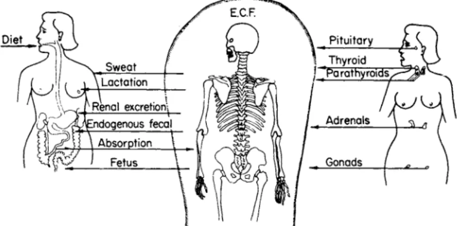
for the purpose of this review, however, these aspects will not be emphasized. The major fraction of the body calcium, the 99% present in the skeleton, is in the form of calcium hydroxyapatite, a mineral crystal functioning as the major structural element of the skeleton and as the major storehouse of calcium for maintaining homeostatic concen- tration in the rest of the body (Fig. 1). The remaining 1% of body calcium is present almost entirely in extracellular fluid and, to a slight extent, as a component of various membrane structures. In man, the concentration of calcium in extracellular fluid is relatively stable, main-
FIG. 1. Metabolic and endocrine control of calcium metabolism in man. E . C . F .
= extracellular fluid.
tained at a level of 2.5 mEq per liter under the influences of the gastro- intestinal system, the kidneys, and the various endocrine systems. Thus, a consideration of calcium metabolism (and secondarily of phosphorus metabolism) must take into account the relative roles of other systems of the body in maintaining integrity of the skeleton and control of extra- cellular fluid concentrations. It is apparent that the large surplus stores of mineral in the skeleton permit the organism to withstand extensive onslaughts on the total body content of these minerals before sig- nificant changes can be seen in the skeletal content of calcium and phosphorus or in the structural integrity of the skeleton.
The past decade has seen significant developments in new techniques and in refinements of old methodology which have permitted new insights into various problems of calcium metabolism in man. The present review will summarize some of these technical advances and their applications to human physiology and pathology.
II. TECHNIQUES
A. Balance Studies
The metabolic balance technique has long been applied to studies of the body economy of macroelements such as calcium, phosphorus, mag- nesium, sodium, potassium, and nitrogen. In 1927, Bauer and Aub (1) described the ward routine and methods then employed in studies of inorganic salt metabolism at the Massachusetts General Hospital. Sub- sequently, in 1945, Reifenstein et al. (2) further amplified this description and went into considerable detail describing the interpretation of data resulting from such studies. These techniques with various modifications
are in use at present in many research units throughout the world.
The following discussion is a description of the techniques for con- ducting a metabolic balance study as evolved by the Metabolic Disease Branch of the National Institute of Arthritis and Metabolic Disease at the National Institutes of Health (3) and further modified in the Clinical Nutrition Unit of the Graduate School of Nutrition at Cornell University.
1. The Metabolic Unit
The research ward consists of five single-bed rooms with connecting toilets under the supervision of five registered nurses and three practical nurses, thus permitting three shifts a day of highly trained professional supervision. There is a separate dietetic kitchen in the same wing under the supervision of a full-time research dietitian, assisted by three spe- cially trained kitchen helpers.
2. Design of Study
In the majority of studies of calcium metabolism the patient serves as his own control. Thus, it is essential that when a study is planned only one variable is changed at a time. A diet history is obtained by the clinical investigator and by the dietitian and, based upon this, a menu is devised providing the amounts of calcium, phosphorus, magnesium, nitro- gen, and calories called for in the specific study and provided by foods that the patient will eat readily. Usually a week of adjustment is re-
quired for minor modifications of a constant menu. If the study is to con- tinue for more than 30 days, two menus as similar in composition as possible are planned, to be fed on alternate days. The patient is urged to consume the full diet, avoiding the pitfalls of "weigh-back" analyses.
Urine is collected in 24-hour periods. The collection jug is refrigerated on the ward throughout the metabolic day; the volume of urine soon after it is passed is measured by at least two members of the nursing personnel before being pooled into the 24-hour collection. Stools are collected into
436 LEO LUTWAK
polyethylene-lined, stainless steel containers that fit the modified toilet seat (4) and are sent to the laboratory directly for pooling. The meta- bolic periods are usually of 6 days duration, with the administration of an insoluble dye marker at the beginning of each period. It has been found (5) that periods may be timed more accurately by taking the first appearance of the dye marker as indicative of the beginning of a new period, rather than using the last appearance to indicate the end of the preceding period. Because of the wide variation in time of stool passage, periods shorter than 6 days do not permit accurate stool collections. The virtues of different stool markers have been recently discussed (5), with the conclusion that the best marker is one that is readily visible to nurs- ing and laboratory personnel and one that presumably has a density equal to that of the bowel contents. In our experience Brilliant Blue has satisfied these criteria most effectively. Carmine is used occasionally, but the red color of this dye is often marked by the intrinsic brown pigments of the stool.
3. Diet and Medications
Fruits and vegetables used in the preparation of study diets may be obtained as constant-composition products by the purchase of dietetic pack canned foods in large quantities from single cannery lots. Milk is provided as powdered dry skim milk reconstituted the night before use with distilled water and allowed to remain in the refrigerator for better palatability. Butter is used in unchanged form since the content of salts and water is relatively constant in a single batch. In some of the diets eggs may be served; under these circumstances a large batch of eggs from a single flock is purchased. These are then cracked and scrambled;
individual weighed portions of the scrambled eggs are frozen until used, thereby maintaining a high degree of palatability. Bread is purchased in large quantities from a single day's baking, using a salt-free formula.
Two varieties of meat are offered. Beef is purchased as a whole tender- loin, stripped free of tendon and fat in the kitchen, and divided into weighed serving portions, frozen until used. Chicken is served as the bone-free, skin-free, and fat-free breast of chicken, obtained from a large lot of a single flock. All of the water used for drinking, food preparation, and mouth wash is glass distilled. The patient is not permitted to use commercial dentifrices, since these have been found to contain large and variable amounts of minerals. A mineral-free dentifrice is prepared, based on urea and quaternary detergents with the addition of appropriate flavoring. Medications used in these studies are purchased in large lots, and representative tablets are analyzed for all of the elements studied under the balance regimen. In studies of calcium metabolism it is often
desirable to increase the calcium content of the diet without changing any of the other constituents. Under these circumstances calcium supplements are administered in the form of calcium lactate (12.97% calcium) or calcium gluconate (9.25% calcium).
4. Management of Patients
Activity of the patient is carefully controlled. The metabolic unit is air-conditioned and maintained within a narrow range of temperature, approximately 72°F all year round. The patient is encouraged to main- tain a minimum level of walking activity and to avoid exercise to the point of active sweating. The patient is not permitted to go out of doors, except for periods in the early evening when accompanied by a member of the professional staff. At the beginning of the study, the proposed protocol is described to the patient by the investigator. The patient is urged to ask questions and discuss problems as they occur. Full-staff conferences, including the investigators, nurses, dietitian, and a member of the laboratory staff, are held weekly to discuss the conduct and prog- ress of the studies; these supplement the daily "rounds" involving the clinicians and the nurses. Although every attempt is made to conclude a research protocol successfully, the patient's welfare still remains a pri- mary concern, and the investigator must be prepared to terminate a study prematurely if the patient develops an intercurrent illness or otherwise becomes incapable of cooperation.
5. Value of Balance Studies
The balance technique is of particular value in determining the net re- sultant of a specific mode of therapy on the balance of a desired nutrieni Obviously, the significance of the data depends upon the accuracy of the metabolic techniques, i.e., reproducibility of preparation of diet, accuracy of urine and fecal collections, and strictness of adherence to controlled conditions, as well as upon the accuracy of the analytic procedures avail- able for the desired component. When evaluated in conjunction with dynamic studies, such as are possible with modern tracer techniques, the metabolic balance study enables the investigator to derive an over-all summary of metabolic pathways.
B. Analytical Techniques
1. Calcium
The classical procedure for the determination of calcium in biological materials is that of precipitation as the oxalate and subsequent titration
438 LEO LTJTWAK
with potassium permanganate (6). Under carefully controlled conditions this method is highly reproducible, and the error introduced by coprecipi- tation of magnesium and other interferences may be kept constant and minimal. However, because of the technical difficulties in precipitation and titration, there have been many attempts to develop more reproduci
ble, simpler procedures requiring less attention to detail. Direct spectro- photometric methods have been suggested, based on the formation of chloranilic acid derivatives (7) and other complex dyes (8, 9). These techniques suffer from the defect that, in the presence of variable amounts of phosphate, magnesium, and protein, the results are variable.
When used for the determination of calcium in serum, where interfering anions are present in relatively constant amounts, these methods are sufficiently accurate. However, in the estimation of calcium in urine, stool, and diet samples, considerable interference results.
A second technique of considerable value is based on the complexo- metric titration of calcium in the presence of specific indicators such as Eriochrome Black T, calcein, murexide, and other metallochrome indi
cators (10-13). With some of these dyes, magnesium in the sample leads to erroneously high values. With all, phosphate, present in variable amounts, will change the end point to a significant extent. Various tech
niques, including ion-exchange resins and precipitation of the phosphate as molybdate complexes (14, 15), have been suggested to avoid this problem, particularly for the estimation of calcium in urine and stool.
A more direct technique of increasing importance is the flame photo
metric estimation of calcium. The primary sources of interference with this technique result from the presence of phosphate in the sample, which depresses the ionization of calcium in the flame, and the presence of sodium, which emits at a wavelength sufficiently close to that for calcium (620 ταμ) to lead to sufficient error. These interferences have been con
trolled in various ways. The procedure of Loken et al. (16) avoids inter
ferences by measuring absorption in the near ultraviolet with a reversed oxyacetylene flame and by adding a masking solution containing excess sodium and phosphate to each sample. Still another modification, used by our laboratory with reproducible results for several years, is based on removal of the interfering substances (17, 18).
An aliquot of urine, serum, or solution of the ash of feces or diet, containing between 50 and 100 /Ag of calcium, is diluted to approximately 10 ml in a centrifuge tube. One milliliter of 2.5% oxalic acid is added, and the solution is brought to pH 4.5 with ammonium hydroxide and hydrochloric acid. Methyl red is used as the indicator. Hydrochloric acid (IN) is added to bring the final pH to about 4.3, and precipitation is allowed to proceed overnight in the refrigerator. The samples are centri-
fuged, and the supernatant is discarded. The precipitate is washed twice with 0.5% ammonium oxalate, with centrifuging and aspiration of the supernatant after each washing. The final precipitate is dissolved in 1 ml of 2 Μ perchloric acid with the aid of a boiling water bath. The dissolved precipitate is diluted to 100 ml with distilled water and 2.0 ml of 1%
Sterox solution. The samples are then atomized in a Coleman Flame Photometer with appropriate standard solutions.
The development of atomic absorption spectroscopy as an economi
cally feasible tool by the C.S.I.R.O. in Melbourne, Australia has brought a new procedure free of manipulation to the metabolic research labora
tory, permitting the determination of minerals in large numbers of samples rapidly and directly. As this apparatus becomes more available, I believe this technique will supplant many of the older procedures in laboratories handling large numbers of samples.
The procedure described by David (19) has resulted in reproducible values in our laboratory. Biological solutions to be analyzed are diluted 5- to 50-fold (depending upon the concentration of calcium) with a solu
tion of 1% lanthanum chloride to prevent phosphate interference. The final solution is then sprayed into an air-acetylene flame, and absorption is measured by a millivolt recorder. Recoveries average close to 100%, and reproducibility results in standard deviations of less than 1%.
With the increasing availability to many laboratories of automatic analytical equipment, attempts have been made to automate many of the procedures for calcium determination. Since most of these are based on direct colorimetric estimation of calcium by complexing with specific dyes, the automatic techniques presently available (20, 21) provide reli
able measurements in serum samples, but are of little value in calcium determinations in urine or digests of stool and diet. In our laboratory we have automated the atomic absorption spectroscopic measurement of calcium by combining a sampler and pump module of the Auto Analyzer with a direct readout Atomic Absorption Spectrometer for rapid, repro
ducible automatic calcium determinations in a variety of substances (22). Still another technique in use in some laboratories is based on the measurement of X-ray fluorescence of samples bombarded with narrow wavelength X-rays (23).
2. Phosphorus
The standard procedure for the determination of inorganic phosphate in biological investigations is based on the original Fiske-SubbaRow method (24), the formation of phosphomolybdic acid and reduction to a blue chromogen by one of several reducing agents, such as aminonaph- tholsulfonic acid, ascorbic acid, or Amidon. All of these techniques
440 LEO LUTWAK.
readily lend themselves to automation and have been employed in this fashion (25).
C. Tracer Studies
1. General
The history of the use of tracers in bone metabolism in attempts to study mechanisms of bone formation and remodeling extends back to the early eighteenth century. In 1736 Belchier (26) observed that the bones of hogs fed diets containing madder extract were tinctured a deep-red color. Subsequently, in 1740 du Hamel (27) reported the results of a series of experiments in which pigeons and chickens were fed madder root at various ages and then sacrificed at periods thereafter. He con- cluded that the rapidly growing bones deposit the scarlet color more quickly than the slower growing bones and to a lesser extent. Further- more, after the madder was removed from the diet, the color gradually disappeared from the bones. This author referred to previous observations published in 1566 by Mizaldus, who had similarly observed the colora- tion of bone after the feediiig of madder root. The discovery of artificial radioactivity led to the use of more physiological tracers in the study of metabolism. In 1935 Chiewitz and Hevesy (28) studied bone metabolism using phosphorus-32. Tracer methodology underwent rapid growth follow- ing the development of nuclear reactors in the 1940's and 1950's. Today we have available for studies of bone physiology specific isotopes of calcium such as calcium-45 and calcium-47, as well as strontium-89 and strontium-90 tracers from which information regarding bone turnover may be derived.
Since the introduction of the use of calcium-45 in 1940 by Campbell and Greenberg (29) this isotope has been utilized in animal and clinical studies for the determination of the kinetics of bone formation (30-33), mechanisms of calcium absorption and excretion (31a,b, 34, 35), and for studies of the role of calcium in soft tissue metabolism (36, 37). Calcium- 45 is a pure beta-emitting isotope with a half-life of 153 days, emitting a beta particle with energy of approximately 0.261 MeV. Counting of this isotope in tracer studies requires preliminary isolation of the calcium from biological fluid and the estimation of the beta activity either in a gas flow counter after plating to infinite thickness (30-33) or in a liquid scintillation spectrometer after appropriate solution in a scintillating me- dium (38).
Because of the technical difficulties involved in biological and clinical studies with calcium-45, the availability of calcium-47 has been greeted with enthusiasm by investigators (39-42). Calcium-47 has a half-life of 4.7 dpys and emits, in addition to a series of beta particles with energies
of 1.94 and of 0.66 MeV, a series of gamma rays with energies of 1.31, 0.83, and 0.48 MeV. Calcium-47 is available today in the form of mix- tures of isotope containing from 1 to 10% of calcium-45. Studies with this isotope are further complicated by the presence of the daughter, scan- dium-47, which also emits beta and gamma radiation and has a half-life of about 3.4 days. Calcium-47 may be detected in biologic materials without preliminary isolation of the calcium by counting the gamma emission with appropriate scintillation crystals or other types of gamma detectors. The major drawback of studies with calcium-47 has been the relatively short half-life of this tracer, which limits studies to short periods of time. An additional advantage of this isotope, however, is that it permits in vivo counting over bones, allowing calculations of local uptake (39, 41).
Clinical studies have also been reported using strontium-85, an iso- tope emitting a pure gamma ray of 0.513 MeV with a half-life of 64 days (39, 42-45). Studies have also been reported using strontium-90 with a half-life of 27.7 years, which produces a beta particle of 0.545 MeV, and using strontium-89 with a half-life of 51 days, which produces a beta particle of 1.46 MeV (46-48). Strontium-85 has all of the ad- vantages of calcium-47 in terms of ease of estimation. It also affords the advantage of calcium-45 in permitting long-term studies because of its long half-life, but has the serious disadvantage that reservations always have to be made in interpretation of studies made with strontium be- cause of the significant differences in its metabolic behavior as compared with calcium. Strontium is preferentially excreted by the kidney in com- parison with calcium (49), while calcium is preferentially absorbed from the intestinal tract (50). Calcium is also preferentially deposited in bone during bone crystal formation and may displace strontium readily from crystals containing the latter (51-53).
In an attempt to avoid the administration of radioactive substances to patients and yet carry out tracer studies, Fraser et al. (54) and Eisen- berg and Gordan (55) have utilized nonradioactive strontium, the natu- rally occurring form of this element, in studies of patients. However, the same reservations concerning the interpretation of data as have been stated for radioactive strontium must be considered.
With the availability of highly enriched, stable isotopes of calcium, such as calcium-46 and calcium-49, it will become possible to carry out kinetic studies in subjects to whom radioactive isotopes cannot be ad- ministered, such as pregnant women and children. The stable isotopes can be estimated in biological tissues by mass spectrometry or neutron acti- vation.
Other bone-seeking substances have been utilized in studies of bone metabolism. It has been shown that tetracycline and its derivatives are
442 LEO LUTWA K
deposited in bone in the fashion similar to that seen with the madder- root dye (56, 57). These substances have been used in studies of bone remodeling and deposition (58, 59). Alizarin (and iodinated derivatives of this substance), the principal component of the madder-root dye, has also been reintroduced in recent years in studies of bone physiology (60, 61).
2. Techniques for Use
a. Clinical Administration. The various isotopes of calcium (and of strontium as well) may be administered either orally or intravenously to the patient. For intravenous administration the tracer substance is dis
solved in physiological saline or isotonic glucose containing a trace amount of chlorobutanol as antiseptic and properly sterilized. The usual dosage for intravenous use of calcium-45 ranges from 1 to 10 ju,c, depend
ing on the size of the patient and the purpose of the study. For oral use, doses of 5 to 20 μο have been used. Calcium-47 may be administered in somewhat higher dosage because of the shorter half-life and thus de
creased radiation dosage. Calcium-47 has been used in the study of bone metabolism in oral doses of 1 to 40 μο, and intravenous doses of 0.5 to 15 /AC.
b. Estimation of Isotopes in Biological Fluids. (1) Calcium-45. The liquid scintillation technique has been utilized in our laboratory for the measurement of calcium-45 (38).
An appropriate sample of blood, urine, or digest of stool is treated with ammonium oxalate to precipitate calcium oxalate under standard conditions. The calcium oxalate precipitate is washed once to remove sub
stances that may quench scintillation, and the final precipitate is dis
solved in 0.3 ml of 30% hydrochloric acid. To this is added 4 ml of absolute ethanol and 6 ml of toluene containing 0.4% diphenyloxazole.
The resulting scintillation mixture is counted in a liquid scintillation spectrometer such as the Packard Tri-Carb.
(2) Calcium-47. Calcium-47, because of the emission of a gamma ray, may be counted in blood, urine, or stool directly without prior sepa
ration. Either a scintillation crystal well or a large-volume liquid scintil
lation counter such as the Packard Armac may be used for this purpose.
When large volumes of material are to be examined, it is desirable to concentrate the calcium in the sample by precipitation as the oxalate to allow more advantageous counting statistics.
3. Applications of the Tracer Techniques
a. Kinetic Studies of Bone Formation (and Resorption). From kinetic analysis of data obtained after the intravenous administration of tracer
it has been possible to calculate, by various mathematical formulas, parameters which may be related to the functions of bone accretion
(presumably including bone formation and exchange of bone calcium), endogenous fecal calcium excretion, and by operating in combination with metabolic balance studies, functions of bone resorption. From oral ad- ministration of isotopes, estimates of calcium absorption have been ob- tained (34, 35, 62). A promising area for the application of bone-seeking isotopes in clinical medicine has been for the detection of localized lesions of bone. In neoplastic disease, metastases to the skeleton may be de- tected and treated long before the lesions become apparent radiologically
(41, 63).
Tracers, both radioactive and nonmineral substances such as tetra- cycline, have proved of considerable aid in studies of microscopic calcium metabolism in bone. Bone biopsy specimens are taken and subjected to autoradiography and microradiography, which permit the direct estima- tion of osteon growth rates and resorption rates in various human dis- eases (57, 58, 64).
b. Calculations. (1) Absorption studies. The calculations utilized in direct absorption studies after the oral feeding of the tracer are relatively simple, involving the serial measurement of the amount of isotope re- coverable in the feces until asymptotic values are achieved. This leads to a rough estimation of the per cent utilizability of the orally adminis- tered tracer and is of considerable value when the tracer is administered in conjunction with a known amount of dietary calcium.
(2) Kinetic analyses. Many mathematical treatments of the data obtained from isotope kinetic studies have been suggested (30-33, 65).
These are all based on analysis of a semilogarithmic plot of the expo- nential curve of disappearance of radioactivity from the blood. As an example of the calculations, the data obtained in the study shown in Fig. 2 will be evaluated. The points on this curve represent experimental observations obtained in blood and urine at different time periods after the intravenous injection of a dose of radioactive calcium-45 into the patient, expressed as the specific fraction of the dose, i.e., counts per minute in the sample divided by total counts per minute injected at time zero per gram of calcium in the sample. The entire curve can be expressed as a series of exponentials in the form of
Specific fraction = Ae~at + Be~bt + Ce~ct. (1) In some instances it is possible to calculate additional exponential
terms to describe the experimentally obtained curve; however, generally the data toward the end of the experiment are not of sufficient precision to permit accurate calculation of additional terms. In this specific ex-
444 LEO LUTWAK
ample the following values were obtained for the constants: A = 0.245, Β = 0.190, C = 0.147, a = 0.231, b = 0.0257, and c = 0.00495. The first term in this exponential equation covers the experimental period from zero time to approximately 20 hours and is assumed to represent initial mixing of the radioactive dose with the entire miscible pool of calcium.
During this period of time, between 5 and 10% of the administered dose may be excreted in the urine. The second exponential term covers the interval from approximately 20 to 120 hours and is interpreted to repre
sent the phases of exchange with surface calcium in the bone crystal.
FIG. 2. Calcium-45 specific fraction curve. Study L-63, F . H . , a 72-year-old female, osteoporosis. See text for explanation.
The third term represents the period from 120 hours to the end of the observations which, in this instance, was 440 hours. This is interpreted as indicating the uptake of radioactive calcium by the bone, including new bone formation as well as exchange with more "mature" bone crystal according to the definition of Neuman (66). The third portion of the curve is used for the subsequent calculations. Extrapolation of the straight line describing this portion of the curve back to zero time yields the specific activity of the pool (P) of calcium at this time:
c
where C is the specific activity at zero time. In this instance 1
(2)
Ρ = 0.147 = 6.803 gm.
The rate (c) of disappearance of isotope from the pool is calculated from the slope of this line by the equation
0.693 ,Q. c = — — (3)
h
In this instance t1/2 = 140 hours and c = 0.00495. The rate of disappear
ance of isotope from the pool may be accounted for by the rate of ex
cretion of isotope in the urine (fcu), the rate of excretion of isotope into the intestine (fcs), and the rate of incorporation of isotope into the bone
( f c b ) , i.e.,
c = ku + k8 + kb. (4)
The rate fcu may be calculated from the observed excretion of isotope in the urine by integrating the values observed for total excretion over the time period for these calculations. Thus
ku=fuX c. (5)
The fractional excretion rate in the urine is calculated as follows:
. _ (total activity in urine) t2 — (total activity in urine) h
In the present example, /„ = 0.428 and ku = 0.0021. Similarly, kB may be calculated from the observed excretion of isotope in the feces:
and
*. = fsXc (7)
f _ (total counts in feces)t2 — (total counts in feces)ti
In the present example, /s = 0.1254 and ks = 0.0006. From the calculated values of fcu and fc8, fcb was calculated by Eq. (4) and found to equal 0.0022. The total pool turnover may be calculated as
Turnover = c Χ Ρ = 0.0337 gm/hr. (9) The amount of tracer incorporated into the bone may be converted to the
amount of calcium incorporated into the bone over this period by
Ca6 = h Χ Ρ = 0.015 gm/hr. (10)
The amount of calcium actually absorbed may be calculated from the observed data.
Total fecal calcium =
endogenous fecal calcium + dietary calcium — absorbed calcium. (11) Endogenous fecal calcium may be calculated from
Endogenous fecal calcium = k8(P) = 0.0042 gm/hr. (12)
446 LEO LUTWAK
In the present experiment the observed values for dietary calcium and fecal calcium are as follows: dietary calcium = 0.1000 gm/hr and fecal calcium = 0.0774 gm/hr. Substituting these values into Eq. (11), ab
sorbed calcium = 0.0268 or 26.8% of the dietary calcium. An estimate of the amount of calcium being resorbed from bone can be arrived at by combining these data: Total pool turnover is equal to the rate of calcium absorbed from the diet, plus the rate of calcium resorbed from the bone:
cXP = Caa 6 e + Car (13)
or in the present instance, 0.0069 gm/hr.
(3) In vivo absorption studies. As recently described (62), calcium-47 can be utilized for in vivo estimations of relative absorption of calcium.
A tracer dose of between 1 and 5 μο, of calcium-47, mixed with a known standard amount of stable calcium in the form of milk, calcium gluco
nate, or other appropriate vehicle, is administered to an individual in a postabsorptive state. The radioactivity appearing in the patient's arm is measured at intervals after the oral administration by utilizing a large- volume scintillation counter such as the Packard Armac. The blood levels of radioactivity resulting from such dosage are insignificant in compari
son to the total counts seen in the arm. Thus, it may be assumed that the radioactivity appearing in the arm is that which has been absorbed from the gut and not excreted via the kidneys. This technique provides a rela
tive estimation of the rate of absorption and the maximal level of ab
sorption of calcium from the gut. Mathematical analyses of these curves result in an equation in the form of Y = A -f- Β χ e~ht — C X e~ct. Com
parison of intravenous studies with oral absorption studies suggests that the term A represents a measurement of maximal retention, that the terms Β and C describe the amounts of calcium being absorbed, and the terms b and c are indicators of the rates of absorption and the rates of transfer of mineral into the bone. Further studies may help to define these parameters more accurately.
c. Critique of Kinetic Studies with Tracer Techniques. The param
eters calculated as accretion rate by Bauer et al. (30), bone formation rate by Heaney and Whedon (31a), and v0+ by Aubert and Milhaud (65) have been used by many investigators as indicators of bone formation.
It must be recognized, as has been demonstrated by autoradiographic studies and by studies with tetracycline, that a variable portion of the bone will exchange with administered tracer and result in disappearance of tracer from the circulation without actual incorporation into new bone substance. The magnitude of these exchange reactions is extremely vari
able from one clinical situation to another. Therefore, estimates of true
new bone formation arrived at by the indirect studies of kinetic analysis of isotope disappearance curves must be considered as approximations containing between 10 and 50% error. In conditions such as osteomalacia, where little new bone formation can be described by histochemical tech- niques, isotope disappearance rates may be quite high, suggesting a high contribution due to exchange processes. The extraordinarily high values obtained in osteitis deformans similarly may be due to high components of exchange.
Terepka et al. (67) have described a technique for the estimation of crystal surface exchange by determining the retention of uranyl ion in patients with metabolic bone disease. They demonstrated high values for this variety of bone uptake of bone-seeking mineral in subjects with diagnoses of osteomalacia and osteitis deformans. By combining studies with uranyl ion and those with calcium-45, more accurate estimates of true new bone formation may be possible.
D. X-ray Techniques
1. Bone Densitometry
In studies of osteoporosis and other demineralizing conditions of the bone which are primarily roentgenographic diagnoses, a quantitative technique for the estimation of bone density would be desirable to permit assessment of the degree of disease and to follow the course of therapy.
It is well recognized that considerable changes in bone density, on the order of 30%, are necessary before demineralization can be detected on nonstandardized X-rays of the skeleton. Techniques for more quantita- tive assessment have been based, to a large extent, on the comparison of X-rays taken under standardized conditions with calibrated standards of known radiographic density such as analyzed ivory or especially pre- pared aluminum alloy wedges. Many of these techniques have been recently reviewed by Garn (68). The majority evaluate the density of X-rays of a phalanx, metacarpal, or os calcis. Unfortunately, the skeleton does not demineralize at a constant rate in every bone. In many clinically significant disorders of demineralization the spine is the primary site of pathology; there is a remarkable lack of correlation between changes in the peripheral bones and the spine. Nordin et al. (69) and Vose and Mack (70) have recently described techniques for quantitative estima- tion of the density of the vertebrae, which appear to be extremely prom- ising. The tomographic procedures described by these authors correct, to a large extent, for variability in the proportion of overlying soft tissue of low density, which obscures details of the vertebrae. By comparison of tomograms obtained under standardized conditions with similar views of
448 LEO LUTWAK
standardized material, it is possible to arrive at objective evaluations of vertebral density.
2. Microradiography
The techniques described by Engstrom and Amprino (71, 72), and further evolved by other workers (73-78), permit the quantitative evalu- ation of the mineral content of histologic biopsy specimens of bone. It is thus possible to describe the degree of mineralization of a section of bone at the microscopic osteon level. There appears to be excellent correlation between data obtained in this fashion and clinical evaluations of the patient.
3. X-ray Diffraction
The armamentarium of the crystallographer has been brought to bear on the problem of mineralization. The recent work of Posner (79) has demonstrated the variability of crystallinity of calcium hydroxyapatite in bone. By combining X-ray diffraction and infrared techniques it has become possible to describe quantitatively the crystal structure of miner- alized tissue. The effects of agents such as fluoride, dietary calcium, and strontium have been evaluated in this fashion (80-82).
III. SPECIFIC APPLICATIONS TO STUDIES
A. Osteoporosis
Among the debilitating disorders of middle age, osteoporosis is one of the most common afflictions. Recent concern with problems of geron- tology has led to re-evaluation of osteoporosis as a possible preventable and treatable dysfunction of physiology rather than as an inevitable accompaniment to aging (83-85). Various surveys in the past few years have suggested that the incidence of osteoporosis severe enough to pro- duce vertebral fracture is on the order of 10% of the population over the age of 50 (86).
Osteoporosis may be defined in terms of clinical and laboratory find- ings as a condition of the skeleton in which the absolute amount of bone has been diminished, but in which the remaining bone is normal in chemical composition.
Osteoporosis may be the stereotyped response of the skeleton to vari- ous stimuli, and thus it is seen in conjunction with many other disorders where the etiology of bone loss may be clearly ascribed to a primary pathology. It has been described, as will be discussed below, in associa- tion with hyperadrenocorticism, hyperthyroidism, hyperparathyroidism,
scurvy, various malignancies, immobilization, and trauma. However, particularly in the aging population, many patients are seen with osteo- porosis and no other demonstrable disease. This has been called idio- pathic, postmenopausal, or senile osteoporosis. It is more common in women than in men, at a ratio of about 4:1. It is usually seen after the age of 50, with an increasing incidence from this point on. The diagnosis is suspected on the clinical complaint of low-back or midthoracic pain due usually to muscle spasm. Historical evidence of loss of height may be obtained. Radiologically, the osteoporotic spine demonstrates a thinning of the trabecular bone, fractures of the vertebral end plates, and collapse of one or more of the lower thoracic or lumbar vertebrae. On gross in- spection of a specimen of bone, osteoporosis is characterized by marked diminution of the supporting trabecular bone and thinning of the cortex.
On microradiography these changes are quite dramatic. Chemical anal- yses of the serum show no abnormalities of calcium, phosphorus, alkaline phosphatase, or protein concentration; analysis of the bone shows that calcium, phosphorus, water, and organic material are present in the same proportion as the normal bone. Urinary calcium may be low, normal, or elevated.
As will be discussed below in the section on endocrine diseases (Sec- tion III,D), it was thought for many years that osteoporosis was pri- marily the result of decreased bone formation associated with decreased gonadal hormone production. However, evidence has accumulated in re- cent years that osteoporosis may be the result of normal bone formation with increased bone resorption (87). Studies based on radioactive calcium distribution have demonstrated normal bone formation in patients with osteoporosis. On the other hand, bone resorption rates as calculated by the indirect techniques described previously are markedly elevated. Sur- veys of the usual dietary calcium intake in osteoporotic patients have demonstrated lower intakes of this mineral than in normal subjects of comparable age and sex. Metabolic balance studies have shown that they are usually in negative calcium balance at their accustomed dietary in- takes of calcium, but that positive balances may result from increases in dietary calcium alone. The evidence accumulated to date indicates that in many patients with osteoporosis, whose long-term dietary intakes have been on the order of 200-400 mg daily, positive balances of calcium may be achieved and maintained for periods up to 3 years by increasing dietary calcium to approximately 2 gm per day. Other patients with similar laboratory and clinical findings do not demonstrate positive balances of calcium when calcium intake alone is increased. In some of these, evidence of borderline intermittent steatorrhea has been obtained, and calcium retention may be initiated by the use of a low-fat diet or by
450 LEO LUTWAK
the administration of vitamin D at doses of 5,000 to 10,000 units daily (88, 89).
In many patients with a long history of negative calcium balance, a regimen promoting positive balance leads to the retention of calcium in amounts considerably in excess of those predicted by the phosphorus balances. It has been suggested that, in persistent dietary calcium de- ficiency, a calcium-deficient bone mineral is present which is capable of retaining large amounts of calcium to replenish the mineral deficits. It is of interest to note that in the majority of patients with long-standing osteoporosis the negative calcium balances occur with positive balances of nitrogen and phosphorus, which emphasizes further that this may be, in fact, a calcium-deficiency disorder (90, 91).
B. Renal Disease
1. Renal Calcinosis and Calculosis
A recent survey in the United States estimated that approximately 9.5 patients per 10,000 population per year are admitted to general hospitals with the diagnosis of renal stones (92). The great majority of urinary calculi in North America contain calcium. The etiology of a proportion of these stones can be readily defined; however, in a majority of the cases, investigative evaluation has proven fruitless. In about half the cases hypercalciuria is present, associated with renal lithiasis. The urinary calcium excretion of an unselected population in the United States ranges from 90 to 350 mg per 24 hours (93). If the urinary calcium ex- cretion is less than 150 mg per day, hypercalciuria cannot be considered the cause for stone formation in an adult. Excretion greater than 150 mg of calcium per day may be considered hypercalciuria and should be evaluated as a potential factor in stone formation. The stones in these patients are usually calcium phosphate, calcium oxalate, or mixtures of the two.
2. Causes of Hypercalciuria
a. Idiopathic. This syndrome occurs primarily in males and is char- acterized by a normal serum calcium, a low serum phosphorus, increased urinary calcium excretion, and renal stones (94, 95). The mechanism remains unclear. Balance studies have shown increased calcium absorp- tion. The hypercalciuria is not diminished by cortisone administration.
Treatment has been largely unsuccessful.
b. Primary Hyperparathyroidism. It was pointed out by Albright (96) that kidney disease (renal stones and calcinosis) without evident bone disease is the most common manifestation of primary hyperpara-
thyroidism. Goldman (97) recently reported kidney stones in 68% and nephrocalcinosis in 5% of patients with primary hyperparathyroidism.
The incidence of hyperparathyroidism may vary from 2 to as high as 10% of patients with renal stones. The diagnosis of primary hyperpara- thyroidism has been made by the repeated finding under reliable condi- tions of hypercalcemia, hypophosphatemia, and, under controlled dietary study, hyperphosphaturia (98). The diagnosis is strengthened by the ex- clusion of other causes of hypercalcemia.
c. Bone Disease. In disuse osteoporosis, hypercalciuria is a frequent complication (99). In a recent study of 1104 paraplegics, 6.8% were found to have renal calculi and 28% vesical calculi (100). In another study, 21 of 44 patients with poliomyelitis developed calcium-containing stones (101). Prevention of lithiasis in these patients depends largely upon prevention of the hypercalciuria due to immobilization. Judicious early mobilization, the use of a rocking bed, frequent turning, adequate hydration, and prevention of urinary tract infections may act as deter- rents under these conditions.
d. Destructive Bone Disease. In primary osseous malignancies, meta- static carcinoma, multiple myeloma, granulomatosis of the bones, and in chronic osteomyelitis, hypercalcemia and hypercalciuria with subsequent stone formation may be seen. Breast carcinoma in particular may pro- duce hypercalcemia with markedly elevated urinary calcium findings.
However, in the majority of these patients the disease is of such rapid progression that renal lithiasis does not become a significant problem.
e. "Milk-Alkali Syndrome." In a small percentage of stone patients a history of excessive intake of milk, "greater than 2 quarts daily," in con- junction with a high intake of absorbable alkali such as sodium bicarbon- ate or calcium carbonate may be obtained. Burnett and his associates (102) have described this syndrome, which consists of hypercalcemia without hypercalciuria or hypophosphatemia, normal alkaline phospha- tase, renal insufficiency with azotemia, alkalosis, calcinosis of soft tissue, and rapid improvement on withholding dietary calcium and absorbable alkali. The syndrome develops rapidly within 7 to 14 days after starting a regimen of high alkali and milk.
f. Hypervitaminosis D. In patients with vitamin D toxicity, hyper- calciuria and occasionally renal stones may appear secondary to the development of hypercalcemia as a result of the action of the vitamin D on the bones and the kidney. The hypercalcemia and hypercalciuria respond to the administration of corticoids (103).
g. Renal Tubular Acidosis. Renal tubular acidosis is a primary dys- function of the renal tubules showing: (1) impaired ability to secrete hydrogen ion and produce ammonia, which results in a urinary pH above
452 LEO LUTWAK
6.0, (2) hyperchloremic acidosis, (3) hypokalemia, (4) hypercalciuria, often with renal calculi and nephrocalcinosis, (5) usually normocalcemia with low serum phosphorus and occasionally increased serum alkaline phosphatase, (6) occasional osteomalacia, and (7) inability to concen- trate urine. Urinary calcium is usually higher than one would expect in
patients with osteomalacia. The renal calculi seen are probably due to hypercalciuria in an alkaline urine. The administration of oral alkali re- stores the chemical homeostasis to normal and prevents further stone formation in the majority of the patients (104).
h. Sarcoidosis. Sarcoidosis is a chronic disorder of unknown cause in- volving the skin, lymph nodes, eyes, salivary glands, lungs, and bones.
The disseminated systemic manifestations suggest a widespread, gener- alized disease. It has recently been suggested that the chronic granulo- mata associated with this disorder are the result of a sensitivity to pine pollen (105). Characteristically the patients will have hyperglobulinemia and hypercalcemia. Approximately 35% of the patients will show ele- vated serum calcium, hypercalciuria, nephrocalcinosis, and often renal stones. Evidence has accumulated that the hypercalcemia and hyper- calciuria are secondary to excessive absorption of calcium from the gastrointestinal tract in a pattern similar to that seen in hypervita- minosis D (106). The administration of corticosteroids will produce a lowering of the serum calcium unlike the phenomenon seen in patients with hyperparathyroidism (107).
i. Primary Hyperoxaluria. This extremely rare condition reflects an error of metabolism in which increased urinary excretion of oxalate is seen unrelated to the ingestion of dietary oxalate. The disorder is char- acterized clinically by the deposition of calcium oxalate in extrarenal sites (such as the bone, heart, spleen, liver, thymus, testes, adrenal, thyroid, pituitary, pancreas, parathyroid, lymph nodes, and blood ves- sels), the formation of renal stones, the presence of nephrocalcinosis, and the finding of hyperoxaluria in the presence of low dietary oxalate and oxalate-forming substances such as glycine. The disorder is frequently familial, occurs predominently in males, and usually results in death before adolescence because of the progressive renal failure secondary to oxalate deposits in the kidney (108).
3. Chronic Renal Disease
The relationship of chronic primary renal disease to bone disorders falls into two distinct categories. The first of these is the complex osteo- dystrophy of rickets, osteitis fibrosa, and osteomalacia associated with chronic azotemic renal failure. The second group includes rickets second-
ary to renal tubular insufficiency of many etiologies, congenital and ac- quired (109).
a. Renal Osteodystrophy. In chronic renal disease with azotemia two forms of bone disease may be seen alone or more usually in combination with each other: rickets or osteomalacia, and osteitis fibrosa. Recent metabolic studies by Stanbury and Lumb (110) and by Dent and his co-workers (111) have led to the hypothesis that the primary defect here is one of malabsorption of calcium from the gut. This defect has been attributed to an "antivitamin-D" activity resulting from the renal dis- ease and leading to a secondary malabsorption of phosphorus and the development of osteomalacia. The relative hypocalcemia produced stimu- lates the parathyroids to secretion leading to the histologic picture of osteitis fibrosa. The calcium and phosphate mobilized from the bones by the action of parathyroid hormone are deposited rapidly into rachitic bone; thus the patient readily demonstrates negative calcium balance.
Therapy of renal osteodystrophy is generally of temporary benefit only, since the chronic underlying kidney disease will lead to the death of the patient within months after osteodystrophy has developed. Vitamin D at doses on the order of 100,000 to 300,000 units daily will increase absorp- tion of calcium and produce healing of the bone lesions.
b. Hypophosphatemic Vitamin D-Refractory Rickets. The second group of renal bone diseases is the so-called hypophosphatemic vitamin D-refractory rickets. The primary defect here appears to be a renal tubular lesion leading to excessive renal phosphate clearance. This syn- drome is seen in conjunction with many other renal tubular disorders.
The congenital variety, which appears to be sex-linked dominant or auto- somal dominant inheritance, is associated with one or more tubular de- fects. Hyperphosphaturia, aminoaciduria, glucosuria, defective urinary acidification, and excessive losses of water and potassium may be seen as in renal tubular acidosis. These findings may be seen singularly or in combination with each other (112). A small group of patients does not present symptoms of bone disease until late adolescence or early adult- hood. The identical syndrome of rickets associated with the complete or partial picture of renal tubular defects has been described in association with hepatolenticular degeneration (Wilson's disease), neurofibromatosis, glycogen storage disease, multiple myeloma, nephrosis, chronic lead poisoning, and in patients with tubular damage secondary to treatment with degraded tetracycline. The mechanism of development of bone dis- ease in the different forms of renal tubular disorder suggests that primary malabsorption of calcium exists in this type of rickets as it does in renal osteodystrophy. Therapy has been aimed at promotion of calcification
454 LEO LUTWAK
by raising serum phosphate. Radiologic evidence of bone healing has been obtained after either repeated intravenous infusions of phosphate or after the oral administration of very high amounts of phosphate over periods of several months (113, 114). Large amounts of vitamin D start- ing at 50,000 units daily and increasing the dose until serum phosphate levels have been raised to 2.5 mg per 100 ml have been used in conjunc- tion with high dietary phosphate.
C. Gastrointestinal Disease
Since, as emphasized previously, normal metabolism of calcium and phosphorus requires normal assimilation of dietary sources of these elements, it is to be expected that primary dysfunctions of the gastro- intestinal system will lead to secondary disorders of the metabolism of these minerals.
1. Malabsorption Syndrome
The absorption of calcium has been shown to take place primarily in the upper portion of the small intestine, with significant amounts being absorbed in the middle and lower portions as well (115). Thus, mal- absorption syndromes associated with small intestinal disease may lead to inability to absorb calcium from the gut. In the clinical condition known as idiopathic steatorrhea, or adult celiac disease, deficiencies of vitamin D and other fat-soluble vitamins have been demonstrated. In addition, in the presence of large amounts of unabsorbable fat in the bowel, calcium is poorly absorbed and is excreted in the form of calcium soaps of the dietary fat. In such patients generalized osteomalacia is frequently seen. Many have been shown to respond clinically to the administration of corticosteroids with the disappearance of steatorrhea and improvement of absorption not only of fat and protein, but also of calcium, frequently with correction of the findings of osteomalacia (116).
It has been recently suggested that in some instances of subclinical mal- absorption syndromes with borderline steatorrhea, malabsorption of cal- cium also occurs and may be responsible for the development not only of osteomalacia, but also of osteoporosis (117). Another significant propor- tion of patients with primary steatorrhea has been shown to have an idiosyncrasy to wheat gluten. When these patients are treated with gluten-free diets, both the steatorrhea and the malabsorption of calcium is reversed (118, 119).
2. "Postgastrectomy Dump Syndrome^
Gastric resection represents a significant cause of disturbances in digestion and absorption. After a latent period of several months up to
1 year following surgery, the patient may develop diarrhea, weight loss, and edema, with a systemic picture of generalized malnutrition. X-ray examination of the intestine with a barium meal reveals a precipitous emptying of the stomach and shortening of the total passage time. His- tologic examination of the gastric mucosa reveals atrophic gastritis;
frequently biopsy of the small intestine will also show atrophy of intesti- nal mucosa and a superficial picture of a malabsorption syndrome.
Chronic parenchymal liver damage often results as well, with associated problems of malabsorption of protein, carbohydrate, fat, and calcium.
The recommended therapy for this complex syndrome has been the feed- ing of many frequent, small meals in an attempt to facilitate absorption and reverse the generalized malnutrition with secondary reversal of the findings of intestinal dystrophy (120).
D. Endocrine Disorders
1. Parathyroid
The influence of the parathyroid gland on serum calcium was clearly demonstrated first by MacCullum and Voegtlin in 1908 (121). Clinical studies thereafter showed the involvement of parathyroid secretions in bone metabolism. The site and mechanism of action of the hormone of this gland, however, remained debatable until recent years. Collip in 1925 (122) prepared the first effective extract of parathyroid tissue, but this was a recognized crude preparation. Effects were demonstrated early on bone (123) and kidney (124); however, these effects were frequently ascribed to the presence of at least two different active principles. In recent years Rasmussen (125) and Aurbach (126) have developed tech- niques for the purification of parathyroid hormone. Physiologic activity of purified preparations of parathyroid hormone has been demonstrated on the absorption of calcium from the intestine (127), in the reabsorption of phosphate by the renal tubules (128), and both in bone formation and bone resorption (129).
Although the incidence of the clinical diagnosis of hyperparathyroid- ism is relatively rare in the general population, the high cure rate ob- tainable by appropriate surgical therapy has led to the development of many clinical tests for making this diagnosis. Initially the disorder was suspected as a disease affecting bone. Increasing interest led to efforts to find ways in which hyperparathyroidism could be detected prior to the establishment of crippling bone demineralization. Albright demonstrated that 80% of the patients with parathyroid disease had concomitant renal calculi, and he suggested investigation of patients with calculi alone (96).
According to various recent compilations, 3 to 10% of the patients with
456 LEO LUTWAK
kidney stones have been found to have hyperparathyroidism (98). Other clinical entities have since been implicated in this diagnosis. Between 1 and 2% of patients with peptic ulcer have been found to have para- thyroid adenomata (130). Pancreatitis in the absence of alcoholism in young individuals is frequently associated with disease of the para- thyroids (131). Acute and chronic mental symptoms such as psychotic depression have also been seen as the sole presenting symptom in hyper- parathyroidism (132). Although considerable effort has been expended in recent years to develop a specific test for the measurement of para- thyroid hormone, there are none that are clinically feasible. Techniques based on immunoassay offer the best possibility for future years (133, 134). At present the diagnosis of hyperparathyroidism must be made on the basis of functional tests of parathyroid activity (98). Of greatest value is the demonstration of persistent hypercalcemia, not due to the presence of sarcoidosis, or metastatic malignancy. Of considerable value, too, are tests based on measurement of tubular reabsorption of phosphate under conditions of constant known phosphate intake. In hyperpara- thyroidism the reabsorption of phosphate is diminished. Studies with radioactive calcium tracers have shown increased bone formation and increased bone resorption in hyperparathyroidism (135).
Recently, Krook and co-workers (136, 137) have suggested that the bone atrophy called "juvenile osteoporosis" and "osteogenesis imper- fecta," frequently seen in felines and monkeys in captivity, may actually be a form of nutritionally induced secondary hyperparathyroidism.
Generalized thinning of the bones has been found in cats on meat diets (138, 139), and similar findings have been produced in rats on low cal- cium diets (140). Krook has suggested that such diets have low C a / P ratios which result in low serum Ca and high serum phosphate which, in turn, stimulate secondary hyperplasia of the parathyroids (137).
In hypoparathyroidism, the metabolism of calcium and phosphorus are depressed. There is decreased absorption of calcium from the intes- tine, decreased tubular reabsorption of phosphate by the kidney, and decreased bone turnover. Hypoparathyroidism is most frequently seen as a result of surgical trauma in thyroidectomy procedures. More rarely, idiopathic hypoparathyroidism has been reported, frequently associated with systemic moniliasis and congenital abnormalities of the skeletal system (141). Marked hypocalcemia with its accompanying symptoms of hyperirritability, is the usual means of making the diagnosis. Hyper- phosphatemia is generally seen in association with the hypercalcemia.
Therapy is aimed at bringing serum calcium levels up to normal and decreasing the serum phosphate. This may be accomplished by the ad- ministration of a diet high in calcium and low in phosphorus, which is
most readily achieved by the use of calcium lactate supplements and aluminum hydroxide gels to prevent phosphate absorption. Replacement therapy is best accomplished by the administration of vitamin D at doses ranging from 10,000 to 100,000 units, depending on individual require- ments. Other agents have not been shown to be more effective than oral vitamin D.
2. Thyroid
In hyperthyroidism there is generalized hypermetabolism involving all systems, including the skeletal, renal, and gastrointestinal. As a re- sult, there is usually hyperabsorption of calcium and phosphorus, in- creased renal excretion of calcium and phosphorus, and increased bone breakdown. Occasionally hypercalcemia has been reported in association with florid hyperthyroidism. Particularly when dietary calcium is in- adequate, marked bone demineralization can occur in long-standing hyperthyroidism, with the resulting clinical picture of osteoporosis. When the hypercalcemia is associated with hyperphosphaturia (142), which may be seen due to rapid tissue breakdown occurring in hyperthyroidism, the differential diagnosis between hyperthyroidism and hyperparathy- roidism becomes of importance. In myxedema, conversely, there is de- creased turnover of the skeletal system as demonstrated by the use of radioactive isotopes (32), as well as decreased absorption of calcium and phosphorus from the intestine. No significant bone pathology has been reported in chronic hypothyroidism.
3. Adrenal Cortex
Hyperadrenocorticism, whether due to intrinsic disease or secondary to therapy with exogenous corticosteroids, has been reported repeatedly to produce disease of the bone. Corticosteroids have been demonstrated to affect calcium and phosphorus metabolism at several levels. These hormones, in excess, increase the glomerular filtration rate and decrease tubular reabsorption both of calcium and of phosphorus (143). Thus, in patients with high adrenal corticosteroid levels there is increased uri- nary loss of calcium. At the level of the gastrointestinal tract the effects are less clear-cut. Under certain circumstances, such as in sarcoidosis, corticosteroids exert an antivitamin D-like activity and produce de- creased absorption of calcium from the gut. Under other situations, such as in malabsorption, corticosteroids improve the absorption of minerals.
In a mixed group of patients with osteoporosis and rheumatoid arthritis under treatment with various corticosteroids it has been shown that cal- cium absorption may be either increased or decreased (144). The mecha- nism of this effect is still unknown. At the level of the bone, corti-
458 LEO LUTWAK
costeroids inhibit both bone formation and resorption as measured by isotopic and balance techniques (145). Generally, the inhibition of bone formation is greater than that of resorption with a net result of decreased bone substance and, hence, the development of osteoporosis. The corti- costeroids exert their effect probably by influencing the synthesis of bone matrix.
4. Estrogens and Androgens
In birds the secretion of calcium for the formation of egg shell has been associated with the estrogen cycle (146). In female birds endosteal bony spicules appear within the marrow cavity in correlation with the development of ovarian follicles. These disappear with the decline in estrogenic activity and the laying down of the egg shell. Rises in serum calcium and phosphorus have been demonstrated to correlate with the egg-producing cycle (147). The demonstration that similar changes in serum calcium and in endosteal bone could be produced in male pigeons by the administration of estrogens indicated that this was a specific action of the hormonal agent (148). Other workers have found that in castrated pigeons estrogen alone did not cause hyperossification, but that the concomitant administration of testosterone produced a synergistic effect resulting in new bone formation (149). The effects of gonadal hor- mone administration in mammals, however, are less striking. Neither estrogen nor androgen has been reported to produce a rise in serum calcium in mammals. In young mice estrogen administration increases endosteal bone formation with inhibition of resorptive processes (150).
In older mice resorption was increased and formation inhibited. Similar results have been described in rats and guinea pigs with thickening of the shaft of the long bones in young animals (151). Continued treatment of these animals with estrogen resulted in secondary inhibition of new bone formation with subsequent increase in porosity. In older animals, estrogen treatment produced no changes in calcium or phosphorus content of bones. Because of the associated development of osteoporosis soon after the menopause in human females, and because of the observations in birds, patients with osteoporosis have been treated for many years by the administration of estrogens and/or androgens (152). It was suggested that the primary defect in osteoporosis was decreased bone matrix syn- thesis due to decreased gonadal hormone production accompanying the menopause, which leads to relative antianabolic hormone excess (153).
Balance studies carried out by various laboratories for the evaluation of the effect of gonadal steroids in calcium retention have been reviewed by Whedon (154), by Henneman and Wallach (155), and by Reifenstein
(156). At the dosages of estrogen used, sufficiently high to produce endo-
metrial changes, significant retentions of calcium were recorded over 30 days of balance measurements. Comparable studies at lower doses of gonadal steroids have not been published, nor have there been any re- ports of follow-up balance studies demonstrating the duration of the mineral retaining effects of gonadal steroids after months or years of administration. Primary and secondary hypogonadism have been found to be associated with osteoporosis. In view of the beneficial effect in some patients, at least, of estrogenic hormones, it must be concluded that these substances play a role, as yet undefined, in the maintenance of skeletal integrity and, hence, of calcium and phosphorus metabolism in some individuals. Effects of androgenic hormones on calcium and phos- phorus metabolism, on the other hand, have not been demonstrated.
5. Other Endocrine Secretions
No specific effect of insulin has been shown on calcium and phos- phorus metabolism. Indirectly, insulin may effect phosphorus metabolism because of its action on the transport of carbohydrates and the hexose phosphorylation system. The osteoporosis associated with acromegaly has been well recognized. It has been suggested that the mechanism of this is increased linear and tubular growth of bone with relative lack of adequate substrate for bone matrix and bone mineral formation. As a result, the remainder of the skeleton is depleted to provide substance for the new growth. Recently it has been demonstrated that the effect of growth hormone to produce hypocalciuria in normal subjects is depend- ent upon the presence of a normal parathyroid (157, 158). Human growth hormone produced no effect on creatinine clearances, which sug- gests that its primary action may be directly on bone.
E. Rickets and Vitamin D
At the turn of the century the most common clinical disorder of mineral metabolism seen in children was that of rickets due to vitamin D deficiency. Despite the increased use of vitamin D supplements in pedi- atric practice, rickets are still seen sporadically in many parts of the world. The effect of vitamin D deficiency is clinically expressed by the appearance of hypocalcemia, hypophosphatemia, and the presence of large amounts of osteoid within the bone which is poorly calcified. Treat- ment is still primarily directed at correction of the nutritional deficiency of vitamin D by the administration of adequate amounts of this sub- stance. Of equal importance today, and probably due to the increased availability of vitamin D supplements, is hypervitaminosis D which leads to hypercalcemia and severe metastatic calcifications in the kidneys,