Article
Comparison of Solution Chemical Properties and Biological Activity of Ruthenium Complexes of Selected β‐Diketone, 8‐Hydroxyquinoline and Pyrithione Ligands
Tamás Pivarcsik 1,2, Gábor Tóth 1, Nikoletta Szemerédi 3, Anita Bogdanov 3, Gabriella Spengler 1,3,*, Jakob Kljun 4, Jerneja Kladnik 4, Iztok Turel 4,* and Éva A. Enyedy 1,2,*
1 MTA‐SZTE Lendület Functional Metal Complexes Research Group, University of Szeged, Dóm Tér 7, H‐6720 Szeged, Hungary; pivarcsik.tamas@gmail.com (T.P.); gabor462600@gmail.com (G.T.)
2 Department of Inorganic and Analytical Chemistry, Interdisciplinary Excellence Centre, University of Szeged, Dóm Tér 7, H‐6720 Szeged, Hungary
3 Department of Medical Microbiology, Albert Szent‐Györgyi Health Center, Faculty of Medicine, University of Szeged, Semmelweis u. 6, H‐6725 Szeged, Hungary;
szemeredi.nikoletta@med.u‐szeged.hu (N.S.); varga‐bogdanov.anita@med.u‐szeged.hu (A.B.)
4 Faculty of Chemistry and Chemical Technology, University of Ljubljana, 1000 Ljubljana, Slovenia;
jakob.kljun@fkkt.uni‐lj.si (J.K.); jerneja.kladnik@fkkt.uni‐lj.si (J.K.)
* Correspondence: spengler.gabriella@med.u‐szeged.hu (G.S.); Iztok.Turel@fkkt.uni‐lj.si (I.T.);
enyedy@chem.u‐szeged.hu (É.A.E.)
Abstract: In this work, the various biological activities of eight organoruthenium(II) complexes were evaluated to reveal correlations with their stability and reactivity in aqueous media. Com‐
plexes with general formula [Ru(η6‐p‐cymene)(X,Y)(Z)] were prepared, where (X,Y) represents either an O,O‐ligand (β‐diketone), N,O‐ligand (8‐hydroxyquinoline) or O,S‐pyrithione‐type ligands (pyrithione = 1‐hydroxypyridine‐2(1H)‐thione) with Cl‒ or 1,3,5‐triaza‐7‐phosphaadamantane (PTA) as a co‐ligand (Z). The tested complexes inhibit the chlamydial growth on HeLa cells, and one of the complexes inhibits the growth of the human herpes simplex virus‐2. The chlorido com‐
plexes with N,O‐ and O,S‐ligands displayed strong antibacterial activity on Gram‐positive strains including the resistant S. aureus (MRSA) and were cytotoxic in adenocarcinoma cell lines. Effect of the structural variation on the biological properties and solution stability was clearly revealed. The decreased bioactivity of the β‐diketone complexes can be related to their lower stability in solution.
In contrast, the O,S‐pyrithione‐type complexes are highly stable in solution and the complexation prevents the oxidation of the O,S‐ligands. Comparing the binding of PTA and the chlorido co‐ligands, it can be concluded that PTA is generally more strongly coordinated to ruthenium, which at the same time decreased the reactivity of complexes with human serum albumin or 1‐methylimidazole as well as diminished their bioactivity.
Keywords: MTT assay; UV‐vis; solution stability; MRSA; albumin binding; ligand effect
1. Introduction
Ruthenium complexes are prominent subjects in the development of chemothera‐
peutic agents, and some of them have entered clinical trials. Imidazolium trans‐[tetrachlorido(dimethylsulfoxide)(1H‐imidazole)ruthenate(III)] (NAMI‐A) was the first Ru(III) complex introduced into such trials [1], while sodium trans‐[tetrachloridobis(1H‐indazole)ruthenate(III)] (NKP‐1339) is at the moment one of the most investigated non‐platinum drugs in clinical development [2]. A novel Ru(II) compound [Ru(4,4′‐dimethyl‐2,2′‐bipyridine)2‐(2‐(2′,2″:5″,2‴‐terthiophene)‐imidazo‐
[4,5‐f][1,10]‐phenanthroline)]Cl2 (TLD‐1433) has also entered a human clinical trial as a potential nontoxic photodynamic therapeutic agent [3]. Organoruthenium(II) complexes
Citation: Pivarcsik, T.; Tóth, G.;
Szemerédi, N.; Bogdanov, A.;
Spengler, G.; Kljun, J.; Kladnik, J.;
Turel, I.; Enyedy, É.A. Comparison of Solution Chemical Properties and Biological Activity of Ruthenium Complexes of Selected β‐Diketone, 8‐Hydroxyquinoline and Pyrithione Ligands. Pharmaceuticals 2021, 14, 518. https://doi.org/10.3390/
ph14060518
Academic Editors: Angelo Maspero, Luca Nardo and Giovanni Palmisano
Received: 26 April 2021 Accepted: 25 May 2021 Published: 27 May 2021
Publisher’s Note: MDPI stays neutral with regard to jurisdictional claims in published maps and institutional affiliations.
Copyright: © 2021 by the authors.
Licensee MDPI, Basel, Switzerland.
This article is an open access article distributed under the terms and conditions of the Creative Commons Attribution (CC BY) license (http://creativecommons.org/licenses
have also attracted great interest in chemotherapeutic studies and represent a versatile platform for the design of antitumor metal‐containing drugs. Currently, the best‐known prototypes of half‐sandwich Ru(II) complexes are 1,3,5‐triaza‐7‐phosphatricyclo‐
[3.3.1.1]decane (PTA) containing Ru(II)‐arene compounds such as [Ru(η6‐p‐cymene) (PTA)Cl2] (RAPTA‐C) with their significant antimetastatic properties being ready for translation into clinical evaluation [4] as well as the 1,2‐ethylenediamine (en)‐containing complexes such as [Ru(η6‐biphenyl)(en)Cl]PF6 (RM175) [5,6].
The chemical properties and pharmacological activity of the Ru(II) half‐sandwich complexes can be fine‐tuned by varying the coordinated arene ring, the bidentate ligand (X,Y), and the co‐ligand (Z) [4,7,8]. These components influence the size of the complex, its lipophilicity and charge as well as the strength of the coordination bond between the bidentate ligand and the metal ion, which has a strong impact on the solution stability. It is also well‐known that the dissociation of the monodentate (often chlorido) co‐ligand (Z) may facilitate the reactions with biological macromolecules such as proteins or DNA.
Numerous organoruthenium(II) complexes have been developed and extensively inves‐
tigated, and some structure‐activity relationship analyses have been also conducted to identify the key structural features [2,9‒14]. These types of comparative studies are highly important for the development of novel, more efficient and selective anticancer complexes.
In this work, series of Ru(η6‐p‐cymene) complexes with various bidentate ligands were investigated (Figure 1). These compounds were selected from a library of com‐
pounds synthesized by the Turel group in last years due to their excellent anticancer potential determined by in vitro cytotoxicity assays on human cancer cell lines. We have thus chosen a β‐diketone ligand (1‐(4‐chlorophenyl)‐4,4,4‐trifluorobutane‐1,3‐dione (Hp‐Cl‐dkt)) with O,O‐donor set, an 8‐hydroxyquinoline (5‐chloro‐7‐iodoquinolin‐8‐ol, clioquinol (HCQ)) bearing N,O‐donor, and two pyrithione‐type ligands (1‐hydroxypyridine‐2(1H)‐thione (pyrithione, HPYR), 2‐hydroxyisoquinoline‐1(2H)‐
thione (HHiQT)) with O,S‐donor set. Either chloride ion or PTA were applied as co‐ligands. Synthesis and cytotoxic activity of complexes 1‒5 and 7‒8 were reported in our previous works [15‒21]. Additionally, complex 6 was newly prepared to perform a comparative study on cytotoxic, antibacterial (including antichlamydia) and antiviral ac‐
tivity of complexes in relation to their solution stability and reactivity. The differences and similarities in the chemical structure of the selected complexes (Scheme 1) raise questions about the effect of the coordinated bidentate ligands and the substitution of chloride with PTA as co‐ligand on the various chemical and biological properties of ru‐
thenium complexes.
Cl Ru
Y X
N P N N
Ru Y
PF6
X
(X,Y):
1:PYR 3:HiQT 5:CQ 7:p-Cl-dkt N
-O S
N
-O S
N O- I
Cl
Cl
O O- CF3
PYR HiQT
CQ
p-Cl-dkt
(X,Y) ligands Ru(6-p-cymene) complexes
(X,Y):
2:PYR 4:HiQT 6:CQ 8:p-Cl-dkt Scheme 1. Structures of the studied [Ru(η6‐p‐cymene)(X,Y)(Z)] complexes 1‒8.
2. Results and Discussion
2.1. Synthesis and Characterization of the Complexes
Complexes 1–5 and 7–8 were prepared according to published procedures [11,15‒21].
Namely, the neutral chlorido complexes 1, 3, 5 and 7 were prepared by the reaction of the corresponding ligand with half‐equivalent of dimeric ruthenium precursor [Ru(η6‐p‐cymene)(μ‐Cl)2]2 in the presence of sodium methoxide. The PTA‐containing complexes 2, 4, 6 and 8 were synthesized from the corresponding chlorido compounds using AgPF6/NH4PF6 to remove the chloride ion and upon the addition of PTA phosphine ligand positively charged complexes were obtained as PF6‒ salts.
Solid state structures 1–3, 5 and 7–8 were characterized by single crystal X‐ray crystallography [11,15‒18,20‒23]. The aromatic p‐cymene is π‐bounded to the metal ion, and the remaining three coordination sites are occupied by the deprotonated bidentate ligand and the chlorido or PTA co‐ligand. The β‐diketone O,O‐donor p‐Cl‐dkt forms a six‐membered ring with ruthenium in which the bonds are delocalized, while a five‐membered chelate ring is formed with the other ligands (PYR, HiQT, CQ).
2.2. Pharmacological Activity of the Complexes 1‒8
Organoruthenium complexes are widely tested for their antitumor activity. As men‐
tioned above some of our investigated complexes in this study have already been tested for their antiproliferation properties towards some cancer cell lines, exhibiting promising ac‐
tivity. However, herein the prepared complexes were tested for the first time against the multidrug resistant Colo 205 and Colo 320 cancer cell lines. It should be noted that multi‐
drug resistance (MDR) is a serious problem in health care regarding microbial infections and cancer. Bacteria and tumor cells are able to develop adaptive strategies for even the most powerful treatments, for this reason the design and screening of possible compounds to reverse or overcome resistance is crucial. The overexpression of MDR membrane trans‐
porters is an important resistance mechanism since these transporters extrude harmful agents out of the cells. The inhibition of these efflux pumps is a promising approach to overcome MDR. This is the reason why the compounds were tested against resistant cancer cells (Colo 320) and resistant bacteria (Staphylococcus aureus) as well for the inhibition of ABCB1 efflux pump.
Moreover, these complexes have previously also not been tested neither for their antibacterial, antichlamydial nor antiviral properties and as such results present novel knowledge on biological potential of tested compounds. There are several reports on an‐
ticancer‐antimicrobial dual therapeutic effect, which can be for instance found in the re‐
view of Alibek et al. [24]. It is well‐known that various infections may take role in initia‐
tion and progression of diseases [25]. On the other hand, it is also known that chemo‐
therapeutics commonly weaken the immune system. Therefore, it would be of great benefit if one compound would show various, still synergistic therapeutics effects [26]. In order to investigate whether any of our prepared complexes possess such properties, the selected compounds were tested not only in in vitro cytotoxicity assay, but also for their antibacterial and antiviral activity.
2.2.1. In Vitro Cytotoxicity of the Complexes 1‒8 on Cancer cells and Inhibition of the ABCB1 Efflux Pump
Ruthenium complexes are known to exert their anticancer activity acting on multi‐
ple molecular targets. Complexes 1–5, 7–8 were previously screened on selected panels of cancer cell lines and their mode of action was studied by evaluating their interactions with potential enzyme targets related to specific types of cancer as well as molecular mechanisms relevant to metallodrug action such as reactive oxygen species (ROS) gen‐
eration. It was found, that complexes 1 and 2 trigger early apoptosis by producing ROS resulting in good selectivity towards cancer cells with IC50 values in the low micromolar range. The compounds along with a series of complexes containing methylated pyrithi‐
one ligands were most effective against lung cancer cells with near nanomolar IC50 val‐
ues. Moreover, compound 1 effectively inhibits aldo‐keto reductases AKR1C1 and AKR1C3 resulting in the very low half maximal effective concentration against hormone dependent MCF‐7 breast cancer cell line (3.8 μM) [15,16,27]. In addition, another study was conducted where chlorido complex 3 and PTA complex 4 with HiQT ligand were screened on an array of cancer cell lines including cisplatin‐ and adriamycin‐resistant strains. Increased lipophilicity of complex 3 resulting from the extension of the aromatic scaffold of the HiQT ligand is reflected in even lower IC50 values on a similar panel of cancer cell lines, while complex 4 was surprisingly inactive at concentrations up to 50 μM [21]. Complex 5 was also reported to be cytotoxic against a series of human cancer cells and induced caspase‐dependent cell death in leukemia cells [17]. Complexes 7 and 8 showed cytotoxicity against ovarian carcinoma CH1 (IC50 values 17 and 8 μM, respec‐
tively) and the osteosarcoma MG63 cells (IC50 values 64 and 41 μM, respectively) [19].
Interestingly, although the chlorido compound 7 was less active in the antiproliferative assay than the PTA complex 8, complex 7 accumulated more efficiently in the investi‐
gated cell lines. Therefore, the differences in the activity can be explained due to various mode of actions, where chlorido complex 7 exerted oxidative stress paralleled by DNA damage induction and apoptotic cell death, whereas for 8 bearing PTA it turned out to inhibit cell cycle in G0/G1 phase.
Herein, the cytotoxic activity of complexes 1–8 was assayed in the chemo‐sensitive Colo 205, and the multidrug resistant Colo 320 human colonic adenocarcinoma cell lines, using colorimetric 3‐(4,5‐dimethylthiazol‐2‐yl)‐2,5‐diphenyl‐tetrazolium bromide (MTT) test. RAPTA‐C and cisplatin were also included for a comparison. Additionally, the cy‐
totoxicity was measured in normal human embryonal lung fibroblast cells (MRC‐5). De‐
termined IC50 values using 72 h incubation time are collected in Table 1. Complexes 2, 4 and 6 with the PTA as a co‐ligand were non‐toxic against the tested cell lines. The same result was obtained for RAPTA‐C, which was expected taking into consideration already published data [4]. However, in case of complex 2, it was previously reported that sub‐
stitution of chlorido with PTA as a co‐ligand did not decrease the activity in case of other types of cancer cells [16].
Table 1. IC50 values of the complexes determined on chemo‐sensitive (Colo 205), multidrug resistant (Colo 320) human colonic adenocarcinoma cell lines and normal human embryonal lung fibroblast cells (MRC‐5) (72 h), in addition to selectivity indexes (S.I., ratio of the indicated IC50 values).
IC50 (μM) S.I.
Colo 205 Colo 320 MRC‐5 MRC‐5/Colo 205 MRC‐5/Colo 320 1 14.04 ± 0.62 3.3 ± 1.3 2.17 ± 0.22 0.15 0.66
2 >100 >100 >100 ‒ ‒
3 17.3 ± 3.1 10.7 ±1.5 7.6 ± 1.6 0.44 0.71
4 >100 >100 81.0 ± 6.2 <0.81 <0.81 5 21.0 ± 2.4 13.74 ± 0.85 2.95 ± 0.71 0.14 0.21
6 >100 >100 >100 ‒ ‒
7 52.4 ± 2.7 29.1 ± 2.0 26.5 ± 3.2 0.51 0.91
8 80.6 ± 1.1 47.8 ± 7.3 17.6 ± 1.5 0.22 0.37
RAPTA‐C >100 >100 >100 ‒ ‒
cisplatin 29.8 ± 1.2 5.58 ± 0.70 0.88 ± 0.09 0.03 0.16
In contrast, for PTA complex 4 with pyrithione ligand with an extended aromaticity it was recently reported to lose the activity once chlorido co‐ligand in complex 3 is re‐
placed by PTA [21]. Herein, complex 1 was the most active among the tested compounds;
although the benzo‐fused analogue complex 3 also shows similar cytotoxicity with a somewhat better selectivity index (i.e., higher ratio of IC50 (MRC‐5)/IC50 (Colo 205 or 320)), and both being more selective than cisplatin. Among complexes 5–8, the
PTA‐containing complexes 6 and 8 were less cytotoxic than their chlorido counterparts 5 and 7, as they showed higher IC50 values.
Among the tested compounds, chlorido complexes 1, 3, 5 and 7 as well as PTA complex 8 exerted significant activity against the multidrug resistant Colo 320 cells. It is important to note, that the resistance of this cell line is primarily mediated by the over‐
expression of the ABC‐transporter P‐glycoprotein, which pumps out xenobiotics from the cytosol. Therefore, the effect of the complexes on this efflux pump was monitored via the rhodamine 123 fluorometric assay. Rhodamine 123 is a non‐toxic, lipophilic, posi‐
tively charged mitochondrial specific fluorescent dye and was reported to be a substrate of P‐glycoprotein encoded by the human ABCB1 gene. This dye is membrane‐permeable and is rapidly taken up by the cells; therefore, it can be effectively used for the screening of efflux pump inhibiting compounds. The intracellular accumulation of rhodamine 123 was followed by fluorometry at 2 μM and 20 μM concentrations of the compounds in Colo 320 cells (Table S1). Then the fluorescence accumulation ratios (FAR) were calcu‐
lated (Figure 1) according to the equation given in the Materials and Methods section.
Verapamil was used as a reference inhibitor compound. Addition of verapamil to Colo 320 cells increased the fluorescence indicating higher level of accumulation of the fluo‐
rescent dye owing to the inhibition of the pump, which resulted in a relatively high FAR value. Most of the tested complexes were characterized by a FAR value around 1. How‐
ever, higher values were obtained for chlorido complexes 3 and 5 with HiQT and CQ ligands, respectively suggesting their ABCB1‐modulating ability (in these cases FAR values are above 2).
Figure 1. ABCB1 modulating activity on multidrug‐resistant Colo 320 colonic adenocarcinoma cells in the presence of complexes 1‒8, RAPTA‐C at 2 μM (stripped bars) and 20 μM (full bars) concen‐
trations. Verapamil (Verap.) was used as a positive control at 20 μM.
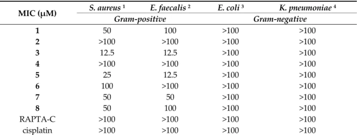
2.2.2. Antibacterial Effect of the Complexes 1‒8
The antibacterial activity of the complexes including the well‐known RAPTA‐C and cisplatin was studied on the Gram‐positive Staphylococcus aureus and Enterococcus faecalis and the Gram‐negative Escherichia coli and Klebsiella pneumoniae strains. The minimum inhibitory concentration (MIC) values are presented in Table 2. All tested Ru complexes had no activity on the Gram‐negative bacteria (MIC > 100 μM), and PTA‐containing complexes 2, 4, 6, 8, RAPTA‐C as well as cisplatin were not active or had quite low ac‐
tivity against the Gram‐positive bacteria. In contrast, chlorido complexes (1, 3, 5, 7) dis‐
played clear antibacterial effect on the tested Gram‐positive bacteria. This finding agrees with our previous results on organoruthenium chlorido complexes with bromo substi‐
tuted 8‐hydroxyquinolines which also showed good antibacterial potential [23]. Herein, among all tested compounds complex 3 was found to be the most active on Gram‐positive organisms. This complex was considerably active on the methicil‐
lin‐resistant S. aureus (MRSA) strain (MIC: 12.5 μM), which is a human pathogen being
responsible for several difficult‐to‐treat hospital‐acquired infections [28]. The Gram‐positive and Gram‐negative bacteria possess different cell wall composition, to‐
gether with various expression of efflux pumps and membrane proteins, which may contribute to their altered susceptibility to investigated Ru complexes possessing differ‐
ent size, charge and solution chemical properties. In Gram‐negative bacteria, the uptake of antibacterial agents depends on the outer membrane (OM). This membrane is an asymmetric bilayer of lipopolysaccharides (LPS) and phospholipids with nonspecific porins and specific channels. In addition, Gram‐negative bacteria express efflux trans‐
porter proteins consisting of an inner‐membrane (IM) pump subunit, a periplasmic adaptor protein and an outer‐membrane channel. These transporters can expel structur‐
ally unrelated drugs from the bacteria cell. The presence of the outer and inner mem‐
branes together with tripartite efflux pumps may result in higher resistance towards an‐
tibacterial compounds compared to Gram‐positive bacteria.
Table 2. Antibacterial activity of the complexes on Gram‐positive and Gram‐negative bacterial strains. MIC: minimum inhibitory concentration.
MIC (μM) S. aureus 1 E. faecalis 2 E. coli 3 K. pneumoniae 4
Gram‐positive Gram‐negative
1 50 100 >100 >100
2 >100 >100 >100 >100
3 12.5 12.5 >100 >100
4 >100 >100 >100 >100
5 25 12.5 >100 >100
6 100 >100 >100 >100
7 50 50 >100 >100
8 50 100 >100 >100
RAPTA‐C >100 >100 >100 >100
cisplatin >100 >100 >100 >100
1 MRA ATCC 43300. 2 ATCC 29212. 3 AG100. 4 ATCC 49619.
2.2.3. Antichlamydia Activity of the Complexes 1‒8
Among the sexually transmitted diseases Chlamydia trachomatis‐related infections are the most common. This Gram‐negative bacterium can replicate only within a host cell.
The various serovars can cause pelvic inflammatory diseases and infertility or lympho‐
granuloma venereum [29,30]. The antichlamydia activity of the complexes 1‒8 and RAPTA‐C was assayed using human cervix carcinoma HeLa 229 cells. We would like to note that to the best of our knowledge this is the first study describing the effect of or‐
ganoruthenium(II) compounds against Chlamydia trachomatis. As a first step, the cyto‐
toxicity of the complexes was measured by MTT assay in this cell line, and based on the results (Figure S1), the maximum non‐toxic concentration of complexes was considered as 100 μM for 1‒4, 6 and RAPTA‐C, 50 μM for 7 and 8, and 25 μM for 5.
Then the Hela cells were infected with C. trachomatis at multiplicity of infection (MOI) 0.2, and were treated with different concentrations of the complexes for 24 h. After 48 h post infection, the cells were lysed and the chlamydial growth reducing effect of the complexes was evaluated by comparing the chlamydial genom concentration to that seen on untreated/infected HeLa cells. The C. trachomatis DNA concentration was measured by direct quantitative PCR (qPCR). The average cycle threshold (Ct) numbers were cal‐
culated and ‒Ct values are shown at the various complex concentrations in Figure 2 for 7 and 8 (and Figure S2 for the rest of the complexes). The maximum C. trachomatis growth corresponded to a DNA concentration of Ct ~19 value as detected by direct qPCR, as well. All of the complexes could inhibit the chlamydial growth on HeLa cells, but the MIC value is higher than 100 μM or higher than the maximum non‐toxic concentration.
The inhibition curves showed that the most effective complexes were 7 and 8
(Figures 2 and S2), as the growth difference between the treated and untreated cells was ca. 101‐ and ca. 155‐fold, respectively.
Since the growth‐related chlamydial DNA concentrations were measured by a qPCR method, the potential direct impact of the complexes on the DNA polymerase of the qPCR [31] was also tested (Figure S3). The obtained results suggest that there is no stim‐
ulation or inhibition during the qPCR.
Figure 2. Antibacterial effect of complexes 7 and 8 against Chlamydia trachomatis.
2.2.4. Antiviral Activity of the Complexes 1‒8
Human herpes simplex virus‐1 (HHSV‐1) and preferentially herpes simplex virus‐2 (HSV‐2) genital infections are common viral sexually transmitted infections. Besides the vesicular lesions of the urogenital and anal regions, human herpes simplex infections may lead to severe complications including encephalitis, meningitis and neonatal herpes infections [32]. The antiviral effect of complexes against HSV‐2 was screened using Vero cells (originally isolated from kidney epithelial cells) to host the growing viruses. First, the cytotoxicity of the complexes against Vero cells was assayed (Figure S4), and it was found that complexes 1‒4 and 7‒8 did not produce significant toxicity at any of the con‐
centrations used. Maximum cytotoxicity was observed at concentrations of 12.5 ‒ 100 μM for complexes 5 and 6, and 100 μM for RAPTA‐C, whereas 6.25 μM was considered as maximum non‐toxic concentration for 5 and 6, and 50 μM for RAPTA‐C. The maximum non‐toxic concentration of complexes 1‒4 and 7‒8 were 100 μM.
Then Vero cells were infected with HSV‐2 (at MOI 0.2) and were treated with the complexes for 24 h. After 24 h post infection, the cells were lysed and the virus yield re‐
ducing effect of the compounds was evaluated by comparing the yield to that seen on untreated Vero cells. The HSV‐2 DNA concentration was measured by direct qPCR. It should be noted that the possible direct impact of the complexes on the DNA polymerase of the qPCR was also monitored in Vero cells, and no stimulation or inhibition was found. The average ‒Ct values obtained at the various complex concentrations are shown in Figure 3 for 1‒4 and 7‒8 (and Figure S5 for the rest of the complexes). The maximum HSV‐2 growth corresponded to a DNA concentration of Ct ~14 value as detected by di‐
rect qPCR. Inhibition curves showed that 7 is the most potent complex (~7.89 (dCT = 21.89–14) qPCR cycles difference, which means the growth difference was ca. 237‐fold), whilst the other complexes did not inhibit the growth of the HSV‐2. The concentration of 7 that decreased the growth of HSV‐2 in the cells and the corresponding DNA content by 50% (IC50), increased the qPCR Ct value by approximately one cycle. Also, the complex 7 concentration that inhibited the HSV‐2 growth by 90% (IC90), raised the Ct value by ~3.32 cycles. In the case of complex 7 the IC50 was ~25 μM and IC90 was between 50–100 μM.
7 8
50 12.5 3.12 0.78 0.19 0.048 concentration / M
-20
-25
-Ctvalue
Figure 3. Antiviral effect of complexes 1‒4 and 7‒8 against herpes simplex virus‐2.
2.3. Solution Speciation of Complexes 1‒8
2.3.1. Solution Chemical Properties of β‐Diketone and 8‐Hydroxyquinoline Complexes 5‒8 The solution stability of the studied Ru(η6‐p‐cymene) complexes was conducted to reveal differences in their solution chemical properties, which may provide explanation for their significantly different biological activity. Solution speciation of numerous
Ru(η6‐p‐cymene) complexes has already been characterized previously among others in‐
cluding complexes of hydroxamates [33], acetylacetonates [34,35], hydroxy‐
(thio)pyr(id)ones [36] with O,O‐donor ligands, and 8‐hydroxyquinoline derivatives [37,38] or pyridinecarboxylic acids [39] with N,O‐donor set. General equilibrium pro‐
cesses which can take place in solution of the half‐sandwich [Ru(η6‐p‐cymene)(X,Y)(Z)]
complexes are shown in Scheme S1. The herein tested complexes 5 and 6 contain coor‐
dinated 8‐hydroxyquinolinato ligand CQ, while complexes 7 and 8 consist of the β‐diketonato ligand p‐Cl‐dkt (Scheme 1), for which the binding strength of these ligands is supposed to be significantly different based on the reported stability constants for analogous complexes. 8‐Hydroxyquinolines were found to form highly stable complexes with this organometallic cation [37,38], while Ru(η6‐p‐cymene) complexes of acety‐
lacetonates were characterized by fairly low stability resulting in the dissociation of the O,O‐ligand at pH 7.4 in diluted solution [34,34]. Therefore, the bidentate ligand CQ is suggested to be bound rather strongly in the complexes 5–6, while a higher extent of complex decomposition of complexes 7–8 bearing the O,O‐ligand p‐Cl‐dkt is probable.
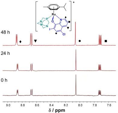
In order to confirm our prediction, time‐dependent UV‐visible (UV‐vis) spectra were recorded for complexes 5–8 at pH 7.4 in aqueous solution (in modified phosphate buffered saline (PBS) buffer—PBS’) and in Eagle’s minimum essential medium (EMEM) used for the cytotoxicity studies in the presence of 2% (v/v) dimethyl sulfoxide (DMSO).
No characteristic UV‐vis spectral changes within 24 h were observed for chlorido 5 and PTA complexes 6 with CQ ligand in PBS’. However, some precipitation occurred in case of 5 at the applied 50 μM concentration. Additionally, 1H‐NMR spectra were recorded for more water‐soluble complexes 6–8. The 1H‐NMR spectra of 6 showed no changes during 48 h (Figure 4). On the contrary, UV‐vis measurements for both 8‐hydroxyquinoline complexes 5–6 displayed slow changes in EMEM at the applied 150 μM concentrations (Figure S6). Complex 5 is involved in a relatively fast initial reaction taking place in a 30 min period, where most probably the chlorido co‐ligand is exchanged to a medium component (e.g., histidine). Then a similar slow process was seen for both complexes with the absorbance decrease at 425 nm and the appearance of a novel band at 560 nm, which might be the result of the partial loss of the arene ring followed by the oxidation of Ru(II) to Ru(III), as it was also reported for analogous 8‐hydroxyquinoline
Ru(η6‐p‐cymene) complexes [37]. Notably, no indication for arene loss was observed at
1 2 3 4 7 8
100 25 6.25 1.56 0.39 0.097 concentration / M -10
-15
-20
-25
-Ctvalue
higher concentration for complex 6 (500 μM) in EMEM according to the 1H NMR spec‐
troscopic measurements (not shown). UV‐vis spectral changes indicate decomposition of both p‐Cl‐dkt containing complexes 7–8 in PBS’ (160 μM), although the PTA co‐ligand in complex 8 significantly slowed down this process (Figure S7). The 1H‐NMR spectra rec‐
orded for complex 8 (c = 500 μM, Figure S8) also confirm only a slow transformation in this medium, namely the formation of the chlorido complex 7 and its partial, slow de‐
composition was found over time. For the complexes these findings confirm the superior solution stability of the 8‐hydroxyquinolino chlorido complex 5 over the acetylacetonato chlorido complex 7, which may explain the lower cytotoxicity and weaker ABCB1 mod‐
ulating activity of complex 7 compared to complex 5. On the one hand, the coordination of PTA stabilizes the complexes, which may result in slower co‐ligand displacement re‐
action in comparison to the chlorido complexes. Such kinetics is not beneficial in terms of the biological effect, as it may hinder the coordination of the donor atom of a target macromolecule. All of this is in good agreement with our previous finding for complexes 7 and 8, where it was reported that PTA delays replacement reactions with soft donor atoms from cellular targets like histidines, cysteines, or methionines and purine bases of DNA [19].
Figure 4. 1H‐NMR spectra of 6 at pH 7.4 (PBS’) in the low field region recorded for the fresh sample, after 24 h and 48 h waiting time. {T = 25.0 °C, ccomplex 6 = 0.5 mM; 10% (v/v) DMSO‐d6}.
2.3.2. Solution Chemical Properties of Pyrithione‐Type Complexes 1‒4
Stability constants for complexes 1‒4 formed with pyrithione (HPYR) and its ben‐
zo‐fused analogue HHiQT (Scheme 1) have not been reported yet. Therefore, we aimed to perform an in‐depth study on the solution speciation of complexes 1‒4 (Scheme 1), since among them 1 and 3 proved to have remarkable anticancer activity and antibacte‐
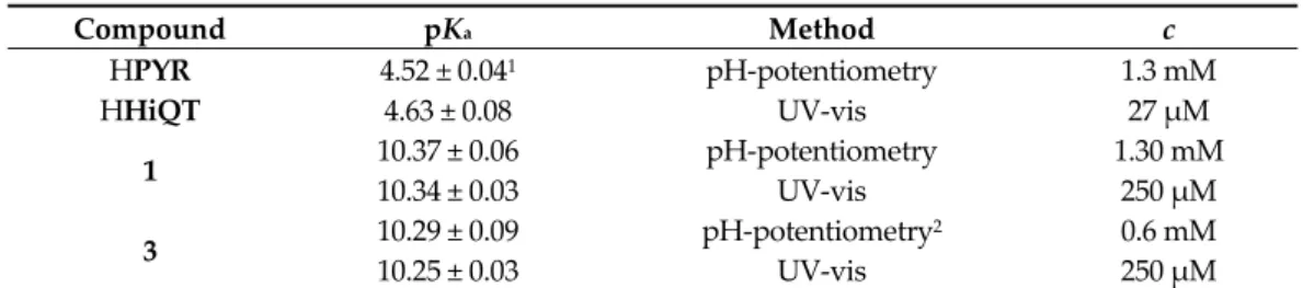
rial effect on Gram‐positive bacteria (Tables 1 and 2). The solution speciation studies of complexes 1‒4 were performed using UV‐vis spectrophotometry and pH‐potentiometry in the presence of 200 mM chloride ions. However, measurements were limited by the insufficient water solubility of ligand HHiQT and the oxygen‐sensitivity of both pyri‐
thiones. The pKa values determined for the ligands are shown in Table 3, where values for HPYR are in a good agreement with previously reported data [40]. In case of HHiQT, the pKa could be determined only by UV‐vis titrations under argon atmosphere, alt‐
hough, oxidation was observed at pH > 5.5 due to O2 traces in the sample.
● ■
♦ ▼
/ ppm 24 h
48 h
0 h
N P N N
Ru
+
O N I
● Cl
■
♦
▼
Table 3. pKa values of ligands HPYR and HHiQT and chlorido complexes 1 and 3 in pure water in the presence of 200 mM chloride ions. {T = 25.0 °C, I = 0.2 M KCl}.
Compound pKa Method c
HPYR 4.52 ± 0.041 pH‐potentiometry 1.3 mM
HHiQT 4.63 ± 0.08 UV‐vis 27 μM
1 10.37 ± 0.06 pH‐potentiometry 1.30 mM
10.34 ± 0.03 UV‐vis 250 μM
3 10.29 ± 0.09 pH‐potentiometry2 0.6 mM
10.25 ± 0.03 UV‐vis 250 μM
1 pKa = 4.49 (I = 0.20 M KCl) [40]. 2 Complex 3 has much better water solubility than its ligand HiQT.
In order to obtain information about the stability of the isolated complexes 1‒4, pH‐dependence of their UV‐vis spectra was monitored in the pH range 2‒11.5. In case of 1 and 3, UV‐vis spectra recorded at pH 2 revealed the formation of highly stable com‐
plexes as the spectra recorded for the metal precursor‒ligand mixture (1:1) were signifi‐
cantly different (for 1 see Figure 5). Then, the 1H‐NMR spectrum was recorded after 24 h where the pH of the solution of complex 1 was decreased to 1.0 to enforce complex dis‐
sociation. Figure S9 shows only a minor fraction of unbound Ru(η6‐p‐cymene) (<3%), which confirms the high solution stability of complex 1. UV‐vis spectra of complexes 2 and 4 stayed unchanged in pH range 2–11 during the monitored 24 h period (not shown).
On the contrary, spectra recorded for complexes 1 and 3 displayed significant changes in the basic pH range (for 1 see Figure 5). This process is fast and the appearance of isos‐
bestic points also suggest that these spectral changes are due to the coordination of hy‐
droxide ions replacing the co‐ligand (see Scheme S1 for the deprotonation of the aqua ligand). For this process, pKa values were calculated for both complexes from pH‐potentiometric and UV‐vis titration data (Table 3), which reflect that hydroxido spe‐
cies are not formed at physiological pH (notably, these pKa values are valid only in the presence of 200 mM chloride ions, thus, are considered as conditional constants). Com‐
plexes 1 and 3 were found to be stable at pH 7.4 in various media (e.g., phosphate buff‐
ered saline (PBS), RPMI‐1640 cell culture medium) and only minor release of p‐cymene was observed after 24 h in case of 1 and 3, respectively [21,41].
Figure 5. UV‐vis absorption spectra recorded for complex 1 in the presence of 200 mM chloride ions at various pH values (solid lines), in addition to the summed spectra of the unbound ligand pyrithi‐
one and [Ru(η6‐p‐cymene)(H2O)3]2+ at pH 2 (dashed line) using the same concentration of the com‐
plex, ligand and metal precursor in the samples. Inserted figure shows the absorbance values of complex 1 at 360 nm plotted against the pH. {T = 25.0 °C, I = 0.2 M KCl, ccomplex 1 = 250 μM; l = 1 cm}.
The high solution stability of 1 and 3 hindered the direct and accurate calculation of their formation constants, and a logK > 8 value is suggested on the basis of the pH‐potentiometric data. In order to compare the stability of 1 and 3 at physiological pH,
0.0 0.2 0.4 0.6 0.8 1.0 1.2
305 355 405 455 505
Absorbance
/ nm pH = 2.05 11.50
0.40 0.45 0.50 0.55
3 5 7 9 11
Abs. at 360 nm
pH
a serial dilution was made and UV‐vis spectra were recorded in a wide concentration range (10 μM–3 mM) (Figure S10). The calculated molar absorbance (ε) values change more significantly with decreasing the concentration in case of complex 1 suggesting the somewhat higher stability of its analogue complex 3 containing pyrithione with an ex‐
tended aromaticity. We attempted to determine the stability constants for 1 and 3 using 1,10‐phenantroline as a competitor ligand, although it failed due to the arene loss of the complexes (see details in Figure S11).
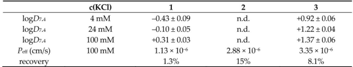
The lipophilicity of chlorido complexes 1 and 3 was characterized via the conven‐
tional shake‐flask method (PTA complexes 2 and 4 were found to be too lipophilic and could not be measured), and distribution coefficients (D7.4) are collected at pH 7.4 at dif‐
ferent chloride ion concentrations in Table 4. Chloride ion concentrations of 4, 24 and 100 mM were chosen according to the chloride content of the nucleus, cytosol and blood se‐
rum, respectively. The lipophilicity of both tested complexes is enhanced with increasing chloride ion concentrations, most probably due to higher fraction of the neutral chlorin‐
ated complexes over the positively charged aqua complexes. Complex 3 is more lipo‐
philic than 1 due to the presence of the additionally condensed benzene ring. Then par‐
allel artificial membrane permeability assay (PAMPA) was used to estimate the ability of the complexes to penetrate membranes by passive diffusion and the effective passive permeability coefficients (Peff) obtained for complexes 1‒3 are shown in Table 4 (in case of 4, there are no data due to formation of precipitate). The assay was performed at pH 7.4 in the presence of 100 mM chloride ion and it shows the permeability order: 3 > 2 > 1.
Therefore, both the conjugation of the benzene ring and the coordination of PTA increase the membrane permeability of the complexes. Despite higher lipophilicity and mem‐
brane permeability of PTA complexes in case of the acetylacetonato type p‐Cl‐dkt com‐
plexes (7, 8), higher accumulation of the chlorido complex 7 was reported [19].
Table 4. Log D7.4 (n‐octanol/water) and effective passive permeability values (Peff) of complexes 1–3 at pH = 7.4. {T = 25.0 °C, 20 mM phosphate buffer}.
c(KCl) 1 2 3
logD7.4 4 mM ‒0.43 ± 0.09 n.d. +0.92 ± 0.06
logD7.4 24 mM ‒0.10 ± 0.05 n.d. +1.22 ± 0.04
logD7.4 100 mM +0.31 ± 0.03 n.d. +1.37 ± 0.06
Peff (cm/s) 100 mM 1.13 × 10⁻⁶ 2.88 × 10⁻⁶ 3.35 × 10⁻⁶
recovery 1.3% 15% 8.1%
Since it was found that the chloride/water exchange has an impact on the lipophilicity of the complexes, we attempted to characterize this equilibrium process (Scheme S1) as it was done for other half‐sandwich organometallic complexes [37,38,42]. Unfortunately, the chloride/water exchange in these complexes was not accompanied by measurable changes in the UV‐vis spectra, unlike the PTA/Cl‒ exchange (Scheme S1). Thus, the possibility of the replacement of PTA in complexes 2 and 4 was monitored spectrophotometrically upon the addition of chloride ions. However, even the addition of a huge excess of chloride ion (>104) did not result in spectral changes, thus this halide ion cannot compete efficiently with PTA.
The reverse reaction was also followed (Figures S12 and S13), and practically the quantita‐
tive replacement of the chlorido ligand in complexes 1 and 3 was observed indicating the much stronger binding of PTA compared to Cl‒.
2.4. Interaction of Complexes 1–4 with Human Serum Albumin
Human serum albumin (HSA) is the most abundant protein in the blood and exerts an important role in the transport and distribution of exogenous and endogenous mole‐
cules. Additionally, binding of antitumor compounds to HSA is of considerable interest due to the enhanced permeability and retention effect [43]. Complexes 1‒4 were selected for a more detailed study. The binding of these complexes on HSA was monitored by UV‐vis spectrophotometry and fluorometry in a PBS’ containing 100 mM chloride ions
according to the blood serum. First, the binding was followed in time spectrophotomet‐
rically, and representative UV‐vis spectra were recorded at half equivalent of HSA as shown in Figure 6 for complexes 1 and 2 (complexes 3 and 4 behaved similarly to 1 and 2, respectively). The spectra recorded for 1 (Figure 6a) displayed a bi‐phasic binding profile, namely, a very fast process was followed by a much slower one. For the Ru(η6‐p‐cymene) triaqua cation, it was suggested that the binding is completed in the first step, and the subsequent slow and minor changes are due to some structural rearrangement of the coordination sphere around the metal center [44]. While, the spectra in case of 2 were almost unchanged in the presence of the protein (Figure 6b) suggesting that there is only a minimal measurable interaction between them during the monitored time (30 min) at the applied concentrations (100 μM complex, 50 μM HSA). Then, spectra were recorded for the complexes at various complex‐to‐HSA ratios (Figure 7a–c). In the case of the chlorido complexes titrations were done with HSA, due to the fast binding; otherwise, 24 h incubation time was applied (in case of PTA complexes). The spectra became constant after the addition of rather low equivalents of the protein (<1) indicating that more than one complex is bound on one HSA molecule. However, in case of 4 a much higher amount of HSA was needed to reach the constant spectra. Based on the spectral changes, the binding affinity of the complexes gives the following order: 3 > 1 > 4 > 2.
Figure 6. Time‐dependence of UV‐vis absorption spectra recorded for (a) complex 1 and (b) complex 2 in the presence of 0.5 equiv. HSA at pH = 7.4. {T = 25.0 °C, ccomplex = 100 μM, pH = 7.40 PBS’ buffer; l = 1 cm}.
Thus, the chlorido complexes are bound stronger (and faster) than the PTA con‐
taining compounds, as in the latter case the strongly bounded PTA should be replaced by the coordinating donor atom of the protein. Binding data of complexes 1 and 2 herein correlate well with data already published for these complexes to HSA and bovine serum albumine (BSA) by HPLC‐ICP‐MS [41] and AAS [16] methods, respecitvely, where chlo‐
rido complex 1 was bound to HSA/BSA in greater extent than PTA complex 2.
Organometallic half‐sandwich complexes are often bound to HSA via coordinative bounds [44,45] and His imidazoles are suggested as main binding sites among the po‐
tential donors of other residues such as Glu, Cys, Asp. In our previous work, the fast in‐
teraction of complexes 7 and 8 with thiol‐containing low molecular mass endogenous compounds Cys, N‐acetylcysteine and glutathione was found on the basis of a mass spectrometry study [46]. The donor atoms generally coordinate at the site of the mono‐
dentate co‐ligand, however, they can also displace the weakly bound bidentate ligand as well. Herein, interaction of the complexes 1–4 towards 1‐methylimidazole (mim), a monodentate binding model of HSA was measured by UV‐vis (Figures 7d–f).
0.00 0.05 0.10 0.15 0.20 0.25
330 380 430 480
Absorbance
/ nm 15 s - 30 min 0 s
0.00 0.05 0.10 0.15 0.20 0.25
330 380 430 480
Absorbance
/ nm 15 s
0 s
50 min
(a) (b)
Figure 7. UV‐vis absorption spectra recorded for (a,d) complex 1, (b,e) complex 3 and (c,f) complex 4 in the presence of various equivalents of HSA or mim, respectively at pH = 7.4. The numbers indicate the range of the cHSA/ccomplex or cmim/ccomplex ratios. In case of PTA complexes 24 h incubation time was used. {T = 25.0 °C, pH = 7.40 PBS’ buffer, ccomplex = 210
μM, cHSA= 6.3‒71.4 μM or cmim = 29‒210 μM (a,b,d,e); ccomplex = 116 μM, cHSA= 6.3‒71.4 μM or cmim = 29‒1160 μM with 8%
(v/v) DMSO (c,f); l = 1 cm}.
The reactions in all cases were very fast (except complex 2 and 4), and the observed spectral changes were similar to those obtained with HSA (Figures 7a–c). There was no indication for the replacement of the bidentate ligand by 1‐methylimidazole. Considering that the latter ligand binds at the coordination site of the co‐ligand, maximum one com‐
plex is able to be bound to 1‐methylimidazole. However, the constant spectra upon ad‐
dition of 1‐methylimidazole to the complex 4 could only be reached at high excess of 1‐methylimidazole due to its lower tendency to interact with this model (and with HSA as well). Based on these spectral changes, conditional binding constants were calculated for the formation of the 1‐methylimidazole adducts. LogK’ = 4.16 ± 0.45, 4.18 ± 0.27 and 3.64 ± 0.02 were obtained for complexes 1, 3 and 4, respectively (notably, complexes 1 and 3 were bound to 1‐methylimidazole almost quantitatively). These results also confirm stronger binding of the chlorido complexes over the PTA species. The protecting role of PTA in ligand‐exchange processes for the complex pair 7 and 8 was also reported re‐
garding their interaction with thiols or protein NCp7 [46,47].
Steady‐state spectrofluorometric measurements were also performed to assay the binding interactions at the binding pockets of HSA located in the IIA and IIIA subdo‐
mains. His residues are found nearby at both binding sites [48], and covalent binding is
(a)
(b)
(c)
0.0 0.4 0.8 1.2 1.6
300 400 500 600
Absorbance
/ nm complex
HSA:complex 0.03-0.34 0.0
0.4 0.8 1.2
300 400 500 600
Absorbance
/ nm complex
HSA:complex 0.05-0.34
0.03
0.0 0.4 0.8 1.2
300 400 500 600
Absorbance
/ nm complex
mim:complex 0.14-1.0
0.0 0.4 0.8 1.2 1.6
300 400 500 600
Absorbance
/ nm complex
mim:complex 0.17-0.87
0.0 0.4 0.8 1.2 1.6
300 400 500 600
Absorbance
/ nm complex HSA:complex
0.06-1.0
0.0 0.4 0.8 1.2 1.6
250 350 450 550
Absorbance
/ nm complex
mim:complex 0.25-10.0
(d)
(e)
(f)
1 1
3 3
4
4
possible here. Trp‐214 quenching and site marker displacement experiments were con‐
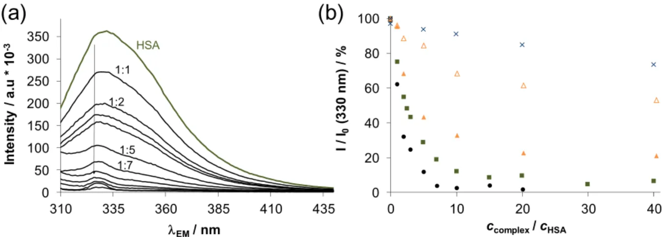
ducted as reported in our former works [44,49]. Warfarin (WF) was applied as site I marker and dansyl glycine (DG) was used for site II. Representative emission spectra for Trp‐214 quenching in case of 1 are shown in Figure 8a, and changes in the intensities at the emission maximum are compared for 1‒4 in Figure 8b. Based on the spectral changes conditional binding constants were computed for both of the quenching and site marker displacement assays (Table 5).
Figure 8. (a) Fluorescence emission spectra obtained for HSA titrated by complex 1 using λEX = 295 nm. (b) Intensity changes (%) at 330 nm at the various HSA‐to‐complex ratios for complex 1 (■), complex 2 (×), complex 3 (●) complex 4 (∆;
▲ (batch samples using 48 h equilibration time)). {T = 25.0 °C, pH = 7.40 PBS’ buffer, cHSA= 1 μM}.
As the binding of the chlorido complexes was found to be relatively fast, the quenching experiment was performed as titration; however, in case of complex 4 the batch method and 48 h equilibration time was used which provided a higher constant indicating that the displacement of PTA is a slow process. The logKQ’ quenching con‐
stants reveal the significantly stronger binding of complexes 3 and 1 compared to 4 and 2.
Complex 2 is hardly bound at site I, while binding of 3 and 1 is considered as a strong interaction here. Displacement experiments were done only with these strongly bound compounds, and the determined constants (Table 5) show that they interact at both binding sites of HSA.
Table 5. Conditional binding constants of the compounds at binding sites I and II of HSA deter‐
mined by spectrofluorometric Trp‐214 quenching (logKQ’) and site marker (WF or DG) displace‐
ment (logKWF’ and logKDG’) measurements. {pH = 7.40 PBS’ buffer; T = 25 °C}.
logKQ’ logKWF’ logKDG’
1 5.81 ± 0.03 (titration) 6.16 ± 0.03 5.80 ± 0.03
2 <4 (titration) ‒ ‒
3 6.18 ± 0.03 (titration) 5.98 ± 0.03 5.61 ± 0.03 4 4.46 ± 0.03
5.39 ± 0.03 (titration) (48 h) ‒ ‒
3. Materials and Methods 3.1. Chemicals
Chemicals and solvents used for the synthesis were purchased from commercial suppliers: Fluorochem (Hadfield, UK), Sigma Aldrich (St. Louis, MO, USA), Strem Chemicals, Inc. (Newburyport, MA, USA) and used as received, except for the phosphine ligand PTA, which was synthesized as reported [50]. [Ru(η6‐p‐cymene)(μ2‐Cl)Cl]2, HPYR, HHiQT, phen, mim, KCl, HCl, KOH, 4,4‐dimethyl‐4‐silapentane‐1‐sulfonic acid (DSS), NaH2PO4, Na2HPO4, KH2PO4, DMSO and HSA (A8763, essentially globulin free) were 0
50 100 150 200 250 300 350
310 335 360 385 410 435
Intensity / a.u * 10-3
EM/ nm HSA
1:1
1:5 1:2
1:7
(a) (b)
0 20 40 60 80 100
0 10 20 30 40
I / I0(330 nm) / %
ccomplex / cHSA