Article
Binding Models of Copper(II) Thiosemicarbazone Complexes with Human Serum Albumin: A Speciation Study
Nóra V. May1, Attila Jancsó2 andÉva A. Enyedy2,3,*
Citation: May, N.V.; Jancsó, A.;
Enyedy, É.A. Binding Models of Copper(II) Thiosemicarbazone Complexes with Human Serum Albumin: A Speciation Study.
Molecules2021,26, 2711. https://
doi.org/10.3390/molecules26092711
Academic Editor: Farkas Etelka
Received: 11 April 2021 Accepted: 3 May 2021 Published: 5 May 2021
Publisher’s Note:MDPI stays neutral with regard to jurisdictional claims in published maps and institutional affil- iations.
Copyright: © 2021 by the authors.
Licensee MDPI, Basel, Switzerland.
This article is an open access article distributed under the terms and conditions of the Creative Commons Attribution (CC BY) license (https://
creativecommons.org/licenses/by/
4.0/).
1 Centre for Structural Science, Research Centre for Natural Sciences, Magyar Tudósok Körútja 2, H-1117 Budapest, Hungary; may.nora@ttk.hu
2 Department of Inorganic and Analytical Chemistry, Interdisciplinary Excellence Centre, University of Szeged, Dóm tér 7, H-6720 Szeged, Hungary; jancso@chem.u-szeged.hu
3 MTA-SZTE Lendület Functional Metal Complexes Research Group, University of Szeged, Dóm tér 7, H-6720 Szeged, Hungary
* Correspondence: enyedy@chem.u-szeged.hu
Abstract:Copper(II) complexes of thiosemicarbazones (TSCs) often exhibit anticancer properties, and their pharmacokinetic behavior can be affected by their interaction with blood transport proteins.
Interaction of copper(II) complexes of an {N,N,S} donorα-N-pyridyl TSC (Triapine) and an {O,N,S}
donor 2-hydroxybenzaldehyde TSC (STSC) with human serum albumin (HSA) was investigated by UV–visible and electron paramagnetic resonance spectroscopy at physiological pH. Asp-Ala-His- Lys and the monodentate N-methylimidazole were also applied as binding models. Conditional formation constants were determined for the ternary copper(II)-TSC complexes formed with HSA, DAHK, and N-methylimidazole based on the spectral changes of both charge transfer and d-d bands.
The neutral N-methylimidazole displays a similar binding affinity to both TSC complexes. The partially negatively charged tetrapeptide binds stronger to the positively charged Triapine complex in comparison to the neutral STSC complex, while the opposite trend was observed for HSA, which demonstrates the limitations of the use of simple ligands to model the protein binding. The studied TSC complexes are able to bind to HSA in a fast process, and the conditional constants suggest that their binding strength is only weak-to-moderate.
Keywords:peptide model; albumin binding; EPR spectroscopy; ternary complexes
1. Introduction
Thiosemicarbazones (TSCs) show wide pharmacological versatility including antibac- terial, antifungal, antiviral, and antitumor activity. Among them, 3-aminopyridine-2- carboxaldehyde thiosemicarbazone (Triapine, Scheme1) is the best-known compound [1,2], and it has been tested in more than 30 phase I and II clinical trials in both solid and hema- tological tumors [3], and a phase III trial is recruiting patients to study the combination of Triapine with cisplatin during radiation [4]. Triapine is administered intravenously, while 4-(2-pyridinyl)-2-(6,7-dihydro-8(5H)-quinolinylid- ene)hydrazide (COTI-2) [5] and di-2- pyridylketone-4-cyclohexyl-4-methyl-3-TSC (DpC) [6] have entered clinical studies as oral drugs. The mechanism of action of anticancer TSCs is often linked to their interaction with endogenous metal ions such as iron and copper [1–3]. They can form metal chelates via the {N,S} donor set; however, more diversified binding modes can occur when an additional coordinating group is present in the TSC molecule. Tridentate {N,N,S} coordination mode is realized withα-N-pyridyl TSCs (e.g., Triapine), and the {O,N,S} binding motif occurs in complexes of 2-hydroxybenzaldehyde TSCs such as the simplest representative, the salicylaldehyde TSC (STSC, Scheme1) [3,7,8]. Numerous metal complexes of TSCs were developed and tested as potential anticancer agents, and copper(II) complexes gained a special interest since they often exhibit higher cytotoxicity, compared to their corresponding ligands [9–14]. Both Triapine and STSC form highly stable and redox-active complexes with
Molecules2021,26, 2711. https://doi.org/10.3390/molecules26092711 https://www.mdpi.com/journal/molecules
Molecules2021,26, 2711 2 of 15
copper(II) [15–18], and X-ray crystallography studies showed that the metal ion adopts a square planar coordination geometry with the tridentate binding of the TSC ligand and a chlorido co-ligand (or water) [9,18,19]. Based on the solution stability and structural studies, complexes with {N,N,S−} and {O−,N,S−} coordination modes predominate at physiological pH in the case of Triapine and STSC, respectively [15,16]. The co-ligands (Cl− or H2O) can be replaced by the donor atoms of endogenous compounds such as proteins in the biofluids, and interaction with human serum albumin (HSA) may have a strong influence on the pharmacokinetic properties of the copper(II) complexes.
Molecules 2021, 26, x FOR PEER REVIEW 2 of 15
since they often exhibit higher cytotoxicity, compared to their corresponding ligands [9–
14]. Both Triapine and STSC form highly stable and redox-active complexes with cop- per(II) [15–18], and X-ray crystallography studies showed that the metal ion adopts a square planar coordination geometry with the tridentate binding of the TSC ligand and a chlorido co-ligand (or water) [9,18,19]. Based on the solution stability and structural studies, complexes with {N,N,S−} and {O−,N,S−} coordination modes predominate at physiological pH in the case of Triapine and STSC, respectively [15,16]. The co-ligands (Cl− or H2O) can be replaced by the donor atoms of endogenous compounds such as proteins in the biofluids, and interaction with human serum albumin (HSA) may have a strong influence on the pharmacokinetic properties of the copper(II) complexes.
Scheme 1. Chemical structures of Triapine, STSC in their neutral forms, and their copper(II) complexes predominating at pH 7.4, and structures of the binding models (DAHK, mim).
HSA is the most abundant protein in the blood with an average concentration of 630 μM, and it greatly augments the transport capacity of serum due to its extraordinary binding affinity toward endogenous and exogenous compounds [20]. Binding to HSA affects the half-life of drugs, and this protein can act as a transport vehicle; thus, HSA is considered as a promising drug delivery system as well [20–22]. It should be noted that the microenvironment of malignant and inflamed tissues such as solid tumors often ex- hibits an increased level of HSA accumulation as a consequence of the enhanced per- meability and retention effect [21]. Fairly diverse scenarios are possible considering the binding modes on HSA and reaction rates for metallodrugs [23–25]. On the one hand, HSA has three main nonspecific binding pockets located in subdomains IIA, IIA, and IB, where compounds can bind via noncovalent bonds [23,24]. On the other hand, HSA has four partially selective metal-binding sites with well-defined metal preferences [25], and the N-terminal site (also known as ATCUN motif) consisting of the tripeptide sequence Asp-Ala-His can efficiently coordinate to copper(II) in a square planar geometry, with the participation of the N-terminal amine, histidine imidazole and two backbone amides [25]. However, copper(II) complexes with vacant coordination sites can bind to HSA via, e.g., accessible surface imidazole nitrogen donors of histidines as well. Coordination of His242 and Lys199 of HSA to the copper(II) complex of a tridentate Schiff base ligand bearing {O,N,O} donor set was reported by Guo et al. [22], and the adduct formation with the protein increased the anticancer activity of the complex. Yang and Liang developed various copper(II)-α-N-pyridinyl TSC-HSA adducts [26,27], which exhibited better se- lectivity and improved capacity of inhibiting tumor growth in in vivo (mice) tests in comparison to the copper(II)-TSC complexes alone. On the basis of X-ray crystallography analysis of the complex–protein adducts, coordination of His nitrogen donors (His242 and His146) was proved [26,27].
Although the binding affinity of HSA toward copper(II) ions was already reported, and conditional constant was determined by Bal et al. at physiological pH [28], no equi- librium constants are available for the binding of copper(II)-TSC complexes to HSA. In
Triapine
N N H NH2
S OH
STSC N
N N H
S NH2 NH2
N
N N S-
NH2 NH2
Cu H2O
N N S-
NH2
Cu OH2
O-
[CuA]+complex
formed with Triapine [CuA] complex formed with STSC
H2N CH C CH2
O
C OH
O
HN CH C CH3
O H
N CH C CH2
O
N NH
HN CH C CH2
OH O
CH2
CH2
CH2
NH2 Asp-Ala-His-Lys (DAHK)
N N
N-methylimidazole (mim)
Scheme 1.Chemical structures of Triapine, STSC in their neutral forms, and their copper(II) complexes predominating at pH 7.4, and structures of the binding models (DAHK, mim).
HSA is the most abundant protein in the blood with an average concentration of 630µM, and it greatly augments the transport capacity of serum due to its extraordinary binding affinity toward endogenous and exogenous compounds [20]. Binding to HSA affects the half-life of drugs, and this protein can act as a transport vehicle; thus, HSA is considered as a promising drug delivery system as well [20–22]. It should be noted that the microenvironment of malignant and inflamed tissues such as solid tumors often exhibits an increased level of HSA accumulation as a consequence of the enhanced permeability and retention effect [21]. Fairly diverse scenarios are possible considering the binding modes on HSA and reaction rates for metallodrugs [23–25]. On the one hand, HSA has three main nonspecific binding pockets located in subdomains IIA, IIA, and IB, where compounds can bind via noncovalent bonds [23,24]. On the other hand, HSA has four partially selective metal-binding sites with well-defined metal preferences [25], and the N-terminal site (also known as ATCUN motif) consisting of the tripeptide sequence Asp- Ala-His can efficiently coordinate to copper(II) in a square planar geometry, with the participation of the N-terminal amine, histidine imidazole and two backbone amides [25].
However, copper(II) complexes with vacant coordination sites can bind to HSA via, e.g., accessible surface imidazole nitrogen donors of histidines as well. Coordination of His242 and Lys199 of HSA to the copper(II) complex of a tridentate Schiff base ligand bearing {O,N,O} donor set was reported by Guo et al. [22], and the adduct formation with the protein increased the anticancer activity of the complex. Yang and Liang developed various copper(II)-α-N-pyridinyl TSC-HSA adducts [26,27], which exhibited better selectivity and improved capacity of inhibiting tumor growth in in vivo (mice) tests in comparison to the copper(II)-TSC complexes alone. On the basis of X-ray crystallography analysis of the complex–protein adducts, coordination of His nitrogen donors (His242 and His146) was proved [26,27].
Although the binding affinity of HSA toward copper(II) ions was already reported, and conditional constant was determined by Bal et al. at physiological pH [28], no equilibrium constants are available for the binding of copper(II)-TSC complexes to HSA. In this work, we investigated the interaction of copper(II) complexes of Triapine and STSC (Scheme1) with
HSA and two His-containing simpler ligands by pH-potentiometry, UV–visible (UV–vis) spectrophotometry, and electron paramagnetic resonance (EPR) spectroscopy to reveal the binding strength and rates, namely, the tetrapeptide Asp-Ala-His-Lys (DAHK) consisting of the native HSA sequence for copper(II) binding and the monodentate N-methylimidazole (mim) were selected as binding models (Scheme1).
2. Results and Discussion
2.1. Solution Speciation in the Binary Systems
Thiosemicarbazones and their metal complexes are generally characterized by insuf- ficient water solubility, and this is the reason why solution equilibrium studies are often performed in mixtures of water and an organic solvent with a relatively lower dielectric constant. The proton dissociation processes of Triapine and STSC and their complex forma- tion with copper(II) ions were characterized in a 30% (w/w) DMSO/H2O solvent mixture in our previous works [15,16], and this medium was also used for the speciation studies pre- sented here. Triapine forms high stability mono-ligand [CuHA]2+, [CuA]+and [CuA(OH)]
complexes (in which A−is the completely deprotonated form of the ligand), and [CuA]+ predominates at pH 7.4 characterized by a {Npyridine,N,S−}{H2O} binding mode (Scheme1).
In this species, the negative charge is localized on the sulfur atom due to the thione–thiol tautomeric equilibrium following the loss of the proton from the hydrazonic-Natom [15].
Notably, at ligand excess, bis-ligand complexes and the dinuclear species [Cu2A3]+are also formed [15]. On the contrary, STSC forms exclusively mono complexes and the predomi- nant species is also [CuA] (Scheme1) at physiological pH; however, in this case, the ligand is coordinated in its dianionic form with an {O−,N,S−} donor set [16]. It is noteworthy that DMSO weakly coordinates to the copper(II) ions, and as a consequence, the complexes of Triapine, 2-formylpyridine thiosemicarbazone, and their N-terminally dimethylated derivatives were characterized by somewhat higher solution stabilities in pure water in comparison to 30% (w/w) DMSO/H2O [29,30].
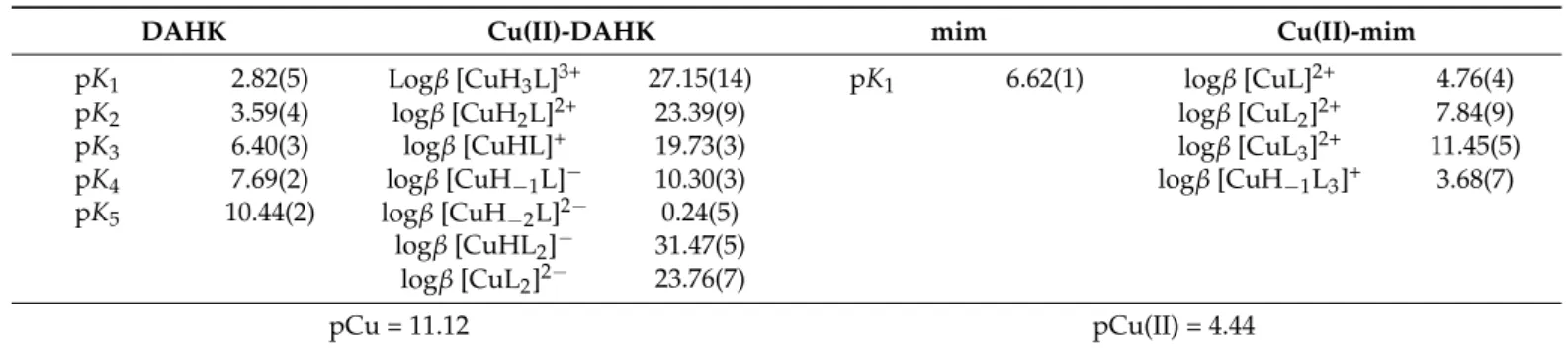
The pKavalues of DAHK and N-methylimidazole (Scheme1) and the overall stability constants of their copper(II) complexes were determined by pH-potentiometric titrations (Table1). DAHK possesses five dissociable protons; however, the dissociation processes overlap resulting in difficulties in the assignment of the pKavalues to the functional groups;
thus, they are considered as macroscopic constants. Based on chemical evidence and data reported for related tri- and tetrapeptides (Asp-Ala-His-NH2, Asp-Ala-His-Lys-NH2) in pure water [31,32], the two lowest pKavalues are assigned to the carboxyl groups of Asp and Lys residues, pK3belongs to the histidine-nitrogen, while pK4and pK5to the N-terminal and the side-chain Lys amino groups, respectively. In the copper(II)-DAHK system formation of mainly mono complexes in different protonation states was found (Table1), similarly to the speciation model obtained for copper(II)-Asp-Ala-His-Lys-NH2
reported by Bal et al. [31]. The C-terminal amide group of Asp-Ala-His-Lys-NH2is not involved in the coordination, and the same probably holds for the C-terminal COOH of DAHK; thus, the speciation models and the overall stability constants can be compared with each other. Bal et al. found four copper(II)-bound nitrogen donors in the square planar [CuH−2L]−dominating in a wide pH range including the physiological pH (L−is the completely deprotonated form of the ligand). These are the N-terminal amino group, the amide nitrogen atoms located between Asp and Ala and between Ala and His, and one His nitrogen [31].
Complexes [CuH3L]3+, [CuH2L]2+, [CuHL]+, [CuH−1L]−, and [CuH−2L]2− were observed with DAHK in the 30% (w/w) DMSO/H2O solvent mixture. On the basis of the overall stability constants, concentration distribution curves were calculated (Figure1), showing the formation of [CuH3L]3+, [CuH2L]2+, and [CuHL]+in the acidic pH range, and [CuH−1L]−becomes the predominating species at pH > 5.5, while [CuH−2L]2−forms in the alkaline medium. Due to the close similarity between the complexation scheme of DAHK and Asp-Ala-His-Lys-NH2[31], the coordination of four nitrogen donors (Asp-N, amide-N of Asp-Ala, amide-N of Ala-His residues, and His-N) can be assumed in the
Molecules2021,26, 2711 4 of 15
complex [CuH−1L]−. It should be noted that at ligand excess bis complexes are also present. Additionally, the conditional stability constant for the DAHK complex was also calculated at pH 7.4 in 30% (w/w) DMSO/H2O, and the obtained value is logK’7.4= 14.2, which shows an acceptable agreement with values 13.6 and 13.7 (in pure water) reported for DAHK by isothermal titration calorimetry [32] and for DAHK-NH2obtained by pH- potentiometry [31], respectively.
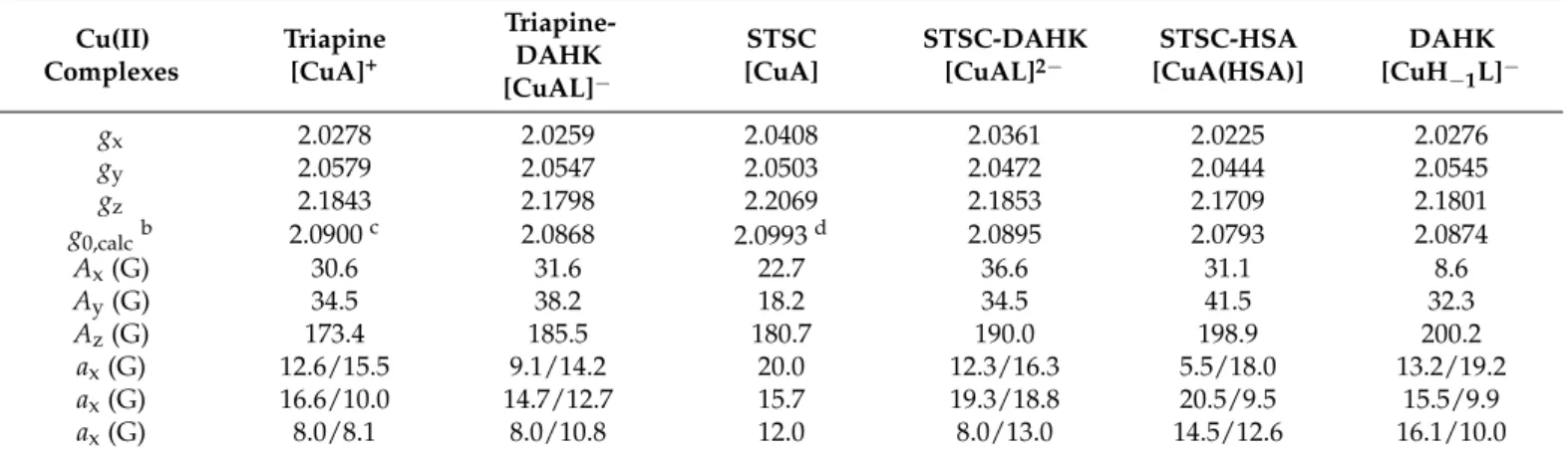
Table 1. pKavalues of DAHK and mim, overall stability constants (logβ) of their copper(II) complexes in 30% (w/w) DMSO/H2O,aand pCu valuesb(calculated at pH 7.4 atcCu(II)=cligand= 100µM). {T= 25#C,I= 0.10 M (KCl)}.
DAHK Cu(II)-DAHK mim Cu(II)-mim
pK1 2.82(5) Logβ[CuH3L]3+ 27.15(14) pK1 6.62(1) logβ[CuL]2+ 4.76(4)
pK2 3.59(4) logβ[CuH2L]2+ 23.39(9) logβ[CuL2]2+ 7.84(9)
pK3 6.40(3) logβ[CuHL]+ 19.73(3) logβ[CuL3]2+ 11.45(5)
pK4 7.69(2) logβ[CuH−1L]− 10.30(3) logβ[CuH−1L3]+ 3.68(7)
pK5 10.44(2) logβ[CuH−2L]2− 0.24(5) logβ[CuHL2]− 31.47(5)
logβ[CuL2]2− 23.76(7)
pCu = 11.12 pCu(II) = 4.44
aUncertainties (SD) of the last digits are in parenthesis.bpCu calculated for Triapine: 7.79, for STSC: 9.40 (in 30% (w/w) DMSO/H2O, 0.1 M KCl) and for HSA: 8.00 (in 0.1 M NaCl, pure water) 7.70 (in 0.1 M NaCl, 0.1 M 2-[4-(2-hydroxyethyl)piperazin-1-yl]ethanesulfonic acid (HEPES) buffered pure water) based on data in Refs. [15,16,28].
Molecules 2021, 26, x FOR PEER REVIEW 4 of 15
medium. Due to the close similarity between the complexation scheme of DAHK and Asp-Ala-His-Lys-NH2 [31], the coordination of four nitrogen donors (Asp-N, amide-N of Asp-Ala, amide-N of Ala-His residues, and His-N) can be assumed in the complex [CuH−1L]−. It should be noted that at ligand excess bis complexes are also present. Addi- tionally, the conditional stability constant for the DAHK complex was also calculated at pH 7.4 in 30% (w/w) DMSO/H2O, and the obtained value is logK’7.4 = 14.2, which shows an acceptable agreement with values 13.6 and 13.7 (in pure water) reported for DAHK by isothermal titration calorimetry [32] and for DAHK-NH2 obtained by pH-potentiometry [31], respectively.
Table 1. pKa values of DAHK and mim, overall stability constants (logβ) of their copper(II) complexes in 30% (w/w) DMSO/H2O,a and pCu values b (calculated at pH 7.4 at cCu(II) = cligand = 100 μM). {T = 25 ○C, I = 0.10 M (KCl)}.
DAHK Cu(II)‒DAHK mim Cu(II)‒mim
pK1 2.82(5) Logβ [CuH3L]3+ 27.15(14) pK1 6.62(1) logβ [CuL]2+ 4.76(4)
pK2 3.59(4) logβ [CuH2L]2+ 23.39(9) logβ [CuL2]2+ 7.84(9)
pK3 6.40(3) logβ [CuHL]+ 19.73(3) logβ [CuL3]2+ 11.45(5)
pK4 7.69(2) logβ [CuH−1L]− 10.30(3) logβ [CuH−1L3]+ 3.68(7) pK5 10.44(2) logβ [CuH−2L]2− 0.24(5)
logβ [CuHL2]− 31.47(5) logβ [CuL2]2− 23.76(7)
pCu = 11.12 pCu(II) = 4.44
a Uncertainties (SD) of the last digits are in parenthesis. b pCu calculated for Triapine: 7.79, for STSC: 9.40 (in 30% (w/w) DMSO/H2O, 0.1 M KCl) and for HSA: 8.00 (in 0.1 M NaCl, pure water) 7.70 (in 0.1 M NaCl, 0.1 M 2-[4-(2-hydroxyethyl)piperazin-1-yl]ethanesulfonic acid (HEPES) buffered pure water) based on data in Refs. [15,16,28].
Figure 1. Concentration distribution curves for the copper(II)‒DAHK (1:1) system. {cDAHK = cCu(II) = 1 mM; 30% (w/w) DMSO/H2O; T = 25 °C; I = 0.10 M (KCl)}.
For N-methylimidazole (Scheme 1), only one pKa (6.62) was determined experi- mentally, attributed to the imidazolium-NH+ moiety (Table 1), and it is somewhat lower than the value reported in pure water (pKa = 7.05 [33]) since the neutral form of the ligand (L) is better solvated in the presence of DMSO. Complexes with various metal-to-ligand ratios, i.e., [CuL]2+, [CuL2]2+, [CuL3]2+, and a mixed hydroxido species [CuH−1L]+ (Table 1), were observed for this monodentate ligand. The calculated EPR parameters are found in Table S1. Notably, the formation of [CuL4]2+ is suggested (instead of [CuH−1L]+) based on the recorded frozen solution EPR spectra (Figure S1).
In order to compare the affinity of the ligands toward copper(II), pCu (‒log[Cu(II)]) values were calculated for Triapine, STSC, DAHK, and N-methylimidazole at pH 7.4 (Table 1). These data reveal the strongest copper(II) binding property of the tetradentate DAHK, and the weakest for the monodentate mim, as was expected.
2.2. Interaction of the Copper(II)-TSC Complexes with HSA, DAHK, and N-Methylimidazol
0 20 40 60 80 100
2 4 6 8 10
% Cu(II)
pH
Cu2+ [CuH-1L]-
[CuH-2L]2- [CuH3L]3+
[CuHL]+ [CuH2L]2+
Figure 1.Concentration distribution curves for the copper(II)-DAHK (1:1) system. {cDAHK=cCu(II)= 1 mM; 30% (w/w) DMSO/H2O;T= 25◦C;I= 0.10 M (KCl)}.
For N-methylimidazole (Scheme1), only one pKa(6.62) was determined experimen- tally, attributed to the imidazolium-NH+ moiety (Table1), and it is somewhat lower than the value reported in pure water (pKa = 7.05 [33]) since the neutral form of the ligand (L) is better solvated in the presence of DMSO. Complexes with various metal-to- ligand ratios, i.e., [CuL]2+, [CuL2]2+, [CuL3]2+, and a mixed hydroxido species [CuH−1L]+ (Table1), were observed for this monodentate ligand. The calculated EPR parameters are found in Table S1. Notably, the formation of [CuL4]2+is suggested (instead of [CuH−1L]+) based on the recorded frozen solution EPR spectra (Figure S1).
In order to compare the affinity of the ligands toward copper(II), pCu (-log[Cu(II)]) values were calculated for Triapine, STSC, DAHK, and N-methylimidazole at pH 7.4 (Table1). These data reveal the strongest copper(II) binding property of the tetradentate DAHK, and the weakest for the monodentate mim, as was expected.
2.2. Interaction of the Copper(II)-TSC Complexes with HSA, DAHK, and N-Methylimidazol Since the donor atoms occupy only three coordination sites in the copper(II) complexes of Triapine and STSC (Scheme1), there are vacant sites where the donor atoms of HSA and its simple binding models can coordinate. In order to characterize the binding affinity
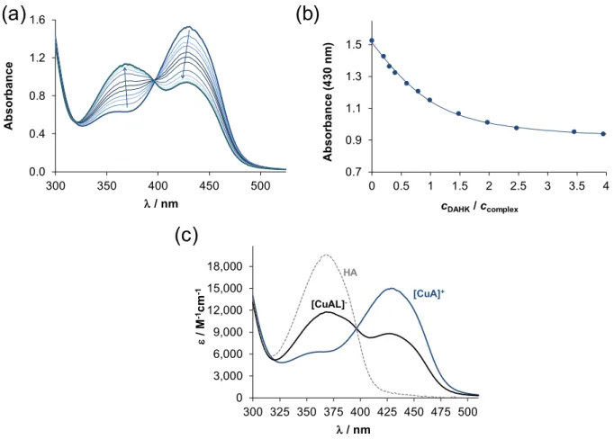
of the protein, DAHK, and N-methylimidazole to these copper(II)-TSC complexes, condi- tional binding constants were determined. The copper(II)-TSC-DAHK/N-methylimidazole ternary systems were attempted to be studied first by pH-potentiometric titrations apply- ing mM concentration of the components. However, precipitate formation at pH > 6.5 in all systems and slow equilibrium processes with DAHK prevented the execution of titrations. Therefore, formation constants for the ternary complexes were determined at pH 7.4 in 30% (w/w) DMSO/H2O by UV–vis spectrophotometry using lower concentra- tions, and the spectra were always recorded after a proper equilibration time based on the preliminary time-dependence assays. The reaction between the copper(II)-TSC complex and the oligopeptide DAHK was found to be relatively slow, since at least 30 min was necessary to reach the equilibrium, and thus, a minimum 4 h waiting time was utilized. By increasing the DAHK content of the samples, significant spectral changes were observed (as an example, see spectra for the Triapine complexes in Figure2a), and the changes ended at ca. four equivalents of the peptide (Figure2b).
Molecules 2021, 26, x FOR PEER REVIEW 6 of 15
Figure 2. (a) UV–vis spectra recorded for the copper(II)‒Triapine‒DAHK (1:1:x) system at pH 7.4 after 4 h equilibration time (x:0–4). (b) Absorbance values at 430 nm plotted against the ratio of DAHK and the copper(II)-Triapine complex. (c) Molar absorbance spectra of the Triapine complex ([CuA]+), the Triapine-DAHK ternary complex ([CuAL]−), and the unbound Triapine molecule (HA). {cCu(II) = cTriapine = 200 μM, cDAHK = 0‒800 μM; 30% (w/w) DMSO/H2O; pH = 7.4 (20 mM HEPES); T = 25 °C; I = 0.10 M (KCl); ℓ = 0.5 cm}.
Table 2. Anisotropic EPR parameters calculated for the various species formed in the binary copper(II)‒
Triapine/STSC/DAHK and ternary copper(II)‒Triapine/STSC (A)‒DAHK (L)/HSA systems in 30% (w/w) DMSO/H2O at pH 7.4 (20 mM HEPES).a {I = 0.10 M (KCl)}.
Cu(II) Complexes
Triapine [CuA]+
Triapine-DAHK [CuAL]−
STSC [CuA]
STSC-DAHK [CuAL]2−
STSC-HSA [CuA(HSA)]
DAHK [CuH−1L]−
gx 2.0278 2.0259 2.0408 2.0361 2.0225 2.0276
gy 2.0579 2.0547 2.0503 2.0472 2.0444 2.0545
gz 2.1843 2.1798 2.2069 2.1853 2.1709 2.1801
g0,calc b 2.0900 c 2.0868 2.0993 d 2.0895 2.0793 2.0874
Ax (G) 30.6 31.6 22.7 36.6 31.1 8.6
Ay (G) 34.5 38.2 18.2 34.5 41.5 32.3
Az (G) 173.4 185.5 180.7 190.0 198.9 200.2
ax (G) 12.6/15.5 9.1/14.2 20.0 12.3/16.3 5.5/18.0 13.2/19.2
ax (G) 16.6/10.0 14.7/12.7 15.7 19.3/18.8 20.5/9.5 15.5/9.9
ax (G) 8.0/8.1 8.0/10.8 12.0 8.0/13.0 14.5/12.6 16.1/10.0
a The experimental errors are ±0.001 for g, ± 1 G for A and aN tensor values. b Isotropic values of the g tensor were calcu- lated via equation: g0 = (gx + gy + gz)/3. c g0 = 2.0958 (obtained from room temperature measurement) [15]. d g0 = 2.0945 (room temperature) [16].
(a) (b)
(c)
0.0 0.4 0.8 1.2 1.6
300 350 400 450 500
Absorbance
λ/ nm
0.7 0.9 1.1 1.3 1.5
0 0.5 1 1.5 2 2.5 3 3.5 4
Absorbance(430 nm)
cDAHK/ ccomplex
,0 3,000 6,000 9,000 12,000 15,000 18,000
300 325 350 375 400 425 450 475 500 ε/ M-1cm-1
λ/ nm
[CuA]+ [CuAL]-
HA
Figure 2.(a) UV–vis spectra recorded for the copper(II)-Triapine-DAHK (1:1:x) system at pH 7.4 after 4 h equilibration time (x:0–4). (b) Absorbance values at 430 nm plotted against the ratio of DAHK and the copper(II)-Triapine complex. (c) Molar absorbance spectra of the Triapine complex ([CuA]+), the Triapine-DAHK ternary complex ([CuAL]−), and the unbound Triapine molecule (HA). {cCu(II)=cTriapine= 200µM,cDAHK= 0-800µM; 30% (w/w) DMSO/H2O; pH = 7.4 (20 mM HEPES);
T= 25◦C;I= 0.10 M (KCl);`= 0.5 cm}.
The appearance of clear-cut isobestic points suggests a single equilibrium process, which is most probably the formation of a copper(II)-Triapine-DAHK ternary complex. On the other hand, the absorbance decrease at 430 nm and the increase at 368 nm might be the consequence of the displacement of the original TSC ligand by the stronger copper(II) binder DAHK (see pCu values in Table1) instead of the formation of a ternary complex.
However, the final spectrum recorded for the ternary system at the applied highest excess of DAHK is rather different from the spectrum of the free Triapine (Figure2c) suggesting the
Molecules2021,26, 2711 6 of 15
formation of mixed-ligand species (notably, DAHK and its copper(II) complex have no or negligible characteristic absorption bands in the monitored wavelength range (Figure S2)).
The S−→Cu2+charge transfer (CT) band located at 430 nm displays significant changes indicating a rearrangement in the coordination sphere. Similar but somewhat smaller spectral changes were observed for the STSC complex (Figure S2). Additionally, UV–vis spectra were recorded in the wavelength range of d-d transitions (not shown) using a longer path length (5 cm). Theλmaxvalue observed in this wavelength range in the absence of the peptide shifted toward lower wavelength values upon the addition of DAHK (625 nm→ 584 nm (Triapine), 583 nm→540 nm (STSC)).
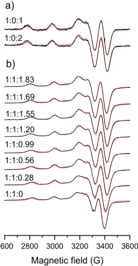
In order to confirm the formation of ternary complexes anisotropic EPR spectra were recorded for the copper(II)-TSC, copper(II)-DAHK, and the copper(II)-TSC-DAHK ternary systems (for Triapine complexes see Figure3, and for STSC Figure S3). EPR parameters were calculated for the various species formed in the binary and ternary systems via the deconvolution of the spectra (Table2). Analysis of these data indicates that the species formed in the ternary systems possess different EPR parameters than the copper(II)-DAHK complex ([CuH−1L]−), considering especiallyAx andAz. Upon the interaction of the copper(II)-TSC complexes with DAHK, thegfactor is decreased in general, while theA value is increased suggesting a stronger ligand field due to the coordination of an additional donor atom.
Molecules 2021, 26, x FOR PEER REVIEW 7 of 15
2600 2800 3000 3200 3400 3600 Magnetic field (G)
1:1:0 1:1:0.28 1:1:0.56 1:1:0.99 1:1:1.20 1:1:1.55 1:1:1.69 1:1:1.83
a)
b)
1:0:1 1:0:2
Figure 3. Experimental (black) and simulated (red) anisotropic EPR spectra recorded at 77 K for the (a) copper(II)‒DAHK and (b) copper(II)‒Triapine‒DAHK systems at pH 7.4 after 4 h equilibration time. Numbers above the spectra indicate the copper(II):Triapine:DAHK ratios. {cCu(II) = cTriapine = 495 μM, cDAHK = 0–908 μM; 30% (w/w) DMSO/H2O; pH = 7.4 (20 mM HEPES); I = 0.10 M (KCl)}.
The interaction of the binary complexes of Triapine and STSC with the monodentate N-methylimidazole was also followed by UV–vis spectrophotometry. The reaction was found to be fairly fast (<5 min), and spectral changes were also observed (Figure 4).
However, the changes in the visible range were much more pronounced than in the wavelength range of the CT bands (c.f. Figure 4a,b). In order to achieve a complete spec- tral change a much higher excess of N-methylimidazole was needed (>10) in comparison to DAHK; however, two overlapping processes were observed (Figure 4c).
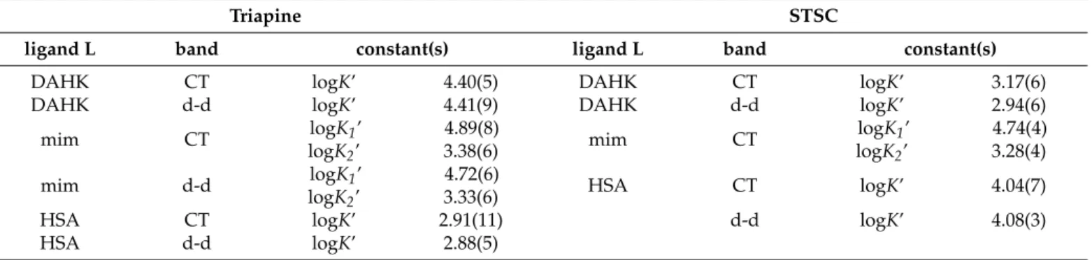
Based on the recorded UV–vis spectra conditional formation constants (logK’) were calculated for the ternary complexes formed with DAHK and N-methylimidazole cov- ering both wavelength ranges (CT and d-d bands) (Table 3) (see representative molar absorbance spectra in Figures 2c and 4d). The constants obtained from the two different wavelength ranges were in good agreement with each other. The obtained results indi- cate that only one DAHK, compared to two N-methylimidazole ligands, can coordinate to the binary TSC complex. These equilibrium constants reflect the similar binding strength of the neutral N-methylimidazole ligand to both TSC complexes. On the con- trary, the binding affinity of the DAHK peptide, which is partially negatively charged at pH 7.4 (32% HL−, 60% H2L, 8% H3L+), is somewhat stronger to the positively charged [CuA]+ Triapine complex, compared to the neutral [CuA] species of STSC. In these ter- nary complexes, N-methylimidazole can coordinate via only the imidazole-nitrogen, while DAHK migth coordinate in a bidentate fashion (e.g., via imidazole and terminal amine nitrogen donors forming a macrochelate) (Scheme S1), which leads to the changes of the S− → Cu2+ CT band, especially in the copper(II)-Triapine-DAHK complex.
Figure 3.Experimental (black) and simulated (red) anisotropic EPR spectra recorded at 77 K for the (a) copper(II)-DAHK and (b) copper(II)-Triapine-DAHK systems at pH 7.4 after 4 h equilibration time. Numbers above the spectra indicate the copper(II):Triapine:DAHK ratios. {cCu(II)=cTriapine= 495µM,cDAHK= 0–908µM; 30% (w/w) DMSO/H2O; pH = 7.4 (20 mM HEPES);I= 0.10 M (KCl)}.
The interaction of the binary complexes of Triapine and STSC with the monodentate N- methylimidazole was also followed by UV–vis spectrophotometry. The reaction was found to be fairly fast (<5 min), and spectral changes were also observed (Figure4). However, the changes in the visible range were much more pronounced than in the wavelength range of the CT bands (c.f.Figure4a,b). In order to achieve a complete spectral change a much
higher excess of N-methylimidazole was needed (>10) in comparison to DAHK; however, two overlapping processes were observed (Figure4c).
Table 2. Anisotropic EPR parameters calculated for the various species formed in the binary copper(II)- Triapine/STSC/DAHK and ternary copper(II)-Triapine/STSC (A)-DAHK (L)/HSA systems in 30% (w/w) DMSO/H2O at pH 7.4 (20 mM HEPES).a{I= 0.10 M (KCl)}.
Cu(II) Complexes
Triapine [CuA]+
Triapine- DAHK [CuAL]−
STSC [CuA]
STSC-DAHK [CuAL]2−
STSC-HSA [CuA(HSA)]
DAHK [CuH−1L]−
gx 2.0278 2.0259 2.0408 2.0361 2.0225 2.0276
gy 2.0579 2.0547 2.0503 2.0472 2.0444 2.0545
gz 2.1843 2.1798 2.2069 2.1853 2.1709 2.1801
g0,calcb 2.0900c 2.0868 2.0993d 2.0895 2.0793 2.0874
Ax(G) 30.6 31.6 22.7 36.6 31.1 8.6
Ay(G) 34.5 38.2 18.2 34.5 41.5 32.3
Az(G) 173.4 185.5 180.7 190.0 198.9 200.2
ax(G) 12.6/15.5 9.1/14.2 20.0 12.3/16.3 5.5/18.0 13.2/19.2
ax(G) 16.6/10.0 14.7/12.7 15.7 19.3/18.8 20.5/9.5 15.5/9.9
ax(G) 8.0/8.1 8.0/10.8 12.0 8.0/13.0 14.5/12.6 16.1/10.0
aThe experimental errors are±0.001 forg,±1 G for A andaNtensor values.bIsotropic values of the g tensor were calculated via equation:
g0= (gx+gy+gz)/3.cg0= 2.0958 (obtained from room temperature measurement) [15].dg0= 2.0945 (room temperature) [16].
Molecules 2021, 26, x FOR PEER REVIEW 8 of 15
Figure 4. UV–vis spectra recorded for the copper(II)‒Triapine‒N-methylimidazole (mim) system at pH 7.4 after 5 min equilibration time in the wavelength range of the (a) CT bands and the (b) d-d bands at various equivalents of mim. (c) Absorbance values at 580 nm plotted against the ratio of mim and the copper(II)-Triapine complex obtained from the d-d range of the UV–vis spectra. (d) Calculated molar absorbance spectra of the Triapine complex ([CuA]+), the Triapine-mim ternary complexes ([CuAL]+ and [CuAL2]+). {30% (w/w) DMSO/H2O; pH = 7.4 (20 mM HEPES); T = 25 °C; I = 0.10 M (KCl);
a: cCu(II) = cTriapine = 102 μM, cmim = 0–1.80 mM; ℓ = 1 cm; b: cCu(II) = cTriapine = 250 μM, cmim = 0–2.50 mM; ℓ = 4 cm}.
Table 3. Conditional stability constants (logK’) for the formation of ternary copper(II)‒STSC‒DAHK/mim/HSA com- plexes in 30% (w/w) DMSO/H2O at pH 7.4 (20 mM HEPES) determined by UV–vis spectrophotometric measurements. In systems of DAHK and HSA 4 h, while with min only 5 min equilibration time was applied. Uncertainties (SD) of the last digits are in parenthesis. {T = 25 °C; I = 0.10 M (KCl)}.
Triapine STSC
ligand L band constant(s) ligand L band constant(s)
DAHK CT logK’ 4.40(5) DAHK CT logK’ 3.17(6)
DAHK d-d logK’ 4.41(9) DAHK d-d logK’ 2.94(6)
mim CT logK1’
logK2’ 4.89(8)
3.38(6) mim CT logK1’
logK2’ 4.74(4) 3.28(4)
mim d-d logK1’
logK2’ 4.72(6)
3.33(6) HSA CT logK’ 4.04(7)
HSA CT logK’ 2.91(11) d-d logK’ 4.08(3)
HSA d-d logK’ 2.88(5)
Interaction between the two copper(II)-TSC complexes and HSA was followed spectrophotometrically and conditional constants were calculated based on the spectral changes at the CT and d-d bands (Table 3). Representative spectra are shown for the copper(II)‒TSC‒HSA systems in the visible range (Figure 5). Similar to the model com- pounds, the binding of HSA also results in significant spectral changes. A leveling of the changes could be reached at a lower excess of HSA with STSC (ca. three equivalents), compared to Triapine. It is noteworthy that the addition of various equivalents of the
0 100 200 300
520 620 720 820
ε/ M-1cm-1
λ/ nm [CuA]+
[CuAL]+ [CuAL2]+
0.20 0.22 0.24 0.26
0 2 4 6 8 10
Absorbance(580 nm)
cmim/ ccomplex
0.0 0.1 0.2 0.3
500 600 700 800 900
Absorbance
λ/ nm + 0 equiv. mim
+ 10 equiv. mim
0.0 0.4 0.8 1.2 1.6
300 350 400 450 500
Absorbance
λ/ nm + 0 equiv.
mim
+ 18 equiv. mim
(a) (b)
(c) (d)
Figure 4. UV–vis spectra recorded for the copper(II)-Triapine-N-methylimidazole (mim) system at pH 7.4 after 5 min equilibration time in the wavelength range of the (a) CT bands and the (b) d-d bands at various equivalents of mim.
(c) Absorbance values at 580 nm plotted against the ratio of mim and the copper(II)-Triapine complex obtained from the d-d range of the UV–vis spectra. (d) Calculated molar absorbance spectra of the Triapine complex ([CuA]+), the Triapine-mim ternary complexes ([CuAL]+and [CuAL2]+). {30% (w/w) DMSO/H2O; pH = 7.4 (20 mM HEPES);T= 25◦C;I= 0.10 M (KCl); a:cCu(II)=cTriapine= 102µM,cmim= 0–1.80 mM;`= 1 cm; b:cCu(II)=cTriapine= 250µM,cmim= 0–2.50 mM;`= 4 cm}.
Molecules2021,26, 2711 8 of 15
Based on the recorded UV–vis spectra conditional formation constants (logK’) were calculated for the ternary complexes formed with DAHK and N-methylimidazole covering both wavelength ranges (CT and d-d bands) (Table3) (see representative molar absorbance spectra in Figures2c and4d). The constants obtained from the two different wavelength ranges were in good agreement with each other. The obtained results indicate that only one DAHK, compared to two N-methylimidazole ligands, can coordinate to the binary TSC complex. These equilibrium constants reflect the similar binding strength of the neutral N-methylimidazole ligand to both TSC complexes. On the contrary, the binding affinity of the DAHK peptide, which is partially negatively charged at pH 7.4 (32% HL−, 60% H2L, 8%
H3L+), is somewhat stronger to the positively charged [CuA]+Triapine complex, compared to the neutral [CuA] species of STSC. In these ternary complexes, N-methylimidazole can coordinate via only the imidazole-nitrogen, while DAHK migth coordinate in a bidentate fashion (e.g., via imidazole and terminal amine nitrogen donors forming a macrochelate) (Scheme S1), which leads to the changes of the S− →Cu2+ CT band, especially in the copper(II)-Triapine-DAHK complex.
Table 3.Conditional stability constants (logK’) for the formation of ternary copper(II)-STSC-DAHK/mim/HSA complexes in 30% (w/w) DMSO/H2O at pH 7.4 (20 mM HEPES) determined by UV–vis spectrophotometric measurements. In systems of DAHK and HSA 4 h, while with min only 5 min equilibration time was applied. Uncertainties (SD) of the last digits are in parenthesis. {T= 25◦C;I= 0.10 M (KCl)}.
Triapine STSC
ligand L band constant(s) ligand L band constant(s)
DAHK CT logK’ 4.40(5) DAHK CT logK’ 3.17(6)
DAHK d-d logK’ 4.41(9) DAHK d-d logK’ 2.94(6)
mim CT logK1’
logK2’
4.89(8)
3.38(6) mim CT logK1’
logK2’
4.74(4) 3.28(4)
mim d-d logK1’
logK2’
4.72(6)
3.33(6) HSA CT logK’ 4.04(7)
HSA CT logK’ 2.91(11) d-d logK’ 4.08(3)
HSA d-d logK’ 2.88(5)
Interaction between the two copper(II)-TSC complexes and HSA was followed spec- trophotometrically and conditional constants were calculated based on the spectral changes at the CT and d-d bands (Table3). Representative spectra are shown for the copper(II)-TSC- HSA systems in the visible range (Figure5). Similar to the model compounds, the binding of HSA also results in significant spectral changes. A leveling of the changes could be reached at a lower excess of HSA with STSC (ca. three equivalents), compared to Triapine.
It is noteworthy that the addition of various equivalents of the copper(II)-Triapine complex to HSA did not result in measurable changes of the circular dichroism spectra of the protein, suggesting that HSA maintains itsα-helical structure upon the interaction (see Figure S4).
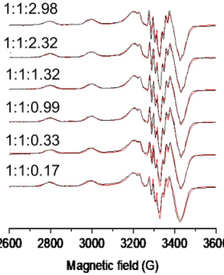
EPR spectra were recorded in the copper(II)-STSC system at various excess of HSA (Figure6), and the EPR parameters (Table2) calculated for the ternary system differ from those of the binary systems. It should be noted that these parameters reveal differences from the data obtained for the ternary complex of DAHK, suggesting a somewhat al- tered coordination.
Molecules2021,26, 2711 9 of 15
Molecules 2021, 26, x FOR PEER REVIEW 9 of 15
copper(II)-Triapine complex to HSA did not result in measurable changes of the circular dichroism spectra of the protein, suggesting that HSA maintains its α-helical structure upon the interaction (see Figure S4).
Figure 5. (a) Visible spectra recorded for the copper(II)‒STSC‒HSA system at pH 7.4 after 4 h equilibration time in the wavelength range of the d-d bands at various equivalents of HSA and b) the calculated molar absorbance spectra of the binary STSC [CuA], and the ternary [CuA(HSA)] complex. {30% (w/w) DMSO/H2O; pH = 7.4 (20 mM HEPES); T = 25 °C; I
= 0.10 M (KCl); cCu(II) = cTSC = 200 μM, cHSA = 0–600 μM; ℓ = 5 cm}.
EPR spectra were recorded in the copper(II)-STSC system at various excess of HSA (Figure 6), and the EPR parameters (Table 2) calculated for the ternary system differ from those of the binary systems. It should be noted that these parameters reveal differences from the data obtained for the ternary complex of DAHK, suggesting a somewhat altered coordination.
Figure 6. Experimental (black) and simulated (red) anisotropic EPR spectra recorded for the cop- per(II)‒STSC‒HSA system at pH 7.4 after 4 h equilibration time at 77 K. Numbers above the spectra indicate the copper(II):STSC:HSA ratios. {cCu(II) = cSTSC = 202 μM, cHSA = 0–600 μM; 30% (w/w) DMSO/H2O; pH = 7.4 (20 mM HEPES); I = 0.10 M (KCl)}.
The molar absorbance spectra calculated for the ternary adducts of the two TSC complexes formed with HSA (Figure 7) indicate a bathochromic shift of the CT bands upon binding to the protein; however, this type of change is different from those ob- served for the adducts formed with DAHK and N-methylimidazole. The conditional binding constants reflect a stronger binding of DAHK to the Triapine complex relative to the binding of HSA, while the trend is the opposite with the STSC complex. All these
0 200 400 600 800 1000 1200
430 530 630 730
ε/ M-1cm-1
λ/ nm [CuA]
[CuA(HSA)]
0.0 0.1 0.2 0.3 0.4 0.5 0.6 0.7
430 530 630 730
Absorbance
λ/ nm [CuA]
+ 3 equiv. HSA
+ 0.2 equiv. HSA
(a) (b)
1:1:2.98 1:1:2.32 1:1:1.32 1:1:0.99 1:1:0.33 1:1:0.17
Figure 5. (a) Visible spectra recorded for the copper(II)-STSC-HSA system at pH 7.4 after 4 h equilibration time in the wavelength range of the d-d bands at various equivalents of HSA andb) the calculated molar absorbance spectra of the binary STSC [CuA], and the ternary [CuA(HSA)] complex. {30% (w/w) DMSO/H2O; pH = 7.4 (20 mM HEPES);T= 25◦C;
I= 0.10 M (KCl);cCu(II)=cTSC= 200µM,cHSA= 0–600µM;`= 5 cm}.
copper(II)-Triapine complex to HSA did not result in measurable changes of the circular dichroism spectra of the protein, suggesting that HSA maintains its α-helical structure upon the interaction (see Figure S4).
Figure 5. (a) Visible spectra recorded for the copper(II)‒STSC‒HSA system at pH 7.4 after 4 h equilibration time in the wavelength range of the d-d bands at various equivalents of HSA and b) the calculated molar absorbance spectra of the binary STSC [CuA], and the ternary [CuA(HSA)] complex. {30% (w/w) DMSO/H2O; pH = 7.4 (20 mM HEPES); T = 25 °C; I
= 0.10 M (KCl); cCu(II) = cTSC = 200 μM, cHSA = 0–600 μM; ℓ = 5 cm}.
EPR spectra were recorded in the copper(II)-STSC system at various excess of HSA (Figure 6), and the EPR parameters (Table 2) calculated for the ternary system differ from those of the binary systems. It should be noted that these parameters reveal differences from the data obtained for the ternary complex of DAHK, suggesting a somewhat altered coordination.
Figure 6. Experimental (black) and simulated (red) anisotropic EPR spectra recorded for the cop- per(II)‒STSC‒HSA system at pH 7.4 after 4 h equilibration time at 77 K. Numbers above the spectra indicate the copper(II):STSC:HSA ratios. {cCu(II) = cSTSC = 202 μM, cHSA = 0–600 μM; 30% (w/w) DMSO/H2O; pH = 7.4 (20 mM HEPES); I = 0.10 M (KCl)}.
The molar absorbance spectra calculated for the ternary adducts of the two TSC complexes formed with HSA (Figure 7) indicate a bathochromic shift of the CT bands upon binding to the protein; however, this type of change is different from those ob- served for the adducts formed with DAHK and N-methylimidazole. The conditional binding constants reflect a stronger binding of DAHK to the Triapine complex relative to the binding of HSA, while the trend is the opposite with the STSC complex. All these
0 200 400 600 800 1000 1200
430 530 630 730
ε/ M-1cm-1
λ/ nm [CuA]
[CuA(HSA)]
0.0 0.1 0.2 0.3 0.4 0.5 0.6 0.7
430 530 630 730
Absorbance
λ/ nm [CuA]
+ 3 equiv. HSA
+ 0.2 equiv. HSA
(a) (b)
1:1:2.98 1:1:2.32 1:1:1.32 1:1:0.99 1:1:0.33 1:1:0.17
Figure 6. Experimental (black) and simulated (red) anisotropic EPR spectra recorded for the copper(II)-STSC-HSA system at pH 7.4 after 4 h equilibration time at 77 K. Numbers above the spectra indicate the copper(II):STSC:HSA ratios. {cCu(II)=cSTSC= 202µM,cHSA= 0–600µM; 30%
(w/w) DMSO/H2O; pH = 7.4 (20 mM HEPES);I= 0.10 M (KCl)}.
The molar absorbance spectra calculated for the ternary adducts of the two TSC complexes formed with HSA (Figure7) indicate a bathochromic shift of the CT bands upon binding to the protein; however, this type of change is different from those observed for the adducts formed with DAHK and N-methylimidazole. The conditional binding constants reflect a stronger binding of DAHK to the Triapine complex relative to the binding of HSA, while the trend is the opposite with the STSC complex. All these findings suggest that conclusions drawn for the binding modes and strength of HSA on the basis of the results obtained with the simplified model compounds should be considered carefully. Besides the coordinative binding, secondary interactions might play a role as well. The conditional constants determined for the HSA adducts suggest a weak-to-moderate binding affinity of the studied TSC complexes to this protein, namely, ~66% (Triapine) and ~15% (STSC) of the complexes are predicted to be unbound under biologically more relevant conditions (e.g.,cHSA= 630µM,ccomplex= 10µM).
Molecules2021,26, 2711 10 of 15
Molecules 2021, 26, x FOR PEER REVIEW 10 of 15
findings suggest that conclusions drawn for the binding modes and strength of HSA on the basis of the results obtained with the simplified model compounds should be con- sidered carefully. Besides the coordinative binding, secondary interactions might play a role as well. The conditional constants determined for the HSA adducts suggest a weak-to-moderate binding affinity of the studied TSC complexes to this protein, namely,
~66% (Triapine) and ~15% (STSC) of the complexes are predicted to be unbound under biologically more relevant conditions (e.g., cHSA = 630 μM, ccomplex = 10 μM).
Figure 7. Calculated molar absorbance UV–vis spectra of the binary copper(II)-TSC ([CuA]) and the copper(II)-TSC-HSA ([CuA(HSA)]) ternary complexes in the wavelength range of the CT bands.
(Triapine: black lines, STSC: orange lines). {30% (w/w) DMSO/H2O; pH = 7.4 (20 mM HEPES); T = 25 °C; I = 0.10 M (KCl); ℓ = 5 cm}.
2.3. Redox Properties of the Copper(II)-TSC Complexes Affected by HSA and DAHK
Since the anticancer activity of the copper(II)-TSC complexes is often related to their redox properties [7,10,29,34], we also investigated whether these features are affected by the binding of HSA (or the model DAHK). Cyclic voltammetric studies were performed for the copper(II)‒Triapine (1:1) system in the absence and presence of DAHK in 30%
(w/w) DMSO/H2O at pH 7.4 (Figure 8). Only the position of the cathodic peak could be observed in the voltammograms due to the irreversible nature of the redox processes.
The reduction peak potential of the copper(II)-Triapine complex corresponds well to the literature data [18], and a significant shift toward the higher potentials is seen as a result of the coordination of DAHK. It suggests the somewhat stronger oxidizing power of the ternary complex, compared to the binary species.
Investigation of the redox reaction of these copper(II) complexes with physiological reductants can provide more direct information about their ability to react with reducing agents than solely the values of reduction peak potentials. Therefore, the reaction of the copper(II)-Triapine complex in the presence of HSA (and DAHK) with ascorbic acid and glutathione (GSH) was followed spectrophotometrically under anaerobic conditions. The copper(II)-Triapine complex alone was already studied in one of our previous works [30,34]. Ascorbic acid, which is a weaker reducing agent than GSH, was not able to re- duce the copper(II) complex either in the absence or in the presence of HSA. On the con- trary, the addition of GSH resulted in significant spectral changes (Figure 9a).
,0 2,000 4,000 6,000 8,000 10,000
340 390 440 490
ε/ M-1cm-1
λ/ nm [CuA(HSA)]
[CuA]+
[CuA(HSA)]
[CuA]
Figure 7. Calculated molar absorbance UV–vis spectra of the binary copper(II)-TSC ([CuA]) and the copper(II)-TSC-HSA ([CuA(HSA)]) ternary complexes in the wavelength range of the CT bands.
(Triapine: black lines, STSC: orange lines). {30% (w/w) DMSO/H2O; pH = 7.4 (20 mM HEPES);
T= 25◦C;I= 0.10 M (KCl);`= 5 cm}.
2.3. Redox Properties of the Copper(II)-TSC Complexes Affected by HSA and DAHK
Since the anticancer activity of the copper(II)-TSC complexes is often related to their redox properties [7,10,29,34], we also investigated whether these features are affected by the binding of HSA (or the model DAHK). Cyclic voltammetric studies were performed for the copper(II)-Triapine (1:1) system in the absence and presence of DAHK in 30%
(w/w) DMSO/H2O at pH 7.4 (Figure8). Only the position of the cathodic peak could be observed in the voltammograms due to the irreversible nature of the redox processes.
The reduction peak potential of the copper(II)-Triapine complex corresponds well to the literature data [18], and a significant shift toward the higher potentials is seen as a result of the coordination of DAHK. It suggests the somewhat stronger oxidizing power of the ternary complex, compared to the binary species.
Molecules 2021, 26, x FOR PEER REVIEW 11 of 15
Figure 8. Cyclic voltammograms of the copper(II)‒Triapine (1:1) (solid line) and the copper(II)‒
Triapine‒DAHK (1:1:1, dashed line; 1:1:2, dotted line) systems at pH 7.4. {cCu(II) = cTriapine = 500 μM, cDAHK = 0, 500 or 1000 μM; 30% (w/w) DMSO/H2O; pH = 7.4 (20 mM HEPES); T = 25 °C; I = 0.10 M (KCl); scan rate: 5 mV/s}.
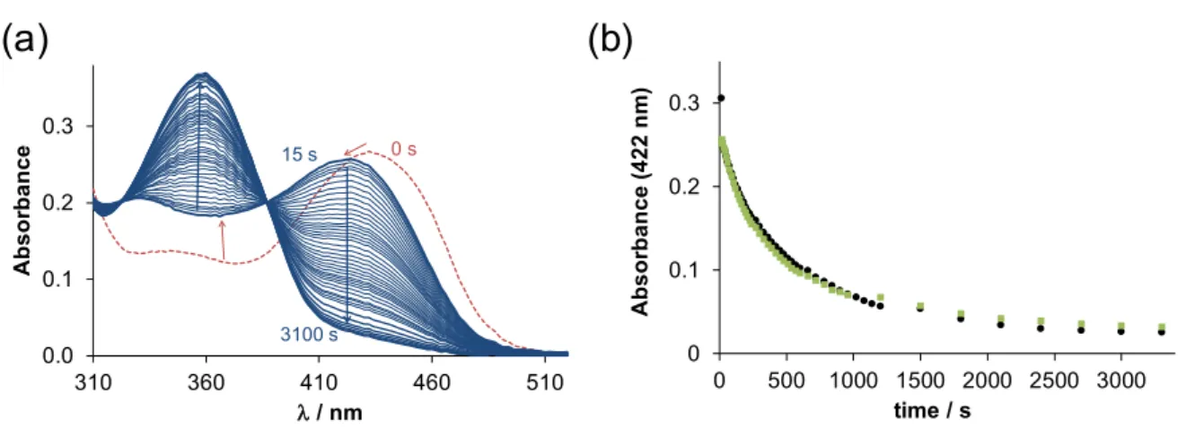
Figure 9. (a) Time-dependent changes of the UV–vis absorption spectra of the copper(II)‒Triapine‒HSA (1:1:1) system in the presence of 50 equiv. GSH at pH 7.4 in pure water under argon atmosphere. (b) Absorbance changes at 422 nm rec- orded for the binary copper(II)‒Triapine (●) and for the ternary copper(II)‒Triapine‒HSA (■) systems. {cCu(II) = cTriapine = cHSA = 25 μM, cGSH = 1250 μM; pH = 7.4 (20 mM HEPES); T = 25 °C; I = 0.10 M (KCl); ℓ = 1 cm}.
For the copper(II)‒Triapine system, it was reported that a ternary Cu(II)-Triapine-GSH complex is formed in the first step after mixing the reactants. Then, the free TSC ligand appears in the solution as a consequence of the dissociation of the generated copper(I) complex since copper(I) tends to form a stable complex with GSH, which is present in a high excess, compared to the TSC. In the presence of one equivalent HSA, the initial spectrum differs from that of the copper(II)-Triapine complex due to the formation of the ternary adduct. Moreover, upon the addition of GSH, the spectrum be- comes identical to that observed without the protein. The rate of the subsequent redox reaction is also very similar (Figure 9b). The same phenomenon was observed in the presence of DAHK. Accordingly, it can be concluded that the redox potential is changed due to the ternary complex formation; however, the donor atoms of the protein (or DAHK) can be replaced by other endogenous ligands such as GSH.
3. Materials and Methods 3.1. Chemicals
Triapine, STSC, HEPES, GSH, ascorbic acid, mim, and HSA (A8763, essentially globulin free) were purchased from Sigma-Aldrich (Hungary), while KCl, HCl, KOH, and DMSO were from VWR (Hungary). DAHK was obtained from GenScript (the Neth- erlands). The concentration of the stock solution of CuCl2 was determined by complex-
-20 -15 -10 -5 0 5 10
-0.45 -0.35 -0.25 -0.15 -0.05 0.05
i / µA
Evs.Ag/AgCl/3M KCl / V
Cu(II):Triapine:DAHK (1:1:1) Cu(II):Triapine (1:1)
Cu(II):Triapine:DAHK (1:1:2)
(a) (b)
0 0.1 0.2 0.3
0 500 1000 1500 2000 2500 3000
Absorbance (422 nm)
time / s 0.0
0.1 0.2 0.3
310 360 410 460 510
Absorbance
λ/ nm 15 s 0 s
3100 s
Figure 8. Cyclic voltammograms of the copper(II)-Triapine (1:1) (solid line) and the copper(II)- Triapine-DAHK (1:1:1, dashed line; 1:1:2, dotted line) systems at pH 7.4. {cCu(II)=cTriapine= 500µM, cDAHK= 0, 500 or 1000µM; 30% (w/w) DMSO/H2O; pH = 7.4 (20 mM HEPES);T= 25◦C;I= 0.10 M (KCl); scan rate: 5 mV/s}.
Investigation of the redox reaction of these copper(II) complexes with physiological reductants can provide more direct information about their ability to react with reducing agents than solely the values of reduction peak potentials. Therefore, the reaction of the copper(II)-Triapine complex in the presence of HSA (and DAHK) with ascorbic acid and glutathione (GSH) was followed spectrophotometrically under anaerobic conditions. The copper(II)-Triapine complex alone was already studied in one of our previous works [30,34].
Ascorbic acid, which is a weaker reducing agent than GSH, was not able to reduce the
![Figure 7. Calculated molar absorbance UV–vis spectra of the binary copper(II)-TSC ([CuA]) and the copper(II)-TSC-HSA ([CuA(HSA)]) ternary complexes in the wavelength range of the CT bands.](https://thumb-eu.123doks.com/thumbv2/9dokorg/761672.33185/10.892.253.573.701.921/figure-calculated-absorbance-spectra-binary-ternary-complexes-wavelength.webp)