https://doi.org/10.1007/s00775-019-01683-0 ORIGINAL PAPER
Binding mechanisms of half‑sandwich Rh(III) and Ru(II) arene complexes on human serum albumin: a comparative study
Orsolya Dömötör1 · Éva A. Enyedy1
Received: 9 May 2019 / Accepted: 27 June 2019 / Published online: 12 July 2019
© The Author(s) 2019
Abstract
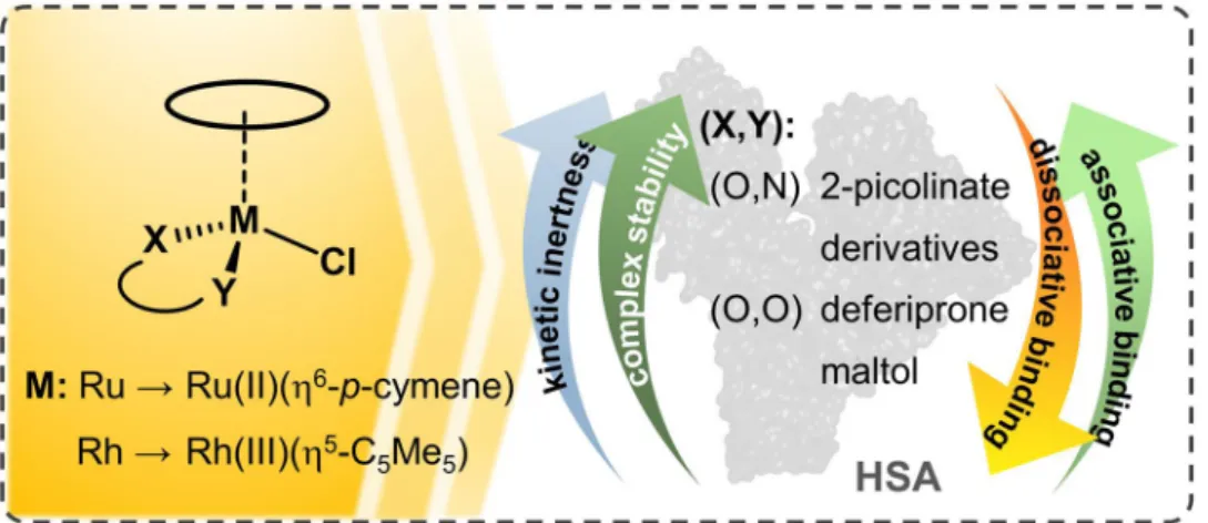
Various half-sandwich ruthenium(II) arene complexes and rhodium(III) arene complexes have been intensively investigated due to their prominent anticancer activity. The interaction of the organometallic complexes of Ru(η6-p-cymene) and Rh(η5- C5Me5) with human serum albumin (HSA) was studied in detail by a combination of various methods such as ultrafiltration, capillary electrophoresis, 1H NMR spectroscopy, fluorometry and UV–visible spectrophotometry in the presence of 100 mM chloride ions. Binding characteristics of the organometallic ions and their complexes with deferiprone, 2-picolinic acid, maltol, 6-methyl-2-picolinic acid and 2-quinaldic acid were evaluated. Kinetic aspects and reversibility of the albumin bind- ing are also discussed. The effect of low-molecular-mass blood components on the protein binding was studied in addition to the interaction of organorhodium complexes with cell culture medium components. The organometallic ions were found to bind to HSA to a high extent via a coordination bond. Release of the bound metal ions was kinetically hindered and could not be induced by the denaturation of the protein. Binding of the Ru(η6-p-cymene) triaqua cation was much slower (ca. 24 h) compared to the rhodium congener (few min), while their complexes interacted with the protein relatively fast (1–2 h). The studied complexes were bound to HSA coordinatively. The highly stable and kinetically inert 2-picolinate Ru(η6-p-cymene) complex bound in an associative manner preserving its original entity, while lower stability complexes decomposed partly or completely upon binding to HSA. Fast, non-specific and high-affinity binding of the complexes on HSA highlights their coordinative interaction with various types of proteins possibly decreasing effective drug concentration.
Graphic abstract
Keywords Albumin · Binding affinity · Capillary electrophoresis · Fluorescence · Ultrafiltration Abbreviations
6-Mepic 6-Methyl-2-picolinic acid BGE Background electrolyte bpy 2,2′-Bipyridine
Electronic supplementary material The online version of this article (https ://doi.org/10.1007/s0077 5-019-01683 -0) contains supplementary material, which is available to authorized users.
Extended author information available on the last page of the article
CZE Capillary zone electrophoresis DG Dansylglycine
dhp 1,2-Dimethyl-3-hydroxy-pyridin-4(1H)-one, deferiprone
HMM High molecular mass HSA Human serum albumin LMM Low molecular mass
maltol 3-Hydroxy-2-methyl-pyran-4(1H)-one PBS’ Modified phosphate buffered saline pic 2-Picolinic acid
PTA 1,3,5-Triaza-7-phosphaadamantane RhCp* Rh(η5-C5Me5)
RM175 [Ru(II)(η6-biphenyl)(ethylenediamine)Cl]+ RuCym Ru(η6-p-cymene)
UV–vis UV–visible WF Warfarin
Introduction
During the development of metal compounds for medici- nal purposes, organometallics have been shown to exhibit promising activity in various diseases [1–5]. In particular, half-sandwich Ru(II)-arene complexes have attracted atten- tion, since some representatives show significant anticancer activity and non-cross resistance with cisplatin. [3, 6–8]. The interest for organoruthenium compounds in cancer treatment became more pronounced as the clinically relevant Ru(III) drug candidates NKP1339 and NAMI-A were reported to exert their anticancer effect in the reduced Ru(II) forms [9]. Among Ru(II) compounds [Ru(II)(4,4′-dimethyl-2,2″- bipyridine)(2-((2′,2′’:5′’,2′’’-terthiophene)-imidazo[4,5-f]
[1,10] phenanthroline))] (TLD1433) has completed a phase 1 clinical trial for treating bladder cancer with photodynamic therapy [10]. [Ru(II)(η6-p-cymene)(PTA)Cl2] (RAPTA-C;
PTA = 1,3,5-triaza-7-phosphaadamantane) and the RAED type [Ru(II)(η6-biphenyl)(ethylenediamine)Cl]+ (RM175) are considered the lead structures for antitumor half-sand- wich Ru(II)-arene compounds [1, 5, 11, 12]. Complexes of the isoelectronic congener Rh(III)(η5-C5Me5) showing promising in vitro anticancer activity have also been inten- sively investigated [13, 14]. Early reports on the mechanism of action of anticancer Ru(III) complexes suggested interac- tion with DNA, similar to cisplatin, which was believed to be ultimately responsible for the anticancer activity. Even in the case of Ru(II)-arene complexes, DNA nucleobases were pri- mary suspects in their cytotoxic activity [15–18]. Although the actual mechanisms of action of these complexes are still not clarified, in recent years, more studies evidenced that DNA is not necessarily the only and/or primary target for ruthenium organometallics. Currently, the role of intra and extracellular proteins as possible targets of organoruthenium and related complexes is widely investigated [19].
Binding of these complexes to proteins is of consider- able interest not only because of the exploration of feasible modes of action but explanation of side effects and pharma- cokinetic behavior becomes possible as well. Amino acid side chains of proteins, similarly to DNA nucleobases, pro- vide versatile coordinating sites for metal ions. The question is how these Ru(η6-arene) and Rh(η5-arenyl) complexes can interact with protein donor groups. Thus, our research inter- ests are in the area of the following: (i) what kind of adducts can be formed, (ii) which are the preferred sites and, (iii) what are the time frame and the extent of these interactions.
If these complexes are administered intravenously, potential first binding partners in vivo are blood constituents such as the non-specific transport protein albumin. In case of oral administration after the absorption from the gastrointestinal tract, the drug also reaches the systemic circulation. In both cases, binding to transport proteins, e.g., to human serum albumin (HSA) has a profound effect on the distribution, metabolism and excretion of a compound. This interaction can be advantageous due to the enhanced permeability and retention effect in solid tumor tissues resulting in the accu- mulation of protein-bound drugs close to the cancer cells [20]. On the other hand, an irreversible protein binding can decrease effective drug concentration and may be respon- sible for adverse effects as well [21, 22]. Notably, the cell culture media used for the in vitro cytotoxic measurements also contain albumin and other serum proteins, thus interac- tion with these proteins in the medium can also affect the original integrity of the complex.
HSA binding of anticancer metallodrugs is a widely investigated area and numerous papers were published for, e.g., Pt(II) [23–26], Ru(III) [27–31] and Ga(III) [32–34]
complexes. In vitro studies on direct albumin binding of half- sandwich organometallic complexes are also reported [35, 36], however, in vivo results are rarely published [37]. Klose et al. have investigated recently the distribution of two half- sandwich p-cymene complexes ([Ru(η6-p-cymene)(plec- statin)Cl] and [Os(η6-p-cymene)(plecstatin)Cl]) in mouse blood serum by size-exclusion chromatography–inductively coupled plasma–mass spectrometry (SEC–ICP–MS). Free drug was not observed in any of the samples indicating rapid protein binding of the metallodrugs. A larger portion of these drugs (ca. 75%) was found in the albumin/transferrin fraction underlying the importance of interactions towards HSA as well [37].
Sadler et al. reported the coordination of the rather acces- sible surface imidazole nitrogen donors (histidines) of HSA to the Ru(II) center in the case of RM175 [38], while adduct formation with cytochrome c was thought to involve the N-terminus or carboxylic acid side chains on the pro- tein [39]. Moreover, several single crystal X-ray structures confirm coordinative binding of various types of Ru(η6-p- cymene) and Rh(η5-C5Me5) complexes towards proteins like
lysozyme, apo-ferritin or histone proteins [40–48], which are not believed to be all the primary targets for these organome- tallic compounds but may provide insight on the types of non- specific adducts formed with proteins. The albumin binding of Rh(η5-C5Me5) and its complexes formed with 1,2-dimethyl- 3-hydroxy-pyridin-4(1H)-one (deferiprone, dhp), ethylenedi- amine and 2,2′-bipyridine (bpy) was formerly investigated in our research group. Our results suggested that the interaction with HSA can proceed in both dissociative (binding of the organometallic fragment only) and non-dissociative manner depending most probably on the thermodynamic stability of the metal complexes [49]. HSA is a key protein in blood transport and may serve as a universal protein model as well.
Herein we provide a comprehensive picture on the interac- tions of various Rh(η5-C5Me5), Ru(η6-p-cymene) complexes with HSA. Studied compounds are the organometallic cations [Rh(η5-C5Me5)(H2O)3]2+ and [Ru(η6-p-cymene)(H2O)3]2+
themselves and their complexes formed with bidentate ligands:
3-hydroxy-2-methyl-pyran-4(1H)-one (maltol), dhp, 2-pico- linic acid (pic), 6-methyl-2-picolinic acid (6-Mepic) and quinoline-2-carboxylic acid (2-quinaldic acid) (Chart 1). Our goal is to provide a comprehensive overview of the binding strength, location, and kinetics and the binding mode and its reversibility. The effect of low-molecular-mass components on the protein binding and the stability of selected complexes in a minimum essential cell culture medium were tested as well.
Efficiency and possible shortages of techniques applied here, namely 1H NMR-, UV–visible and fluorescence spectrosco- pies; ultrafiltration–UV–visible and capillary zone electropho- resis, are critically discussed as well.
Experimental
Chemicals[Rh(η5-C5Me5)(µ-Cl)Cl]2, [Ru(η6-p-cymene)(µ-Cl)Cl]2, maltol, dhp, pic, 6-Mepic, 2-quinaldic acid, bpy warfarin (WF), dansylglycine (DG), sodium dodecyl sulfate (SDS), citric acid, sodium lactate, l-amino acids: histidine (His), methionine (Met), cysteine (Cys), serine (Ser), and inorganic salts KCl, KH2PO4, NaH2PO4, Na2HPO4 were products of Sigma-Aldrich or Reanal in puriss quality. HSA (A8763, essentially globulin free) and Roswell Park Memorial Insti- tute (RPMI) 1640 cell culture medium was purchased from Sigma-Aldrich.
Stock solutions and sample preparation
Milli-Q water was used for preparation of stock solutions and samples. Exact concentrations of stock solutions of the ligand and the organometallic cations were determined by pH-potentiometric titrations according to procedure pub- lished in our former work [50]. Organometallic complexes
were obtained by mixing the corresponding ligand and metal precursor ([Rh(η5-C5Me5)(µ-Cl)Cl]2, [Ru(η6-p-cymene) (µ-Cl)Cl]2) in 1:0.5 molar ratio in water; solutions were equilibrated for 24 h. HSA stock solutions were prepared in modified phosphate buffered saline (PBS′), pH 7.40 con- taining 12 mM Na2HPO4, 3 mM KH2PO4, 1.5 mM KCl and 100.5 mM NaCl. Concentration of the K+, Na+ and Cl‒ ions corresponds approximately to that of the human blood serum. Residual citrate content of HSA was removed by repeated ultrafiltration of the protein stock solution, and its concentration was calculated from its UV absorption:
λ280 nm(HSA) = 36,850 M−1 cm−1 [51]. Stock solutions of WF and DG were prepared as described previously [31].
Samples containing RhCp* and RuCym compounds were incubated for 2 and 24 h, respectively, prior to the meas- urements, or the reaction kinetic was followed. All samples were prepared in PBS’ and incubated at 25 ± 0.1 °C and measurements were carried out at this temperature.
N O
OH
N O
OH
OH2
H2O Rh H2O OH2
H2O Ru H2O
N O
OH N
O HO
O O HO [Ru(η6-p-cymene)(H2O)3]2+
(RuCym)
2+ 2+
pic
6-Mepic
2-quinaldic acid
dhp maltol
(X,Y) = (O,O)
[Rh(η5-C5Me5(H2O)3]2+
(RhCp*)
(X,Y) = (O,N)
H2O M X Y
Chart 1 Chemical structures and abbreviations of the organometal- lic triaqua cations of Ru(η6-p-cymene) (RuCym) and Rh(η5-C5Me5) (RhCp*), general formula of the studied complexes and structures of the complex forming bidentate ligands in their neutral forms:
1,2-dimethyl-3-hydroxy-pyridin-4(1H)-one (deferiprone, dhp), 3-hydroxy-2-methyl-pyran-4(1H)-one (maltol), 2-picolinic acid (pic), quinoline-2-carboxylic acid (2-quinaldic acid) and 6-methyl-2-pico- linic acid (6-Mepic)
1
H NMR spectroscopy
1H NMR spectroscopic studies were carried out on a Bruker Avance III HD instrument. All spectra were recorded with the WATERGATE water suppression pulse scheme using DSS internal standard. To study the interaction with HSA and the RPMI 1640 components 1H NMR spectra were recorded for samples containing 1 mM organometallic com- pound and 0.5 mM HSA or 1.25-fold diluted RPMI 1640 in PBS’ at 10% (v/v) D2O content.
Spectrofluorometry
Fluorescence spectra were recorded on a Hitachi-F4500 fluorometer in 1 cm quartz cell. Samples contained 1 μM HSA and various HSA-to-metal compound ratios (from 1:0 to 1:20) were used. In the site marker displacement experi- ments, the HSA-to-site marker (WF or DG) ratio was 1:1 and the concentration of the compounds varied. The excita- tion wavelengths were 295, 310 or 335 nm depending on the type of the experiment and the emission was read in the range of 310–650 nm. The conditional binding constants (log K′) were computed with the computer program PSE- QUAD [52] as described in our previous works [31, 53]. The effect of low-molecular-mass (LMM) components on the albumin binding of Ru and Rh compounds was monitored in samples containing 1 μM HSA, 10 μM metal complex and LMM component mixture or only one of these components corresponding to their blood serum concentration: Ser (~ 100 μM), His (~ 77 μM), Met (~ 77 μM) and Cys (~ 33 μM), cit- rate (~ 99 μM) and lactate (~ 1.5 mM). Samples containing Cys were kept in a strictly oxygen-free atmosphere. Calcula- tions always were based on data obtained from at least two independent measurements. Corrections for self-absorbance and inner filter effect were necessary, since the emitted light was partly absorbed by the compounds. Corrections were carried out according to the following equation [54]:
where Icorr and Imeas are the corrected and measured fluo- rescence intensities, AEX and AEM are the absorbance values at the excitation and emission wavelengths in the samples, respectively.
UV–visible spectrophotometry
A Hewlett Packard 8452A and an Agilent Carry 8454 diode- array spectrophotometer were utilized to record the UV–vis- ible (UV–vis) spectra in the interval 190–820 nm. The path length (l) was 1 cm. For the reaction kinetic studies, a tan- dem cuvette with two chambers was used. Measurements were performed at various metal complex concentrations Icorr = Imeas × 10(AEX+AEM)∕2,
(between 20 and 200 μM), and various protein-to-complex ratios (from 0.02:1 to 2:1) were applied. UV–vis spectra presented in this paper are subtracted by the spectrum of reference albumin sample in all cases in favor of the bet- ter interpretation of the results. Reference spectra for the complexes and their components (ligand and organometal- lic cation) were recorded as well. Excel Solver (Microsoft Office 2016) was utilized for deconvolution of the absorb- ance spectra.
Ultrafiltration
Samples were separated by ultrafiltration through 10 kDa membrane filters (Millipore, Amicon Ultra-0.5), in low- and high-molecular-mass (LMM and HMM) fractions with the help of an Eppendorf MiniSpin plus centrifuge (rela- tive centrifugal force ∼ 1500g, 5–10 min). Three different types of experiments were carried out: samples contained (1) 50 μM HSA and 50–430 μM organometallic compound;
(2) ~ 500 μM organometallic cation and 8–50 μM HSA; (3) reversibility of the protein binding was followed by con- secutive washing and filtration steps and then by filtration of samples after addition of 0.5% (m/m) SDS. The concen- tration of the non-bound compounds in the LMM fractions was determined by UV–Vis spectrophotometry by compar- ing the recorded spectra to those of reference samples with- out the protein. When the complex partly decomposed due to the protein binding, the free ligand was also detected in the LMM fraction. In this case, the recorded spectrum was deconvoluted with the use of the molar absorbance spectra of the ligand and metal complex by Excel Solver (Microsoft Office 2016). The HMM fraction was studied in a similar way for some samples as well.
Capillary zone electrophoresis
Measurements were performed on a G7100 capillary elec- trophoresis system (Agilent Technologies, Waldbronn, Ger- many) equipped with a diode-array detector (210–600 nm).
For all experiments, fuse silica capillaries of 48 cm total length (50 μm inner diameter) were used (BGB Analytik).
Phosphate buffer (20 mM, pH 7.40) was the background electrolyte (BGE). The conditioning process of new capil- laries started with HCl (0.1 M) and H2O followed by NaOH (0.1 M) and H2O for 20 min each and was completed with flushing of the BGE for 40 min. A similar procedure was applied for daily preparation of the capillary although with- out purging with HCl. To ensure the steady baseline, the capillary was flushed with BGE (2 min) before each run and was rinsed with NaOH (0.1 M; 1.5 min), H2O (1.5 min), and BGE (2 min) after each separation. The sample tray and the capillary were kept at a constant temperature of 25 °C. Sam- ples were injected hydrodynamically at 50 mbar for 15 s,
and voltage of 30 kV was applied for the separation process producing a current of ca. 44 μA. Sample run time was set to 6 min. Electropherograms were recorded and evaluated by the program ChemStation (Agilent Technologies). The detection wavelength was set to 280 or 260 nm depending on the samples studied. Samples contained ca. 200 μM orga- nometallic compound and various concentrations of HSA (10–100 μM) in PBS′. The peak integrals of the non-bound complex and ligand were used to obtain their concentrations using external calibration. UV–Vis spectra belonging to all peaks were collected as well.
Results
Interaction of Rh(η5‑C5Me5) compounds with RPMI 1640 cell culture medium
Prior to the investigation of the interactions of biomolecules with organorhodium and organoruthenium compounds, the knowledge of their actual forms at physiological condi- tions is of critical importance to get adequate conclusions.
Dissolving the organometallic precursor ([Rh(η5-C5Me5) (µ-Cl)Cl]2 in water at pH 7.40 the triaqua cation [Rh(η5- C5Me5)(H2O)3]2+ and its hydrolysis products [(Rh(η5- C5Me5))2(μ-OH)2]+ and [(Rh(η5-C5Me5))2(μ-OH)3]2+ are formed; their actual quantity depends on the analytical concentration of the dissolved precursor. In the presence of 0.1 M chloride ion, corresponding to the chloride concentra- tion in blood serum, aqua and hydroxido ligands are partly displaced by chloride ions. Namely, a mixture of species is present in the solution. The ruthenium precursor behaves similarly. For this reason, these species (if there is no need for closer definition) are referred generally as RhCp* for Rh(η5-C5Me5) and RuCym for Rh(η6-p-cymene) species.
RhCp* and RuCym hydrolyze in about 23% and 100%, respectively, at physiological conditions at 50 μM concen- tration [50, 55].
The complexes of the bidentate ligands presented in Chart 1 were obtained by mixing the precursors and the ligands in 0.5:1 ratio in water and by this way the aqua complexes formed (Chart 1). Although, in chloride-con- taining media, chlorido complexes are present to some extent as well, therefore the leaving group is generally not specified in our complex structures and are referred to as, e.g., RhCp*(dhp) or RuCym(pic). Dhp as an (O,O) donor ligand forms somewhat lower stability complexes with the two organometallic cations compared to the (O,N) donor pic. Dhp complexes of both RhCp* and RuCym dissociate in about 4–5% at pH 7.40 and 50 μM complex concentration at I = 0.2 M KCl, while pic complexes are more stable and less than 0.5% dissociate under the same conditions [56–58]. These values may somewhat differ at
physiological (100 mM) chloride ion concentration, as chloride ions can compete with the coordinating ligands and suppress the complex formation. RuCym(maltolato) as an example for a low stability complex (29% decomposed under the condition vide supra) was also selected [59], and complexes of picolinate derivatives 6-Mepic and 2-qui- naldic acid formed with RhCp* characterized by higher lipophilicity than the pic complex were involved in our investigations as well [60] (constants regarding the com- plex stability, chloride affinity and lipophilicity data are listed in Table S1).
In vitro cytotoxicity studies are always carried out in cultured media, which are produced in different composi- tions according to the actual use. Some compounds are essential and universal components of these mixtures.
All kinds of minimum essential media (MEM or Eagle’s MEM) contain phosphate and bicarbonate buffered saline, inorganic ions Ca2+, Mg2+, K+, Na+, essential l-amino acids (histidine, (iso)leucine, lysine, methionine, pheny- lalanine, threonine, tryptophan, valine) and some non- essential l-amino acids (e.g., arginine, cystine, tyrosine).
While phosphate was proven to be a ‘safe’ buffer for these organometallic compounds in our preliminary measure- ments, hydrogencarbonate and amino acids are potential ligands of organorhodium and organoruthenium cations and can be effective competitors of the original ligand set in this kind of complexes [61–64]. The medium applied here was RPMI 1640, containing 20 alpha amino acids, vitamins, glucose, glutathione and phenol red.
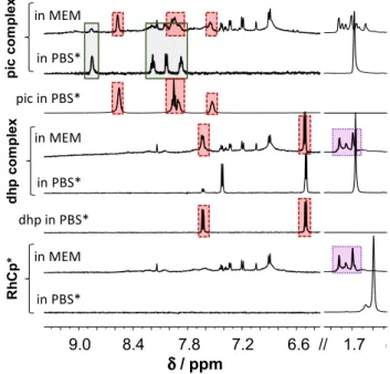
1H NMR spectra in Fig. 1 show the interaction of organorhodium ion and its dhp or pic complexes with RPMI 1640 components. The original RhCp*(dhp) com- plex is completely dissociated, dhp and (most probably) coordinating water as well are replaced by RPMI 1640 components, which results in the appearance of the same peak set for C5Me5 methyl protons like in case of RhCp*
itself, and no mixed-ligand complex formation can be observed. A similar result was obtained with related com- plex RhCp*(acetylacetonato) in our former work [65]. The pic complex reacts somewhat differently: about 75% of the ligand is liberated here as well, but the rest is coordinated to the metal ion. Splitting and shifting of the C5Me5− pro- ton signal, however, suggest mixed-ligand complex for- mation with medium component(s). In cytotoxicity meas- urements, the tested compounds are dissolved typically in micromolar concentrations in cell medium, and more pronounced decomposition of the original complexes is expected under these conditions. Consequently, cytotoxic- ity measured for lower stability complexes (e.g., the dhp or acac complexes) did not exceed the sum of efficacy measured for organorhodium ion (coordinated to medium components) and the ligand (if it is active) separately [56, 65].
Cultured media often contain ca. 10% heat-inactivated fetal bovine serum. Serum proteins including albumin as potential reaction partners for metal complexes are present in these samples as well. Studying the interaction of these organometallic compounds with proteins, especially with human serum albumin is important in other aspects as well, which are discussed in the next sections in detail.
Binding of (Rh(η5‑C5Me5) and (Ru(η6‑p‑cymene) to human serum albumin
Binding of anticancer metallodrugs to HSA is of consid- erable interest, as it may have a significant effect on the biodistribution, toxicity and side effects. Moreover, HSA- and HSA-bound drugs can accumulate in malignant and inflamed tissues such as solid tumors as a consequence of the enhanced permeability and retention effect, which can be exploited for tumor targeting as well [20]. Organome- tallic fragments RhCp* and RuCym (Chart 1) per se exert no antitumor activity, however a deeper knowledge on their interaction with albumin is a mandatory prerequisite for understanding the binding mechanism of their complexes to this protein.
The binding mode and binding strength of the organorho- dium ion (without coordinated ligands) on HSA were inves- tigated in our former work [49]. However, albumin binding
of organoruthenium ion was not characterized adequately in solution, since only its binding at the hydrophobic site I (IIA subdomain) was reported [57]. This section provides a detailed kinetic, quantitative and qualitative description on the HSA binding of RhCp* and RuCym species.
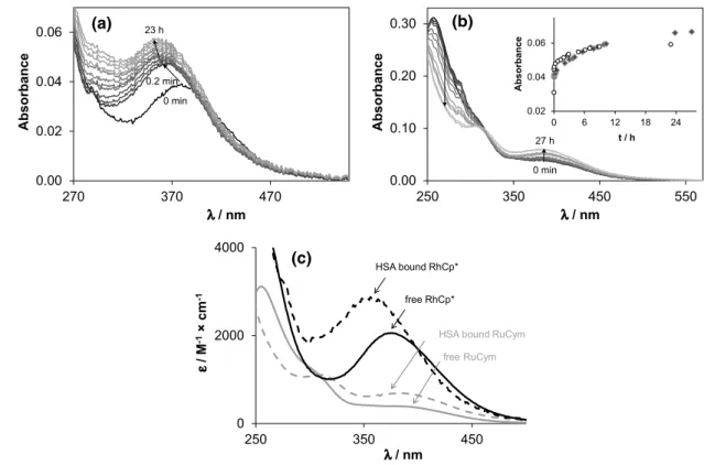
Both organometallic cations displayed considerable changes in their UV–vis spectra in Fig. 2 in the presence of HSA. RhCp* showed a bi-phasic binding profile, where a fast (~ 5 min) process is followed by a much slower one (~ 24 h). Based on our ultrafiltration, and CZE studies, the binding is completed in the first few minutes, and the sub- sequent spectral changes in Fig. 2a can be attributed most probably to structural rearrangement of the coordination sphere around the metal ion. On the contrary, binding of RuCym to the protein takes place about 24 h (Fig. 2b) con- firmed by 1H NMR spectroscopic, ultrafiltration and CZE time-dependent measurements. The observed remarkably different binding rates of the two organometallic cations partly can be explained by their different solution spe- ciation at pH 7.40. RuCym is present in 100% as dimeric hydroxido species [(RuCym)2(μ-OH)3]+, which is known to be kinetically more inert compared to the aqua cation, while in case of RhCp* there is ca. 55% non-hydrolyzed [Rh(η5-C5Me5)(H2O)3]2+ in solution under this condition [50, 55, 66]. Furthermore, as a result of the markedly increased trans effect of the anionic pentamethylcyclo- pentadienyl ligand in comparison to the neutral p-cymene ligand, typical substitution rates for RhCp* complexes are several orders of magnitude higher (~ 102–103 s−1 for H2O exchange in monoaqua complexes) than for analo- gous Ru(II)-arene anticancer compounds (~ 10−3–10−1 s−1) [67, 68]. In subsequent experiments, samples containing RuCym or RhCp* were always measured after 24 h and 1 h waiting time, respectively.
It should be mentioned that commercially available lyo- philized HSA may contain residues of citric acid as anti- coagulant additive of the original blood stock. The pres- ence of citrate can be detected by 1H NMR spectroscopy (Fig. S1). Citrate is a coordinating ligand of RuCym [69]
(and probably of RhCp* as well), consequently peaks of the citrate complexes could be observed in 1H NMR spectra of citrate-contaminated protein samples (Fig. S1). Further- more, we have found that presence of citric acid increases the binding rate of RuCym towards albumin about four to fivefold. Therefore, citrate content of HSA stocks was always checked by 1H NMR spectroscopy and, if it was necessary, HSA stock solutions were purified by ultrafiltration.
HSA is able to accommodate 8–9 eq RhCp* as it was reported in our former work [49], although these numbers do not seem to be the upper limit of available binding sites for RhCp*. The results of ultrafiltration studies presented in Fig. 3 reveal nearly quantitative binding of even 20 eq of cations on HSA and a maximal number of ca. 24 ± 3 RhCp*
1.6 1.7 1.8 f1 (ppm) 6.6
7.0 7.4 7.8 8.2 8.6 9.0
4 f1 (ppm)
in MEM in PBS*
in MEM in PBS*
piccomplexRhCp*
pic in PBS*
in MEM in PBS*
dhpcomplex
dhp in PBS*
9.0 8.4 7.8 7.2 6.6 // 1.7 δ/ ppm
Fig. 1 Stability of the organometallic ion RhCp* and its pic and dhp complexes in RPMI 1640 medium. Peaks in solid, dotted and dashed frames denote the original complex, the forming complexes without the original ligand, and the unbound ligands, respectively [ccomplex = clig = 1 mM, in RPMI 1640 or PBS’ medium, 10% D2O, 25 °C]
can be bound by the protein. Practically quantitative bind- ing of RuCym can be observed up to 9 eq of metal ion, and
binding capacity of HSA is saturated with ca. 14 ± 1 eq of RuCym.
Both RhCp* and RuCym may bind to HSA in a coordina- tive manner, but a striking difference is seen in the number of coordination sites. Coordination of His imidazole nitro- gen is suggested by Sadler et al. in the interaction of HSA with RM175 complex [38]. HSA contains 16 His residues as possible coordination sites, however, it is less, than the max- imal number of bound RhCp*, and some of these histidine nitrogens may be not easily accessible for the organometallic cations. Interaction with sulfur donor side chains of Met and Cys can be assumed as well based on the works of Briš et al.
and Hu et al. [70, 71]. Free thiol groups of small ligands Cys, glutathione and N-acetyl-cysteine show high affinity towards RuCym(PTA) complexes [70], while the single free thiol group in HSA (Cys-34) was reported to undergo oxida- tion during its interaction with [RuCym(ethylenediamine) Cl]+ resulting in the formation of sulfinato complex with- out dissociation of the ethylenediamine ligand [71]. In the same work surface-exposed His-128, His-247, His-510 and Met-298 are also denoted as coordination sites of Ru(arene) complexes [71]. X-ray single crystal structures reported for protein–RuCym complex adducts often show coordination of amino acids His or Glu to the metal center [40–46], and
0.00 0.02 0.04 0.06
270 370 470
Absorbance
λ/ nm
0 min 23 h
0.2 min
(a)
0.00 0.10 0.20 0.30
250 350 450 550
Absorbance
λ/ nm
0.02 0.04 0.06
0 6 12 18 24
Absorbance
t / h
(b)
27 h 0 min
0 2000 4000
250 350 450
ε/ M-1×cm-1
λ/ nm
HSA bound RhCp*
free RuCym free RhCp*
HSA bound RuCym
(c)
Fig. 2 Albumin binding kinetics of the organometallic ions RhCp*
(a) and RuCym (b) followed by UV–Vis spectrometry, and calcu- lated molar absorbance (ε) spectra of the HSA-free and HSA-bound forms of RhCp* and RuCym (c). Inset shows the absorption changes
at 350 nm for RhCp* (empty circle) and at 390 nm for RuCym (filled diamond), respectively. Spectra depicted here are subtracted by the spectrum of HSA [cRh = 20 µM and cHSA = 10 µM (a); cRu = 100 µM and cHSA = 33 µM (b); PBS’; 25 °C]
0 5 10 15 20 25 30
0 10 20 30 40 50 60
[bound M] / cHSA
cM/ cHSA 4 h 24 h
Fig. 3 Binding capacity of HSA for RhCp* (filled triangle) and RuCym (filled square) determined by ultrafiltration–UV–Vis meas- urements. Empty symbol (empty square) denotes the bound equiva- lents of RuCym after 4 h incubation time. Slopes of the fitted lines are: s = 0.971 (R2 = 0.999) for RhCp* and s = 0.969 (R2 = 0.997) for RuCym [cM is varied at < 10 cM/cHSA (50–430 μM) and cM ≈ 600 uM at > 10 cM/cHSA; incubation time: 1 h for Rh and 24 h for Ru com- pound; in PBS’; 25 °C]
coordination of Lys, Arg, Asp and Cys side chains occurs as well [41, 46, 47]. Coordination of one or two of these side chains is typical, and the loss of p-cymene ligand can be detected in some cases as well [40, 41, 47]. Apo-ferritin coordinated [(NHis,NHis,OGlu)] is the only reported struc- ture, where coordination sphere of RuCym is saturated by the donor groups of the protein [41]. To the best of our knowledge, there is only one structure reported on protein- coordinated RhCp*; here two organorhodium fragments are bound to His-112 and His-121 imidazoles, respectively, in streptavidin [48]. Namely, mono or bidentate coordination seems to be feasible in HSA, while binding of HSA donor groups in tridentate mode is most probably sterically less favored. Involvement of S-donor Cys-34, Met (6 residues in HSA); O-donor glutamate (62), aspartate (36) or N-donor lysine (59) or arginine (24) side chains is feasible in binding of high excess of RuCym and RhCp*.
It is difficult to get a clue on the binding environment of the organometallic cations in solution; however, UV–vis spectra can provide some information about the coordina- tion sphere(s) forming around the organometallic cations inside the protein. Individual molar spectrum of the ‘albu- min-bound’ forms can be obtained both in steady-state UV–vis measurements (Fig. 2c) and via examining albu- min-bound fraction of ultrafiltered or electrophoretically separated samples (Fig. S2). As Fig. 2c shows, dinuclear [(RuCym)2(μ–OH)3]+ transforms into a typical mononu- clear complex bound form, and two charge-transfer (CT) bands develop with λmax = 310 nm (εmax = 1100 M−1cm−1) and 382 nm (εmax = 670 M−1cm−1), which did not depend significantly on the Ru-to-HSA ratio applied. Notably, the recorded spectra did not indicate the loss of p-cymene ligand from the Ru(II) center (see details in Refs. [72, 73]). More detailed qualitative information can be obtained on the albu- min-bound form of RhCp* (Fig. 2c), where λmax = 356 nm and εmax = 2800 M−1 cm−1 values of the forming protein complex can be compared to those of various complexes of bidentate ligands studied formerly in our research group (Table S2) [49, 50, 56, 60, 65, 72, 74, 75]. The correlation diagram presented in Fig. 4 (and Fig. S3) illustrates well the effect of each donor groups on the λmax and εmax values:
λmax increases while εmax decreases tendentiously by chang- ing the donor sets from (N,N) to (O,N) and then to (O,O).
Additionally, coordination of the chlorido ligand in the third position induces red shifted λmax. According to the corre- lation diagram, spectral parameters of HSA-bound RhCp*
are close to the (N,N)(Cl)-type complexes (like complexes of ethylenediamine, bpy and their derivatives) raising the possibility of (Nimidazole,Namide)-type coordination. These spectral parameters are practically unaltered between 1:1 and 8:1 Rh-to-HSA ratio, but increasing the Rh excess from 8:1 to 20:1 results in a gradual shift to λmax = 373 nm and εmax = 2270 M−1 cm−1, indicating the involvement of other
types, probably (O,N), (O,O) and/or (O) donor sites into the binding of RhCp* on HSA as well. Another explanation can be the appearance of monodentate N-donor binding sites, which consequently resulted in red shifted λmax [76] as well.
Reaction rates of coordinating ligands with RuCym are strongly affected by the type of the donor atoms. Complex formation with (O,O) or (O,O,O) donor ligands is a fast process at physiological pH, while (N) and (N,N,N) donor ligands chelate the RuCym rather slowly (hours to days) [59, 69, 76, 77]. Reaction rate with (O,N) donor bearing ligands mainly depends on the actual protonation state of the given donor atom, thus it also depends on the actual pH, and the interaction may take place from few minutes to several hours [57, 72]. In this view, slow coordination of protein (N)-donor group(s) to RuCym can be assumed.
Spectrofluorometric studies were carried out to investi- gate the binding of RuCym at hydrophobic binding pockets site I (in subdomain IIA) and site II (in subdomain IIIA).
Binding of RhCp* was formerly investigated by Trp-214 quenching (at site I) and site marker displacement experi- ments using WF and DG to follow interaction at sites I and II, respectively. High-affinity binding and fast binding rate (few min) were found for RhCp* at both sites [49]. RuCym displays slow binding at site I, as it was observed in the global binding studies (vide supra) as well and exerted affin- ity at binding pocket I comparable to that of RhCp* (see Table 1). RuCym additionally interacts with free DG and quenches its fluorescence. Therefore, the binding at site II is observed but data can not be quantitatively evaluated. Since aqua complexes are partly chlorinated and can hydrolyze at pH 7.40, all protein binding constants determined here are
1000 3000 5000 7000
350 370 390 410
εmax/ M-1×cm-1
λmax/ nm
(O,S) thiomaltol
(O,N) 8-HQ deriv.s
(O,N) picolinates (N,N)
(O,O) [(RhCp*)2(OH)3]+
’HSA bound’
Fig. 4 Correlation diagram for the λmax and εmax values of various bidentate [RhCp*(L)Cl] complexes and dimeric hydroxido species [(RhCp*)2(μ-OH)3]+ and HSA-bound RhCp* at 1:1 HSA-to-Rh ratio (correlation diagram for the aqua complexes ([RhCp*(L)(H2O)]) are presented in Fig. S3; see exact values for each complexes in Table S2)
regarded as conditional stability constants and valid only under the given conditions. Considering the extremely high number of binding sites on HSA (which cannot be included in the calculations), the binding constants calculated here reflect the competitiveness of sites I and II against the other sites together with their affinity for the free metal com- pounds. Namely, higher constants could be expected if the binding event at isolated sites could be studied. Slow binding of RuCym at site I indicates the involvement of (N) donor His-242 lining binding site I. Most probably RhCp* binds to the same His residue here, while the nature of coordinat- ing donor group at site II is in question, since no His can be found among the residues evolving in this pocket [78].
Interaction of Rh(η5‑C5Me5) and Ru(η6‑p‑cymene) complexes of dhp, pic and some related ligands with human serum albumin
Selection of the complexes was mainly governed by their well-characterized and somewhat different solution chemi- cal properties and not by the otherwise poor cytotoxic effi- cacy of the complexes. These selected complexes, however, are good models of in vitro active compounds, e.g., (O,O) donor flavone and naphtoquinone-type complexes of Ru(η6- p-cym) or (O,N) donor 8-hydroxyquinoline or Schiff-base complexes of both metal ions [72, 80–85]. It was shown previously that interaction of the studied complexes with HSA is possible, both by loss of the bidentate ligand or by
the loss of only chlorido leaving group [49]. The former one is considered as dissociative binding as the original ligand becomes unbound, while the latter one is referred here to as associative binding, i.e., binding of the ‘non-dissociated’
complex to the protein. Complex stability seems to play a key role in the type of the protein binding [49]. This way, it is useful to take a look at the reported solution chemical behavior of the studied complexes, since they could have a profound effect on their protein binding (Table S1). As it was mentioned (vide supra), the complex stability follows the following trend: maltol < dhp < picolinates at pH 7.4 (Table S2).
First of all, global binding kinetics of the complexes towards albumin was studied by UV–Vis spectometry.
RhCp* complexes interacted relatively fast with HSA and reaction rates did not depend on the complex-to-HSA ratios (Fig. S4a and b). The dhp and pic complexes were bound within 10 and 60 min, respectively, to the protein.
RuCym(dhp) at the same time displays a completely differ- ent behavior, a bi-phasic binding profile is characteristic with a fast, 10 min process and a much slower > 24 h phase. The equilibrium state could not be achieved even after 2 days, but longer studies are not reasonable from the physiological point of view; therefore subsequent samples were measured after 24 h waiting time. Samples with RuCym(pic) ultrafil- tered after 4 h and 24 h indicated binding of the complex in a fairly similar extent in both cases without the release of
Table 1 Conditional binding constants (log K′) of the compounds at binding sites I and II of HSA determined by spectrofluorometric quenching (log KQ′) and marker displacement log KWF′ and log KDG′ measurements [pH 7.40 (PBS′); 25 ºC]
a log KQ′ = 5.25 reported for the related organometallic cation [Ru(η6-toluene)(H2O)3]2+ [57]
b Reported in Ref. [65]
c Could not be calculated because RuCym interacts with DG
d log KQ′ = 4.16 reported for the complex [Ru(η6-toluene)(pic)(H2O)]+ [57]
e Reported binding affinities of pic: log KQ′ = 4.18, log KWF′ = 4.05, log KDG′ = 3.65 [79]
f Reported in Ref. [49]
Site I Site II
Trp-214 quenching WF competition DG competition
RuCyma 5.60b – –
5.7 ± 0.1 5.9 ± 0.1 nmc
Complexed by
Maltol 5.4 ± 0.1 5.8 ± 0.1 5.7 ± 0.1
dhp 5.7 ± 0.1 5.7 ± 0.1 5.5 ± 0.1
picd,e 4.3 ± 0.1 4.2 ± 0.1 4.2 ± 0.1
RhCp*f 5.8 6.1 5.8
Complexed by
dhpf 5.9 6.2 5.8
pice 5.1 ± 0.1 5.6 ± 0.1 5.1 ± 0.1
6-Mepic 5.4 ± 0.1 5.9 ± 0.1 5.6 ± 0.1
2-Quinaldic acid 5.5 ± 0.1 5.8 ± 0.1 5.5 ± 0.1
Ethylenediaminef Weak Weak Weak
bpyf Weak Weak Weak
pic. Based on these findings, the Rh and Ru samples were incubated for 2 h and 24 h, respectively.
Mainly dissociative binding of RhCp*(dhp) was reported based on ultrafiltration and 1H NMR experi- ments in our former work. Our new results obtained by UV–vis and CZE measurements were compared to that of previous ultrafiltration data. Figure 5 demonstrates the effect of increasing amounts of HSA on the UV–Vis
spectra of RhCp*(dhp). Decomposition of the complex and appearance of free dhp and albumin-bound RhCp*
were observed. According to former investigations, dhp itself does not interact with HSA [79]. Isobestic points in Fig. 5 indicate the predominance of only one kind of equilibrium process. The original recorded spectra could be deconvoluted to the spectra of HSA, HSA- bound RhCp*, free dhp and free RhCp*(dhp) according to the hypothesized equilibrium RhCp*(dhp) + HSA ⇌ RhCp* − HSA + dhp. Deconvolution of the spectra gave a fairly good fit even at high complex excess (see Fig.
S5). The CZE method turned out to be a particularly use- ful tool for both quantitative and qualitative evaluations of the interaction. Samples for CZE measurements were prepared in PBS′, but 20 mM phosphate (pH 7.40) was chosen for BGE to avoid peak splitting and broadening caused by the fast equilibration of aqua and chlorido com- plex forms in the electrophoretic capillary. Accordingly, the aqua complexes [M(ligand)(H2O)]+ (M=RhCp* or RuCym) were detected in CZE experiments. Each species separated well in the capillary: aqua complexes are + 1 charged, dhp is neutral, pic is present in its anionic form and HSA is negatively charged under the given conditions.
Calculated fractions computed from the peak areas of the
0.0 0.1 0.2 0.3
250 300 350 400 450
Absorbance
λ/ nm
0 eq. HSA 0.04 0.110.25 1.0
Fig. 5 UV–Vis absorption spectra of the HSA‒RhCp*(dhp) system recorded at different HSA-to-complex ratios and summed spectrum (gray dashed line) calculated for system containing free dhp and HSA-bound RhCp*. Spectra are subtracted by the corresponding absorbance spectrum of HSA [ccomplex = 20 μM, cHSA = 0–20 μM; in PBS’, 25 °C. See further details in the “Experimental” Section]
0 20 40 60 80 100
0.0 0.2 0.4 0.6 0.8 1.0
Molar fraction %
cHSA/ ccomplex free complex
free dhp = bound metal compound
Molarfraction%
(a)
0 20 40 60 80 100
0.0 0.2 0.4 0.6 0.8 1.0
[compound] / ccomplex%
cHSA/ ccomplex free pic free complex bound metal compound
Molarfraction%
(b)
0 20 40 60 80 100
0.0 0.2 0.4 0.6 0.8 1.0
[compound] / ccomplex%
cHSA/ ccomplex free dhp free complex bound metal compound
Molarfraction%
(c)
0 20 40 60 80 100
0.0 0.2 0.4 0.6 0.8 1.0
[compound] / ccomplex%
cHSA/ ccomplex
free pic free complex bound metal compound
Molarfraction%
(d)
Fig. 6 Molar fractions obtained for albumin-bound and unbound compounds in the HSA–RhCp*(dhp) (a), –RhCp*(pic) (b), – RuCym(dhp) (c) and –RuCym(pic) (d) systems calculated on the basis of UV–Vis titration (×), CZE (filled triangle, empty triangle) and ultrafiltration–UV–Vis (filled square, empty square) measure- ments. Dashed curves were fitted for CZE data, and fractions of
bound metal compound (dotted line) were calculated on this basis (x(bound metal comp.) = 100 – x(free complex). Molar fraction (%) is given as: [compound]/c(metal complex) × 100 [ccomplex = 20 μM (UV–vis), 200 μM (CZE), 0–430 μM (ultrafiltration); PBS′, 25 °C;
see more details in the “Experimental” Section]
recorded electropherograms for unbound complex/ligand and then for the protein-bound complex are depicted in Fig. 6a. These fractions are in fairly good agreement with the results of UV–vis studies. Interestingly, ultrafiltration data revealed much lower quantities of free RhCp*(dhp) compared to CZE and UV–vis experimental data indicat- ing the binding of non-dissociated RuCym(dhp) on HSA as well. The ultrafiltration experimental setup, however, differs from the other two methods basically, since the metal complex concentration was varied here in contrast to CZE and UV–Vis studies, where the complex concen- tration was kept constant, and that of albumin was varied.
Careful analysis of the absorbance spectra of the ‘protein peak’ in the electropherograms confirms the binding of RhCp* without the dhp ligand on HSA (Fig. S6). There- fore, stacking of the complex to the filter is probable in ultrafiltration measurements resulting in overestimated quantities of bound RhCp*(dhp). Free fractions of dhp (which is equal to the bound fraction of organometallic ion) are well comparable in all three methods.
The interaction of RuCym(dhp) with HSA appears to be basically different from that of RhCp*(dhp). The bi-phasic binding profile mentioned before is due to the fast binding of the complex on HSA, and then slow release of dhp took place in the second phase as it is shown in Fig. 7. This pro- cess did not lead to the complete release of dhp, ca. 20% of the ligand was liberated after 24 h incubation in CZE experi- ment in a sample with 1:10 HSA-to-complex ratio. Namely, release of dhp occurs here as well, however, this phenom- enon is more likely a kinetically governed process and only 20–25% free dhp could be detected even after 24 h by both spectroscopic and separation techniques (1H NMR, CZE, ultrafiltration) at various HSA-to-complex ratios. Kinetic curves for this binding event were recorded at various
HSA-to-complex ratios by UV–vis, which show increasing rates of dhp release at higher HSA concentrations (Fig. S4c).
Notably, at lower HSA concentrations, the free portions of the RuCym(dhp) complex obtained in ultrafiltration experiments are almost negligible compared to those of in CZE samples (Fig. 6c). In other words, ultrafiltration pre- dicts remarkably high, ca. 98% binding of the metal com- pounds (intact complex and organometallic ion together) at 1:0.12 HSA-to-complex ratio, while CZE provides only ca. 76%. UV–vis spectra in Fig. 8 recorded for the RuCym(dhp)–HSA system show only moderate absorbance increase in the wavelength range where free dhp absorbs light (λmax = 278 nm). This refers to the predominant binding of non-dissociated RuCym(dhp), and shows high similarities to those obtained for the albumin bound compound frac- tion in the CZE studies (Fig. S6c). Spectra are not altered between 300 and 550 nm at 0.2–1 eq of HSA, namely five- fold excess of metal complex seems to be bound quantita- tively to albumin, and even at 20-fold excess (0.05 eq. HSA) spectra show stronger similarities to the protein-bound form than to the spectrum of the complex not bound to the pro- tein (Fig. 8). More detailed evaluation of the spectra is not possible, since both the Ru complex and the RuCym bind parallel on HSA, the individual spectrum of the HSA-bound complex is unknown. In all, CZE and UV–vis data suggest the presence of some free RuCym(dhp) in samples con- taining less than 0.2 eq of HSA, while negligible extent of free complex was registered in ultrafiltration experiments.
Complex might stick on the filter in the latter case, although both RhCp* and RuCym complexes of dhp filtered alone displayed only 9% and 6% loss of compound in the filtered samples, respectively. RuCym(maltolato) complex displayed a similar behavior like RuCym(dhp) in ultrafiltration experi- ments, except this complex bound considerably slower, as a results of the decomposition of the complex in ca. 29%
39 44 49 54 59
1 2 3
Abs. at 280 nm
migr. time / min
0 10 20 30
0 10 20 30
Free dhp / %
incubation time / h
29 h 24 h 7 h 1 h 0.5 h 3 min
■
▲
■
▲
○
Fig. 7 Electropherograms recorded for HSA–RuCym(dhp) 1:10 system after various incubation times and electropherograms of the complex and the ligand alone; symbols denote the free complex (gray triangle), free dhp (black square) and HSA (white circle) peaks.
Right side: calculated fractions of dhp released as a function of time based on CZE (black square) measurements [ccomplex = 215 μM; PBS’
buffer, 25 °C]
0.0 0.1 0.2 0.3 0.4
250 300 350 400 450 500
Absorbance
λ/ nm
0 eq. HSA 0.05 eq.
0.13 eq.
0.2-1 eq.
free dhp + bound RuCym
Fig. 8 UV–Vis absorbance spectra of RuCym(dhp) in the absence and presence of various eq of HSA indicated in the figure. Dashed spectrum was calculated as sum of free dhp and HSA-bound organoruthenium ion. Spectra are subtracted by the spectrum of HSA [ccomplex = 29 μM, PBS′, 25 °C]
forming hydrolyzed [(RuCym))2(μ-OH)3]+ species under the experimental conditions.
Taking a look at Fig. 6/b, the interaction between HSA and RhCp*(pic) seems to take place according to the same scenario that was seen in case of RuCym(dhp). At the same time, there are important differences between the two sys- tems: (i) binding of RhCp*(pic) proceeds much faster and (ii) pic itself can bind to the protein via intermolecular bonding [79]. The binding of pic to HSA is relatively weak;
two low-affinity binding sites were identified formerly by spectrofluorometric measurements (see Table 1). Ultrafil- tration studies confirmed the binding of at least one pic on HSA as well [79]. This way, the detected free pic in Fig. 6b does not correlate directly with the dissociated amount of RhCp*(pic) upon binding to HSA. Ultrafiltration and CZE studies provide similar fractions of free pic, while fractions of free complex differ somewhat at higher HSA-to-complex ratios. 1H NMR spectra in Fig. 9 indicate predominant
binding of the metal species in complexed form at 1:0.5 complex-to-HSA ratio and C5Me5− proton signals are dif- ferent from those of the bound organometallic cation (not shown here). In the case of RuCym(pic), signals of unbound complex were predominant in 1H NMR spectra of Fig. 9 in the presence of albumin after 24 h incubation time. CZE and ultrafiltration experiments confirmed lower affinity binding of this complex, and non-dissociated complex is assumed to bind mainly on the protein. UV–Vis studies showed minimal spectral changes in the presence of HSA, implying small, or no rearrangement in the coordination sphere of Ru(II) ion.
Combining the results of the four techniques applied in this work, we can conclude that binding of the pic complex is thermodynamically less favored than in case of the other metal complexes studied. Similar tendencies were reported for RhCp*(ethylenediamine) and RhCp*(bpy) complexes in our former work, which were bound to a lower extent and retained their original structure [49].
Distribution diagrams in Fig. 6 provide further insight into the binding mechanism of the complexes. For exam- ple, stabilization of free ligand fraction at ca. 20% for RuCym(dhp) and 25% for RhCp*(pic) in the presence of increasing amounts of HSA indicates an associative ligand exchange mechanism as shown in Chart 2, that was ascer- tained for RuCym(dhp). Namely, metal complexes bind coordinatively (covalently) to the protein by displacement of the aqua (or chlorido) leaving group by protein donor atoms, and release of ligand at particular sites occurs while the protein coordinates with additional function(s) to the organometallic fragment to achieve thermodynamic equi- librium. The equilibrium state is strongly affected by the solution stability of the original metal complex at pH 7.4 and the stability of the forming HSA–metal ion adduct.
This conception can be extended for albumin binding of RuCym(pic) and most probably for RhCp*(dhp) as well.
Although the organometallic cation is ultimately bound in case of this complex, the maximal number of binding sites is much lower, about 8 ± 1 compared to the 24 ± 3 available sites in the case of RhCp* (Table 2). This finding confirms
(a)
HSA+(b) (b)
HSA+pic pic
5.5 0
.
7 6.5 6.0 7.5
5 .
9 9.0 8.5 8.0 HSA
HSA+(a)
9.5 8.5 7.5 6.5 5.5 δ/ ppm
Fig. 9 1H NMR spectra recorded for HSA and HSA–RhCp*(pic) (a) or RuCym(pic) (b) or pic systems (dotted frame denotes HSA- bound pic complex, solid frame shows non-bound complex signals) [ccomplex = 1 mM, cHSA = 0.5 mM, PBS′, 10% D2O]
Z Ru
Z
N O HO
Cl/H2O/Z
ligand dissociation donor atom substitution
Ru O
N O H2O Ru
O N O
Z Z Z
associative binding
Ru O
N
Z O
Z Z
Chart 2 Presumed mechanisms of binding of the studied metal com- plexes on HSA. The initial step is monodentate coordination of a HSA donor atom displacing the leaving group (H2O here) in the com- plex. Dissociation of the bidentate ligand (dhp) is possible if a second
donor atom of HSA is in coordinating position and is strong enough to replace the ligand. The binding of RuCym(dhp) is shown here, Z = donor atom of HSA
![Fig. 4 Correlation diagram for the λ max and ε max values of various bidentate [RhCp*(L)Cl] complexes and dimeric hydroxido species [(RhCp*) 2 (μ-OH) 3 ] + and HSA-bound RhCp* at 1:1 HSA-to-Rh ratio (correlation diagram for the aqua complexes ([Rh](https://thumb-eu.123doks.com/thumbv2/9dokorg/1074175.71898/8.892.472.795.89.356/correlation-diagram-various-bidentate-complexes-hydroxido-correlation-complexes.webp)
![Table 1 Conditional binding constants (log K′) of the compounds at binding sites I and II of HSA determined by spectrofluorometric quenching (log K Q ′) and marker displacement log K WF ′ and log K DG ′ measurements [pH 7.40 (PBS′); 25 ºC]](https://thumb-eu.123doks.com/thumbv2/9dokorg/1074175.71898/9.892.266.819.87.408/conditional-constants-compounds-determined-spectrofluorometric-quenching-displacement-measurements.webp)


![Fig. 9 1 H NMR spectra recorded for HSA and HSA–RhCp*(pic) (a) or RuCym(pic) (b) or pic systems (dotted frame denotes HSA-bound pic complex, solid frame shows non-HSA-bound complex signals) [c complex = 1 mM, c HSA = 0.5 mM, PBS′, 10% D 2 O] Z Ru](https://thumb-eu.123doks.com/thumbv2/9dokorg/1074175.71898/12.892.94.414.90.363/spectra-recorded-systems-denotes-complex-complex-signals-complex.webp)


