Accepted Manuscript
Comparative solution studies and cytotoxicity of gallium(III) and iron(III) com- plexes of 3-hydroxy-2(1H)-pyridinones
Éva A. Enyedy, János P. Mészáros, Gabriella Spengler, Muhammad Hanif, Christian G. Hartinger
PII: S0277-5387(19)30257-8
DOI: https://doi.org/10.1016/j.poly.2019.04.010
Reference: POLY 13870
To appear in: Polyhedron Received Date: 15 January 2019 Accepted Date: 8 April 2019
Please cite this article as: E.A. Enyedy, J.P. Mészáros, G. Spengler, M. Hanif, C.G. Hartinger, Comparative solution studies and cytotoxicity of gallium(III) and iron(III) complexes of 3-hydroxy-2(1H)-pyridinones, Polyhedron (2019), doi: https://doi.org/10.1016/j.poly.2019.04.010
This is a PDF file of an unedited manuscript that has been accepted for publication. As a service to our customers we are providing this early version of the manuscript. The manuscript will undergo copyediting, typesetting, and review of the resulting proof before it is published in its final form. Please note that during the production process errors may be discovered which could affect the content, and all legal disclaimers that apply to the journal pertain.
Comparative solution studies and cytotoxicity of gallium(III) and iron(III) complexes of 3-hydroxy-2(1H)-pyridinones
Éva A. Enyedy,a,* János P. Mészáros,a Gabriella Spengler,b Muhammad Hanif,c Christian G. Hartingerc*
Dedicated to Prof. Akhil R. Chakravarty on the occasion of his 65th birthday
a Department of Inorganic and Analytical Chemistry, Interdisciplinary Excellence Centre, University of Szeged, Dóm tér 7, H-6720 Szeged, Hungary
b Department of Medical Microbiology and Immunobiology, University of Szeged, Dóm tér 10, H-6720 Szeged, Hungary
c The University of Auckland, School of Chemical Sciences, Private Bag 92019, Auckland 1142, New Zealand
Keywords: Oxygen-donor ligands; Solution stability; Cytotoxicity; Chelators; Equilibrium
* Corresponding authors.
E-mail addresses: enyedy@chem.u-szeged.hu (É.A. Enyedy), c.hartinger@auckland.ac.nz (C.G.
Hartinger).
ABSTRACT
The stoichiometry and stability constants of the gallium(III) and iron(III) complexes of two alkoxycarbonylmethyl-3-hydroxy-2(1H)-pyridinone ligands were determined by means of pH-potentiometry, UV-Vis spectrophotometry and 1H and 71Ga NMR spectroscopy in aqueous solution. The cytotoxicity of one of the gallium(III) complexes was also measured in multidrug resistant/non-resistant human colonic adenocarcinoma cell lines. Iron(III) forms complexes with the studied 3-hydroxy-2-pyridinones of higher stability than gallium(III), while the obtained pFe values are significantly lower (pFe: 14.95, 15.06; pH 7.4, cM = 1 M, cL = 10 M) compared to those of typical iron binders such as deferiprone or transferrin. The moderate gallium(III) and iron(III) binding ability of the compounds stands for lower solution complex stability compared to that of analogous bidentate non-substituted 3-hydroxy-2- pyridinone or 3-hydroxy-4-pyridinone (O,O) donor ligands. Tris-ligand complexes of the general formula [ML3] (M = Ga, Fe) predominate at physiological pH for both ligands. No interaction with cell culture medium components was observed in the millimolar concentration range of gallium(III) complexes, however they can suffer significant decomposition at biologically relevant low concentrations leading to negligible cytotoxic activity. The redox potential of the studied iron–3-hydroxy-2-pyridinone complex (E1/2 =
‒597 mV at pH 7.4) falls into the range that is typical of iron(III) complexes with conventional bidentate (O,O) donor-containing chelators.
Introduction
Gallium, ruthenium, and copper complexes are often considered as attractive alternatives to platinum-based compounds such as cisplatin and oxaliplatin. Different types of platinum complexes are still widely used anticancer drugs, despite patients experiencing adverse effects and their lack of activity against certain types of cancer [1-5]. Ga(NO3)3 exerts antineoplastic effects in particular the treatment of lymphoma and bladder cancer and the anticancer activity can be enhanced by complexation to lipophilic ligands [5,6]. Promising anticancer gallium(III) compounds are the six-coordinate tris-ligand neutral complexes tris(3-hydroxy-2- methyl-4H-pyran-4-onato)Ga(III) (Ga-maltolate) [1,7] and tris(8-quinolinolato)Ga(III) (KP46) which have reached clinical trials [8,9]. Their solution stability differs significantly and as a consequence of the ca. 8 orders of magnitude higher stability constant of KP46 [10], it is able to preserve its original entity without decomposition [11], suggesting a transferrin (Tf)-independent gallium uptake mechanism. However, a preference of KP46 for Tf over human serum albumin (HSA) was suggested based on capillary electrophoresis-mass spectrometry studies at physiological HSA:Tf:Ga(III):Fe(III) ratios [12]. On the other hand, Tf is able to displace maltol significantly from its Ga(III) complex in the blood serum, leading to Tf-receptor-mediated endocytosis pathway [1,7]. Besides these complexes, many Ga(III) compounds have been prepared and tested in vitro and in vivo such as complexes of thiosemicarbazones and pyridoxal isonicotinyl hydrazones [1,7]. The supposed mode of action of these iron-targeting Ga(III) complexes, except to KP46, corresponds to the similarity of Ga(III) to Fe(III) in terms of charge, ionic radius, electronegativity, electron affinity and coordination geometry. However, Ga(III) is redox inactive, thus it can interfere with the cellular iron metabolism.
Hydroxypyridinone-based chelators have been extensively studied for Fe(III) sequestration, especially in iron overload diseases [13,14]. Compounds from this family have also been investigated as potential Ga(III) binders for molecular imaging in positron emission tomography using 68Ga or for scintigraphic studies with 67Ga-labeled species [13,15,16]. In our previous work [17,18], a series of half-sandwich Ru(II)(6-p-cymene) complexes of alkoxycarbonylmethyl-3-hydroxy-2(1H)-pyridinone ligands with an (O,O) bidentate donor set was prepared, and characterized in solution and solid state. The anticancer activity of these complexes was evaluated in CH1 adenocarcinoma human cells representing moderate cytotoxicity (IC50 = 232-240 M). In this work, complex formation equilibrium processes of two representative ligands of this series, namely N-[(ethoxycarbonyl)methyl]-3-hydroxy-2-
(1H)-pyridinone (EHP) and N-[(ethoxycarbonyl)methyl]-3-hydroxy-4-methyl-2-(1H)- pyridinone (EHMP) (Chart 1), with Ga(III) and Fe(III) were studied. In addition to the aqueous solution studies, the in vitro cytotoxicity of the Ga(III) complexes in multidrug resistant and doxorubicin-sensitive human colon adenocarcinoma cell lines was studied. As complexation of Ga(III) and Fe(III) has been widely characterized with other 3-hydroxy-2- pyridinone and 3-hydroxy-4-pyridinone type ligands in solution [16,19-23], stoichiometry and stability of EHP and EHMP complexes were compared to the analogous hydroxypyridinone complexes.
N OH O O
O R
EHP R = H EHMP R = Me
X O
OH
maltol X = O deferiprone X = NMe
MH2P N
OH O
4 5 6
Chart 1. Chemical structures of compounds used in this study in their neutral HL forms, i.e., EHP = N-[(ethoxycarbonyl)methyl]-3-hydroxy-2-(1H)-pyridinone, and EHMP = N- [(ethoxycarbonyl)methyl]-3-hydroxy-4-methyl-2-(1H)-pyridinone, as well as the structures of related compounds maltol, deferiprone and MH2P used for comparison and the NMR numbering scheme.
Materials and Methods
Materials
The ligands EHP and EHMP (Chart 1) were prepared as described previously [18]. The purity and hydrolytic stability of the ligands was verified and the exact concentrations of the stock solutions prepared were determined using the software Hyperquad [24]. To prepare the Ga(III) and Fe(III) stock solutions, GaCl3 and FeCl3 were dissolved in known amounts of HCl. Their concentrations were determined by complexometry via the EDTA complexes.
Accurate strong acid content of the metal stock solutions were determined by pH- potentiometric titration.
pH-potentiometry and cyclic voltammetry
The pH-metric measurements for determination of the protonation constants of the ligands and the overall stability constants of the metal complexes were carried out at 25.0 ± 0.1 ºC in water and at an ionic strength of 0.20 M (KCl, Sigma-Aldrich) in order to keep the activity coefficients constant. The titrations were performed with carbonate-free KOH solution (0.20 M). Both the base and the HCl used were Sigma-Aldrich products and their concentrations were determined by pH-potentiometric titrations. An Orion 710A pH-meter equipped with a Metrohm combined electrode (type 6.0234.100) and a Metrohm 665 Dosimat burette were used for the pH-metric measurements. The electrode system was calibrated to the pH = −log[H+] scale by means of blank titrations (strong acid vs. strong base: HCl vs. KOH), as suggested by Irving et al. [25]. The average water ionization constant, pKw, was 13.76 ± 0.01 at 25.0 ºC, I = 0.20 M (KCl), which corresponds well to the literature data [26]. The reproducibility of the titration points included in the calculations was within 0.005 pH units.
The pH-metric titrations were performed in the pH range 2.0−11.5. The initial volume of the samples was 10.0 cm3. The ligand concentration was 2−4 mM and metal-to-ligand ratios of 1:1 – 1:10 were used. The samples were degassed by bubbling purified argon through them for ca. 10 min prior to the measurements and it was also passed over the solutions during the titrations.
The protonation constants of the ligands were determined with the computer program Hyperquad [24]; and PSEQUAD [27] was utilized to establish the stoichiometry of the complexes and to calculate the stability constants. MpLqHr is defined for the general equilibrium:
pM + qL + rH MpLqHr (Eq. 1)
(MpLqHr) = [MpLqHr]/[M]p[L]q[H]r (Eq. 2)
where M denotes the metal ion and L the deprotonated ligand. The log values of the Ga(III) ([Ga(OH)]2+: -2.46; [Ga(OH)2]+: -5.92; [Ga(OH)3]: -10.63; [Ga(OH)4]-: -16.87) and Fe(III) ([Fe(OH)]2+: -3.21; [Fe(OH)2]+: -6.73; [Fe2(OH)2]4+: -4.09; [Fe3(OH)4]5-: -7.58) hydroxido complexes were taken from the literature [28,29]. The calculations were always made from the experimental titration data measured in the absence of any precipitate.
Cyclic voltammograms of the iron(III) complexes of EHP and deferiprone (Sigma- Aldrich) were measured at 25.0 ± 0.1 °C and at an ionic strength of 0.2 M (KCl) on samples containing 1.0 mM metal ion and 3 mM ligand at pH 7.4 in aqueous solution. Samples were purged for 15 min with argon before recording the cyclic voltammograms. Measurements were performed on a conventional three-electrode system under argon atmosphere and a PC
controlled Autolab-PGSTAT 204 potentiostat. Platinum working and auxiliary electrodes and an Ag/AgCl/KCl (3 M) reference electrode were used. The electrochemical measuring system was calibrated with K3[Fe(CN)3].
UV-Vis spectrophotometric and NMR spectroscopic titrations
A Hewlett Packard 8452A diode array spectrophotometer was used to record the UV-Vis spectra in the range of 200–800 nm. The path length was 1 cm. Stability constants were calculated with the computer program PSEQUAD [27]. The spectrophotometric titrations were performed on samples of the ligand alone or with Fe(III) or Ga(III); the concentration of ligand was 100 M and the metal-to-ligand ratios were 1:1, 1:2 and 1:3 over the pH range 2.0–11.5 at an ionic strength of 0.20 M (KCl) and at 25.0 ± 0.2 ºC. Measurements for Ga(III)/Fe(III)–ligand systems at 1:1 metal-to-ligand ratio were also carried out by preparing individual samples in which KCl was partially or completely replaced by HCl and pH values, varying in the range ca. 1.0–2.0, were calculated from the HCl content.
1H and 71Ga NMR spectroscopic studies were carried out on a Bruker Ultrashield 500 Plus instrument. 4,4-Dimethyl-4-silapentane-1-sulfonic acid was used as a 1H NMR standard.
To record the 1H NMR spectra, the ligands were dissolved in a 10% (v/v) D2O/H2O mixture in concentrations of 2−4 mM and were titrated in the absence or presence of Ga(III)at a 1:3 metal-to-ligand ratio at I = 0.20 M (KCl). For the 71Ga NMR spectroscopic titrations, a GaCl3
solution (3 mM) was prepared in 10% (v/v) D2O/H2O with or without 12 mM EHP.
Cell lines, culture conditions and cytotoxicity tests in cancer cell lines
The human colon adenocarcinoma cell lines Colo 205 (doxorubicin-sensitive; ATCC-CCL- 222) and Colo 320/MDR-LRP (multidrug resistant overexpressing ABCB1 (MDR1)-LRP;
ATCC-CCL-220.1) were purchased from LGC Promochem, Teddington, UK. The cells were cultured in RPMI 1640 medium supplemented with 10% heat-inactivated fetal bovine serum (FBS), 2 mM L-glutamine, 1 mM Na pyruvate and 100 mM 4-(2-hydroxyethyl)-1- piperazineethanesulfonic acid (HEPES). These cell lines were incubated at 37 C, in a 5%
CO2, 95% air atmosphere. The semi-adherent human colon cancer cells were detached with Trypsin-Versene (EDTA) solution for 5 min at 37 C.
MRC-5 human embryonal lung fibroblast non-cancerous cell lines (ATCC CCL-171) were purchased from LGC Promochem, Teddington, UK. This cell line was cultured in Eagle’s Minimal Essential Medium (EMEM, containing 4.5 g/L glucose) supplemented with a
non-essential amino acid mixture, a selection of vitamins and 10% heat-inactivated FBS. The cell lines were incubated at 37˚C, in a 5% CO2, 95% air atmosphere.
MRC-5, Colo 205 and Colo 320 cells were used to determine the effect of EHP and a solution containing Ga(III) and EHP at a metal-to-ligand ratio of 1:3 at pH 7.4 in buffered aqueous solution on cell growth. The assay was performed with increasing concentrations of compounds in 96-well flat-bottomed microtiter plates. The compounds were diluted in 100 μL of medium.
The adherent human embryonal lung fibroblast cells were cultured in 96-well flat- bottomed microtiter plates, using EMEM supplemented with 10% heat-inactivated FBS. The density of the cells was adjusted to 1×10⁴ cells in 100 μL per well, the cells were seeded for 24 h at 37 C, 5% CO2, then the medium was removed from the plates containing the cells, and the dilutions of compounds previously made in a separate plate were added to the cells in 100 μL.
In case of the adenocarcinoma cells, two-fold serial dilutions of compounds were prepared in 100 μL of RPMI1640. The semi-adherent cells were treated with Trypsin-Versene (EDTA) solution. They were adjusted to a density of 6×10³ cells in 100 μL of RPMI1640 medium, and were added to each well, with the exception of the medium control wells. The final volume of the wells containing compounds and cells was 200 μL.
The culture plates were incubated at 37 °C for 72 h. At the end of the incubation period, 20 μL of MTT (thiazolyl blue tetrazolium bromide, Sigma) solution (from a stock solution of 5 mg/mL) were added to each well. After incubation at 37 °C for 4 h, 100 μL of sodium dodecyl sulfate (SDS; Sigma) solution (10% in 0.01 M HCI) were added to each well and the plates were further incubated at 37 °C overnight. Cell growth was determined by measuring the optical density (OD) at 540/630 nm with a Multiscan EX ELISA reader (Thermo Labsystems, Cheshire, WA, USA). Inhibition of the cell growth was determined according to the formula below:
IC50 =100 100 (Eq. 3)
control medium
OD control cell
OD
control medium
OD sample OD
Results are expressed in terms of IC50 values, defined as the inhibitory dose that reduces the growth of the cells exposed to the tested compounds by 50%.
Results and discussion
Solution equilibria of Fe(III) and Ga(III)complexes of EHP and EHMP
For the complete description of the equilibrium processes in the metal ion‒ligand systems the proton dissociation constants of the ligands are needed. The proton dissociation processes of EHP and EHMP (Chart 1) in aqueous solution were already studied previously with pH- potentiometric, UV spectrophotometric and 1H NMR spectroscopic titrations [18]. These alkoxycarbonylmethyl-3-hydroxy-2(1H)-pyridinone ligands feature ester linkages, which however, were found to be stable against hydrolysis in the pH range 2.0−10.4 in aqueous solution. pKa values were determined and found to be similar as published earlier [18]. The proton dissociation constants are attributed to the deprotonation of the hydroxyl functional group, showing the higher basicity of the methylated derivative.
Table 1. pKa values and overall stability constants (log) of the Ga(III) and Fe(III) complexes of EHP, EHMP and MH2P for comparison (T = 25.0 ºC, I = 0.20 M (KCl)). Based on the listed constants, pGa and pFe values were calculated at cM = 1 M; M:L = 1:10; pH = 7.4.
EHP EHMP MH2P a
pKa 8.60b 9.20b 8.89
log [GaL]2+ c 10.31 ± 0.09 11.05 ± 0.07 11.20 log [GaL2]+ 19.39 ± 0.06 20.89 ± 0.02 21.10 log [GaL3] 27.52 ± 0.03 29.55 ± 0.02 29.66
pGad 18.76 18.76 18.76
log [FeL]2+ c 11.22 ± 0.05 12.09 ± 0.06 11.80 log [FeL2]+ 20.26 ± 0.02 21.76 ± 0.03 21.63 log [FeL3] 27.93 ± 0.03 29.80 ± 0.08 29.99
pFe e 14.95 15.06 16.03
a Data taken from ref. [30]. b Data taken from ref. [18].
c Determined by UV-Vis spectrophotometric measurements (pH = 1.0−2.5). d pGa (deferiprone) = 17.8 [30], 19.40 calculated with log-s taken from ref. [31]; pGa (maltol) = 18.76 [10], pGa (Tf) = 20.3 [19]. e pFe (deferiprone) = 20.5 [20], 19.31 calculated with log-s taken from ref. [31]; pFe (maltol) = 16.98 [23], pFe (Tf) = 20.3 [21].
The complex formation processes of the ligands with Ga(III) and Fe(III) ions were studied primarily by pH-potentiometric titrations in aqueous solution. The coordination of EHP and EHMP starts already at pH < 2 in case of both metal ions; therefore the overall formation constants of the complexes of the type [ML]2+ (M = Ga, Fe; L = EHP, EHMP)
following the changes of the ligand bands (Ga(III)) or the charge-transfer bands (Fe(III)). KCl was partially or completely replaced by HCl in order to adjust the pH and keep the ionic strength constant in the samples. The stability constants for [ML]2+ complexes (see Table 1) was calculated by fitting the spectra recorded between pH 1.0 and 2.0 for Fe(III) and 2.5 for Ga(III). After keeping these log [ML]2+ values constant, the overall stability constants of the [ML2]+ and [ML3] species weredetermined by pH-potentiometry at pH < 9 or 8 in the case of Ga(III) or Fe(III), respectively. Data collected at pH > 9 was neglected due to the probable hydrolysis of the ester bond in the ligands and formation of precipitate in the Fe(III)–ligand systems. The stoichiometries of the metal complexes and the overall stability constants furnishing the best fits to the experimental data are listed in Table 1. In the [ML]2+, [ML2]+ and [ML3] complexes the bidentate (O,O) coordination mode is the most feasible, similar to other hydroxypyridinone compounds [19-21].
0.0 0.2 0.4 0.6 0.8
0.0 0.2 0.4 0.6 0.8 1.0
2 4 6 8 10
Absorbance (312 nm)
molar fraction of EHMP
pH HL
L-
[GaL]2+
[GaL2]+ [GaL3]
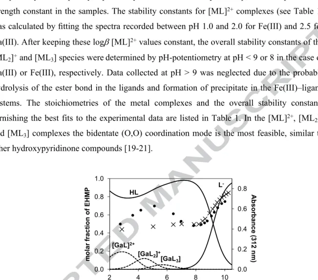
Fig. 1. Concentration distribution curves for the Ga(III)–EHMP system calculated on the basis of the stability constants together with the pH-dependence of the absorbance values at 312 nm (●) and for the ligand alone (×). (cEHMP = 100 M; Ga:L = 1:3; T = 25.0 °C, I = 0.20 M (KCl))
In order to confirm the speciation models obtained by pH-potentiometry, UV spectrophotometric, 1H and 71Ga NMR spectroscopic titrations were applied for the Ga(III) containing systems. The pH-dependent UV spectral changes were significantly different in the acidic pH-range (pH ~2−7) in the presence of Ga(III) compared with the absence of the metal ion as shown for EHMP complexes in Fig. 1. This finding corresponds well with the complex formation processes predicted on the basis of the stability constants obtained by pH- potentiometry. At pH > ~9, however, similar spectra with those of the ligands alone were observed owing to complex dissociation resulting in the release of the ligands and formation of [Ga(OH) ]−.
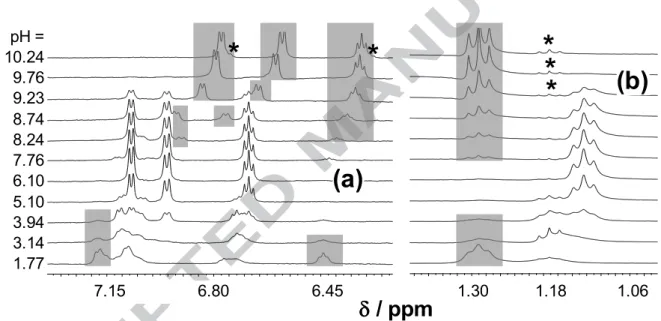
Peaks belonging to the free and Ga-bound ligands were distinguishable in the 1H NMR spectra due to the slow exchange processes on the NMR time-scale (Fig. 2 for EHP). The coordination to Ga(III) is accompanied by significant electronic shielding effects as compared with the HL ligand forms (Fig. 2 and Table 2). Upfield shifts of the aromatic ring, CH3(4), side chain CH2(q) and CH3(t) protons are observed except to CH(5) of EHP and CH(6) of EHMP in meta and ortho position to the pyridine-N atom, respectively. These latter protons appear at higher ppm by ~0.2 units in case of coordination to Ga(III). It is also worth to mention that the proton dissociation processes practically do not affect the chemical shifts of the side chain CH2(q) and CH3(t) protons of the ligands alone [18], although significant upfield shifts (0.15–0.19 ppm) are seen when the ligands are bound to the metal.
6.45 6.80
7.15 ppm
1 2 3 4 5 6 7 8 9 10 11 9.76
8.74
1.06 1.18
1.30
1.42 ppm
1 2 3 4 5 6 7 8 9 10 11 9.76
8.74
7.15 6.80 6.45 1.30 1.18 1.06
(a)
(b)
/ ppm
* *
* *
*
pH = 10.24 9.76 9.23 8.74 8.24 7.76 6.10 5.10 3.94 3.14 1.77
Fig. 2. Low (a) and high (b) field regions of the 1H NMR spectra of the Ga(III)–EHP system recorded at the indicated pH values. Grey boxes indicate signals assigned to the non-bound ligand, and the signals for the hydrolysis product ethanol and carboxylate are marked by stars (cEHP = 4 mM; Ga:L = 1:3; T = 25.0 °C, I = 0.20 M (KCl), 10% D2O).
Table 2. 1H NMR chemical shift (ppm) values of the peaks of ligands EHP and EHMP in their [GaL3] complexes and values of the free ligands for comparison (T = 25.0 ºC, I = 0.20 M (KCl), 10%
D2O).
EHP EHMP
/ ppma [GaL3] HLb L− b [GaL3] HLb L− b
CH(4) (d) 7.070 7.179 6.767 − − −
CH3(4) (s) − − − 2.177 2.187 2.087
CH(5)c 6.685 6.440 6.313 6.983 7.112 6.725
CH(6) (d) 6.954 7.086 6.562 6.615 6.403 6.316
CH2 (q)d 4.088 4.264 4.250 4.062 4.256 4.242
CH3 (t)d 1.124 1.271 1.271 1.115 1.266 1.266
a The signal of -CH2C=O (s) was underneath the water peak.
b Data taken from ref. [18].
c CH(5) EHP: (d/d), EHMP: (d).
d Data of ethanol for comparison: CH2 (q) = 3.65 ppm; CH3 (t) = 1.17 ppm.
Based on these data it can be assumed that the [GaL3] complexes predominate in the pH range 5.5−7.0 in the millimolar concentration range, while considerable dissociation is observed at higher pH values. Integrated areas of the 1H NMR signals of all corresponding protons of the pH-dependent non-bound ligand peaks and those of the complexes (non pH- dependent) were collected and are depicted for EHP in Fig. 3 together with the summed concentration distribution curves calculated with the help of the stability constants. Strong correlation between the data of the two independent methods was found at pH < 9. Most probably the partial hydrolysis of the ester of the ligand becomes significant at pH > 9 as the signals of the hydrolysis product ethanol appear undoubtedly in the spectra with increasing intensity (Fig. 2).
0.0 0.2 0.4 0.6 0.8 1.0
1.5 3.5 5.5 7.5 9.5
molar fraction ofEHP
pH
[metal-free ligand]
[bound ligand]
Fig. 3. Summed concentration distribution curves for the Ga(III)-bound (black line) and free ligand (grey line) species in the Ga(III)–EHP system calculated on the basis of the stability constants and 1H NMR peak integrals of the CH3(t) peaks: bound (×), free (●) ligand and ethanol (○). The grey background shows the pH range where the hydrolysis of the ligand is already considerable (cEHP = 4 mM; Ga:L = 1:3; T = 25.0 °C, I = 0.20 M (KCl), 10% D2O).
The complex formation of EHP with Ga(III) ions was also monitored by 71Ga NMR spectroscopic titrations (Fig. S1). Among the various Ga(III)-hydroxido species only the octahedral [Ga(H2O)6]3+ and the tetrahedral [Ga(OH)4]‒ can be detected by 71Ga NMR, because of their highly symmetric local environment around the metal center [32]. While a peak attributed to [Ga(H2O)6]3+ was seen in the acidic pH range in case of GaCl3, in the presence of EHP practically no signals were detectable due to complexation, as it was expected under the given conditions (Fig. S2). The extent of the formation of gallate in the basic pH range was somewhat lower due to complex formation, although the peak integrals could not be used for quantifications.
In order to clarify the hydrolytic stability of the tris-ligand Ga(III)complexes [GaL3],
1H NMR spectra were recorded over 168 h at pH 7.4 and found to be practically unchanged (Fig. S3).
The formation of the Fe(III) complexes with the ligands EHP and EHMP was accompanied by the appearance of an intense purple color, which was already observed at pH
~1. Therefore, complexation could be followed by UV-Vis spectrophotometric titrations and the development of characteristic CT bands was seen in the visible wavelength range. The position of the max values shows strong pH dependence due to the changes of the coordination environment of the metal ion. max was at 594 nm and a shoulder appeared at 412 nm in case of EHP and at 602 and 408 nm, respectively, for EHMP in a strongly acidic
pH at 1:3 metal-to-ligand ratio and were located at 508 and 416 nm for EHP and at 506 and 414 nm for EHMP (Fig. 4a). These bands reach maxima at pH 6.4−7.4 above which a sudden decrease in the absorbance values occurs, most probably as a consequence of precipitation.
During the pH-potentiometric titrations, precipitation also occurred in the samples independent of the applied metal-to-ligand ratios at pH > ~8.5. Therefore, log values of the [FeL2]+ and [FeL3] complexes were calculated for the data collected below this pH (Table 1).
0.00 0.02 0.04 0.06 0.08 0.10
0.0 0.2 0.4 0.6 0.8 1.0
2 4 6 8 10
Absorbance(508nm)
molar fraction of Fe(III)
pH
[FeL]2+
[FeL2]+
[FeL3]
Fe3+ [Fe(OH)2]+
b)
0.00 0.03 0.06 0.09
340 390 440 490 540 590 640
Absorbance
/nm pH = 1.95
4.63 6.46-7.40
8.055.71 8.78
9.96
a)
Fig. 4. (a) UV-Vis spectra of the Fe(III)–EHP system at various pH values. (b) Concentration distribution curves for the Fe(III)–EHP system calculated on the basis of the stability constants together with the pH dependence of the absorbance values at 508 nm (×). The grey box shows the pH range where the hydrolysis of the ligand is already considerable (cEHP = 0.10 mM; Fe:L = 1:3; T = 25.0 °C, I = 0.20 M (KCl)).
Based on the overall stability constants, concentration distribution curves were calculated for the conditions used in the UV-Vis spectrophotometric titrations, as illustrated for the Fe(III)–EHP system in Fig. 4b, and the pH-dependent absorbance values at a chosen wavelength are shown. The development of the charge transfer bands corresponds well to the calculated speciation up to pH ~ 8. At higher pH values, the absorbance decreased possibly due to the increasing concentration of neutral [FeL3] (or a mixed hydroxido [FeL2(OH)2]), which has limited water solubility. The rate of this process shows pH-dependence and becomes faster at higher pH values (Fig. S4).
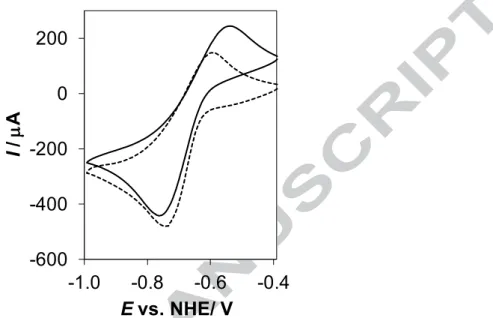
Cyclic voltammograms (Fig. 5) indicate quasi-reversible electrochemical processes for the iron complexes of EHP (E = 104 mV) and deferiprone (E = 149 mV). The redox potential determined for the iron complexes of EHP (E1/2 = ‒0.597 ± 0.019 V vs. NHE) at physiological pH is similar to that of deferiprone (E1/2 = ‒0.627 ± 0.004 V vs. NHE; ‒0.620 V [22]). This relatively negative redox potential of the EHP–Fe(III/II) redox couple suggests a
preference of the 3-hydroxy-2-pyridinone-type ligand towards Fe(III) over Fe(II), similarly to other (O,O) donor iron chelators [20,22]. Therefore, the redox cycling of the iron–EHP complexes is unlikely to occur under aerobic condition.
-600 -400 -200 0 200
-1.0 -0.8 -0.6 -0.4
I/ A
Evs. NHE/ V
Fig. 5. Cyclic voltammogram for the iron complexes of EHP (solid line) and deferiprone (dashed line) at pH 7.4 (cligand = 3 mM; Fe:L = 1:3; T = 25.0 °C, I = 0.20 M (KCl); scan rate: 10 mV/s).
Comparison of the stability of complexes of EHP and EHMP with other related ligands Direct comparison of the stability constants of the Ga(III) and Fe(III) complexes formed with EHP and EHMP (Table 1) reveals that the presence of the extra electron- donating methyl group located adjacent to the coordination site results in higher log values.
It suggests a slightly higher metal binding ability of EHMP, although the pKa of EHMP is also higher. In order to compare the ligand preferences logK*-derived constants were calculated for the neutral [ML3]-type species, which predominate at physiological pH. To obtain the logK* values, the overall stability constants of the [ML3] species were corrected by the different ligand basicities according to the following competition reaction:
M3+ + 3 HL [ML3] + 3 H+ (Eq. 4)
logK* = logβ([ML3]) – 3 × pKa(HL) (Eq. 5)
The logK* values can provide information about the chelate stability in the [ML3] complexes (Fig. 6). A higher logK* implies more favored metal complex formation over the protonation.
The logK* values were compared to those of the related model compound 3-hydroxy-1-
methyl-2(1H)-pyridinone (MH2P [30]; see stability constants in Table 1), deferiprone as a prominent representative of the 3-hydroxy-4-pyridinones [20,30], and maltol [10,23] as a 3- hydroxy-4-pyrone. Consequently, the chelate stability order for both Fe(III) and Ga(III) is the following: EHP < EHMP < MH2P < maltol < deferiprone. Thus it can be concluded that EHP and EHMP possess only moderate binding ability towards the selected metal ions, revealing higher stability for Fe(III) than Ga(III).
Another way frequently used to compare the relative affinities of ligands towards a given metal ion is the calculation of pM value introduced by Raymond et al. [33], which is defined as the negative logarithm of the equilibrium concentrations of the unbound metal ion.
Therefore, higher pM values reflect the stronger metal binding ability of the ligand under a given condition, but are comparable only in the case of a given metal ion. The pM values obtained at pH 7.4 (Table 1) show the same ligand trend as it was seen for the logK* values.
Notably, pFe for EHP and EHMP are significantly lower compared to those of the well- known iron binders deferiprone (20.5 [20], 19.31 [31]) and transferrin (20.3 [21,34]). While the similar pGa values computed for EHP, EHMP, MH2P and maltol show that all the metal ions are in the unbound fraction under the conditions applied (cGa = 1 M; Ga:L = 1:10, pH = 7.4). All these results fit within the generally accepted stability trend, namely the higher ligand effectiveness of the 3-hydroxy-4-pyridinones over the 3-hydroxy-2-pyridinones [19,20].
0.0 1.2 2.4 3.6 4.8 6.0
EHP EHMP MH2P deferiprone maltol
logK*[ML3]
Fe(III) Ga(III) Fe(III) Ga(III)
Fig. 6. Derived stability constants (logK*) for the [ML3] complexes of Fe(III) and Ga(III)with EHP, EHMP, MH2P, deferiprone and maltol. Data for MH2P, deferiprone and maltol were taken from refs.
[10,20,23,30].
The in vitro cytotoxicity of EHP and its in situ formed Ga(III) complex using a 1:3 metal-to- ligand ratio was measured in doxorubicin-sensitive (Colo 205) and multidrug resistant (Colo 320) human colon adenocarcinoma cells using the thiazolyl blue tetrazolium bromide (MTT) method and compared to the activity in non-carcinogenic human embryonal lung fibroblast cells (MRC-5). Cisplatin was used as a positive control (IC50 values: 10.1 ± 0.3 M (Colo 205); 4.78 ± 0.11 M (Colo 320); 2.61 ± 0.07 M (MRC-5)). The ligand and its Ga(III) complex did not show cytotoxic activity (IC50 > 100 μM) in the tested cell lines. In previous work on the Ru(II)(6-p-cymene) complexes of EHP and EHMP we observed weak cytotoxic activity for the complexes in CH1 human ovarian cancer cells (IC50 values of 232 and 242 M, respectively), as well as for the ligands [17].
The Ga(III) complexes of hydroxypyrones and hydroxypyridinones often show moderate cytotoxicity [5,7,19], however, the complexes of maltol and deferiprone possess higher solution stability compared to those of EHP and EHMP as described in the section
‘Comparison of the stability of complexes of EHP with EHMP and with other related ligands’. Therefore, the solution stability of the Ga(III) complex of EHP in the presence of cell culture medium components was monitored by 1H NMR spectroscopy. Cancer cells were grown in cell culture medium RPMI1640 supplemented with 10% heat-inactivated fetal bovine serum (FBS). This modified medium contains numerous inorganic salts, amino acids and various other small biomolecules (e.g. folic acid, L-glutamic acid) and various serum proteins (including transferrin) from FBS and some of them are considered as potential binders of Ga(III). The recorded 1H NMR spectra (Fig. 7a) show that ~9% of the EHP complex [GaL3] dissociates at the applied 1 mM concentration at pH 7.4 leading to the detection of a small portion of the unbound ligand. This was expected on the basis of the stability constants determined as the concentration distribution curves (Fig. 7b). The level of complex decomposition did not increase in either medium used, i.e., in RPMI1640 or RPMI1640+FBS (Fig. 7a), when using the same conditions as above. When the concentration of the complex was decreased a significant decomposition could be expected (Fig. 7b) which is also suggested to be responsible for the inactivity of the complex against the cancer cells.
Notably complete binding of Ga(III) to transferrin is predicted in the blood serum based on the stability constants of the Ga(III)-transferrin [34] and Ga(III)-EHP complexes at concentration lower than 50 M.
6.4 6.5 6.6 6.7 6.8 6.9 7.0 7.1 7.2 7.3
7.4 f1 (ppm)
1 2 3 4 5 6
ge3 6 6
20 5 5
ge1 4 4
eh1 3 3
30 2 2
rpmi1640
1 1 0.0
0.2 0.4 0.6 0.8 1.0
-5 -4.5 -4 -3.5 -3
molar fraction of [GaL3]
-log c[GaL3]
[GaL3]
[Ga(OH)3] [Ga(OH)4]-
EHP
RPMI1640+FBS RPMI1640 [GaL3] [GaL3] in RPMI1640 [GaL3]
in RPMI1640+FBS
7.4 7.2 7.0 6.8 6.6 6.4
/ ppm
a) b)
Fig. 7. 1H NMR spectra of the media used for the cytotoxicity test, EHP (3 mM) and its [GaL3] complex (1 mM) alone and in the media at pH 7.4 (a). Concentration distribution of the [GaL3] complex of EHP in the concentration range from 1 mM down to 10 M at pH 7.4 (b).
Conclusions
Solution equilibrium studies on Ga(III) and Fe(III) complexes of the (O,O) donor 3-hydroxy- 2-pyridinone derivatives EHP and EHMP were performed in aqueous solution by pH- potentiometric and spectroscopic (UV-Vis: Fe(III); 1H NMR: Ga(III)) methods. These bidentate ligands form [ML]2+, [ML2]+ and [ML3] type species with Ga(III) and Fe(III) ions, and the neutral [ML3] complexes predominate at physiological pH. EHP and EHMP form complexes of higher stability with Fe(III) than with Ga(III), and the presence of the methyl substituent on the aromatic ring in EHMP results in a stronger metal binding ability compared to EHP. The studied 3-hydroxypyridin-2-one derivatives form lower stability complexes than deferiprone (as a representative of 3-hydroxy-4-pyridinone) and maltol (a 3- hydroxy-4-pyrone) in all cases. The [GaL3] complex of EHP suffers 60% decomposition at 100 M concentration based on the determined stability constants. This moderate stability probably contributes to the low cytotoxicity (IC50 > 100 μM) of the [GaL3] complex of EHP in doxorubicin-sensitive (Colo 205) and multidrug resistant (Colo 320) human colon adenocarcinoma cell lines. However, this complex does not show significant decomposition at 1 mM concentration even in the cell culture medium supplemented with 10% fetal bovine serum. On the other hand the complex is predicted not to preserve its original entity under more diluted conditions and the coordinating ligand is supposed to be exchanged partly or completely by endogenous bioligands, such as human serum transferrin in the concentration
range lower than 50 M. The Fe(III) complexes of EHP and EHMP have lower solution stability than that of deferiprone, and the redox potentials show stronger preference towards Fe(III) than to Fe(II), as is seen in case of traditional (O,O) donor iron chelators.
Acknowledgements
This work was supported by National Research, Development and Innovation Office, (Hungary) through projects GINOP-2.3.2-15-2016-00038, FK 124240 and Ministry of Human Capacities (Hungary) grant 20391-3/2018/FEKUSTRAT.
References
[1] L.R. Bernstein, T. Tanner, C. Godfrey, B. Noll, Metal-Based Drugs 7 (2000) 33–
47. doi: 10.1155/MBD.2000.33
[2] G.N. Kaluderovic, R. Paschke, Curr. Med. Chem. 18 (2011) 4738−4752. doi:
10.2174/092986711797535308
[3] V. Brabec, O. Hrabina, J. Kasparkova, Coord. Chem. Rev. 351 (2017) 2–31. doi:
10.1016/j.ccr.2017.04.013
[4] M.A. Jakupec, M. Galanski, V.B. Arion, C.G. Hartinger, B.K. Keppler, Dalton Trans. (2008) 183–194. doi: 10.1039/B712656P
[5] C.R. Chitambar, Metallo-Drugs: Development and Action of Anticancer Agents, Gallium Complexes as Anticancer Drugs (Eds. A. Sigel, H. Sigel, E. Freisinger, R.K.O. Sigel), Walter de Gruyter GmbH, Berlin, Germany, in Metal Ions Life Sci.
18, 2018, pp, 281–301. doi: 10.1515/9783110470734-016
[6] P. Collery, B.K. Keppler, C. Madoulet, B. Desoize, Crit. Rev. Oncol. Hematol. 42 (2002) 283–296. doi: 10.1016/S1040-8428(01)00225-6
[7] C.R. Chitambar, Pharmacol. Res. 115 (2017) 56–65. doi:
10.1016/j.phrs.2016.11.009
[8] P. Collery, M.A. Jakupec, B. Kynast, B.K. Keppler, Preclinical and early clinical development of the oral gallium complex KP46 (FFC11), in: Metal Ions in Biology and Medicine (Eds. M.C. Alpoim, P.V. Morais, M.A. Santos, L.
Cristovao, J.A. Centeno, P. Collery), John Libbey Eurotext, Paris, 2006, pp. 521–
524.
[9] A.R. Timerbaev, Metallomics 1 (2009) 193–198. doi: 10.1039/B902861G
[10] É.A. Enyedy, O. Dömötör, E. Varga, T. Kiss, R. Trondl, C.G. Hartinger, B.K.
Keppler, J. Inorg. Biochem. 117 (2012) 189–197. doi:
10.1016/j.jinorgbio.2012.08.005
[11] É.A. Enyedy, O. Dömötör, K. Bali, A. Hetényi, T. Tuccinardi, B.K. Keppler, J.
Biol. Inorg. Chem. 20 (2015) 77–88. doi: 10.1007/s00775-014-1211-9
[12] M. Groessl, A. Bytzek, C.G. Hartinger, Elecrophoresis 30 (2009) 2720–2727. doi:
10.1002/elps.200800745
[13] Z.D. Liu, R.C. Hider, Med. Res. Rev. 22 (2002) 26–64. doi: 10.1002/med.1027 [14] G. Crisponi, V. M. Nurchi, M.A. Zoroddu, Thalassemia reports (2014) doi:
10.4081/thal.2014.2046
[15] R. Cusnir, C. Imberti, R.C. Hider, P.J. Blower, M.T. Ma, Int. J. Mol. Sci. 18 (2017) 116. doi: 10.3390/ijms18010116
[16] S. Chaves, A.C. Mendonça, S.M. Marques, M.I. Prata, A.C. Santos, A.F. Martins, C.F. Geraldes, M.A. Santos, J Inorg Biochem. 105 (2011) 31–38. doi:
10.1016/j.jinorgbio.2010.09.012
[17] M. Hanif, H. Henke, S.M. Meier, S.Martic, M. Labib, W. Kandioller, A. Jakupec, V.B. Arion, H.B. Kraatz, B.K. Keppler, C.G. Hartinger, Inorg. Chem. 49 (2010) 7953–7963. doi: 10.1021/ic1009785
[18] E.A. Enyedy, G.M. Bognár, T. Kiss, M. Hanif, C.G. Hartinger, J. Organomet.
Chem. 734 (2013) 38–44. doi:10.1016/j.jorganchem.2012.10.042
[19] M.A. Santos, S.M. Marques, S. Chaves, Coord. Chem. Rev. 256 (2012) 240−259.
doi: 10.1016/j.ccr.2011.08.008
[20] A. Cilibrizzi, V. Abbate, Y.-L. Chen, Y. Ma, T. Zhou, R.C. Hider, Chem. Rev.
118 (2018) 7657–7701. doi: 10.1021/acs.chemrev.8b00254
[21] R.J. Motekaitis, A.E. Martell, Inorg. Chim. Acta 183 (1991) 71–80. doi:
10.1016/S0020-1693(00)82997-7
[22] Y. Ma, X. Kong, Y. Chen, R.C. Hider, Dalton Trans. 43 (2014) 17120−17128.
doi: 10.1039/c4dt02687j
[23] A. Stefanovic, J. Havel, L. Sommer, Collect. Czech. Chem. Commun. 33 (1968) 4198–4214. doi: 10.1135/cccc19684198
[24] P. Gans, A. Sabatini, A. Vacca, Talanta 43 (1996) 1739–1753. doi: 10.1016/0039- 9140(96)01958-3
[25] H.M. Irving, M.G. Miles, L.D. Petit, Anal. Chim. Acta 38 (1967) 475–482. doi:
[26] SCQuery, The IUPAC Stability Constants Database, Academic Software (Version 5.5), R. Soc. Chem., 1993–2005.
[27] L. Zékány, I. Nagypál in Computational Methods for the Determination of
Stability Constants (Ed.: D. L. Leggett), Plenum Press, New York, 1985, pp. 291–
353.
[28] E. Farkas, E. Kozma, T. Kiss, I. Toth, B. Kurzak, J. Chem. Soc. Dalton Trans. 0 (1995) 477–481. doi: 10.1039/DT9950000477
[29] C. F. Baes, R. E. Mesmer, The Hydrolysis of Cations, Wiley, New York, 1976.
doi: 10.1002/bbpc.19770810252
[30] E.T. Clarke, A.E. Martell, Inorg. Chim. Acta 196 (1992) 185−194. doi:
10.1016/S0020-1693(00)86122-8
[31] M.A. Santos, S. Gama, L. Gano, G. Cantinho, E. Farkas, Dalton Trans. 0 (2004) 3772−3781. doi: 10.1039/B409357G
[32] Q. Zhou, C. Henoumont, L. Vander Elst, S. Laurent, R.N. Muller, Contrast Media Mol. Imaging 6 (2011) 165–167. doi: 10.1002/cmmi.441
[33] K.N. Raymond, C.J. Carrano, Acc. Chem. Res. 12 (1979) 183–190. doi:
10.1021/ar50137a004
[34] W.R. Harris, V.L. Pecoraro, Biochemistry 22 (1983) 292–299. doi:
10.1021/bi00271a010
Graphical abstract
The solution speciation of Ga(III) and Fe(III) complexes of two (O,O) donor bearing alkoxycarbonylmethyl-3-hydroxy-2(1H)-pyridinone ligands was characterized. Moderate stabilities were observed with [ML3] complexes predominating at physiological pH.
Significant decomposition of the Ga(III) complexes occurs at low concentration leading to negligible cytotoxic activity in human adenocarcinoma cells.
Graphical abstract
The solution speciation of Ga(III) and Fe(III) complexes of two (O,O) donor-bearing alkoxycarbonylmethyl-3-hydroxy-2(1H)-pyridinone ligands was characterized. Moderate stabilities were observed with [ML3] complexes predominating at physiological pH.
Significant decomposition of the Ga(III) complexes occurs at low concentration leading to negligible cytotoxic activity in human adenocarcinoma cells.

![Table 2. 1 H NMR chemical shift (ppm) values of the peaks of ligands EHP and EHMP in their [GaL 3 ] complexes and values of the free ligands for comparison (T = 25.0 ºC, I = 0.20 M (KCl), 10%](https://thumb-eu.123doks.com/thumbv2/9dokorg/1074591.71969/12.892.161.797.188.568/table-chemical-values-ligands-complexes-values-ligands-comparison.webp)

![Fig. 6. Derived stability constants (logK*) for the [ML 3 ] complexes of Fe(III) and Ga(III) with EHP, EHMP, MH2P, deferiprone and maltol](https://thumb-eu.123doks.com/thumbv2/9dokorg/1074591.71969/16.892.99.780.530.968/fig-derived-stability-constants-complexes-ehmp-deferiprone-maltol.webp)
![Fig. 7. 1 H NMR spectra of the media used for the cytotoxicity test, EHP (3 mM) and its [GaL 3 ] complex (1 mM) alone and in the media at pH 7.4 (a)](https://thumb-eu.123doks.com/thumbv2/9dokorg/1074591.71969/18.892.117.802.138.419/fig-nmr-spectra-media-used-cytotoxicity-complex-media.webp)
