ARTICLE
a.Department of Inorganic and Analytical Chemistry, Interdisciplinary Excellence Centre, University of Szeged, Dóm tér 7, H-6720 Szeged, Hungary; E-mail:
meszaros.janos@chem.u-szeged.hu; enyedy@chem.u-szeged.hu
b.MTA-SZTE Lendület Functional Metal Complexes Research Group, University of Szeged, Dóm tér 7, H-6720 Szeged, Hungary
c. Institute of Enzymology, Research Centre for Natural Sciences, Magyar Tudósok körútja 2, H-1117 Budapest, Hungary
d.Department of Physiology, Semmelweis University, Tűzoltó utca 37-47, H-1094 Budapest, Hungary
e. Institute of Cancer Research, Medical University of Vienna, Borschkegasse 8a, A- 1090 Vienna, Austria
f. Centre for Structural Science, Research Centre for Natural Sciences, Magyar tudósok körútja 2, H-1117 Budapest, Hungary
g.Institute of Organic Chemistry, Research Centre for Natural Sciences, Magyar tudósok körútja 2, H-1117 Budapest, Hungary
†Electronic Supplementary Information (ESI) available: 1H and 13C NMR spectra of complexes, list of NMR peaks, ESI-MS spectra, crystallographic data and additional spectra about aqueous stability (PDF). See DOI: 10.1039/x0xx00000x The crystallographic data files for the complexes have been deposited with the Cambridge Crystallographic Database as CCDC 2038629-2038634.
Received 00th January 20xx, Accepted 00th January 20xx DOI: 10.1039/x0xx00000x
Half-sandwich organometallic Ru and Rh complexes of (N,N) donor compounds: effect of ligand methylation on solution speciation and anticancer activity
János P. Mészáros,a,b,* Veronika F.S. Pape,c,d Gergely Szakács,c,e Gábor Németi,a Márk Dénes,f Tamás Holczbauer,f,g Nóra V. May,f Éva A. Enyedya,b,*
A series of half-sandwich polypyridyl complexes was synthesized and compared focusing on structural, cytotoxic and aqueous solution behaviour. The formula of the synthesized complexes is [M(arene)(N,N)Cl]Cl, where M: Ru or Rh, arene:
p-cymene, toluene or C5Me5‒, (N,N): 2,2’-bipyridine (bpy), 4,4’-dimethyl-2,2’-bipyridine (dmb), 1,10-phenanthroline (phen) or 2,9-dimethyl-1,10-phenanthroline (neo). The structures of five half-sandwich complexes were determined by X-ray crystallography. It was found that introducing methyl groups next to the coordinating nitrogen atoms of the bidentate ligand causes steric congestion around the metal centre which changes the angle between ligand planes. The ligands and the Rh complexes showed significant cytotoxicity in A2780 and MES-SA cancer cell lines (IC50 = 0.1–56 μM) and in the cisplatin-resistant A2780cis cells. Paradoxically, phen and dmb as well as their half-sandwich Rh complexes showed increased toxicity against multidrug resistant MES-SA/Dx5 cells. In contrast, coordination to Ru caused loss of toxicity.
Solution equilibrium constants showed that the studied metal complexes have high stability, and no dissociation was found for Ru and Rh complexes even at micromolar concentrations in a wide pH range. However, in case of Ru complexes a slow and irreversible decomposition, namely arene loss was also observed, which was more pronounced in light exposure in aqueous solution. In case of neo, the methyl groups next to the nitrogen atoms significantly decrease the stability of complexes. For Rh complexes, the order of the stability constants corrected with ligand basicity (log K*): 9.78 (phen) > 9.01 (dmb) > 8.89 (bpy) > 3.93 (neo). The coordinated neo resulted in an enormous decrease in the chloride ion affinity of Ru compounds. Based on the results, a universal model was introduced for the prediction of chloride ion capability of half-sandwich Rh and Ru complexes. It combines the effects of the bidentate ligand and the M(arene) part using only two terms, performing multilinear regression procedure.
Introduction
Based on the success of cisplatin, complexes of other platinum group metal ions were developed and introduced into clinical trials.
Ru(III) complexes, namely NAMI-A and BOLD-100 (formerly known
as KP-1339)1 and the Ru(II) containing TLD1433 entered clinical trials.1 The proposed mechanism is that they are activated by reduction. Based on this idea several Ru(II) complexes have been synthesized, possessing an organometallic half-sandwich structure having a bidentate ligand and a monodentate leaving group. Early examples contained ethylenediamine and halide ion as ligands (the so-called RAED complexes),1,2 which were followed by the Os(II), Ir(III) and Rh(III) analogues and with different bidentate ligands.
Some of these half-sandwich complexes showed remarkable cytotoxic activity and several structure-activity relationship analyses were conducted to identify the key chemical parameters.1,3 The mechanism of action of these compounds show a wide variety, as the RAED complexes are capable of DNA-binding,4 enzyme inhibition (cathepsin B, thioredoxin reductase),5,6 and the complexes of phenylazopyridines with [Ru/Os(6-p- cymene)(H2O)3]2+ show catalytic GSH oxidation.7,8 The latter is the primary mechanism of the ‘catalytic metallodrugs’.9 The group of Sadler proved the occurrence of intracellular catalysis by half- sandwich complexes of (N,N) donor ligands, such as ethylenediamine, 2,2’-bipyridine (bpy) and 1,10-phenanthroline (phen).10 The reaction of these organometallic complexes with the NAD+/NADH pair was found in the presence of formate ions.
Polypyridines (phen and bpy) and their myriad derivatives are common ligands for half-sandwich complexes. Detailed solution behaviour and/or biological data were reported with Ru,3,11-13 Rh,14 Ir14-16 and Os16,17. These ligands usually show similar or higher cytotoxic activity than their metal complexes.12 However, complex formation in general might have advantageous effects on the selective cytotoxicity on cancer cells, since it can change the overall charge, lipophilicity and size, which affect the pharmacokinetics and can also result in altered mechanism of action. Different biological effects were found for Ru/Rh polypyridyl complexes. DNA intercalation was reported in the coordinatively saturated and kinetically inert tris-polypyridyl Ru and Rh complexes, in which mostly phen and its derivatives are the ligands.18,19 On the other hand, taking into consideration the viscosity measurements, cytotoxicity data and ultrafiltration measurements, DNA- intercalation is not likely to occur and DNA is not believed to be the target molecule of half-sandwich complexes [Ru/Rh(arene)(phen)(H2O)]2+.20-22 There are also examples for topoisomerase I and II inhibitors,23 octahedral and half-sandwich cholinesterase inhibitors.24
In the field of cancer treatment, one of the major impediments is the appearance of resistance to chemotherapeutic agents. Cellular mechanisms promoting multidrug resistance (MDR) often rely on the elevated expression of ATP-binding cassette proteins, which pump a wide variety of drug molecules from the cell.25-27 Drug resistant cells resort to further mechanisms in the case of compounds that are not recognized as transported substrates. In the case of cisplatin, cells become resistant as a result of elevated glutathione concentrations and increased DNA-repair.26 Interestingly, the majority of RAED compounds seemed to overcome cisplatin resistance in the A2780cis cell lines model.3 Similarly, a Ru cyclopentadienyl complex containing 4,4’-dimethyl- 2,2’-bipyridine (dmb) showed comparable activity in parental and in cisplatin-resistant cancer cells.28 In our former studies, we reported half-sandwich complexes with high stability, in which the ligands were 8-hydroxyquinoline derivatives.29,30 The ligands substituted at the 7th position showed preferential toxicity in otherwise multidrug resistant MES-SA/Dx5 and Colo320 cell lines, and this characteristic persisted after combination with the half-sandwich organometallic [Rh(5-C5Me5)(H2O)3]2+ triaqua cation.29,30 However, complex formation with [Ru(6-arene)(H2O)3]2+ resulted in a decrease in cytotoxicity and the loss of preferential toxicity.29,30
In this study, we selected four polypyridines and their half-sandwich Ru and Rh complexes to reveal possible relationships between their
Scheme 1 Synthesis procedure of the complexes.
structure, aqueous solution behaviour and anticancer activity against parental and drug resistant cancer cell lines. We synthesized novel Ru(6-toluene) to investigate the effects of the exchanges of p-cymene to toluene, and 2,9-dimethyl-1,10-phenanthroline (neocuproine, neo) complexes were also prepared to reveal the effect of methylation close to the coordinating nitrogens.
Results and discussions
Synthesis and characterization of complexes
As illustrated in Scheme 1, synthesis of complexes with the general formula [M(arene)(N,N)Cl]Cl, where M: Ru or Rh, arene: p-cymene (p-cym), toluene (tol), C5Me5‒
, (N,N): bpy, dmb, phen and neo, was performed according to previously described methods.11,31-33 Among the listed complexes, Ru(6-tol) complexes and neo complexes are new compounds. In this work, all the [M(arene)(N,N)Cl]Cl complexes were obtained as orange solids with moderate-to-excellent yields (45-95%) using methanol (MeOH) as solvent. Notably, the neo complexes were isolated in the lowest yields, which could be improved by using an excess of the ligand.
[Ru(6-p-cym/tol)(ethylenediamine)Cl]Cl complexes are well-known compounds,2 they were also synthesized with the same method for further solution chemical and comparative purposes.
1H and 13C NMR spectra recorded in CD3OD confirmed complex formation, as shown in Figures S1-S14. Deuteration of the C5Me5
ligand (-CH2D and -CHD2 groups) was detected in the 1H and 13C NMR spectra, as three peaks are shown in Figure S7 with the same intensity next to the peak of the methyl groups. ESI-MS spectra were recorded only for the novel complexes, and the results confirmed the stoichiometry of each complex (Figures S15-S21).
Stability and photosensitivity of the complexes were investigated in water (at pH 7.4). Notably, the Rh complexes were stable in water for at least 7 days, as the yellow colour and the 1H NMR spectra remained unchanged (see Figure S22 as an example). In order to investigate the photostability of the Ru complexes parallel samples of [Ru(6-tol)(bpy)(H2O)]2+, [Ru(6-p-cym)(bpy)(H2O)]2+ and [Ru(6- p-cym)(phen)(H2O)]2+ were prepared and followed in time by UV- visible (UV-Vis) spectrophotometry. One of the parallel samples was protected from light, while the other was exposed to diffuse solar irradiation. The starting solutions had yellow colour, which is typical for half-sandwich complexes. Spectra of the complexes (shown in Figure S23) remained unchanged in dark after one day, except for [Ru(6-tol)(bpy)(H2O)]2+, which showed signs of decomposition after 18 h (Figure S23.a). Namely, the sample turned blue-green, and a new band developed in the UV-visible (UV-Vis) spectra, first after 18 h with max = 588 nm, and after 6 days another band appeared with max = 644 nm. When exposed to light for a longer period of time, all samples showed signs of this process, which is most probably linked to the irreversible decomposition of the half- sandwich structure (also known as arene loss). The decomposition of [Ru(6-p-cym)(bpy)(H2O)]2+ is slower than that of the toluene analogue, which appears only after more than 1 day. The product has the same max at 588 nm (Figure S23.b), which is indicative of the loss of arene. The product of [Ru(6-p-cym)(phen)(H2O)]2+ has a
max = 658 nm, where a tiny amount (Amax~0.03) appears only in light after 4 days (Figure S23.c). These experiments suggest that the
metal−carbon bond in the organometallic moiety can break and both the type of the metal ion and the arene have important roles in the stabilization: the bond between Ru(II) and p-cymene is more stable than between Ru(II) and toluene, while the bond between Rh(III) and C5Me5-
is the strongest in this set of complexes. In case of Ru complexes formed with other (N,N) bidentate ligands, the loss of the arene ligand was reported earlier.34 In our previous studies, arene loss induced by ligand excess or by another coordinating bidentate ligand was observed.29,30 Stability studies of the bpy complexes was followed in the cell culture medium Roswell Park Memorial Institute Medium 1640 (RPMI 1640) completed with fetal bovine serum (FBS) in dark. Under these conditions no arene ligand dissociation was detected (Figure S24).
Reactions of the complexes in the cell culture medium Dulbecco’s Modified Eagle Medium (DMEM) was also followed for a week by
1H NMR spectroscopy. This medium provides physiological pH and contains inorganic salts, amino acids, sugar and vitamins, which may interact with these complexes. Figure S25 shows the important ranges of the recorded spectra, focused on the interactions with biomolecules. In the case of [Rh(5-C5Me5)(phen)(H2O)]2+ and [Ru(6-p-cym)(phen)(H2O)]2+ complexes after 5 h new peaks appeared showing the formation of mixed ligand complexes. After this period, no big changes in the spectra could be observed after one week, except in the case of neo complexes, which showed a slow reaction and changed continuously. The complex of [Ru(6-p- cym)(neo)(H2O)]2+ partially dissociated even in the phosphate buffer, indicating its lower stability at pH = 7.40. The measurements were repeated in RPMI 1640 medium completed with FBS (Figure S26). New sets of peaks were observed only for the [Rh(5- C5Me5)(phen)(H2O)]2+ complex compared to the spectra measured in DMEM.
The structures of (N,N) donor bidentate ligands complexes formed with Ru(6-arene) and Rh(5-C5Me5) organometallic cations are well-known and several examples show the complexes in chlorinated3,12,13,20 or in aqua form35,36 in the solid structures. The different arene ligands did not change the piano-stool shaped structure. However, Ru−N and Ru−ring centroid distances can vary in these complexes. Generally, the structures represent mono complexes, in which the ligands are bound to the metal centre through two nitrogen atoms.
After counter ion exchange (Cl- to CF3SO3), single crystals for complexes [Rh(5-C5Me5)(dmb)Cl](CF3SO3) (crystal I), [Ru(6- tol)(dmb)Cl](CF3SO3) (crystal II) and [Ru(dmb)3](CF3SO3)2×2 H2O (crystal III) were obtained, which were subjected to X-ray crystallographic structure determination. Crystal data and structure refinement parameters are collected in Table S1. The results proved the presence of half-sandwich structure of the [Rh(5- C5Me5)(dmb)Cl]+ and [Ru(6-tol)(dmb)Cl]+ complexes, similarly to the other [Ru(6/5-arene)(polypyridyl)Cl]+ complexes, e.g. [Ru(6-p- cym)(dmb)Cl]+.37 However, a dark red crystal was also isolated that showed evidence of arene loss and the formation of [Ru(dmb)3](CF3SO3)2×2 H2O with octahedral structure. This irreversible reaction occurred in organic solvents and in water as well (vide supra), as the colour of solutions turned to green-blue over time. The [Rh(5-C5Me5)(dmb)Cl](CF3SO3) complex crystallized
Fig. 1 Molecular structures of a) [Rh(5-C5Me5)(dmb)Cl](CF3SO3) (I), b) [Ru(6-tol)(dmb)Cl](CF3SO3) (II) c) [Ru(dmb)3].2(CF3SO3)×2 H2O (III), d) [Ru(6-tol)(neo)Cl]Cl×2 MeOH (IV), e) [Ru(6-p-cym)(neo)Cl](CF3SO3) (V) and f) [Rh(5-C5Me5)(neo)Cl](CF3SO3) (VI). Hydrogen atoms, solvent molecules and counter ions are omitted for clarity. Displacement ellipsoids are drawn at 50% probability level.
in the orthorhombic and both Ru complexes in the monoclinic crystal systems in Pbcn, P21/n and P21/c space groups, respectively, with the inclusion of a CF3SO3-
counter ion (and two water molecules in the latter) per asymmetric unit. The ORTEP representation of the compounds is depicted in Figure 1.a,b,c.
Crystal structures of [Ru(6-tol)(neo)Cl]Cl×2 MeOH (crystal IV), [Ru(6-p-cym)(neo)Cl](CF3SO3) (crystal V) and [Rh(5- C5Me5)(neo)Cl](CF3SO3) (crystal VI) could be also obtained (Figure 1.d,e,f). Crystal data and structure refinement parameters for neo complexes are collected in Table S2. Crystal IV and V crystallized in the monoclinic and crystal VI in the triclinic crystal systems in P21/c (IV, V) and P-1 (VI) space groups. The asymmetric unit contains one complex and one counter ion in crystal V, two extra MeOH molecules in crystal IV and two complexes with two counter ions in crystal VI. Selected bond lengths and angles are collected in Tables S3-S4. In the the neo complexes, the neo ligand is not fully planar. The angle enclosed by the planes of the outer ring is bended for IV and V (12.7° and 9.8°, respectively) and the most bent ring was found in crystal VI where this angle is 17.2° and 17.7° for molecules 1 and 2. The bar chart in Figure S27 shows the metal ion−N atom distances, which are longer for neo complexes and are the shortest in the tris-dmb complex.
The structures of [Ru(6-tol)(dmb)Cl](CF3SO3) and [Ru(6-p- cym)(dmb)Cl](BF4)37 are practically identical as the average Ru−N bond lengths are 2.088 Å vs. 2.085 Å, the Ru−ring centroid distances are 1.686 Å vs. 1.685 Å. The two Ru complexes of neo also show a strong similarity, the change of arene has no effect on the
a) b)
c) d)
e) f)
geometrical parameters (Figure S28.a). The geometrical changes are negligible when p-cymene is substituted to toluene ligand.
All neocuproine containing structures differ from the analogous phen complexes,13,20 as the steric congestion around the metal ion is caused by the methyl groups in the 2nd and 9th positions. For instance the Rh−N bond length changed from 2.11 Å to 2.13 Å, in case of exchanges of phen to neo change. A spectacular proof of the steric hindrance is the finding that the planes of the arene ligand and of the bidentate ligand are not the same. The difference between [Ru(6-p-cym)(neo)Cl]+ and [Ru(6-p-cym)(phen)Cl]+ complexes is shown in Figure S28.b.13 In this example there is a 19°
alteration between the plane of phen and neo. The best visualization for this steric congestion is provided by the ligand solid angles calculated by the Olex2 software.38,39 Figure S29 shows the ligand solid angles in the complex of [Ru(6-p-cym)(neo)Cl]+, from two different views. Less overlap is present between the ligands around Rh(III) than in the two Ru(II) complexes (see more details in the legend of Figure S29). For crystallization of all neocuproine complexes we also tried to perform anion exchange using Ag(CF3SO3) salt. However, during the crystallization procedure, red crystals of the precursor [M(arene)Cl2]2 dimer and colourless crystals of [Ag(neo)2](CF3SO3) appeared in these samples, most probably as a consequence of the low stability of the neocuproine complexes (notably they were also characterized by the lowest yields in synthesis). Scheme S1 shows side reactions of neocuproine complexes.
In vitro anticancer activity
The anticancer activity of four related polypyridyl ligands (phen, neo, bpy, dmb) and their half-sandwich Ru(6-p-cym), Ru(6-tol) and Rh(5-C5Me5) complexes was investigated against the uterine sarcoma cell line MES-SA and its doxorubicin resistant counterpart MES-SA/Dx5, as well as against the ovarian cancer cell line A2780 and its cisplatin resistant counterpart A2780cis. A2780cis cells show an increased ability to repair DNA and have higher intracellular concentrations of glutathione,26 while MES-SA/Dx5 cells overexpress P-gp,26,40 which results in multidrug resistance.
The paradoxical toxicity of phen against P-glycoprotein (P-gp)- expressing MDR cells (MDR-selective toxicity) was reported earlier.41 A summary of the literature data on the in vitro toxicity of the half-sandwich complexes of the selected ligands on other cancer cell lines is shown in the Electronic Supplementary Information.13,14,42,43
The obtained IC50 values are shown in Tables 1-2 and in Figure S30.
The relative toxicity of the complexes against parental and drug resistant cancer cell lines was compared, and the selective toxicity was expressed as resistance ratio (RR = IC50 (resistant cell) / IC50
(sensitive cell)). Based on the determined IC50 values (Tables 1-2), the ligands phen, neo and dmb displayed significant toxicity, reaching submicromolar IC50 values in some cases. Neocuproine has a superior cytotoxic effect, it is comparable with doxorubicin (and 10 times higher than phen) in MES-SA cells, while 20 times more active than cisplatin in A2780 cells. The toxicity of the [Rh(5- C5Me5)(N,N)Cl]+ complexes were similar or slightly lower compared to that of their corresponding ligands. Surprisingly, the Ru
complexes exhibited in all cases weaker cytotoxicity than the Rh congeners.
Table 1 In vitro cytotoxic effects (72 h) (IC50 values in M) in parental (MES- SA) and multidrug resistant (MES-SA/Dx5) cell lines treated with polypyridine ligands and their half-sandwich complexes in addition to the corresponding organometallic precursors. Resistance ratio (RR = IC50(MES- SA/Dx5)/IC50(MES-SA)) values are also represented. N. d. = not determined (in cases where both IC50 > 100 M).
MES-SA MES-
SA/Dx5 RR
phen 4 ± 1 1.30 ± 0.01 0.33
[Rh(5-C5Me5)(phen)Cl]Cl 8 ± 2 2.0 ± 0.9 0.25 [Ru(6-tol)(phen)Cl]Cl > 100 > 100 n. d.
[Ru(6-p-cym)(phen)Cl]Cl 24 ± 12 > 100 >4.2
neo 0.37 ± 0.08 0.30 ± 0.04 0.81
[Rh(5-C5Me5)(neo)Cl]Cl 1.4 ± 0.1 2.66 ± 0.09 1.90 [Ru(6-tol)(neo)Cl]Cl 2.1 ± 0.2 4.2 ± 0.7 2.00 [Ru(6-p-cym)(neo)Cl]Cl 4 ± 1 7 ± 1 1.75
bpy 66 ± 19 50 ± 15 0.75
[Rh(5-C5Me5)(bpy)Cl]Cl > 100 69 ± 25 <0.7 [Ru(6-tol)(bpy)Cl]Cl > 100 > 100 n. d.
[Ru(6-p-cym)(bpy)Cl]Cl > 100 > 100 n. d.
dmb 46 ± 7 15 ± 2 0.33
[Rh(5-C5Me5)(dmb)Cl]+ 41 ± 6 16 ± 6 0.39 [Ru(6-tol)(dmb)Cl]+ > 100 > 100 n. d.
[Ru(6-p-cym)(dmb)Cl]+ 63 ± 11 > 100 >1.59 [Rh(5-C5Me5)Cl2]2 > 100 > 100 n. d.
[Ru(6-tol)Cl2]2 > 100 > 100 n. d.
[Ru(6-p-cym)Cl2]2 > 100 > 100 n. d.
doxorubicin 0.35 ± 0.06 3 ± 0.9 8.57
Table 2 In vitro cytotoxic effect (72 h) (IC50 values in M) of polypyridine ligands and their half-sandwich complexes in addition to the organometallic precursors in sensitive (A2780) and cisplatin resistant (A2780cis) human ovarian cancer cell lines. Resistance ratio (RR = IC50(A2780cis)/IC50(A2780)) values are also represented. N. d. = not determined (in cases where both IC50 > 100 M).
A2780 A2780cis RR
phen 0.14 ± 0.03 2.5 ± 0.2 17.9
[Rh(5-C5Me5)(phen)Cl]Cl 0.28 ± 0.09 10 ± 3 35.7 [Ru(6-tol)(phen)Cl]Cl 38 ± 12 > 100 >2.6 [Ru(6-p-cym)(phen)Cl]Cl 11 ± 2 > 100 >9.1
neo 0.13 ± 0.03 9 ± 2 69.2
[Rh(5-C5Me5)(neo)Cl]Cl 0.4 ± 0.2 1.2 ± 0.3 3.0 [Ru(6-tol)(neo)Cl]Cl 0.6 ± 0.1 1.5 ± 0.3 2.5 [Ru(6-p-cym)(neo)Cl]Cl 2 ± 1 6 ± 4 3.0
bpy 2.4 ± 0.8 39 ± 20 16.3
[Rh(5-C5Me5)(bpy)Cl]Cl 7 ± 3 > 100 >14.3 [Ru(6-tol)(bpy)Cl]Cl 11.2 ± 0.9 > 100 >8.9 [Ru(6-p-cym)(bpy)Cl]Cl 19 ± 5 > 100 >5.3
dmb 0.13 ± 0.07 35 ± 6 269
[Rh(5-C5Me5)(dmb)Cl]+ 0.4 ± 0.2 57 ± 10 143 [Ru(6-tol)(dmb)Cl]+ ~100 > 100 >1.0 [Ru(6-p-cym)(dmb)Cl]+ 13 ± 3 57 ± 9 4.4 [Rh(5-C5Me5)Cl2]2 45 ± 6 > 100 >2.2 [Ru(6-tol)Cl2]2 > 100 > 100 n. d.
[Ru(6-p-cym)Cl2]2 > 100 > 100 n. d.
cisplatin 2.5 ± 0.8 17 ± 8 6.8
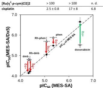
Fig. 2Effect of the P-gp inhibitor TQ on pIC50 values in MES-SA and MES- SA/Dx5 cells. Doxorubicin and compounds with paradoxical cytotoxicity are shown. ‘Rh-dmb’ and ‘Rh-phen’ represent the corresponding [Rh(5- C5Me5)(N,N)(H2O)]2+ complexes. Points above the diagonal represent paradoxical behaviour; points on the diagonal show compounds, whose cytotoxic effect is the same on both cell lines; points under the diagonal display lower toxicity in MES-SA/Dx5 than in MES-SA. △: without TQ; ●: co- incubated with TQ. Arrows show the effect of TQ on the activity, which is decreased in the case of compounds with paradoxical toxicity (red) potentiated by P-gp, while increased for non-selective compounds (green).
As compared to A2780 parental cells, the cisplatin-resistant A2780cis cells were found to be cross-resistant to all investigated compounds (c.f. IC50 values in Table 2). However, the RR values are smaller for the neocuproine complexes unlike the ligand or the cisplatin itself. Interestingly, ligands phen, dmb and their Rh complexes showed higher cytotoxicity in MES-SA/Dx5 cell lines than in the MES-SA cells (RR < 0.5 in Table 1), suggesting that these compounds may show MDR-selective activity.25 However, despite the similar structures of neo and phen or bpy and dmb, neo and bpy showed no preferential toxicity against MES-SA/Dx5 cells. The toxicity of MDR-selective compounds is exacerbated by the function of P-gp.41,44
To test the relevance of P-gp function in the selective toxicity of phen, dmb and their Rh complexes, the in vitro toxicity assays were repeated in the presence of the P-gp inhibitor tariquidar (TQ) in
MES-SA and MES-SA/Dx5 cell lines (Figure 2, Table S5). As expected, the IC50 of doxorubicin significantly decreased (pIC50 increased), in line with the inhibition of drug efflux. In contrast, phen, dmb and their Rh complexes show decreased toxicity in the presence of TQ, proving that their activity is potentiated by P-gp. The most prominent example is dmb, where the IC50 in MES-SA/Dx5 cells changes from 15 M to 72 M upon addition of TQ, respectively.
As a conclusion, phen, dmb and their Rh complexes were found to be cytotoxic, and showed preferential cytotoxicity in the MES- SA/Dx5 cell-line. Exchange of Rh to Ru results in the loss of activity and selectivity, as it was found in our previous publications for ligands with (N,O) donor set.29,30 In line with the results reported for these 8-hydroxyquinoline complexes14,15 irreversible arene loss was found for Ru complexes, and a somewhat higher cell accumulation was detected in the case of Rh. These findings might contribute to the different anticancer properties of the Ru and Rh polipyridyl complexes as well. In case of the Ru complexes, which were characterized in this work, arene loss was demonstrated even without an extra ligand (vide supra), which might be responsible for the observed loss of cytotoxicity in parental and/or resistant cell lines. Since the methylation of ligands (phen and bpy to neo and dmb), and the type of the metal centre (Ru or Rh) strongly affect the toxicity in the MES-SA and the MDR counterpart MES-SA/Dx5 cell lines, further studies were performed in order to better understand the differences in the solution behaviour of these complexes. The A2780cis cells showed cross-resistance for these compounds, however, cells were less cross-resistant for the complexes of neocuproine, as the RR values indicate.
Deprotonation of ligands and hydrolysis of half-sandwich cations Hydrolysis of the organometallic cations and the protonation constants of the ligands influence the solution speciation of metal complexes. The hydrolytic processes of half-sandwich cations ([Rh(5-C5Me5)(H2O)3]2+, [Ru(6-p-cym)(H2O)3]2+ and [Ru(6- tol)(H2O)3]2+) were already investigated in detail earlier.45,46 Proton dissociation constants of bpy, phen and ethylenediamine (Table 3) are also known under various conditions,47,48 and were re- determined herein by pH-potentiometry (see detailed description of this method in ESI). Due to the limited water solubility of dmb and neo, only spectrophotometric titrations were feasible for these ligands in chloride-free medium (I = 0.20 M KNO3) using lower concentrations.
Table 3 Proton dissociation constants (Ka (H2L) and Ka (HL)) for (N,N) ligands, stability (K [M(arene)(L)]), proton dissociation (Ka [M(arene)(L)]) and water- chloride exchange (K’ (H2O/Cl-)) constants of complexes of phen, neo, bpy, dmb and en. {T = 25.0 °C; I = 0.20 M (KNO3)}
M(arene) Constant phen neo bpy dmb en
pKa(HL) 4.92a 5.77(1) 4.41b 5.31(1) H2L: 7.25,
HL: 10.01b Rh(5-C5Me5)
log K [M(arene)(L)] 14.70(3)c 9.70(3)d 13.30(2)c 14.32(2)c 15.04b
pKa [M(arene)(L)] 8.58a 8.88(1)e 8.61b 8.40(1)f 9.58b
log K’ (H2O/Cl-) 2.92a 2.76(1)g 2.58b 2.36(1)g 2.14b
Ru(6-p-cym)
log K [M(arene)(L)] >12.8 8.21(4)e >12.5 >12.8 14.85(5)h
pKa [M(arene)(L)] 7.59(1)f 7.62(1)e 7.48(1)f 7.55(1)f 8.14(2)e
log K’ (H2O/Cl-) 1.79(1)g 0.93(1)g 1.83(1)g 2.02(1)g 1.51(5)g
Ru(6-tol)
log K [M(arene)(L)] >13.0 8.19(8)e >13.0 >13.2 14.90(6)h
pKa [M(arene)(L)] 7.39(1)f 7.55(1)e 7.39(1)f 7.47(1)f 8.04(2)e
log K’ (H2O/Cl-) 1.68(1)g 0.87(1)g 1.62(1)g 1.88(1)g 1.69(5)g
4.0 5.0 6.0 7.0
4.0 5.0 6.0 7.0
pIC50(MES-SA/Dx5)
pIC50(MES-SA)
+TQ
doxorubicin dmb
Rh-dmb Rh-phen
+TQ
phen +TQ
+TQ
+TQ
a See Ref. 22. b See Ref. 49. c1H NMR, displacement measurements. d1H NMR, pH = 0.7-2.1. e1H NMR titrations, pH = 2.0-11.5. f UV-Vis titration, pH = 2- 11.5. g UV-Vis, c(Cl-)=0.0-0.2 M. h1H NMR, c(M(arene)) = 100 M, c(ethylenediamine) = 0-7.3 mM.
Considering the different ionic strengths, values are in good agreement with literature data.47,48 We find that methylation in ortho and in para positions increases the pKa. Based on the pKa
values it can be concluded that the polypyridyl ligands are mainly in their neutral form at physiological pH.
Determination of formation constants for complexes [M(arene)(N,N)(H2O)]2+
Based on the single-crystal X-ray structures and previous solution equilibrium studies on similar complexes,3,13,22,49 these bidentate (N,N) donor ligands form mono complexes with half-sandwich organometallic triaqua cations. Scheme S2 shows the occurring equilibrium processes in aqueous solutions for these complexes.
Although the complex formation equilibrium is reached with (O,O) donors within minutes, hours or days are needed in case of the (N,N) donors.22 Complex formation rates at pH = 0.7 and 2.1 are compared in Figures S31-S33, which show that the [Ru(6- arene)(H2O)3]2+ triaqua cations react much slower than [Rh(5- C5Me5)(H2O)3]2+ with the same ligand (h versus min). Generally, it can be concluded that the reaction rate is lower at more acidic pH.
However, the pH cannot be increased beyond a certain limit to accelerate the reaction, since the hydrolysis of the organometallic cations produces inert hydroxido complexes.
Complexes of bpy, dmb and phen are present in aqueous solutions at pH = 2 and 0.7 exclusively, there is no sign of unbound organometallic cation or ligand under the given conditions (I = 0.20 M KNO3, aqueous solution) based on the 1H NMR spectroscopic measurements. (Notably, pH 0.7 is the lowest pH where the ionic strength can be kept constant.) With decreasing concentrations (from 1.2 mM to 20 M) of [Ru(6-tol)(bpy)(H2O)]2+ and [Ru(6-p- cym)(phen)(H2O)]2+, the molar UV-Vis spectra remain unchanged (Figure S34), which also confirmed the high stability of the complexes. Since measurements with decreasing pH (down to 0.7) or concentration (down to 20 M) do not give any information about the stability constants owing to the lack of complex dissociation, displacement studies were performed with ethylenediamine. For this purpose, stability constants for [Ru(6- arene)(ethylenediamine)(H2O)]2+ were determined (the constant of [Rh(5-C5Me5)(ethylenediamine)(H2O)]2+ was reported earlier).49 Samples were prepared at pH = 3.0 using different ethylenediamine-to-Ru ratios (up to 14-fold ligand excess) and measured by 1H NMR spectroscopy. Figure S35 shows extremely slow complex formation under the conditions used.
When ~80-fold excess of ethylenediamine was used, signals of mixed ligand complexes appeared in the 1H NMR spectra instead of the free polypyridine ligand. Figure S36 shows examples for [Rh(5- C5Me5)(bpy)(H2O)]2+ – ethylenediamine and [Ru(6-tol)(bpy)(H2O)]2+
systems, in which two peaks indicated the monodentate coordination of ethylenediamine. This ternary complex formation process hindered the determination of the stability constant.
Fig. 3 a) Selected 1H NMR spectra of the [Rh(5-C5Me5)(bpy)(H2O)]2+-HQCl- Pro (1:x) system at different HQCl-Pro concentrations (x = 0-22.5). Intensity is decreased in < 2.0 ppm region for the sake of clarity. I.: spectrum of [Rh(5-C5Me5)(bpy)(H2O)]2+; II.: spectrum of unbound bpy at pH 6.19; III.:
spectrum of [Rh(5-C5Me5)(L)(H2O)]+, where L is the deprotonated (coordinated) form of HQCl-Pro. Peaks signed with * belong to the unbound HQCl-Pro; ■ shows the peak of bpy used for calculations; dashed rectangle shows [Rh(5-C5Me5)(L)(H2O)]+; solid rectangle shows [Rh(5- C5Me5)(bpy)(H2O)]2+. b) Measured and fitted (dashed line) percentages of [Rh(5-C5Me5)(L)(H2O)]+; ■ is calculated from integrals of unbound-bound bpy peaks, ◊ is calculated from C5Me5 peaks. {c([Rh(5-C5Me5)(H2O)3]2+) = c(bpy) = 200 M; c(HQCl-Pro) = 0-4.53 mM; pH = 6.51; solvent: 90% H2O / 10% D2O; T = 25.0 °C; I = 0.20 M (KNO3)}
An 8-hydroxyquinoline derivate ((S)-5-chloro-7-((proline-1- yl)methyl)8-hydroxyquinoline (HQCl-Pro))30 was used for the competition experiments, however, its use was limited only to the Rh-containing compounds, as the Ru complexes seem to lose the arene ligands when a competitor ligand is added. A successful displacement of bpy from its Rh complex is shown in Figure 3, at 22.5-fold excess of HQCl-Pro ~70% of the liberated bpy. From this data a log K = 13.30(2) was calculated (Table 3). Bpy was employed for the displacement of dmb and phen ligands. In these measurements 1H NMR spectroscopy was used, since the peak separation of these similar compounds is satisfactory on NMR spectra (Figures S37 and S38). To determine the stability constants of the Ru complexes, we attempted to study a potential displacement between the organometallic cations. Unfortunately, there was no reaction between [Rh(5-C5Me5)(polypyridine)(H2O)]2+
and [Ru(6-p-cym)(H2O)3]2+, even at 10-fold excess of the latter. In all, only minimum values could be provided (Table 3).
0 25 50 75
0 10 20
Ratio of bound HQCl-
c(HQCl-Pro)/c(bpy)
(ppm)
9.1 8.6 8.1 7.6 1.7
22.5 15.5 9.2 4.2 1.1 I.
II.
III.
* * * *
* * * *
b) a)
HQCl-Pro/bpyRatio of bound HQCl-Pro (%)
The behaviour of neocuproine complexes is different due to the steric effect of the methyl groups next to the donor atoms, namely the complex formation reaction is much slower as compared with phen. Samples containing [Ru(6-arene)(H2O)3]2+ and neocuproine in 1:1.2 ratio were followed by 1H NMR spectroscopy for 15 days (Figure 4.a). An extra set of peaks belongs to an intermediate complex which is most probably a sandwich-type complex is (Figure 4.b,c). The solution stability of metal complexes also changed compared to phen. Free neocuproine occurs next to its Rh complex (pH = 0.7, ~50%, Figure S39.a) in the equilibrium, as well as next to the Ru complexes (pH = 2.5, ~15%, Figures 5.a and S40.a).
Based on the samples measured for samples with acidic pH, molar fractions for bound and unbound neocuproine can be calculated and used for the determination of stability constants (Figure S39 shows this in case of Rh). The Ru-containing complexes have smaller stability constants (Table 3). The lower complex stability constants and the stronger bias of [Ru(arene)(H2O)3]2+ towards hydrolysis ensure the lower aqueous stability of [Ru(arene)(neo)(H2O)]2+
complexes compared to [Rh(5-C5Me5)(H2O)3]2+. The lower stability may originate from the shorter distance between the Ru metal ion and arene ligand compared with the Rh‒C5Me5 distance (1.697 Å vs.
1.780 Å), which may cause greater steric repulsion around the Ru centre. To quantify this steric repulsion, the overlaps between ligand solid angles (calculated by Olex238) were listed in the legend of Figure S29. The bigger the overlap, the less the aqueous stability of complex.
Fig. 4 a) Time-dependence of complex formation of [Ru(6-tol)(H2O)3]2+ with neo at pH = 6.0 followed by 1H NMR spectroscopy (only region of ligand is shown). Time of reaction is shown on the left. Assignment: bidentate metal complex: ■, free neo: ♠, ‘sandwich intermediate’: ♥. Arrows show the unusually big shifts of ‘sandwich intermediate’ from free ligand, which
differences are shown in b) (in ppm). c) Proposed structure of the ‘sandwich intermediate’. {c([Ru(6-tol)(H2O)3]2+) = 200 M; c(neo) = 245 M; pH= 6.0 (20 mM phosphate); I = 0.20 M (KNO3); T = 25.0°C}
Comparison of the stability constants of Rh complexes is not feasible due to the different basicity of the ligands. The derived stability constants are calculated as the equation shows below:
log K* = log K [M(arene)(L)(H2O)]2+ ‒ pKa(HL)
With this transformation, the log K* [Rh(5-C5Me5)(N,N)(H2O)]2+
values show the following trend: 9.78 (phen) > 9.01 (dmb) > 8.89 (bpy) > 3.93 (neo). While methylation far from the coordinating nitrogen atoms (bpy vs. dmb) causes slight difference, the methylation next to the coordinating nitrogen atoms results in a huge difference. However, except neocuproine, there are no significant differences in the stability of these Rh complexes. The loss of preferential cytotoxicity in MES-SA/Dx5 cells of [Rh(5- C5Me5)(neo)(H2O)]2+ can be partly explained by the probable dissociation of neo-complex in the cell. However, the small stability difference between bpy and dmb cannot be the reason of the different behaviour against MDR cell lines. Moreover, the question arises whether the reactivity of the coordinated water molecule can be tuned using steric control of a bulky bidentate ligand.
Reactions of the coordinated water molecule: deprotonation and substitution to chloride ion
In half-sandwich complexes, next to the arene hapto ligand and a bidentate (N,N) donor ligand, the coordination sphere is completed by a water molecule, which can either lose a proton or can be substituted by another ligand (for example Cl- in Scheme S2). Chen and co-workers found that the reaction of RAED complexes with nucleobases is slower at higher pH or in the presence of chloride or phosphate anions.50 At higher pH, deprotonation occurs, which is characterized by the pKa[M(arene)(L)] constant. With the knowledge of pKa values one can calculate not only the actual average charge (between +2 and +1) of the compound at a given pH, which has an effect also on lipophilicity, but also the molar fraction of the generally less reactive [M(arene)(L)(OH)]+ mixed hydroxido complex.
Increasing the pH, the effect of the deprotonation process on the UV-Vis spectra is unambiguous, as the pH-dependent spectra (Figure 5) of the example ([Ru(6-tol)(dmb)(H2O)]2+) shows. From the absorbance change pKa values can be computed. Determination of this constant is also feasible by performing 1H NMR titrations, where the changes of the chemical shifts are used for calculations.
In a previously reported article the pKa of [Ru(6-p- cym)(phen)(H2O)]2+ was determined as 7.32 in pure D2O,42 which is in good agreement with the constant reported here. This type of measurement was used in the case of [Ru(6-p-cym)(neo)(H2O)]2+
and interestingly, in the region of aromatic p-cymene protons a singlet appears, which belongs to the arene in the complex (Figure 6). This peak belongs to protons, which are generally not magnetically equivalent in other complexes. As the pH increases, this singlet splits into two doublets.
(ppm)
8.7 8.4 8.1 7.8 7.5
15 d 8 d
4.5 d
9 h
0.55 0.51 0.26
0.04
a)
b) c)
Fig. 5 a) UV-Vis spectra of the [Ru(6-tol)(dmb)(H2O)]2+ recorded at pH = 5.1- 9.9. b) Absorbance change at 364 nm in dependence of the pH. {c([Ru(6- tol)(dmb)(H2O)]2+) = c(dmb) = 200 M; I = 0.20 M (KNO3); ℓ = 1 cm; T=25.0°C}
As shown in Table 3, methylation of the polypyridine ligands has an influence on the deprotonation of the coordinated water molecule.
Namely, the methyl groups increase the pKa values of the complexes in all cases. Ru complexes have stronger OH- affinity than the Rh analogues, as the lower pKa values indicate it. Due to the lower complex stability, at physiological pH [(Ru(arene))2(OH)3]+ also appears in the case of [Ru(arene)(neo)(H2O)]2+ complexes.
Chloride ions can potentially replace the coordinating water molecule, as shown in the single-crystal XRD structures (Figure 1), and this process is characterized by the water-chloride exchange constant (K’(H2O/Cl-)). Cl- is present in aqueous solutions, e.g. in biofluids, where the concentration drops from 103 mM to 24 mM and 4 mM, entering from the blood serum to cytoplasm and nucleus.51 The coordination of Cl- affects the actual charge and the presence of chloride ion in medium can suppress the deprotonation process of the complexes to a more basic pH region.49 The Cl- affinity of [Rh(5-C5Me5)(phen)(H2O)]2+ and [Rh(5- C5Me5)(bpy)(H2O)]2+ was already reported by our group.22,49 Upon addition of chloride ions, changes similar to deprotonation can be observed in the UV-Vis spectra (as shown for [Ru(6- tol)(phen)(H2O)]2+ in Figure 7.a). From the spectral changes the water-chloride exchange constants were calculated (Table 3).
Methylation of the ligands has an effect on the water-chloride exchange constant as well. While for Rh complexes only a slight decrease can be seen, the effect for Ru complexes is remarkable.
The Cl- affinity of dmb complexes is higher than that of the bpy complexes. The Ru(II)-arene complexes of neocuproine have
significantly low constants: log K’(H2O/Cl-) = 1.79→0.93 and 1.68→0.87 (Table 3). The Cl--dependent spectra of [Ru(6-p- cym)(neo)(H2O)]2+ show smaller spectral changes (A~0.05) in
Fig. 6 a) Aromatic region of selected 1H NMR spectra of the [Ru(6-p- cym)(neo)(H2O)]2+ system recorded at pH = 2.0-11.4 after 34 days. Peaks of complex I are signed with *. Appearance of ‘sandwich intermediate’ is shown in the inset. b) Measured (neo H at position 5 and 6: □; all protons of ligand: ×) and fitted (dashed line) ratio of formed complex. c) Measured (○) and fitted (dashed line) chemical shift values of aromatic p-cymene protons at different pH-values. {c([Ru(6-p-cym)(H2O)3]2+) = c(neo) = 500 M; solvent:
90% H2O / 10% D2O; T = 25.0°C; I = 0.20 M (KNO3); c(phosphate) = 20 mM}
Figure S40.b. Most probably a steric repulsion is the reason. The methyl groups of neo hinder the strong interaction between the chloride ion and Ru.
For [Ru(arene)(ethylenediamine)(H2O)]2+ complexes, a larger uncertainty can be seen in the constants. This is in connection with the phosphate ion coordination to RAED complexes based on the a)
b)
0.0 0.2 0.4 0.6
330 380 430 480
Absorbance
(nm)
pH = 5.1 pH = 9.9
0.15 0.25 0.35 0.45
5.0 6.5 8.0 9.5
Absorbance at 364
pH
Absorbanceat364 nm
(ppm) 7.6 5.8 5.4
(ppm)
8.7 8.2 7.7 6.1 5.8 5.5 5.2
*
*
*
*
*
*
*
*
11.40 9.90 8.97 7.91 7.47 7.18 6.68 5.82 3.08 2.04
b) a)
c)
pH
85 90 95 100
2.5 3.5 4.5 5.5
5.7 6.0 6.3
2 6 10
R a ti o o f bound M( a re n e ) (%)
pH
pH
(C H , p -cym ) (p p m)
abovementioned work of Chen et al.,50 and on our measurements showed in details in Figures S41-S42.
Figure 7.c shows the ratio of chlorinated and aqua forms of complexes in the solution at different chloride ion concentrations.
Fig. 7 a) UV-Vis spectra of [Ru(6-tol)(phen)(H2O)]2+ recorded at various chloride ion concentrations. b) Absorbance change at 346 nm plotted against the concentration of the chloride ion. c) Ratio of the aqua and chlorinated forms of complexes (c = 200 M) with the highest and lowest chloride affinity measured in the studied group of compounds. Chloride concentrations are representing the different biofluids. Constants from Table 3 were used for calculation. {c([Ru(6-tol)(H2O)3]2+) = c(phen)
= 182 M; pH = 6.0; ℓ = 1 cm; T = 25.0°C}
Our calculations show the contrasting behaviour of two complexes, which possess the highest and the lowest chloride ion affinity ([Rh(5-C5Me5)(phen)(H2O)]2+ and [Ru(6-tol)(neo)(H2O)]2+, respectively). As seen in Figure 7.c, the fraction of the chlorinated form of the neo complex is minimal at c(Cl-) = 4 mM, and it increases to 43% at c(Cl-) = 100 mM. On the contrary, the [Rh(5- C5Me5)(phen)(H2O)]2+ complex shows an opposite behaviour, as the chlorinated form predominates under all considered chloride ion concentrations.
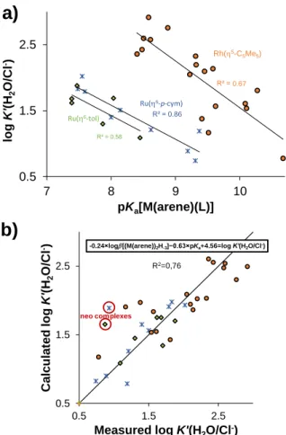
The importance of the knowledge of chloride ion affinity was already mentioned above and in previous reviews.51 In our earlier work, a linear relationship between the pKa[M(arene)(L)] and the log K’(H2O/Cl-) constants was found for Rh(5-C5Me5) complexes with (O,O), (O,N), (N,N) and (O,S) donor bidentate ligands. To complete this model, already published and the newly determined
constants even for Ru(arene) complexes were integrated (except for neo complexes because of the strong steric effect on this
Fig. 8 a) The measured log K’(H2O/Cl-) values of [M(arene)(L)] complexes in the function of their pKa[M(arene)(L)] constants (I = 0.20 M (KNO3)). b) Multilinear regression for calculating log K’(H2O/Cl-) based on the shown equation. Used data are collected in Table S6.22,29,30,45,46,49,52-56
Outlier points are circled.
constant; all used data are summarized in Table S6). The displayed constants (Figure 8.a) are in three different groups with their own fitting lines. However, applying multilinear regression and the log [(M(arene))2H-3] constants arranged the values in one group (Figure 8.b). The equation of this regression is:
-0.24×log [(M(arene))2H-3] − 0.63×pKa[M(arene)(L)] + 4.56 = log K’(H2O/Cl-) The first term of this equation takes the M(arene) part into consideration, while the second term describes the effect of bidentate ligand. This model is universal, as it can predict the chloride ion affinity of different half-sandwich M(arene) complexes, if the log β[(M(arene))2H‒3] and pKa[M(arene)(L)] constants are known. The main drawback is that only chloride ion free pKa[M(arene)(L)] values are acceptable in this model.
After determining those constants, which govern the speciation in solution, we can compare them and find connections with cytotoxicity. In Figure 9 the basicity corrected stability constant, the
0%
25%
50%
75%
100%
100 mM 24 mM 4 mM 100 mM 24 mM 4 mM
Distribution of M(arene)(L)
c
[Rh(5-C5Me5)(phen)Cl]+ Aqua species
[Ru(6-tol)(neo)(H2O)]2+
Chlorinated form
0% 25% 50% 75% 100%
100 mM 24 mM 4 mM 100 mM 24 mM 4 mM
Distribution of M(arene)(L)
c(Cl-)
[Rh(5-C5Me5)(phen)Cl]+
[Rh(5-C5Me5)(phen)(H2O)]2+
[Ru(6-tol)(neo)(H2O)]2+
Chlorinated form
c(Cl-)
a)
b)
0 0.6 1.2 1.8
300 350 400 450
Absorbance
(nm)
c(Cl-)=0.08 M
c(Cl-)=0.00 M
Absorbanceat346 nm
0.40 0.50 0.60
0 0.03 0.06 0.09
Absorbance at 346
c(Cl-) (M)
0% 25% 50% 75% 100%
100 mM 24 mM 4 mM 100 mM 24 mM 4 mM
Distribution of M(arene)(L)
c(Cl-)
[Rh(5-C5Me5)(phen)Cl]+
[Rh(5-C5Me5)(phen)(H2O)]2+
[Ru(6-tol)(neo)(H2O)]2+
[Ru(6-tol)(neo)Cl]+
c(Cl-)
0% 25% 50% 75% 100%
100 mM 24 mM 4 mM 100 mM 24 mM 4 mM
Distribution of M(arene)(L)
c(Cl-)
[Rh(5-C5Me5)(phen)Cl]+
[Rh(5-C5Me5)(phen)(H2O)]2+
[Ru(6-tol)(neo)(H2O)]2+
[Ru(6-tol)(neo)Cl]+
c(Cl-) c)
0.5 1.5 2.5
0.5 1.5 2.5
Calculated logK'(H2O/Cl-)
Measured log K'(H2O/Cl-)
R2=0,76
-0.24 log[(M(arene))2H-3]−0.63 pKa+4.56=log K’(H2O/Cl-)
neo complexes
R² = 0.6659
R² = 0.86 R² = 0.58
0.5 1.5 2.5
7 8 9 10
logK'(H2O/Cl-)
pKa[M(arene)(L)]
Rh(5-C5Me5)
Ru(6-p-cym) Ru(6-tol)
a)
b)
0.67
 (I), b) [Ru( 6 -tol)(dmb)Cl](CF 3 SO 3 ) (II) c) [Ru(dmb) 3 ].2(CF 3 SO 3 )×2 H 2 O (III), d) [Ru( 6 -tol)(neo)Cl]Cl×2 MeOH (IV), e) [Ru( 6 -p-cym)(neo)Cl](CF 3 SO](https://thumb-eu.123doks.com/thumbv2/9dokorg/998031.61744/3.892.525.777.110.477/fig-molecular-structures-rh-dmb-cl-iii-meoh.webp)

![Fig. 3 a) Selected 1 H NMR spectra of the [Rh( 5 -C 5 Me 5 )(bpy)(H 2 O)] 2+ -HQCl- -HQCl-Pro (1:x) system at different HQCl--HQCl-Pro concentrations (x = 0-22.5)](https://thumb-eu.123doks.com/thumbv2/9dokorg/998031.61744/6.892.491.774.151.538/selected-spectra-hqcl-hqcl-different-hqcl-hqcl-concentrations.webp)
![Fig. 4 a) Time-dependence of complex formation of [Ru( 6 -tol)(H 2 O) 3 ] 2+ with neo at pH = 6.0 followed by 1 H NMR spectroscopy (only region of ligand is shown)](https://thumb-eu.123doks.com/thumbv2/9dokorg/998031.61744/7.892.79.399.608.996/time-dependence-complex-formation-followed-spectroscopy-region-ligand.webp)
![Fig. 6 a) Aromatic region of selected 1 H NMR spectra of the [Ru( 6 -p- -p-cym)(neo)(H 2 O)] 2+ system recorded at pH = 2.0-11.4 after 34 days](https://thumb-eu.123doks.com/thumbv2/9dokorg/998031.61744/8.892.100.364.151.508/fig-aromatic-region-selected-nmr-spectra-recorded-days.webp)