For Peer Review
TiO2 and Ag-TiO2 nanohybrid films on titanium dental implant material: an in vitro cell culture study
Journal: Journal of Biomedical Materials Research: Part B - Applied Biomaterials Manuscript ID Draft
Wiley - Manuscript type: Original Research Report Date Submitted by the Author: n/a
Complete List of Authors: Masa, Roland; University of Szeged, Faculty of Dentistry, Department of Oral Biology and Experimental Dental Research
Györgyey, Agnes; University of Szeged Faculty of Dentistry, Department of Prosthodontics
Deák, Ágota; Universitiy of Szeged, Deparment of Physical Chemistry and Materials Science
Janovak, Laszlo; University of Szeged, Deparment of Physical Chemistry and Materials Science
Tóth, Zsolt; University of Szeged Faculty of Dentistry, Department of Oral Biology and Experimental Dental Research
Heszlerné Kopniczky, Judit; University of Szeged, Department of Optics and Quantum Electronics
Pelsőczi-Kovács, István; University of Szeged, Department of Prosthodontics
Ungvári, Krisztina; University of Szeged Faculty of Dentistry, Department of Oral Biology and Experimental Dental Research
Dekany, Imre; University of Szeged, Deparment of Physical Chemistry and Materials Science; University of Szeged, MTA-SZTE Supramolecular and Nanostructured Materials Research Group; University of Szeged, Department of Medical Chemistry, Faculty of Medicine
Turzó, Kinga; University of Szeged, Faculty of Dentistry, Department of Oral Biology and Experimental Dental Research
Keywords: nanoparticle, silver, MG63 osteoblasts, epithelial cells, titanium (alloys)
For Peer Review
TiO2 and Ag-TiO2 nanohybrid films on titanium dental implant material: an in vitro cell culture study
Roland Masa1*, Ágnes Györgyey1, Ágota Deák3, László Janovák3, Zsolt Tóth1, Judit Kopniczky6, István Pelsőczi-Kovács2, Krisztina Ungvári1, Imre Dékány3, 4, 5, Kinga Turzó1
1Department of Oral Biology and Experimental Dental Research, Faculty of Dentistry, University of Szeged, H-6720 Szeged,Tisza Lajos krt. 64, Hungary
2Department of Prosthodontics, Faculty of Dentistry, University of Szeged, H-6720 Szeged, Tisza Lajos krt. 64, Hungary
3MTA-SZTE Supramolecular and Nanostructured Materials Research Group, University of Szeged, H-6720, Szeged, Dóm tér 8, Hungary
4Deparment of Physical Chemistry and Materials Science, University of Szeged, H-6720, Szeged, Rerrich B. tér 1, Hungary
5Department of Medical Chemistry, Faculty of Medicine, University of Szeged, H-6720, Szeged, Dóm tér 8, Hungary
6Department of Optics and Quantum Electronics, University of Szeged, H-6720 Szeged, Dóm tér 9, Hungary
Abstract
Failure of dental implants is caused mainly by peri-implant infections resulting in loss of supporting bone. Our aim was to examine the attachment and proliferation of primary epithelial and MG63 osteosarcoma cells on Ti dental implants coated with two photocatalytic nanohybrid films, which could eliminate bacterial contamination. Commercially pure (CP4) sand blasted, acid etched and machined Ti discs were used as control surfaces. Two copolymer based layers were investigated: 60 % TiO2/ 40 % copolymer and 60% AgTiO2/ 40
% copolymer ([Ag] = 0,001 m/m %). Surface properties were examined by scanning electron microscopy (SEM) and profilometry. In vitro attachment and proliferation of epithelial and MG63 cells were investigated via MTT and visualized with fluorescence microscopy. SEM and profilometry revealed significant changes in surface roughness and morphology of the AgTiO2 layer compared to the control surface. MTT results demonstrated that the attachment (24h) of the epithelial cells was significantly higher on the Ag-TiO2 coated samples than on the control surfaces, whereas MG63 cells did not show any difference in attachment between the groups. No significant difference was found in the proliferation of the cells on the covered samples (72h, 168h). After one-week, epithelial cells showed slightly increased survival as compared to MG63 cells. The potential antibacterial effect of the silver containing nanohybrid layer makes it a promising surface coating.
Keywords: nanoparticles, silver, MG63 osteoblasts, epithelial cells, titanium (alloys) 2
3 4 5 6 7 8 9 10 11 12 13 14 15 16 17 18 19 20 21 22 23 24 25 26 27 28 29 30 31 32 33 34 35 36 37 38 39 40 41 42 43 44 45 46 47 48 49 50 51 52 53 54 55 56 57 58
For Peer Review
1. Introduction
In spite of their widespread application, dental implants are still in the focus of intense research. Dental implants belong to the most frequently used biomaterials, since they have become the first choice for tooth replacement 1-3. The last few decades have seen the development of a host of surface modifications aimed at shortening the healing period and accelerating osseointegration. These modifications have ranged from the macro to the micro scale, but research is increasingly concentrating on the nano scale 4 .
Titanium is still the number one material for dental implants, thanks to its excellent physical, mechanical and chemical properties. A high weight to strength ratio, excellent corrosion resistance and biocompatibility are well known properties of this metal 5,6. Commercially pure (CP 1 - 4) titanium is still the most widely used material for implants, but titanium alloys, such as Ti6Al4V (CP 5) also have great potential 7.
Although the success rate of titanium implants is generally high, peri-implant inflammation, the most common factor leading to implant failure, is still a problem. In the case of peri-implant mucositis, only the soft tissue is affected and it is reversible with efficient conservative therapy. The more progressive form is peri-implantitis, which is an irreversible disease of the soft and hard tissues that surround the implant 8. Of all the implantation cases, 10% ends up in peri-implantitis, provided that the patient has not suffered from periodontal disease previously. Periodontal disease in the patient’s history increases this to approximately 20% 9.
Peri-implantitis is treated either in a conservative (manual debridement, antibiotics, laser supported and photodynamic therapy) or a surgical (resective or regenerative) way. The ultimate purpose is to clean the contaminated implant surface and remove the bacterial biofilm. Both approaches have their drawbacks, though. Mechanical root scaling and air polishing can modify the surface characteristics of the implant, the use of antibiotics can lead to antibiotics resistance, while a resective surgical therapy can result in extensive postoperative recession 8. Since there is no ideal treatment for peri-implantitis, prevention should be assigned priority. Antibacterial surfaces represent a promising approach to reach that end. Such surfaces can prevent the attachment of first colonizing bacteria, inhibit the formation of biofilm and kill bacteria by releasing free radicals or even an antibacterial agent
10,11
. 2
3 4 5 6 7 8 9 10 11 12 13 14 15 16 17 18 19 20 21 22 23 24 25 26 27 28 29 30 31 32 33 34 35 36 37 38 39 40 41 42 43 44 45 46 47 48 49 50 51 52 53 54 55 56 57 58 59
For Peer Review
Photocatalytic surfaces are widely studied as antibacterial surfaces 12,13. The principle of photocatalysis is electron excitation in the catalyzator molecule, resulting in redox reactions with the surrounding adsorbed water which leads finally reactive oxidizing species (hydroxide, superoxide radicals) releasing. These free radicals can induce the destruction of the adsorbed organic pollutants and bacterial contamination 14,15. Because of its high photoactivity, titanium dioxide (TiO2) is one of the most intensively studied photocatalysts 16. It is also a promising semiconductor, because of its low toxicity, chemical inertness and low price. However, due to its large electron band gap energy, TiO2 can only be excited by UV light (λ < 380 nm) 17,18. It is possible, though, to shift its light absorption into the visible range by the application of metallic and non-metallic substances and elements.
Veres et al. used nanosilver to modify and improve the photocatalytic activity of TiO2. They used polyacrilate resin [poly(ethyl acrylate-co-methylacrylate; p(EA-co-MMA)]
to provide a polymer bed for TiO2 and Ag-modified TiO2 nanoparticles, and they prepared thin hydrophilic photocatalytic film coatings with photodeposition 19. This polymer matrix started to decompose after one hour of UV irradiation, which led to higher free catalyst concentration on the surface.
As a result of nanoparticles’ small (10-9 m) size, they have specific properties. After oral, cutaneous exposition or inhalation uptake, they can penetrate across different barriers into the whole human body. Intracellular uptake can occur by endocytosis, and they can interact with proteins or even with the nucleic acids 20. The opinions about Ag nanoparticles (AgNP-s) are not unequivocal. Some authors reported high bactericidal activity and biocompatibility as compared to other nanoparticles 21-23. On the other hand, depending on the size and concentration, AgNP-s show considerably higher toxicity than silver ions. For instance, they exert their toxic effect immediately upon contact in cancer cell lines 24.
We studied two newly developed photocatalysts/polymer nanohybrid films: TiO2- copolymer and Ag-TiO2-copolymer containing 0.001% silver nanoparticles. Both were applied with the spray coating technique, and activated with LED light (λ = 405 nm).The photocatalytic antibacterial activity of TiO2 and Ag-TiO2 coatings was established previously, and it was found that the role of photocatalytic activity was dominant over the antibacterial effect of silver ions themselves 25. Györgyey et al. investigated the antibacterial effect of 0.5%
Ag- containing TiO2 and TiO2 copolymer films on titanium implant material. Their results suggested that this type of surface modification might be of good use in the therapy of peri- implantitis 26.
2 3 4 5 6 7 8 9 10 11 12 13 14 15 16 17 18 19 20 21 22 23 24 25 26 27 28 29 30 31 32 33 34 35 36 37 38 39 40 41 42 43 44 45 46 47 48 49 50 51 52 53 54 55 56 57 58
For Peer Review
Our main goal was to examine the attachment and proliferation of primary human epithelial cells and MG63 osteosarcoma cells on two nanohybrid surface coatings: TiO2- copolymer and Ag-TiO2-copolymer containing 0.001% silver nanoparticles. In vitro cell culture experiments have several advantages compared to in vivo animal studies. Cell- biomaterial interactions can be examined in detail, they are maintained under controlled conditions, have better reproducibility and are even cost-efficient. Primary cells are more convenient models for in vivo simulation compared to immortalized cells. They have a finite life span and their characteristics in culture can change over time. Therefore earlier passages are recommended for experiments with primary cells. Their diversity is a big advantage compared to cancer cell lines, which origins from only one person. Reactions from different donors in different ages and genetics could be observed. According to De Saint Jean et al.
primary epithelial cells showed higher sensitivity than immortalized cell lines to inflammatory cytokines 27. Endosseus dental implants are placed into the alveolar bone, but the transgingival parts are surrounded by soft tissues. For long term success, tight and healty peri-implant soft tissue seal is required. Primary epithelial cells were used for the evaluation of soft tissue response. Based on the literature polished Ti discs were used as control samples for promoting better epithelial cell adhesion and spreading 28,29. On the other hand, one of the most popular cell culture models for in vitro osseointegration studies is MG63 human osteoblast–like cell 30. This tumor cell line shows osteoblast-like characteristics, hence it is an appropriate model for studying integrin expression and osteocalcin production, but not so suitable for examining alkaline phosphatase activities. The MG63 cells represent a suitable model for the in vitro study of osseointegration 31. The response of epithelial cells as primary cells imitates the response of the peri-implant tissues better than that of the MG63 cells. If these coatings can preserve their antibacterial properties in spite of saliva and/or blood contamination during the surgical placement procedure and osseointegration occurs, the photocatalytic properties of the films could serve as promising therapy against peri-implant infections.
2. Materials and methods
2.1 Preparation of Ti samples
Ti sample discs (1.5 mm thick and 9 mm in diameter) were cut from commercially pure (CP 4) titanium rods (Denti® System Ltd. Hungary). Epithelial cell culture studies were carried out with these discs with machined surface (Ra< 0.5 µm). For the MG63 cell culture studies sandblasted and acid-etched discs were used to model the surface of the dental 2
3 4 5 6 7 8 9 10 11 12 13 14 15 16 17 18 19 20 21 22 23 24 25 26 27 28 29 30 31 32 33 34 35 36 37 38 39 40 41 42 43 44 45 46 47 48 49 50 51 52 53 54 55 56 57 58 59
For Peer Review
implant’s fixture. Before the treatment, all samples were cleaned in acetone and then in 70%
ethanol ultrasonically, and finally rinsed in ultrapure water. After this preparation process, the sandblasted and acid-etched discs were coated with the copolymer-based nanohybrid layers.
2.2 Preparation of Ag-TiO2 copolymer films
Polished Ti discs for epithelial cells and sandblasted, acid-etched Ti discs for MG63 cells served as control samples. Two photocatalytic layers: 60 % TiO2/ 40 % copolymer and 60% AgTiO2/ 40 % copolymer ([Ag] = 0.001 m/m %) were investigated. We reduced the amount of nanosilver from 0.5 % to 0.001% compared to our previous experiments 26. A hydrophilic polyacrylate resin [poly (ethyl-acrylate comethyl-methacrylate) (p(EA-co-MMA)) binder material (obtained from Evonik Industries, Germany) was used providing the polymer bed for TiO2 and Ag photocatalyst nanoparticles. The process of producing these TiO2 and Ag-TiO2 nanohybrid layers was described in the study of Veres et al 19.
Before Ti discs were coated with these photocatalyst layers, the suspensions were sonicated for 15 minutes, then sprayed onto the Ti discs with an AD-318 spray gun (Alder, USA) and dried at high temperature (120 °C). 2 ± 0.05 mg polymer suspension was sprayed onto each Ti discs. After this process, the discs were sterilized in hot air sterilizer at 180 °C for 45 minutes. The last step was to illuminate the coated discs with UV-light for 60 minutes in order to decompose the upper layer of the polymer film to uncover the silver and the TiO2
nanoparticles 19.
2.3 Scanning electron microscopy
The morphological characteristics of the coatings were investigated before UV treatment, without cells. A field emission scanning electron microscope (SEM) (Hitachi S4700, Japan) was applied to record images with ×500, ×1000 and ×5000 magnifications in secondary electron imaging mode. The samples were tilted at 45° for better surface visualization.
2.4 Profilometry measurements
Average surface roughness (Ra (µm)) of sandblasted and acid-etched Ti, TiO2 and Ag- TiO2 copolymer coated Ti discs was measured with a Veeco, Dektak 8 Advanced Development Profiler® (Veeco Instruments, USA). 500×500 µm2 areas were recorded at three different places, on two samples of each group. The resolution along the scanning direction 2
3 4 5 6 7 8 9 10 11 12 13 14 15 16 17 18 19 20 21 22 23 24 25 26 27 28 29 30 31 32 33 34 35 36 37 38 39 40 41 42 43 44 45 46 47 48 49 50 51 52 53 54 55 56 57 58
For Peer Review
was 0.17 µm and the spacing was 6.33 µm between the scanned lines. The vertical resolution was 4 nm. Macro-roughness line profiles were recorded also. Profiles were analyzed on computer, using Vision 3D Image and Analysis Software (Veeco Instruments, USA).
2.5 Cell culture studies
Two different cell culture models were used: primary human epithelial cells and MG63 osteosarcoma cells. Gingival tissues were obtained from healthy adult patients undergoing routine dento-alveolar surgery. Epithelial cells were isolated from inflammation- free mucosa after obtaining the written consent from the patients. The study protocol conformed to the tenets of the Declaration of Helsinki in all respects, and it was approved by the Regional Research Ethics Committee for Medical Research at the University of Szeged.
The process of primary epithelial cell isolation was described in detail in Ungvári et al 32. One frozen ampoule of MG63 osteoblast-like cells was placed into a water bath (37
°C) for a few minutes. When the contents of the ampoule thawed, we centrifuged the cells in phosphate-buffered saline (PBS) (PAA Laboratories GmbH, Germany) and fetal bovine serum (FBS) (PAA Laboratories GmbH, Germany). Cells were pipetted into a 25 cm2 flask and were passaged at least three times before the investigations.
The complete culture medium for oral epithelial cells consisted of keratinocyte serum- free medium (KSFM) with L-glutamine (Gibco BRL, Eggstein, Germany), supplemented with recombinant epidermal growth factor 2.5µg/500 ml (Gibco BRL, Eggstein, Germany), bovine pituitary extract 25mg/500 ml (Gibco) and 1 % Antibiotic Antimycotic Solution (penicillin, streptomycin, amphotericin B, Sigma-Aldrich GmbH, Germany).The medium for MG63 cells consisted of Eagle’s Minimal Essential Medium (EMEM) (Sigma-Aldrich GmbH, Germany), supplemented with 10 % FBS, 1 % L-glutamine, 1 % nonessential amino acids (Sigma- Aldrich GmbH, Germany), and 1 % Antibiotic Antimycotic Solution (penicillin, streptomycin, amphotericin B, Sigma-Aldrich GmbH, Germany). The culture medium was refreshed three times per week. Cultures were grown under standard conditions in humidified atmosphere (5 % CO2) at 37 °C.
Cells were inoculated onto the Ti discs in a density of 104 cells / well, and cultured in 48-well plates. Plate wells (henceforth plate) were used as control surface with the same amount of cells. The attachment of the cells was determined after 24 h, and the proliferation rate was measured after 72 and 168 hours via 3-(4,5-dimethylthiazol-2-yl)-2,5- diphenyltetrazolium bromide (MTT) (Sigma-Aldrich GmbH, Germany) assay. The 2
3 4 5 6 7 8 9 10 11 12 13 14 15 16 17 18 19 20 21 22 23 24 25 26 27 28 29 30 31 32 33 34 35 36 37 38 39 40 41 42 43 44 45 46 47 48 49 50 51 52 53 54 55 56 57 58 59
For Peer Review
experiments were repeated four times, and four samples were used for each assay. Beside the MTT measurements, we also captured fluorescent images, one sample per each group, for the visualization of the cells. Experiments with two different types of cells were carried out in 4 replicates.
2.5.1 MTT assay
The viability of cultured cells was assessed by a common colorimetric assay, which determines the living cell number by the reduction of MTT 33. Cells were inoculated into 48- well plates at a density of 104 cells /well, and grown on the Ti sample discs in culture medium for 24, 72 and 168 h. The culture medium was removed and replaced with 1mg/ml MTT solution in EMEM/KSFM. After four hours of incubation under standard culturing conditions, this medium was removed from each well. The formazan crystals, which are the crystallized form of the dye, produced by the living cells, were solubilized with 0.04 mM HCl in absolute isopropanol and 10 % sodium dodecylsulfate (SDS). The optical density was determined at 540 nm (OD540) by an Organon Teknika Reader 530 spectrophotometer (Anthos Labtec Instruments GmbH, Austria). Tissue culture plates without discs served as positive control as well.
2.5.2 Visualization with fluorescent microscopy
We used fluorescent dyes for the visualization of the cells. Representative images of the discs were taken after 24, 72 and 168 h incubation with MG63 and epithelial cells, in parallel with the MTT assay. The cells were fixed on the surface with 4% formaldehyde. Cell nuclei were labeled with Bisbenzimide Hoechst 33342 dye (blue, Merck Millipore, Germany) and the cytoskeleton with Phalloidin–Tetramethylrhodamine B isothiocyanate (TRITC- phalloidin, red, Sigma-Aldrich GmbH, Germany). The images were taken with a Nikon Eclipse 80i fluorescent microscope (Nikon Corporation, Japan) at a magnification of ×100. In each case, we took two images with two different filters (DAPI, ex. 320,520 nm; TRITC, ex.
510-560 nm) with the position of the samples unaltered. To produce composite pictures, we used ImageJ 1.47v software (National Institutes of Health, USA).
2.6 Statistical analyses 2
3 4 5 6 7 8 9 10 11 12 13 14 15 16 17 18 19 20 21 22 23 24 25 26 27 28 29 30 31 32 33 34 35 36 37 38 39 40 41 42 43 44 45 46 47 48 49 50 51 52 53 54 55 56 57 58
For Peer Review
After normality testing, the data yielded by optical densitometry were compared via one-way analysis of variance (ANOVA), followed by Tukey’s HSD, LSD and Scheffé post hoc tests (SPSS 21, Chicago, Illinois, USA). The level of significance was set at α=0.05.
3. Results
3.1 SEM images of the surfaces
SEM images (Fig.1) revealed significant differences in the surface morphology and roughness of the nanohybrid layers as compared to the uncovered Ti surface. The acid-etched, sandblasted Ti discs exhibited typical surface characteristics. The TiO2 copolymer film showed an amorphous surface pattern, similar to the control. The silver-containing polymer film was different. On these surfaces characteristic grains appeared.
3.2 Surface profilometry
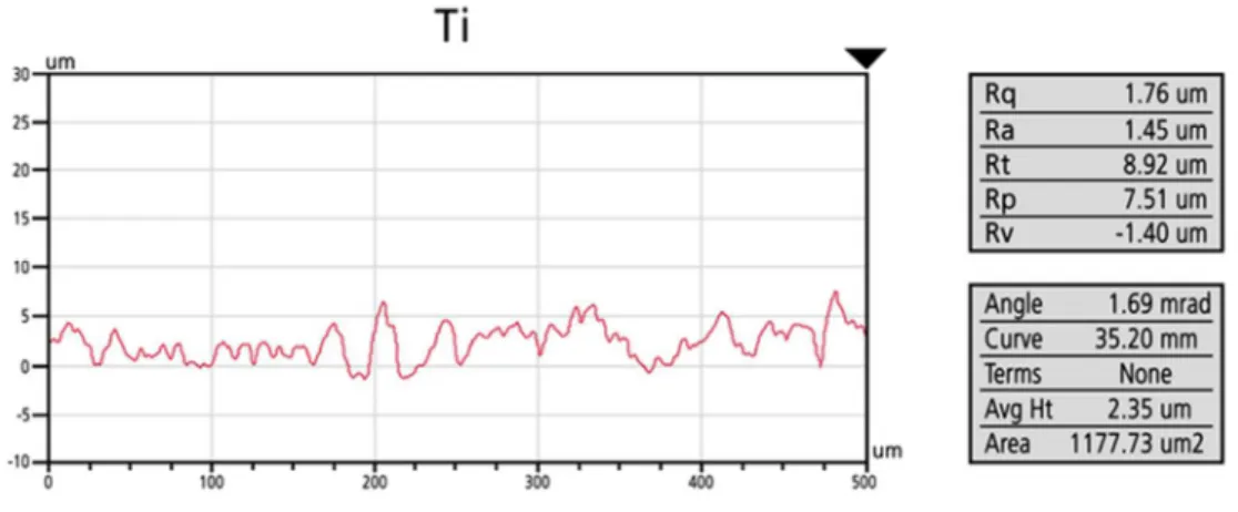
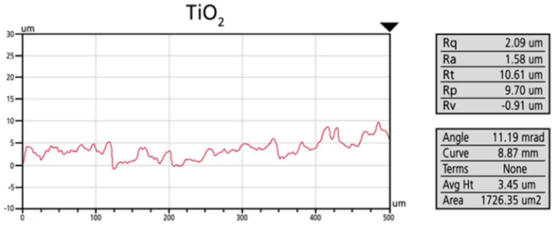
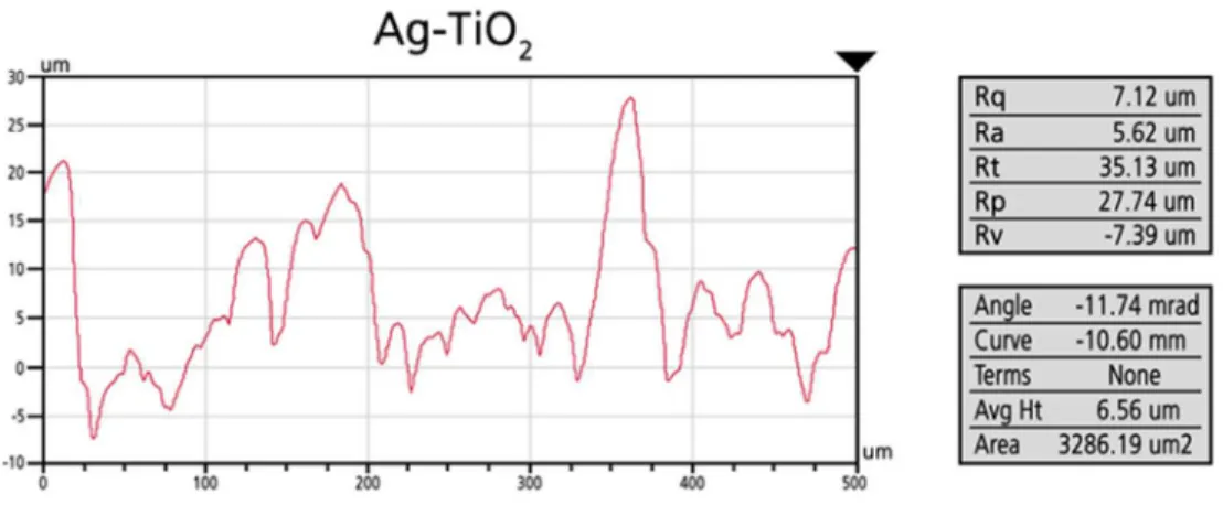
Profilometry images (Figs. 2 and 3) confirmed the above mentioned differences in surface morphology and roughness. Uncovered Ti (sandblasted-, acid-etched) and TiO2
copolymer coated discs had similar roughness values (for Ti: Ra = 1.26 µm; for TiO2: Ra = 1.79 µm), but Ag-TiO2 coated samples had considerably rougher surfaces (for Ag-TiO2:Ra = 5.76 µm), with considerable height differences along the scan direction.
3.3 MTT assay
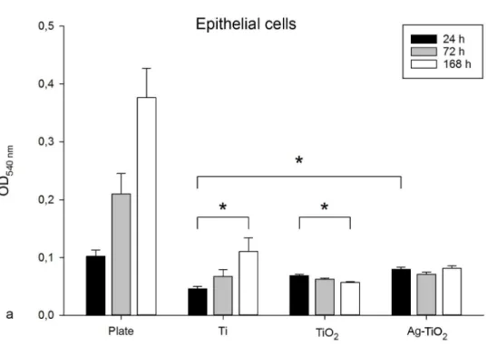
The results of the MTT measurements related to cell attachment (24h observation) and cell proliferation (72 and 168h) are shown on the bar graphs in Fig. 4
Attachment of epithelial cells on the Ag-TiO2 modified surface was significantly higher (p = 0.013) compared to the polished control Ti surface. The level of attachment almost reached the control plate values. No significant differences were found in the attachment of cells between the two copolymer-coated discs. The early proliferation of epithelial cells was similar on all of the investigated discs. No significant differences were found between the polished and either of the the polymer- coated groups after 168h.
From a different perspective, we observed a significant increase in the cell count on the polished Ti surface after one week, while, at the same time point, a significant decrease 2
3 4 5 6 7 8 9 10 11 12 13 14 15 16 17 18 19 20 21 22 23 24 25 26 27 28 29 30 31 32 33 34 35 36 37 38 39 40 41 42 43 44 45 46 47 48 49 50 51 52 53 54 55 56 57 58 59
For Peer Review
was found in the cell count of TiO2 -modified samples. Epithelial cells did not show a growing tendency on the Ag-TiO2 -coated discs, but mitosis and apoptosis was well balanced.
First, the OD540 values of MG63 cells on different surfaces were compared at the same time points (24 h, 72 h and 168 h). No significant difference was found in the attachment (24 h) of the MG63 cells between the treated and the control surfaces. However, significantly more cells (p = 0.03) attached to the surface of the nanosilver- modified discs compared to the TiO2 -modified discs. Early proliferation of the cells (72 hours) was significantly higher on the control discs than on the TiO2 –copolymer and nanosilver coated samples (p < 0.001). After 168 hours, there was a considerable increase in the proliferation rate of MG63 cells on the uncoated Ti surfaces, in contrast to the TiO2 and Ag-TiO2 -modified surfaces, where the cells did not show any growing tendency. No statistical differences were found between the copolymer- treated groups after 72 and 168 hours. We found the smallest number of cells on the TiO2 polymer- coated surfaces.
Secondly the OD540 values of each group were evaluated over time. The definite increase in the cell number of the plate and the untreated Ti discs proves the viability of MG63 cells. In case of the two control groups there was a significant increase in the cell number between the three investigation points (p < 0.001). According to our results these cells did not show any sign of proliferation on the polymer coated discs. There was no significant difference in the cell number of the modified samples under the investigated period (24, 72 and 168 h).
3.4 Fluorescent images
Composite images of epithelial and MG63 cells are presented in figure 5. The actin cytoskeleton is red (TRITC-phalloidin) and the DNA content of cell nucleus is blue (Hoechst 33342).
We found no considerable difference in the attachment of epithelial cells on the three investigated groups, but the cells covered the surface of the silver- modified discs more profusely. Typical polygonal cell morphology with a few filopodia was characteristic in all three groups. After 72 h we observed an increased proliferation on the polished and on the Ag-TiO2 samples with well-spread morphology, whereas a decrease was observable on the TiO2 polymer modified samples. On the Ag-TiO2 samples, not every cytoskeleton was noticeable between the grains, in spite of the good visibility of cell nuclei. One week later, 2
3 4 5 6 7 8 9 10 11 12 13 14 15 16 17 18 19 20 21 22 23 24 25 26 27 28 29 30 31 32 33 34 35 36 37 38 39 40 41 42 43 44 45 46 47 48 49 50 51 52 53 54 55 56 57 58
For Peer Review
polygonal cells covered the whole surface of polished Ti discs, with several filopodia. The cell counts remained unchanged on the Ag-modified surfaces, while just a few poorly spread cells were detected on the TiO2-copolymer-coated discs. A remarkable number of rounded cells were observed on the polymer- covered discs, signifying cell detachment.
The attachment of MG63 cells was similar in the three investigated groups. Cells showed flat morphology with short and little processes. They covered the surfaces dispersedly, without any orientation. We found the most cells with rounded morphology on the silver- modified surface. After 72 h, we observed a higher cell density on the untreated Ti discs. Cells formed bigger groups, and they had more and stronger processes. Their characteristic triangle shape was noticeable. MG63 cells did not show any sign of proliferation on the TiO2 and Ag-TiO2 samples. They had some processes, but we did not observe any mitotic cells. After 168 h the cells covered almost the whole surface of the uncovered Ti sample discs, and we found multiple layers of cells near the edge of the discs.
On the TiO2 and Ag-TiO2 copolymer discs, only a few cells were observed.
4. Discussion
In our study the viability (attachment and proliferation) of epithelial cells and MG63 osteosarcoma cells was compared. Our results showed that the attachment of epithelial cells on the Ag-TiO2 modified samples was significantly higher compared to the polished Ti control samples. However, epithelial cells did not show a growing tendency on the Ag-TiO2 - coated samples, while the rate of mitosis and apoptosis was well balanced. MTT test showed that MG63 cells were not capable of proliferation on the TiO2 and Ag-TiO2-coated Ti discs.
We have found decreasing cell numbers. Nevertheless, we found living cells on both of them after one week incubation.
Using polymer-embedded nanoparticles on the surface of titanium dental implants is a relatively new approach, which means that literature on this topic is scarce. Still, it is possible to interpret our findings with the help of similar studies.
Regarding the surface roughness of the discs, our results were not in accordance with the results of previous studies30,34,35. They found that MG63 cells preferred rougher sufaces (Ra = 4-5 µm). Increased surface roughness led to increased differentiation and decreased cell proliferation. Although the average roughness of the silver modified discs was almost ideal, 2
3 4 5 6 7 8 9 10 11 12 13 14 15 16 17 18 19 20 21 22 23 24 25 26 27 28 29 30 31 32 33 34 35 36 37 38 39 40 41 42 43 44 45 46 47 48 49 50 51 52 53 54 55 56 57 58 59
For Peer Review
MG63 cells could not proliferate. This inhibitory effect could be explained by the presence of the nanoparticles.
The attachment of epithelial cells on the nanoparticle- modified surfaces was relatively high, but the proliferation was partially inhibited. In vitro epithelial cell responses of nanoscale modified dental implants were investigated by several authors 36. Rossi et al.
reported better epithelial attachment on the nanoporous sol–gel-derived TiO2 surface-treated implants as compared to untreated titanium implants in vivo 37. In an in vitro biocompatibility study of Ag-embedded TiO2-nanotubes on Ti dental implants Mei et al. found that epithelial cell viability and proliferation was better on a silver modified nanotube surface (plasma implanted at 1kV) than on pure titanium 38.
The Ag-TiO2 and TiO2 modified titanium implant surfaces in this study showed exerted considerable inhibitory effect on the proliferation of MG63 cells. A number of studies have investigated the effect of Ag nanoparticles on osteoblast-like cells. De Giglio et al. used silver nanoparticles immobilized on Ti implant material in hydrogel coating. They found that there was a progressive release of silver from the coating, which led to long-term antibacterial activity. In accordance with our results, the MTT tests showed reduced MG63 osteoblast-like cell viability after 24 and 48 h of incubation. Interestingly, after 7 days the cell proliferation increased 39. Moaddab and colleagues reported that nano-Ag influenced negatively the growth of G292 cancer cell line. This phenomenon was concentration dependent (IC50 value of 3.42 µg/ mL) and had great selectivity to cancer cells. These findings might indicate the potential of nano-silver in the field of anti-cancer therapies 40.
It must be taken into consideration, however, that some other studies came to the opposite conclusion. In a recent study, Ferraris et al. investigated the viability of MG63 osteoblast-like cells on different titanium surfaces. CP Ti and Ti6Al4V samples were treated with acid etching and controlled oxidation process in hydrogen peroxide, which was completed with silver ions. These nanostructured surfaces showed good antibacterial properties and good viability of MG63 cells on CP Ti surfaces with low concentration of silver (0.001M), but Ti6Al4V surfaces were more cytotoxic with the same amount of silver 41. Tilmaciu et al. investigated MC3T3-E1 preosteoblast cell viability and bacterial adhesion on nanocoatings with minimal silver loading. They found that these monolayers significantly inhibited the adhesion of E. coli and S. epidermidis, while not significantly interfering with the attachment and proliferation of MC3T3-E1 cells 42.
We found decreasing cell counts (both cell types) on the TiO2-polymer modified discs.
This result is in contrast with the literature. Ivask et al. concluded that TiO2 nanoparticles, 2
3 4 5 6 7 8 9 10 11 12 13 14 15 16 17 18 19 20 21 22 23 24 25 26 27 28 29 30 31 32 33 34 35 36 37 38 39 40 41 42 43 44 45 46 47 48 49 50 51 52 53 54 55 56 57 58
For Peer Review
among other metal nanoparticles (Al2O3, Fe3O4, MgO, SiO2, WO3), had no toxic side effect on epithelial and fibroblast cells 43. Similarly, Choi et al. found that TiO2 films on Ti alloys were not toxic to L-929 mouse fibroblast cells 44. A cytotoxicity study was carried out with different metal oxide nanoparticles by Jeng et al., who found that TiO2 nanoparticles had no measurable effect on Neuro-2A cells under 200 µg/mL 45. In all of these studies, cancer cell lines were used as cell culture models. One possible explanation to this discrepancy in the proliferation of epithelial cells that in this study primary cells were investigated. High sensitivity to the polymer matrix might also be an explanation.
The viability of the epithelial cells was more promising on the Ag-TiO2 modified discs according to the fluorescents pictures. The difference between the OD540 values of the MTT assay and the fluorescent images is one that is difficult to explain without going into speculations. MG63 cells proved to be still too sensitive to nanosilver, in spite of reduced AgNP concentration.
5. Conclusions
Taking all aspects of our findings into consideration, new nanoscale surface modifications of titanium dental implants have great antibacterial potential and they also appear to be biocompatible. However, the observed effect on cell proliferation indicates that the process of cell-nanoparticle interaction needs to be better understood, biocompatibility investigations with other cell types and increasing the investigation time are also needed.
6. Acknowledgments
This project was supported by TÁMOP - 4.1.1.C-13/1/KONV-2014-0001 project, entitled
“Coordinated, practice-oriented, student-friendly modernization of biomedical education in three Hungarian universities (Pécs, Debrecen, Szeged), with focus on the strengthening of international competitiveness” and sustained by the European Union and co-financed by the European Regional Development Fund. We are grateful to Denti System Ltd. Hungary for providing the titanium sample discs and to the Department of Oral Surgery (Faculty of Dentistry, University of Szeged) for providing the gingival tissues. The authors also thank to Dr. Gábor Braunitzer for the valuable discussions and advices on the interpretation of our results.
2 3 4 5 6 7 8 9 10 11 12 13 14 15 16 17 18 19 20 21 22 23 24 25 26 27 28 29 30 31 32 33 34 35 36 37 38 39 40 41 42 43 44 45 46 47 48 49 50 51 52 53 54 55 56 57 58 59
For Peer Review
References
1. Williams DF. Implants in dental and maxillofacial surgery. Biomaterials 1981;2(3):133- 146.
2. Parr GR, Gardner LK, Toth RW. Titanium: the mystery metal of implant dentistry.
Dental materials aspects. The Journal of Prosthetic Dentistry 1985;54(3):410-414.
3. Lemons JE. Dental implant biomaterials. Journal of the American Dental Association (1939) 1990;121(6):716-719.
4. Yeo I-S. Reality of dental implant surface modification: a short literature review. The Open Biomedical Engineering Journal 2014;8:114-119.
5. Wang RR, Fenton A. Titanium for prosthodontic applications: a review of the literature.
Quintessence International (Berlin, Germany: 1985) 1996;27(6):401-408.
6. Lautenschlager EP, Monaghan P. Titanium and titanium alloys as dental materials.
International Dental Journal 1993;43(3):245-253.
7. Schiff N, Grosgogeat B, Lissac M, Dalard F. Influence of fluoride content and pH on the corrosion resistance of titanium and its alloys. Biomaterials 2002;23(9):1995-2002.
8. Smeets R, Henningsen A, Jung O, Heiland M, Hammächer C, Stein JM. Definition, etiology, prevention and treatment of peri-implantitis – a review. Head & Face Medicine 2014;10:34.
9. Mombelli A, Müller N, Cionca N. The epidemiology of peri-implantitis. Clinical Oral Implants Research 2012;23 Suppl 6:67-76.
10. Tiller JC, Liao CJ, Lewis K, Klibanov AM. Designing surfaces that kill bacteria on contact. Proceedings of the National Academy of Sciences of the United States of America 2001;98(11):5981-5985.
11. Norowski PA, Bumgardner JD. Biomaterial and antibiotic strategies for peri-implantitis:
a review. Journal of Biomedical Materials Research. Part B, Applied Biomaterials 2009;88(2):530-543.
12. Xu M-f, Lin S, Chen X-m, Peng Y-z. Studies on characteristics of nanostructure of N- TiO2 thin films and photo-bactericidal action. Journal of Zhejiang University. Science.
B 2006;7(7):586-590.
13. Mills A, Le Hunte S. An overview of semiconductor photocatalysis. Journal of Photochemistry and Photobiology A: Chemistry 1997;108(1):1-35.
14. Wang W, Huang G, Yu JC, Wong PK. Advances in photocatalytic disinfection of bacteria: Development of photocatalysts and mechanisms. Journal of Environmental Sciences 2015;34:232-247.
15. Pelaez M, Nolan NT, Pillai SC, Seery MK, Falaras P, Kontos AG, Dunlop PSM, Hamilton JWJ, Byrne JA, O'Shea K and others. A review on the visible light active titanium dioxide photocatalysts for environmental applications. Applied Catalysis B:
Environmental 2012;125:331-349.
16. Foster HA, Ditta IB, Varghese S, Steele A. Photocatalytic disinfection using titanium dioxide: spectrum and mechanism of antimicrobial activity. Applied Microbiology and Biotechnology 2011;90(6):1847-1868.
17. Ohtani B, Ogawa Y, Nishimoto S-i. Photocatalytic Activity of Amorphous−Anatase Mixture of Titanium(IV) Oxide Particles Suspended in Aqueous Solutions. The Journal of Physical Chemistry B 1997;101(19):3746-3752.
2 3 4 5 6 7 8 9 10 11 12 13 14 15 16 17 18 19 20 21 22 23 24 25 26 27 28 29 30 31 32 33 34 35 36 37 38 39 40 41 42 43 44 45 46 47 48 49 50 51 52 53 54 55 56 57 58
For Peer Review
18. Rothenberger G, Moser J, Graetzel M, Serpone N, Sharma DK. Charge carrier trapping and recombination dynamics in small semiconductor particles. Journal of the American Chemical Society 1985;107(26):8054-8059.
19. Veres Á, Janovák L, Bujdosó T, Rica T, Fodor E, Tallósy SP, Buzás N, Nagy E, Dékány I. Silver and Phosphate Functionalized Reactive TiO2/Polymer Composite Films for Destructions of Resistent Bacteria Using Visible Light. Journal of Advanced Oxidation Technologies 2012;15(1):205-216
20. Geiser M, Rothen-Rutishauser B, Kapp N, Schürch S, Kreyling W, Schulz H, Semmler M, Im Hof V, Heyder J, Gehr P. Ultrafine particles cross cellular membranes by nonphagocytic mechanisms in lungs and in cultured cells. Environmental Health Perspectives 2005;113(11):1555-1560.
21. Akhavan O, Ghaderi E. Self-accumulated Ag nanoparticles on mesoporous TiO2 thin film with high bactericidal activities. Surface and Coatings Technology 2010;204(21–
22):3676-3683.
22. Li Q, Mahendra S, Lyon DY, Brunet L, Liga MV, Li D, Alvarez PJJ. Antimicrobial nanomaterials for water disinfection and microbial control: Potential applications and implications. Water Research 2008;42(18):4591-4602.
23. Zhang R, Zhang W, Bai X, Song X, Wang C, Gao X, Tian X, Liu F. Report: Discussion on the development of nano Ag/TiO2 coating bracket and its antibacterial property and biocompatibility in orthodontic treatment. Pakistan Journal of Pharmaceutical Sciences 2015;28(2 Suppl):807-810.
24. Sambale F, Wagner S, Stahl F, Khaydarov RR, Scheper T, Bahnemann D, Investigations of the Toxic Effect of Silver Nanoparticles on Mammalian Cell Lines.
Journal of Nanomaterials 2015:e136765.
25. Tallósy SP, Janovák L, Ménesi J, Nagy E, Juhász Á, Balázs L, Deme I, Buzás N, Dékány I. Investigation of the antibacterial effects of silver-modified TiO2 and ZnO plasmonic photocatalysts embedded in polymer thin films. Environmental Science and Pollution Research International 2014;21(19):11155-11167.
26. Györgyey Á, Janovák L, Ádám A, Kopniczky J, Tóth KL, Deák Á, Panayotov I, Cuisinier F, Dekany I, Turzo K. Investigation of the in vitro photocatalytic antibacterial activity of nanocrystalline TiO2 and coupled TiO2/Ag containing copolymer on the surface of medical grade titanium. J Biomater Appl 2016;31(1):55-67.
27. De Saint Jean M, Baudouin C, Di Nolfo M, Roman S, Lozato P, Warnet JM, Brignole F. Comparison of morphological and functional characteristics of primary-cultured human conjunctival epithelium and of Wong–Kilbourne derivative of Chang conjunctival cell line. Experimental Eye Research 2004;78(2):257-274.
28. Klinge B, Meyle J, Working G. Soft-tissue integration of implants. Consensus report of Working Group 2. Clinical Oral Implants Research 2006;17 Suppl 2:93-96.
29. Atsuta I, Ayukawa Y, Furuhashi A, Ogino Y, Moriyama Y, Tsukiyama Y, Koyano K.
In vivo and in vitro studies of epithelial cell behavior around titanium implants with machined and rough surfaces. Clinical Implant Dentistry and Related Research 2014;16(5):772-781.
30. Bächle M, Kohal RJ. A systematic review of the influence of different titanium surfaces on proliferation, differentiation and protein synthesis of osteoblast-like MG63 cells.
Clinical Oral Implants Research 2004;15(6):683-692.
31. Clover J, Gowen M. Are MG-63 and HOS TE85 human osteosarcoma cell lines representative models of the osteoblastic phenotype? Bone 1994;15(6):585-591.
32. Ungvári K, Pelsöczi IK, Kormos B, Oszkó A, Rakonczay Z, Kemény L, Radnai M, Nagy K, Fazekas A, Turzó K. Effects on titanium implant surfaces of chemical agents 2
3 4 5 6 7 8 9 10 11 12 13 14 15 16 17 18 19 20 21 22 23 24 25 26 27 28 29 30 31 32 33 34 35 36 37 38 39 40 41 42 43 44 45 46 47 48 49 50 51 52 53 54 55 56 57 58 59
For Peer Review
used for the treatment of peri-implantitis. Journal of Biomedical Materials Research.
Part B, Applied Biomaterials 2010;94(1):222-229.
33. Mosmann T. Rapid colorimetric assay for cellular growth and survival: application to proliferation and cytotoxicity assays. Journal of Immunological Methods 1983;65(1- 2):55-63.
34. Lincks J, Boyan BD, Blanchard CR, Lohmann CH, Liu Y, Cochran DL, Dean DD, Schwartz Z. Response of MG63 osteoblast-like cells to titanium and titanium alloy is dependent on surface roughness and composition. Biomaterials 1998;19(23):2219-2232.
35. Bannister SR, Lohmann CH, Liu Y, Sylvia VL, Cochran DL, Dean DD, Boyan BD, Schwartz Z. Shear force modulates osteoblast response to surface roughness. Journal of Biomedical Materials Research 2002;60(1):167-174.
36. Mendonça G, Mendonça DBS, Aragão FJL, Cooper LF. Advancing dental implant surface technology – From micron- to nanotopography. Biomaterials 2008;29(28):3822- 3835.
37. Rossi S, Tirri T, Paldan H, Kuntsi-Vaattovaara H, Tulamo R, Närhi T. Peri-implant tissue response to TiO2 surface modified implants. Clinical Oral Implants Research 2008;19(4):348-355.
38. Mei S, Wang H, Wang W, Tong L, Pan H, Ruan C, Ma Q, Liu M, Yang H, Zhang L and others. Antibacterial effects and biocompatibility of titanium surfaces with graded silver incorporation in titania nanotubes. Biomaterials 2014;35(14):4255-4265.
39. De Giglio E, Cafagna D, Cometa S, Allegretta A, Pedico A, Giannossa LC, Sabbatini L, Mattioli-Belmonte M, Iatta R. An innovative, easily fabricated, silver nanoparticle- based titanium implant coating: development and analytical characterization. Analytical and Bioanalytical Chemistry 2013;405(2-3):805-816.
40. Moaddab S, Ahari H, Shahbazzadeh D, Motallebi AA, Anva AA, Rahman-Nya J, Shokrgozar MR. Toxicity study of nanosilver (Nanocid®) on osteoblast cancer cell line.
Int. Nano Lett. 2011; 1(1):11-16
41. Ferraris S, Venturello A, Miola M, Cochis A, Rimondini L, Spriano S. Antibacterial and bioactive nanostructured titanium surfaces for bone integration. Applied Surface Science 2014;311:279-291.
42. Tilmaciu CM, Mathieu M, Lavigne JP, Toupet K, Guerrero G, Ponche A, Amalric J, Noel D, Mutin PH. In vitro and in vivo characterization of antibacterial activity and biocompatibility: a study on silver-containing phosphonate monolayers on titanium.
Acta Biomater 2015;15:266-77.
43. Ivask A, Titma T, Visnapuu M, Vija H, Kakinen A, Sihtmae M, Pokhrel S, Madler L, Heinlaan M, Kisand V and others. Toxicity of 11 metal oxide nanoparticles to three mammalian cell types in vitro. Curr Top Med Chem 2015;15(18):1914-29.
44. Choi J-Y, Kim K-H, Choy K-C, Oh K-T, Kim K-N. Photocatalytic antibacterial effect of TiO2 film formed on Ti and TiAg exposed to Lactobacillus acidophilus. Journal of Biomedical Materials Research Part B: Applied Biomaterials 2007;80B(2):353-359.
45. Jeng HA, Swanson J. Toxicity of metal oxide nanoparticles in mammalian cells. Journal of Environmental Science and Health. Part A, Toxic/Hazardous Substances &
Environmental Engineering 2006;41(12):2699-2711.
. 2
3 4 5 6 7 8 9 10 11 12 13 14 15 16 17 18 19 20 21 22 23 24 25 26 27 28 29 30 31 32 33 34 35 36 37 38 39 40 41 42 43 44 45 46 47 48 49 50 51 52 53 54 55 56 57 58
For Peer Review
Figure legends:
Figure 1. SEM images of the surfaces without cells before UV irradiation (×500 and ×5000 magnification)
Figure 2. Characteristic line profiles of the uncovered Ti, TiO2 copolymer coated and Ag- TiO2 coated surfaces (Ra (µm))
Figure 3. Average surface roughness values of the samples (Ra (µm)). Data are given as mean± SEM. Asterisk denotes siginificant difference at p< 0.05.
Figure 4. 24, 72 and 168 h OD540 values of epithelial cells (a) and MG63 cells (b) incubated with MTT on the control discs (plate and uncovered Ti) and on the TiO2 and Ag-modified TiO2 polymer covered discs. Data are presented as mean ± SEM.
Figure 5. Fluorescent images of primary epithelial (a) and MG63 cells (b) on Ti, TiO2 and Ag-TiO2 surfaces at 24 h, 72 h and 168 h at a magnification of ×100.
2 3 4 5 6 7 8 9 10 11 12 13 14 15 16 17 18 19 20 21 22 23 24 25 26 27 28 29 30 31 32 33 34 35 36 37 38 39 40 41 42 43 44 45 46 47 48 49 50 51 52 53 54 55 56 57 58 59
For Peer Review
Figure 1. SEM images of the surfaces without cells before UV irradiation (×500 and ×5000 magnification)
SEM images (Fig.1) revealed si 124x115mm (300 x 300 DPI)
2 3 4 5 6 7 8 9 10 11 12 13 14 15 16 17 18 19 20 21 22 23 24 25 26 27 28 29 30 31 32 33 34 35 36 37 38 39 40 41 42 43 44 45 46 47 48 49 50 51 52 53 54 55 56 57 58
For Peer Review
Figure 2. Characteristic line profiles of the uncovered Ti, TiO2 copolymer coated and Ag-TiO2 coated surfaces (Ra (µm))
Profilometry images (Figs. 2 a 59x25mm (300 x 300 DPI)
2 3 4 5 6 7 8 9 10 11 12 13 14 15 16 17 18 19 20 21 22 23 24 25 26 27 28 29 30 31 32 33 34 35 36 37 38 39 40 41 42 43 44 45 46 47 48 49 50 51 52 53 54 55 56 57 58 59
For Peer Review
Figure 2. Characteristic line profiles of the uncovered Ti, TiO2 copolymer coated and Ag-TiO2 coated surfaces (Ra (µm))
Profilometry images (Figs. 2 a 59x25mm (300 x 300 DPI)
2 3 4 5 6 7 8 9 10 11 12 13 14 15 16 17 18 19 20 21 22 23 24 25 26 27 28 29 30 31 32 33 34 35 36 37 38 39 40 41 42 43 44 45 46 47 48 49 50 51 52 53 54 55 56 57 58
For Peer Review
Figure 2. Characteristic line profiles of the uncovered Ti, TiO2 copolymer coated and Ag-TiO2 coated surfaces (Ra (µm))
Profilometry images (Figs. 2 a 59x25mm (300 x 300 DPI)
2 3 4 5 6 7 8 9 10 11 12 13 14 15 16 17 18 19 20 21 22 23 24 25 26 27 28 29 30 31 32 33 34 35 36 37 38 39 40 41 42 43 44 45 46 47 48 49 50 51 52 53 54 55 56 57 58 59
For Peer Review
Figure 3. Average surface roughness values of the samples (Ra (µm)). Data are given as mean± SEM.
Asterisk denotes siginificant difference at p< 0.05.
Profilometry images (Figs. 2 a 109x80mm (300 x 300 DPI)
2 3 4 5 6 7 8 9 10 11 12 13 14 15 16 17 18 19 20 21 22 23 24 25 26 27 28 29 30 31 32 33 34 35 36 37 38 39 40 41 42 43 44 45 46 47 48 49 50 51 52 53 54 55 56 57 58
For Peer Review
Figure 4. 24, 72 and 168 h OD540 values of epithelial cells (a) and MG63 cells (b) incubated with MTT on the control discs (plate and uncovered Ti) and on the TiO2 and Ag-modified TiO2 polymer covered discs. Data are
presented as mean ± SEM.
The results of the MTT measure 157x110mm (300 x 300 DPI)
2 3 4 5 6 7 8 9 10 11 12 13 14 15 16 17 18 19 20 21 22 23 24 25 26 27 28 29 30 31 32 33 34 35 36 37 38 39 40 41 42 43 44 45 46 47 48 49 50 51 52 53 54 55 56 57 58 59
For Peer Review
Figure 4. 24, 72 and 168 h OD540 values of epithelial cells (a) and MG63 cells (b) incubated with MTT on the control discs (plate and uncovered Ti) and on the TiO2 and Ag-modified TiO2 polymer covered discs. Data are
presented as mean ± SEM.
The results of the MTT measure 158x110mm (300 x 300 DPI)
2 3 4 5 6 7 8 9 10 11 12 13 14 15 16 17 18 19 20 21 22 23 24 25 26 27 28 29 30 31 32 33 34 35 36 37 38 39 40 41 42 43 44 45 46 47 48 49 50 51 52 53 54 55 56 57 58
For Peer Review
Figure 5. Fluorescent images of primary epithelial (a) and MG63 cells (b) on Ti, TiO2 and Ag-TiO2 surfaces at 24 h, 72 h and 168 h at a magnification of ×100.
Composite images of epithelial 105x72mm (300 x 300 DPI)
2 3 4 5 6 7 8 9 10 11 12 13 14 15 16 17 18 19 20 21 22 23 24 25 26 27 28 29 30 31 32 33 34 35 36 37 38 39 40 41 42 43 44 45 46 47 48 49 50 51 52 53 54 55 56 57 58 59
For Peer Review
Figure 5. Fluorescent images of primary epithelial (a) and MG63 cells (b) on Ti, TiO2 and Ag-TiO2 surfaces at 24 h, 72 h and 168 h at a magnification of ×100.
Composite images of epithelial 105x72mm (300 x 300 DPI)
2 3 4 5 6 7 8 9 10 11 12 13 14 15 16 17 18 19 20 21 22 23 24 25 26 27 28 29 30 31 32 33 34 35 36 37 38 39 40 41 42 43 44 45 46 47 48 49 50 51 52 53 54 55 56 57 58