Regulation of epithelial and endothelial cell plasticity under certain pathological conditions
Ph.D. Thesis
Ákos Gasparics, M.D.
Semmelweis University
Doctoral School of Basic and Translational Medicine
Supervisor: Attila Sebe, M.D., Ph.D.
Official reviewers: Balázs Antus, M.D., Ph.D., D.Sc.
Anna-Mária Tőkés, Ph.D.
Chair of examination comittee: György Reusz, M.D., D.Sc.
Members of examination committee: Péter Vörös, M.D., Ph.D.
Kálmán Tory, M.D., PhD
Budapest
2018
2
1. Introduction
Phenotypic change of somatic cells is termed cell plasticity. During this process, somatic cells change their morphology and function. In a wider sense, stem cell differentiation belongs to this process too, similarly to the activation of facultative stem cells. Dedifferentiation, transdifferentiation, or phenotypic changes of cells are subtypes of cell plasticity. The aim of this process is to maintain homeostasis in the body. However, malfunction of regulatory mechanisms may result in malignant transformation, or dysfunctional adaptation of cells.
Phenotypical change in epithelial and mesenchymal cells typically occurs under physiological conditions (e.g. embryonic development). The two cell types can transform into each other, phenomena called „epithelial- mesenchymal transition” (EMT), and „mesenchymal- epithelial transition” (MET). EMT is a physiological process in embryonic development, wound healing, but it may emerge in pathological conditions, such as tissue fibrosis and metastasis formation. Endothelial cells are also able to transform into mesenchymal cells through
„endothelial mesenchymal transition” (EndMT). In our study we examined two models of these highly similar processes.
The global and US prevalence of chronic kidney disease (CKD) is as high as 13%.
Irrespective of the pathological cause and disease, CKD is marked by progressive renal fibrosis. Alongside primary glomerular and congenital renal diseases, endemic diabetes and hypertension are the leading causes of CKD.
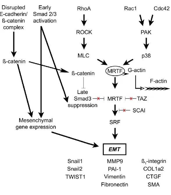
In the long term, CKD may lead to end stage renal disease (ESRD), and patients may require renal replacement therapy or kidney transplantation. With regard to the burden and prevalence of CKD, it is important to study the process. Histological findings of CKD are characterized by tubulointerstitial fibrosis, glomeruli and capillaries are affected as well. In a tubular cell model, the „two hit” model of EMT was established. Initial weakening of epithelial integrity („first hit”, caused by hypoxia, physical injury, ureteral obstruction, etc.) is followed by TGF- β1 exposition, and these two hits result in a focal EMT. Increased TGF-β1 and extracellular matrix (ECM) production leads to the spreading of EMT and loss of epithelial contacts in neighboring cells. During the final stage of transition, α-smooth muscle actin (α-SMA) expression will be displayed, as a marker of myofibroblast cells. Serum response factor (SRF) is an effector in the EMT signaling pathway, and Myocardin related transcription factor A and B (MRTF-A, B) act as activating cofactors of SRF. Recently, suppressor of cancer cell invasion (SCAI) protein was identified, as an inhibiting cofactor or MRTFs.
3
In the majority of patients with cancer metastasis formation leads to a terminal illness.
During metastasis tumor cells produce a variety of factors to destroy/damage endothelial barriers. To infiltrate the central nervous system (CNS), tumor cells need to pass through the blood-brain barrier (BBB). The exact mechanisms of tumor cell extravasation still remain to be elucidated.
The endothelial barrier function is impaired by cytokines (e.g. TGF-β) originated from cancer cells. VE-cadherin complexes are interrupted, which changes the morphology of endothelial cells. Cytoskeletal remodeling is also induced by cancer cells. These changes can be observed in the process of EndMT as well, hence we presumed that endothelial cells are not „victims”
of metastasis formation, but play an active role in extravasation. Expression patterns of endothelial cells change during metastasis, their phenotype shifts towards a mesenchymal state. On the basis of these changes, we hypothesized that cancer cells induce an EndMT to challenge the blood blain barrier.
4
2. Objectives
In previous works, our team identified novel signaling pathways regulating TGF and injury caused EMT and SMA expression. MRTF dependent signalization and protein expression emerged as central regulators of this process. SCAI was identified as an inhibitor of MRTF in tumor models. SCAI is also expressed in the kidney, therefore we sought to investigate its role in the process of EMT and renal fibrosis.
Aims of the study:
1. To investigate the effect of SCAI on TGF-β1 or Angiotensin II treatment in a renal fibrosis model (in vitro), and assess the changes of EMT markers during the transition.
2. To investigate the effect of SCAI on MRTF-A, MRTF-B, Rho, Rac, Cdc42 mediated signaling in a renal fibrosis model (in vitro).
3. To investigate the effect of TGF-β1 treatment on SCAI expression (in vitro), and to study the effect of UUO or allograft rejection on SCAI expression (in vivo).
4. To investigate whether the EndMT or induced pluripotent stem cell (iPS) reprogramming caused phenotypical changes are linked to SCAI expression changes (in vitro).
Metastatic extravasation causes EndMT- like changes in endothelial cells. We hypothesized, that in the vessels of the brain or lungs – where endothelial cells are organized in compact layers – EndMT is involved in metastatic extravasation.
Aims of the study:
1. To investigate the effect of TGF-β1 treatment on brain endothelial cells (in vitro).
2. To investigate whether tumor cells are able to induce EndMT on brain endothelial cells in a TGF-β dependent manner (in vitro).
5
3. Methods
3.1 Cell culture
LLC-PK1 (CL4) proximal tubular epithelial cells were a kind gift of Dr. R. Harris.
LLC-PK1 cells stably expressing GFP-SCAI were created by FuGene6 transfection (Roche, Mannheim, Germany). Cells were sorted based on GFP fluorescence using the fluorescence- activated cell sorting Aria High Speed Cell Sorter (Becton-Dickinson, San Jose, CA).
Samples were analyzed by a FACSCalibur flow cytometer with CellQuest acquisition software (Becton Dickinson Immunocytometry Systems, San Jose, CA).
Primary rat brain endothelial cells (RBECs) were isolated from two week old Wistar rats (Toxi-Coop, Budapest, Hungary). Isolation of primary cerebral endothelial cells was carried out in strict accordance with the national and international recommendations for the care and use of laboratory animals. The protocol was reviewed and approved by the Regional Animal Health and Food Control Station of Csongrád County (Permit Number: XVI/2980/2012).
Human umbilical vein endothelial cells (HUVECs) were isolated as in our previous experiments. Human umbilical cord veins were obtained from the 2nd Department of Obstetrics and Gynaecology, Semmelweis University, after obtaining written informed consent for use of these samples in research approved by the Semmelweis University Regional and Institutional Committee of Science and Research Ethics, Budapest, Hungary (TUKEB 126/2014).
B16/F10 murine melanoma cells were kept in RPMI medium.
A2058 human melanoma cells were kept in MEM medium.
SK-BR3, MCF-7 és MDA-MB231 human breast cancer cells were maintained in DMEM.
Human iPS cells were reprogrammed as described previously.
3.2 Transient transfection and luciferase promoter activity measurements
LLC-PK1 cells were grown on 6-well plates and transfected using 2.5 mL of FuGene6 (Roche, Mannheim, Germany) reagent/1 mg DNA. Firefly and Renilla luciferase activities were measured by the Dual-Luciferase Reporter Assay Kit (Promega) using a Victor X3 2030 Multilabel Plate Reader (PerkinElmer, Waltham, MA), according to the manufacturer’s instructions.
6 3.3 UUO in mice
UUO experiments were carried out by Péter Hamar and his team at the Institute of Pathophysiology, Semmelweis University. Male C57BL/6 mice, obtained from Charles River, were bred at the animal facility of Semmelweis University. Animals were kept on regular rodent chow and given water ad libitum. All animal experiments were performed according to the institutional regulations, the Hungarian law on animal care and protection [1998/XVIII, 243/1998(XII.31)], and were approved by the local Ethical Committee for Animal Experimentation (22.1/4261/003/2009).
3.4 Kidney transplantation rejection model
Experiments were carried out by Péter Hamar and his team at the Institute of Pathophysiology, Semmelweis University. Male Lewis (LEW, RT1I) and Brown-Norway (BN, RT1n) rats were obtained from Charles River (Munich, Germany, through Akronom Kft., Budapest, Hungary). Lewis-Brown-Norway (LEW x BN F1, LBN) hybrid rats were bred at the animal facility of Semmelweis University. The rats were housed under standard conditions, and received rat chow and water ad libitum. All experimental procedures were in accordance with the Guide for the Care and Use of Laboratory Animals published by the US National Institutes of Health. The experimental protocol was reviewed and approved by the Institutional Ethical Committee for Animal Care and Use of Semmelweis University Budapest, Hungary (XIV-I-001/2012–4/2012).
3.5 RT-qPCR analysis
Cells were washed once with PBS and total RNA was isolated using TRIzol (Invitrogen), following the instructions of the manufacturer. 2 μg RNA was reverse transcribed (High Capacity cDNA Reverse Transcription Kit, Applied Biosystems, Forster City, CA, USA) using random primers. PCR reactions were performed on a BioRad CFX thermalcycler (BioRad, Hercules, CA, USA) using the Maxima SYBR Green PCR Master Mix (Thermo) and 95 °C for 15 s and 60 °C for 60 s for 40 cycles. Specificity and efficiency of the PCR reaction was confirmed with melting curve and standard curve analysis, respectively. Mean values are expressed with the formula 2-ΔΔCt. Three parallels were measured for each experimental point, and the experiment was repeated two times.
7 3.6. Western blot
Cells were scraped into lysis buffer. Protein concentration was determined using the
BCA Protein Assay (Pierce Thermo Scientific, Rockford, IL). Samples were mixed in a 1:1 ratio with two times Laemmli buffer and boiled for 5 minutes. Equal amounts of protein were separated on 12% SDS-polyacrylamide gel and transferred to nitrocellulose membranes (Bio- Rad, Budapest, Hungary). Membranes were blocked with Tris-buffered saline, containing 0.1% Tween 20 and 5% skim milk for an hour, and then incubated overnight with the primary antibody (in Tris-buffered saline-Tween 20 plus 0.5% skim milk), extensively washed, and incubated with the corresponding peroxidase-conjugated secondary antibody. Blots were visualized by the electrochemiluminescence detection system (Thermo Scientific, Waltham, MA, USA).
3.7 Gene microarray data analysis
A set of gene expression profiles was downloaded from Gene Expression Omnibus (GEO) of the National Center for Biotechnology Information (NCBI). Data was derived from HUVECs and 1205Lu melanoma cells grown alone or in co-culture (accession number: GSE8699).
Gene expression levels between HUVECs grown alone and HUVECs co-cultured with the melanoma cells were analyzed. EndMT marker gene expression levels were compared in the two datasets. Genes with a detection p-value <0.05 were considered if presenting a minimum fold change threshold of 1.5.
3.8 Statistical analysis
For transfection and luciferase promoter assay, duplicate measurements were performed, and experiments were repeated at least three times. Results are presented as mean ± SE. Relative promoter activities (luciferase activity) were compared between groups.
For RT-PCR three parallels were measured for each experimental point, and the experiment was repeated two times. For statistical analysis we used Mann-Whitney U test.
For Western blot assays triplicate measurements were performed, our results are presented as representative figures each.
8
4. Results
4.1 Role of SCAI during EMT of proximal tubular epithelial cells
4.1.1 SCAI inhibits TGF-β1 induced α-SMA, calponin, CTGF protein expression.
TGF-β1 treatment in LLC-PK1 cells induced SMA, CTGF and calponin expression, while in LLC-PK1/SCAI cells SMA, CTGF or calponin expression barely occurred. TGF-β1 treatment reduced E-cadherin expression, while in LLC-PK1/SCAI these effects were mitigated.
4.1.2 SCAI inhibits MRTF-A, MRF-B induced SMA promoter activity.
In LLC-PK1 cells cotransfection of SCAI with MRTF-A or MRTF-B inhibited the SMA promoter activation induced by these factors.
4.1.3 SCAI inhibits RhoA, Rac, Cdc42 induced SMA promoter activity.
In LLC-PK1 cells the activation of the SMA promoter induced by the constitutively active forms of RhoA, Rac, Cdc42 was diminished by the cotransfection of SCAI.
4.1.4 SCAI inhibits TGF-β1, MRTF-A, MRTF-B induced SMA promoter activity in a CArG dependent manner.
To test whether SCAI interfered with SMA promoter activation via CArG domains, we used the p152-SMA-Luc promoter construct, which contains a 152-bp long sequence of the SMA promoter containing the two CArGs and the TGF-β control element, but lacking both Smad- binding elements and the E-box. The activation of the 152-bp SMA promoter induced by TGF-β treatment or MRTF-A and MRTF-B cotransfection was inhibited by SCAI cotransfection.
4.1.5 TGF-β1 treatment reduces SCAI-, promotes MRTF expression in LLC-PK1 cells.
3 days of TGF-β1 treatment reduced SCAI expression, and promoted MRTF-A and MRTF-B expression in LLC-PK1 cells.
4.1.6 SCAI expression is reduced in kidneys of UUO mice
To characterize the expression patterns of SCAI in the context of renal fibrosis in vivo, the unilateral ureteral obstruction (UUO) model was used. In parallel to a marked expression of SMA in the obstructed kidneys a reduced SCAI expression was detected.
9
4.1.7 SCAI expression is reduced during kidney allograft rejection
In a rat model of renal transplant rejection samples were examined at 4. and 7. days post transplantation. SCAI protein expression decreased, while SMA expression increased in allograft samples.
4.1.8 SCAI inhibits angiotensin II induced SMA promoter activity.
Co-transfection of DN-MyoC reduced the SMA promnoter activation induced by angiotensin II. The CCG-1423 is a specific inhibitor of MRTF dependent signaling. Pretreatment with CCG-1423 prevented angiotensin II induced SMA promoter activation. Co-transfection with SCAI prevented angiotensin II induced SMA promoter activation as well.
4.1.9 SCAI mRNA expression is reduced in EndMT.
Cancer cell conditioned/activated medium (ACM) treatment reduced SCAI mRNA expression in HUVECs. In parallel to the expression of Nanog and E-cadherin, SCAI expression was also increased during the reprogramming of fibroblasts to iPS cells.
4.2 EndMT in primary rat brain endothelial cells (RBEC)
4.2.1 TGF-β1 treatment results in EndMT.
TGF-β1 treatment of RBECs decreased claudin-5, occludin and VE-cadherin protein expression, while SMA, β1-integrin, fibronectin, N-cadherin expression increased.
4.2.2 TGF-β1 induced SMA-expression requires TGFβ receptor and ROCK.
SB-431542 is a specific inhibitor of TGF-βR1 kinase. Pretreatment with SB-431542 inhibited TGF-β1 induced SMA expression in RBECs. Pretreatment with a specific Rho kinase inhibitor (Y-27632) inhibited TGF-β1 induced SMA expression as well.
4.2.3 B16/F10 ACM induces EndMT through TGF-β signaling in RBECs.
ACM treatment reduced claudin-5 protein expression, and led to fibronectin and SMA expression and Smad 2/3 phosphorylation. These effects were mitigated in the presence of the specific TGF-β1R1 kinase inhibitor SB-431542.
10
4.2.4 Various cancer cell line ACM are able to induce SMA expression through TGF- β signaling in RBECs.
Treatment of RBECs with A2058 human melanoma ACM, MCF-7 human breast adenocarcinoma ACM, MDA-MB231 human breast adenocarcinoma ACM induced SMA expression while the pretreatment with SB-431542 prevented SMA expression in all cases.
4.2.5 MDA-MB231, SK-BR3 ACM induced SMA expression through TGF-β signaling in HUVECs.
MDA-MB231 and SK-BR3 human breast adenocarcinoma ACM treatments induced SMA expression in HUVECs. SB-431542 prevented SMA expression in both cases.
In a public Gene Expression Omnibus (GEO) database, co-culture with melanoma cells led to expressional changes characteristic to EndMT in HUVECs. Gene expression profiles of HUVECs and HUVECs co-cultured with 1205Lu human metastatic melanoma cells were analyzed and compared. In HUVECs cocultured with melanoma cells there was a marked decrease in expression levels of several endothelial markers. In parallel, co-cultured HUVECs exhibited elevated expression levels of EndMT markers.
11
5. Conclusions
Role of SCAI in the context of EMT and renal fibrosis
1. SCAI inhibited TGF-β1 induced SMA promoter activity in LLC-PK1 cells. In LLC-PK1 cells, the presence of SCAI prevented the expression of SMA, calponin, and CTGF induced by TGF-β1 treatment, and E-cadherin expression was preserved. SCAI inhibited TGF-β1 induced SMA promoter activation induced by several signaling molecules (Figure 1).
Similarly to pretreatments with a specific MRTF inhibitor and the expression of a dominant negative form of MRTF, the cotransfection of SCAI diminished the SMA promoter activation induced by angiotensin II. These findings suggest, that angiotensin acts through MRTFs to activate the SMA promoter, which effect is mitigated by SCAI.
2. SCAI inhibited MRTF-A, MRTF-B, RhoA, Rac, Cdc42 induced SMA promoter activity.
Using the p152-SMA-Luc promoter construct, SCAI co-transfection resulted in reduced SMA promoter activation induced by MRTF-A or MRTF-B. These findings suggest that SCAI acts as a cofactor of MRTFs, and its effect is CArG dependent.
3. In vitro TGF-β1 treatment, and in vivo renal fibrosis in UUO and allograft rejection models led to reduced SCAI expression, and elevated SMA expression.
4. ACM treatment of HUVEC cells reduced SCAI mRNA expression, while SCAI expression was elevated during iPS reprogramming of fibroblasts. These findings suggest that SCAI expression may be linked to endothelial/epithelial phenotype.
12
Figure 1: Role of SCAI during EMT
13 EndMT and metastasis
1. TGF- β1 treatment reduced claudin-5, occludin, VE-cadherin, and increased SMA, β1- integrin, N-cadherin protein expression in RBECs. These effects were TGFβR and ROCK dependent.
2. Melanoma, and several other cancer cell ACM treatments reduced claudin-5, and induced fibronectin and SMA protein expression. Smad 2/3 phosphorylation induced by ACM suggested that these effects are mediated via TGF-β signaling. Conditioned medium alone did not induce SMA expression, while the presence of a TGFβR inhibitor prevented ACM induced SMA expression, strengthening the hypothesis of a TGF-β dependent signaling events occurring during these effects.
Another primary endothelial cell type (HUVEC) was also subjected to treatments with ACM.
ACM treatment induced SMA expression, which effect was TGF-β1 dependent. Based on findings in databases, co-culture with 1205Lu melanoma cells generated EndMT-like gene expression pattern changes in HUVECs.
14
6. Bibliography of the candidate’s publications
6.1 Publications related to the theme of the Ph.D. thesis
1. Fintha A, Gasparics Á, Fang L, Erdei Z, Hamar P, Mózes MM, Kökény G, Rosivall L, Sebe A. (2013) Characterization and role of SCAI during renal fibrosis and epithelial- mesenchymal transition. Am J Pathol. 182(2):388-400. IF: 4,602
2. Krizbai IA, Gasparics A, Nagyőszi P, Fazakas C, Molnár J, Wilhelm I, Bencs R, Rosivall L, Sebe A. (2015) Endothelial-mesenchymal transition of brain endothelial cells: possible role during metastatic extravasation. PLoS One. 10(3):e0119655. IF: 3,057
3. Gasparics A, Rosivall L, Krizbai IA, Sebe A. (2016) When the endothelium scores an own goal: endothelial cells actively augment metastatic extravasation through endothelial- mesenchymal transition. Am J Physiol Heart Circ Physiol. 1; 310(9):H1055-63.
IF: 3,348
4. Gasparics, Á. and Sebe, A. (2018), MRTFs- master regulators of EMT. Dev. Dyn., 247(3):396-404. IF: 2,507
5. Gasparics Á, Kökény G, Fintha A, Bencs R, Mózes MM, Ágoston EI, Buday A, Ivics Z, Hamar P, Győrffy B, Rosivall L, Sebe A. (2018) Alterations in SCAI Expression during Cell Plasticity, Fibrosis and Cancer. Pathol Oncol Res. 24(3):641-651. IF: 1,935
6.2. Other publications
1. Varga P, Berecz B, Gasparics Á, Dombi Z, Varga Z, Jeager J, Magyar Z, Rigó J Jr, Joó JG, Kornya L. (2017) Morbidity and mortality trends in very-very low birth weight premature infants in light of recent changes in obstetric care. Eur J Obstet Gynecol Reprod Biol. 211:134-139. IF: 1,809
2. Varga P, Berecz B, Pete B, Kollár T, Magyar Z, Jeager J, Görbe ÉR, Rigó J, Joó JG, Gasparics Á.. (2018) Trends in Mortality and Morbidity in Infants Under 500 Grams Birthweight: Observations from Our Neonatal Intensive Care Unit (NICU) Med Sci Monit.
24:4474-4480. IF: 1,894