Article
The Relationship between Antibiotic Susceptibility and pH in the Case of Uropathogenic Bacteria
Annamária Kincses , Bálint Rácz, Zain Baaity , Orsolya Vásárhelyi, Erzsébet Kristóf, Ferenc Somogyvári * and Gabriella Spengler *
Citation: Kincses, A.; Rácz, B.; Baaity, Z.; Vásárhelyi, O.; Kristóf, E.;
Somogyvári, F.; Spengler, G. The Relationship between Antibiotic Susceptibility and pH in the Case of Uropathogenic Bacteria.Antibiotics 2021,10, 1431. https://doi.org/
10.3390/antibiotics10121431
Academic Editor: Khondaker Miraz Rahman
Received: 30 September 2021 Accepted: 20 November 2021 Published: 23 November 2021
Publisher’s Note:MDPI stays neutral with regard to jurisdictional claims in published maps and institutional affil- iations.
Copyright: © 2021 by the authors.
Licensee MDPI, Basel, Switzerland.
This article is an open access article distributed under the terms and conditions of the Creative Commons Attribution (CC BY) license (https://
creativecommons.org/licenses/by/
4.0/).
Albert Szent-Györgyi Health Center, Department of Medical Microbiology, Albert Szent-Györgyi Medical School, University of Szeged, Semmelweis utca 6, 6725 Szeged, Hungary; kincses.annamaria90@gmail.com (A.K.);
racz.balint@med.u-szeged.hu (B.R.); baaity.zain@med.u-szeged.hu (Z.B.); orsolya.vasarhelyi.91@gmail.com (O.V.);
krstf.erzsebet@gmail.com (E.K.)
* Correspondence: somogyvari.ferenc@med.u-szeged.hu (F.S.); spengler.gabriella@med.u-szeged.hu (G.S.);
Tel.: +36-62-545-115 (F.S. & G.S.)
Abstract:Urinary tract infections (UTIs) are common bacterial infections caused mainly by enteric bacteria. Numerous virulence factors assist bacteria in the colonization of the bladder. Bacterial efflux pumps also contribute to bacterial communication and to biofilm formation. In this study, the phenotypic and genetic antibiotic resistance of clinical UTI pathogens such asEscherichia coli,Klebsiella pneumoniae, andProteus mirabiliswere determined by disk diffusion method and polymerase chain reaction (PCR). Following this, different classes of antibiotics were evaluated for their antibacterial activity at pH 5, 6, 7 and 8 by a microdilution method. Gentamicin (GEN) was the most potent antibacterial agent againstE. colistrains. The effect of GEN on the relative expression ofmarRand sdiAgenes was evaluated by quantitative PCR. The slightly acidic pH (pH 6) and GEN treatment induced the upregulation ofmarRantibiotic resistance andsdiAQS activator genes in bothE. coli strains. Consequently, bacteria had become more susceptible to GEN. It can be concluded that antibiotic activity is pH dependent and so the artificial manipulation of urinary pH can contribute to a more effective therapy of multidrug resistant bacterial infections.
Keywords:urinary tract infection (UTI); multidrug resistance; quorum sensing; efflux pump; biofilm;
pH dependence
1. Introduction
Urinary tract infections (UTIs) are one of the most frequently encountered bacterial infections in everyday health care. There are various classifications of UTIs: lower (lim- ited to the bladder) or upper (pyelonephritis), complicated (in patients with a catheter, structural or functional abnormality or pregnant), or uncomplicated (none of the above).
The diagnosis depends on the urinary symptoms and on the urine culture positive for an uropathogen exceeding a given threshold (which varies according to gender and the presence of a urinary catheter). Both patient and bacterial factors contribute to the develop- ment of UTIs. Anatomical or functional abnormalities, genetic predisposition, and certain behaviors (e.g., sexual intercourse) can increase susceptibility to uropathogens [1]. Several virulence factors aid the bacteria in the colonization of the bladder and the evasion of the immune system. These include the urease enzyme, adhesins, biofilm formation, toxins, and iron acquisition systems [2].
The dominant bacterial species involved in uncomplicated and complicated UTIs is uropathogenicEscherichia coli(UPEC). Other species includeKlebsiella pneumoniae,Staphy- lococcus saprophyticus,Enterococcus faecalis, group BStreptococcus(GBS),Proteus mirabilis, Pseudomonas aeruginosa,Staphylococcus aureus, andCandidaspp. [3]. Commonly used antibi- otics for treating UTIs are trimethoprim sulfamethoxazole, third generation cephalosporins, ciprofloxacin, and ampicillin. However, as a result of emerging multidrug resistance,
Antibiotics2021,10, 1431. https://doi.org/10.3390/antibiotics10121431 https://www.mdpi.com/journal/antibiotics
Antibiotics2021,10, 1431 2 of 10
antibiotic treatment is becoming more difficult. This is more pronounced in the case of the Enterobacteriaceae family, which has resistance to third-generation cephalosporines and other antibiotics [4] through its production of extended spectrumβ-lactamases (ESBL), class Cβ-lactamases (AmpC enzymes) and carbapenemases.
Emerging multidrug resistance and the high recurrence rate of UTIs pose a signifi- cant threat, particularly to women, infant boys, and older men. The use of prophylactic antibiotics is no longer sustainable. However, vaccine therapies and anti-virulence factor therapies could be promising strategies [5]. Another attractive approach to battle multidrug resistance is single-dose aminoglycoside therapy, saving carbapenems for treatment of severe infections. Aminoglycosides are excreted in high concentrations in urine and, with a single parenteral dose, patient non-adherence can be avoided [6,7].
Urinary pH levels vary broadly (pH 4.5–8) and can be easily modified by diet or medications. Modification of the urinary pH could play an important role both in the treatment and in the prevention of UTIs, since pH is an essential factor in the colonization and proliferation of uropathogenic bacteria and modifies the efficacy of antibiotics [8–10].
Siderocalin (SCN), a lipocain-type molecule produced also by uroepithelial cells, has a key function in the host defense system. It binds iron-binding siderophores (for example, enterobactin) released by microorganisms, however SCN activity levels can be influenced by pH and metabolites [11].
Quorum sensing (QS) is a cell–cell communication system which regulates gene expression based on population density. It enables bacteria to form biofilms and express various virulence factors, which can contribute to increased drug resistance [12]. QS is also involved in the pathogenesis of UTIs, especially catheter-associated urinary tract infections (CAUTIs). It regulates motility and biofilm formation, allowing the colonization of the bladder [13]. Urine composition is a crucial host factor that can alter the risk of a UTI; a pH less than 5, organic acids, and high urea content make the environment less ideal for bacterial growth. Moreover, urea in the urine is also able to inhibit the expression of QS related genes [14]. Efflux pumps (EPs) are membrane proteins that are mostly associated with antibiotic resistance, however they may also have a major role in the formation of biofilm and in QS regulation. EPs may take part in the efflux of antibiotics and metabolic intermediates, along with extracellular polymeric substances and QS molecules. They can influence aggregation and indirectly regulate biofilm-associated genes. Therefore, the development of molecules with efflux pump inhibitory activity may appeal in order to reverse multidrug resistance in bacteria and also as anti-biofilm agents [15].
2. Results
2.1. Antibiotic Susceptibility Test
In vitro antibiotic susceptibility tests were conducted on six UTI bacterial isolates and MIC breakpoints were determined according to EUCAST guidelines [16]. Fifteen antibacterial agents were used for the susceptibility testing (cefuroxime, ceftriaxone, cef- tazidime, ceftazidime/avibactam, trimethoprim/sulfamethoxazole, ertapenem, imipenem, meropenem, gentamicin, tobramycin, amikacin, ciprofloxacin, norfloxacin, ampicillin, and amoxicillin-clavulanic acid).
E. coli32313 was susceptible to all antibiotics but resistant to trimethoprim/sulfametho xazole, gentamicin, ciprofloxacin, and norfloxacin.E. coli33504 was completely susceptible to all antibiotics.K. pneumoniae33443 was resistant to ampicillin, ciprofloxacin, and nor- floxacin.K. pneumoniae33163 was susceptible to ceftazidime/avibactam and intermediate for tobramycin and amikacin but was resistant to the other antibiotics.P. mirabilis33877 was resistant to ampicillin and trimethoprim/sulfamethoxazole but was susceptible to all other tested antibiotics.P. mirabilis32470 was susceptible to ceftazidime/avibactam and intermediate to amikacin but was resistant to the other antibiotics tested. All strains were susceptible to ertapenem, meropenem, and imipenem.
The results of phenotypic and genetic investigations are presented in Tables1and2, respectively.
Table 1.Phenotypic antibacterial susceptibility results.
Antibiotic E. coli32313 E. coli33504 K. pneumoniae 33443
K. pneumoniae 33163
P. mirabilis 33877
P. mirabilis 32470
Ampicillin 22 mm S 25 mm S 6 mm R 0 mm R 0 mm R 0 mm R
AMC 22 mm S 25 mm S 23 mm S 8 mm R 28 mm S 0 mm R
Cefuroxime 22 mm S 25 mm S 20 mm S 0 mm R 28 mm S 0 mm R
Ceftriaxone 30 mm S 34 mm S 24 mm S 8 mm R 30 mm S 15 mm R
Ceftazidime 27 mm S 30 mm S 24 mm S 8 mm R 30 mm S 8 mm R
CZA 26 mm S 28 mm S 24 mm S 24 mm S 30 mm S 28 mm S
TMP/SMX 0 mm R 30 mm S 18 mm S 0 mm R 0 mm R 0 mm R
Ertapenem 35 mm S 35 mm S 30 mm S 30 mm S 30 mm S 30 mm S
Imipenem 30 mm S 30 mm S 30 mm S 30 mm S 30 mm S 30 mm S
Meropenem 30 mm S 30 mm S 30 mm S 30 mm S 30 mm S 30 mm S
Gentamicin 13 mm R 22 mm S 20 mm S 0 mm R 24 mm S 15 mm R
Tobramycin 20 mm S 22 mm S 20 mm S 15 mm I 24 mm S 14 mm R
Amikacin 20 mm S 22 mm S 20 mm S 25 mm I 24 mm S 22 mm I
Ciprofloxacin 0 mm R 30 mm S 16 mm R 0 mm R 35 mm S 0 mm R
Norfloxacin 0 mm R 30 mm S 16 mm R 0 mm R 35 mm S 0 mm R
AMC: amoxicillin-clavulanic acid, TMP/SMX: Trimethoprim Sulfamethoxazole (sumetrolim), CZA: Ceftazidime/Avibactam.
Table 2.ESBL, quinolone, and sulfonamide resistance genes in UTI bacterial isolates.
Resistance Type Gene
Escherichia coli Proteus mirabilis Klebsiella pneumoniae
32313 33504 32470 33877 33163 33443
ESBL
tem − − + − + +
shv − − + + − −
oxa − − + − − −
ctx-m − − + − − −
Fluoroquinolones
qnrA − − − − − −
qnrD − − − − − −
qnrB − − − − − −
qnrS − − − − − −
oqxAB − − + + + +
aac(69)-Ib-cr + − + − + −
qepA − − − − − −
qnrC − − − − − −
Resistance type
sul1 − − − − − −
sul2 + − + + + +
sul3 − − − + − −
2.2. Genetic Investigation
By PCR 3tem, 2shv, 1ctx-mand 1oxaamplicons were found for ESBL screening.
Furthermore, 4oqxABand 3aac(69)-Ib-crand quinolone resistance genes were detected. In addition, 4sul2and 1sul3sulfonamide resistance genes were identified. The results of the genetic investigations are presented in Table2.
2.3. Antibacterial Activity
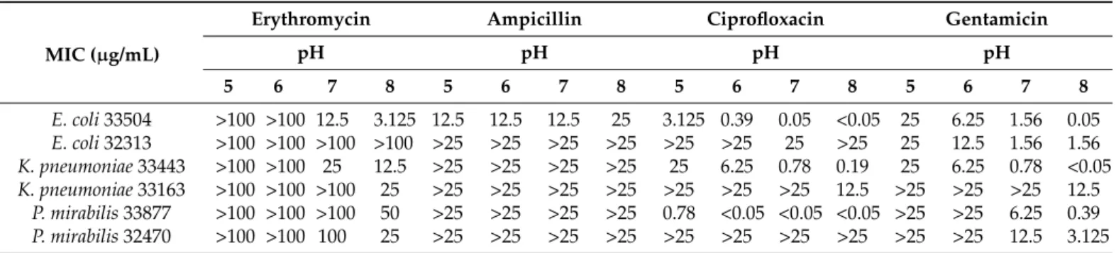
After the determination of resistance genes, different classes of antibiotics, namely erythromycin (ERY), ampicillin (AMP), ciprofloxacin (CIP), and gentamicin (GEN), were evaluated for their antibacterial activity. Since urinary pH could have an impact on the treatment of UTIs, the activity of antibiotics at different pH values is a critical issue. This antibiotic evaluation was performed at pH 5, 6, 7 and 8 by microdilution method on clinical strains ofE. coli,P. mirabilis, andK. pneumoniae. The results show that ERY had no antibacterial effect at pH 5 and 6. At pH 7 and 8, significant activity was observed on sensitiveE. coli33504 andK. pneumoniae33443 strains. ERY prevented the growth of tested bacteria most effectively in alkaline environment (pH 8; Table3).
Antibiotics2021,10, 1431 4 of 10
Table 3. Minimal inhibitory concentrations for erythromycin, ampicillin, ciprofloxacin and gentamicin onE. coli, K.
pneumoniaeandP. mirabilisstrains.
MIC (µg/mL)
Erythromycin Ampicillin Ciprofloxacin Gentamicin
pH pH pH pH
5 6 7 8 5 6 7 8 5 6 7 8 5 6 7 8
E. coli33504 >100 >100 12.5 3.125 12.5 12.5 12.5 25 3.125 0.39 0.05 <0.05 25 6.25 1.56 0.05 E. coli32313 >100 >100 >100 >100 >25 >25 >25 >25 >25 >25 25 >25 25 12.5 1.56 1.56 K. pneumoniae33443 >100 >100 25 12.5 >25 >25 >25 >25 25 6.25 0.78 0.19 25 6.25 0.78 <0.05 K. pneumoniae33163 >100 >100 >100 25 >25 >25 >25 >25 >25 >25 >25 12.5 >25 >25 >25 12.5
P. mirabilis33877 >100 >100 >100 50 >25 >25 >25 >25 0.78 <0.05 <0.05 <0.05 >25 >25 6.25 0.39 P. mirabilis32470 >100 >100 100 25 >25 >25 >25 >25 >25 >25 >25 >25 >25 >25 12.5 3.125
AMP had no effect on the tested strains (MIC greater than 100µg/mL) exceptE. coli 33504. Here, AMP showed potent antibacterial activity at pH 5–7 (MIC: 12.5µg/mL;
Table3). pH dependence was also detected for CIP onE. coli33504,K. pneumoniae33443, K. pneumoniae33163, andP. mirabilis33877 by showing higher antibacterial activity at pH 7 and 8 (Table3). GEN was the most active antibiotic at alkaline pH of all tested strains (Table3).
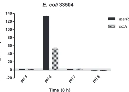
2.4. Relative Expression of marR and sdiA Genes
GEN was the most potent antibacterial agent againstE. colistrains (33504 and 32313) and, for this reason, the effect of GEN on the relative expression ofmarRandsdiAgenes in bothE. colistrains was evaluated. TheE. coli marRgene encodes a repressor of the marRABoperon, a regulatory locus controlling multiple antibiotic resistance. In addition, sdiAencodes the transcription factor SdiA, a LuxR homolog that can respond to acyl- homoserine lactone (AHL), which, in turn, is related to quorum sensing. As shown by Figures1and2, GEN treatment in a pH 6 environment induced a significant stress response in bothE. colistrains with themarRandsdiAgenes being upregulated compared to the other pH levels. In contrast,marRandsdiAgenes at pH 8 and in the presence of GEN were downregulated in the tested strains (Figures1and2).
Antibiotics 2021, 10, x. https://doi.org/10.3390/xxxxx www.mdpi.com/journal/antibiotics
AMP had no effect on the tested strains (MIC greater than 100 μg/mL) except E. coli 33504. Here, AMP showed potent antibacterial activity at pH 5–7 (MIC: 12.5 μg/mL; Table 3). pH dependence was also detected for CIP on E. coli 33504, K. pneumoniae 33443, K.
pneumoniae 33163, and P. mirabilis 33877 by showing higher antibacterial activity at pH 7 and 8 (Table 3). GEN was the most active antibiotic at alkaline pH of all tested strains (Table 3).
2.4. Relative Expression of marR and sdiA Genes
GEN was the most potent antibacterial agent against E. coli strains (33504 and 32313) and, for this reason, the effect of GEN on the relative expression of marR and sdiA genes in both E. coli strains was evaluated. The E. coli marR gene encodes a repressor of the mar- RABoperon, a regulatory locus controlling multiple antibiotic resistance. In addition, sdiA encodes the transcription factor SdiA, a LuxR homolog that can respond to acyl-homoser- ine lactone (AHL), which, in turn, is related to quorum sensing. As shown by Figures 1 and 2, GEN treatment in a pH 6 environment induced a significant stress response in both E. coli strains with the marR and sdiA genes being upregulated compared to the other pH levels. In contrast, marR and sdiA genes at pH 8 and in the presence of GEN were down- regulated in the tested strains (Figures 1 and 2).
E. coli 32313
pH 5
pH 6
pH 7
pH 8 -10
0 10 20 30
40 marR
sdiA
Time (8 h)
Relative gene expression level
Figure 1. Relative gene expression levels of marR and sdiA genes in the presence of gentamicin in Escherichia coli 32313 after 8 h exposure. The line denotes the threshold value, which was set at a two-fold increase in transcripts.
Figure 1.Relative gene expression levels ofmarRandsdiAgenes in the presence of gentamicin in Escherichia coli32313 after 8 h exposure. The line denotes the threshold value, which was set at a two-fold increase in transcripts.
E. coli 33504
pH 5
pH 6
pH 7
pH 8 -20
0 20 40 60 80 100 120 140
marR sdiA
Time (8 h)
Relative gene expression level
Figure 2. Relative gene expression levels of marR and sdiA genes in the presence of gentamicin in Escherichia coli 33504 after 8 h exposure. The line denotes the threshold value, which was set at a two-fold increase in transcripts.
3. Discussion
The results demonstrate that the genetic data agrees mostly with the phenotypical investigations, although there are some differences between the two methods. In E. coli 32313 clinical strain, having taken into consideration that the aac(69)-Ib-cr gene is also re- sponsible for concurrent aminoglycoside and fluoroquinolone resistance induction [17], the genetic background of fluoroquinolone and the sulfonamide resistance agrees with the phenotype. P. mirabilis 33877 strain has one fluoroquinolone resistance gene present (oqxAB), which does not correspond to the phenotypic investigation. Finally, K. pneu- moniae 33443 contained the sul2 gene, which was phenotypically inactive.
There are two possible reasons for these differences. The first possible reason is that the genetic investigation was not quantitative and there were insufficient copies of the gene. The genetic investigation could be quantified using more sensitive quantitative real- time PCR. Additionally, oqxAB confers low to intermediate resistance to quinolones [16].
According to the literature, there is no complete agreement between phenotypical and genetic methods, therefore these differences could have been caused by multiple factors, for example, by the lack of promoter regions (an IS26 element in the case of oqxAB) [18,19].
It was demonstrated that the acidic pH and promethazine treatment induced a significant stress response in E. coli. Moreover, the genes marB, marR, acrA, acrB, soxS, ftsI and sdiA were up-regulated at an acidic pH compared to the treatment at a neutral pH [20].
In this study, the activity of the antibiotics of different classes was studied by broth microdilution method at pH 5, 6, 7 and 8 on sensitive and resistant UTI bacterial strains.
It can be concluded that the activity of ERY, CIP, and GEN was more effective in an alka- line environment on the tested strains. Furthermore, AMP showed a more potent efficacy at acidic and neutral pH levels on E. coli 33504. Urine pH can be modified to prevent cer- tain urological diseases [10]. Patient urinary pH can be acidified by ascorbic acid and am- monium chloride while becoming more alkaline with sodium bicarbonate or potassium citrate [10]. The results confirmed that the activity of antimicrobial drugs is pH dependent.
This enables the artificial manipulation of urinary pH to contribute to a more effective therapy of urinary tract infections, especially in cases of infections caused by multidrug resistant bacteria. Additionally, this technique could reduce the cost of treatment.
The slightly acidic pH (pH 6) and GEN treatment induced the upregulation of marR antibiotic resistance and sdiA QS activator genes in both E. coli strains, increasing bacterial Figure 2.Relative gene expression levels ofmarRandsdiAgenes in the presence of gentamicin in Escherichia coli33504 after 8 h exposure. The line denotes the threshold value, which was set at a two-fold increase in transcripts.
3. Discussion
The results demonstrate that the genetic data agrees mostly with the phenotypical investigations, although there are some differences between the two methods. InE. coli 32313 clinical strain, having taken into consideration that theaac(69)-Ib-crgene is also responsible for concurrent aminoglycoside and fluoroquinolone resistance induction [17], the genetic background of fluoroquinolone and the sulfonamide resistance agrees with the phenotype.P. mirabilis33877 strain has one fluoroquinolone resistance gene present (oqxAB), which does not correspond to the phenotypic investigation. Finally,K. pneumoniae 33443 contained thesul2gene, which was phenotypically inactive.
There are two possible reasons for these differences. The first possible reason is that the genetic investigation was not quantitative and there were insufficient copies of the gene. The genetic investigation could be quantified using more sensitive quantitative real- time PCR. Additionally,oqxABconfers low to intermediate resistance to quinolones [16].
According to the literature, there is no complete agreement between phenotypical and genetic methods, therefore these differences could have been caused by multiple factors, for example, by the lack of promoter regions (an IS26 element in the case ofoqxAB) [18,19].
It was demonstrated that the acidic pH and promethazine treatment induced a significant stress response inE. coli. Moreover, the genesmarB,marR,acrA,acrB,soxS,ftsIandsdiA were up-regulated at an acidic pH compared to the treatment at a neutral pH [20].
In this study, the activity of the antibiotics of different classes was studied by broth microdilution method at pH 5, 6, 7 and 8 on sensitive and resistant UTI bacterial strains. It can be concluded that the activity of ERY, CIP, and GEN was more effective in an alkaline environment on the tested strains. Furthermore, AMP showed a more potent efficacy at acidic and neutral pH levels onE. coli33504. Urine pH can be modified to prevent certain urological diseases [10]. Patient urinary pH can be acidified by ascorbic acid and ammonium chloride while becoming more alkaline with sodium bicarbonate or potassium citrate [10]. The results confirmed that the activity of antimicrobial drugs is pH dependent.
This enables the artificial manipulation of urinary pH to contribute to a more effective therapy of urinary tract infections, especially in cases of infections caused by multidrug resistant bacteria. Additionally, this technique could reduce the cost of treatment.
The slightly acidic pH (pH 6) and GEN treatment induced the upregulation ofmarR antibiotic resistance andsdiAQS activator genes in bothE. colistrains, increasing bacterial susceptibility. A possible explanation for this could be the pH-dependent activity of siderocalin (SCN) protein, which is produced by uroepithelium. This has the ability to
Antibiotics2021,10, 1431 6 of 10
bind the iron-binding enterobactin. In a previous study, the activity of SCN increased at pH > 6.45. The elevated pH facilitated host-derived ferric-aryl complex assembly in SCN, leading to the iron starvation of uropathogenicE. coli(UPEC) [11]. This observation suggests thatE. coliat pH 6 is more susceptible to the antimicrobial agents that caused the over-expression ofmarRandsdiAgenes. It needs to be highlighted that our study represents in vitro results lacking the response of the host to UPEC, therefore host factors should also be included in further in vivo studies.
4. Materials and Methods 4.1. Bacterial Strains
Clinical strains ofEscherichia coli33503, 32313; Klebsiella pneumoniae33443, 33163;
Proteus mirabilis3387, 32470 were provided by the Institute of Clinical Microbiology at the University of Szeged and were included for the investigations. The species identities of the clinical isolates were confirmed by both MALDI-TOF MS and conventional biochemical methods.
4.2. Determination of Minimum Inhibitory Concentrations by Microdilution Method
The minimum inhibitory concentrations (MICs) of antibiotics (erythromycin (ERY), ampicillin (AMP), ciprofloxacin (CIP), and gentamicin (GEN)) were determined by mi- crodilution method in 96-well plates according to the Clinical and Laboratory Standards Institute (CLSI) guidelines using MHB at pH 5, 6, 7 and 8 [21]. The bacterial strains were separately cultured in media of pH 5 to pH 8 overnight at 37◦C and the bacterial culture grown at the appropriate pH was applied in the assay.
4.3. Disk Diffusion
The antibiotic susceptibilities of clinical isolates were determined by Kirby–Bauer’s disk diffusion method. Susceptibility and resistance were determined according to the Clinical and Laboratory Standards Institute criteria [21].
Briefly, a suspension of the bacteria equal to a 0.5 McFarland standard was prepared in phosphate-buffered saline (PBS, pH 7.2) from an overnight culture. Using a swab, strains were inoculated onto a Mueller–Hinton agar (MHA; Bio-Rad, Hercules, CA, USA) plate.
Tested antimicrobials were ampicillin (10µg), amoxicillin-clavulanic acid (20/10µg), cefuroxime (30µg), ceftriaxone (30µg), ceftazidime (10µg), ceftazidime/avibactam (30/20µg), trimethoprim sulfamethoxazole (1.25/23.75µg), ertapenem (10µg), imipenem (10µg), meropenem (10µg), gentamicin (10µg), tobramycin (10µg), amikacin (30µg), ciprofloxacin (5µg), and norfloxacin (10µg). The susceptibility disks were purchased from Biolab Inc.
(Budapest, Hungary). The plates were incubated for 16 to 18 h at 35◦C, and inhibition zones were determined visually.
4.4. Bacterial DNA Purification
The bacterial DNA was extracted by the QIAamp®DNA Blood Mini Kit (QIAGEN Inc, Chatsworth, CA, USA) following the manufacturer’s instructions. One milliliter of log- phase culture suspension, at a concentration of 107CFU/mL, was used for the preparation.
To trigger lysis of the bacterial cell wall, a preincubation step with 20 mg/mL lysozyme (in 20 mM Tris HCl, pH 8.0, 2 mM EDTA, 1.2% Triton X-100) was applied. The spin protocol was followed after incubation at 30◦C for 30 min. The final concentration of DNA was quantified using NanoDrop™ Lite spectrophotometer (Thermo Fisher Scientific™, Waltham, MA, USA) equipment. DNA samples were stored at−20◦C until further use.
4.5. Gene Targets
Three groups of antibiotic resistance genes were investigated in the genetic analy- sis. ESBL genes wereBlaTEM,BlaSHV,BlaOXA, andBlaCTX; plasmid-mediated quinolone resistance genes wereqnrA,qnrD,qnrB,qnrS,oqxAB,aac(60)-Ib-cr,qepA, andqnrC. Finally, sulfonamide genes weresul1,sul2, andsul3.
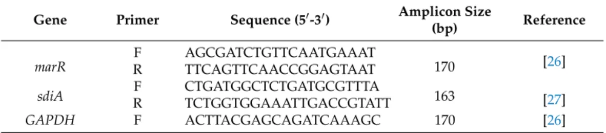
4.6. Primers
Primer sets previously published in the literature were used with slight modifica- tion [22–25]. The written melting temperature (Tm) of the primers was 60◦C in all cases.
However, although rare, some of the primers had higher calculated melting temperatures than published. These were modified by leaving some bases on the 50end of the original sequences. Thus, the specificity was unchanged but the differences in Tm were less than 1◦C. Primer sequences resulting amplicon lengths and references are listed in Tables4–6.
The published primer sequences are listed, and the modifications are underlined.
Table 4.ESBL resistance genes and primers.
Gene Primer Sequence (50-30) Amplicon Size
(bp) Reference
TEM F CATTTCCGTGTCGCCCTTATTC
800
[22]
R CGTTCATCCATAGTTGCCTGAC
SHV F AGCCGCTTGAGCAAATTAAAC
R ATCCCGCAGATAAATCACCAC 713
OXA F GGCACCAGATTCAACTTTCAAG
R GACCCCAAGTTTCCTGTAAGTG 564
CTX-M F TTTGCGATGTGCAGTACCAGTAA
544 [23]
R CGATATCGTTGGTGGTGCCATA
Table 5.Plasmid-mediated quinolone resistance genes and primers [24].
Gene Primer Sequence (50-30) Amplicon Size (bp)
qnrA F CAGCAAGAGGATTTCTCACG
R AATCCGGCAGCACTATTACTC 630
qnrD F CGAGATCAATTTACGGGGAATA
R AACAAGCTGAAGCGCCTG 581
qnrB
F GGCTGTCAGTTCTATGATCG
R GAGCAACGATGCCTGGTAG 488
degR SAKCAACGATGCCTGGTAG
qnrS F GCAAGTTCATTGAACAGGGT
R TCTAAACCGTCGAGTTCGGCG 428
oqxAB F CCGCACCGATAAATTAGTCC
R GGCGAGGTTTTGATAGTGGA 313
aac(60)-Ib-cr F TTGGAAGCGGGGACGGAM
R ACACGGCTGGACCATA 260
qepA F GCAGGTCCAGCAGCGGGTAG
R CTTCCTGCCCGAGTATCGTG 218
qnrC F GCAGAATTCAGGGGTGTGAT
R AACTGCTCCAAAAGCTGCTC 118
Table 6.Sulfonamide resistance genes and primers [26].
Gene Primer Sequence (50-30) Amplicon Size (bp)
sul1 qF TGTCGAACCTTCAAAAGCTG
qR TGGACCCAGATCCTTTACAG 113
sul2 qF ATCTGCCAAACTCGTCGTTA
qR CAATGTGATCCATGATGTCG 89
sul3 qF GGTTGAAGATGGAGCAGATG
qR GCCTTAATGACAGGTTTGAGTC 111
4.7. PCR Conditions
The same conditions and equipment were used in each PCR assay. A BIO-RAD CFX 96 instrument (Bio-Rad, Hercules, CA, USA) was used for PCR reaction. Each reaction was performed in 10µL containing 5µL MMX (Fermentas Probe/ROX qPCR MasterMix,
Antibiotics2021,10, 1431 8 of 10
Fermentas, Lithuania), 1µL template DNA, 0.5µM forward and reverse primers. The PCR cycling parameters were 1 cycle at 95◦C for 3 min, 40 cycles denaturation 95◦C for 15 s, annealing at 60◦C for 20 s and elongation at 72◦C for 1 min. The PCR fragments were separated by electrophoresis on 1.5% agarose gels containing GelRed Nucleic Acid Stain (10,000×in water; Biotium Inc., Hayward, CA, USA) and visualized by UV illumination (Bio-Rad Molecular Imager®GelDoc™ XR+ system with ImageLab™ Software, Bio-Rad Laboratories, Inc., Hercules, CA, USA). Data were evaluated and compared with DNA ladder 100–1000 bp (Bioline, London, UK) DNA marker.
4.8. Bacterial RNA Purification
E. coli33504 andE. coli32313 strains were cultured overnight in LB broths of pH 5 to pH 8 at 37◦C with shaking (OD600:0.6). Bacterial suspensions were prepared with and without GEN (12MIC) in LB medium at pH 5 to pH 8 and incubated at 37◦C with shaking. The total RNA was isolated after 8 h of culturing. RNA preparation was carried out in an RNase-free environment using NucleoSpin RNA kit (Macherey Nagel, Germany) according to the manufacturer’s instructions. Purified RNA was stored in RNase-free water in nuclease- free collection tubes and was maintained at−20◦C until quantification was performed.
The concentration of the extracted RNA templates was assessed by spectrophotometry at 260 nm (Bio-Rad, Hercules, CA, USA, SmartSpec™ Plus).
4.9. Relative Gene Expression Analyses by Real-Time Reverse Transcriptase Quantitative Polymerase Chain Reaction (RT-qPCR)
The relative gene expression levels were determined at pH 5 to pH 8 in the presence and absence of GEN. BothE. colistrains were cultured in LB at pH 5 to pH 8 and total RNA was isolated after 8 h of culturing. The relative expression levels of themarRmul- tiple antibiotic resistance regulator and thesdiAquorum sensing activator genes were determined by RT-qPCR. This involved the CFX96 Touch real-time PCR detection system (BioRad, Hercules, CA, USA), strictly following the manufacturer’s recommendations for the SensiFASTTMSYBR No-ROX One-Step Kit (Bioline GmbH, Luckenwalde, Germany).
Briefly, each well of the 96-well microtiter plate contained 20µL as follows: 10µL of the 2× SensiFASTTMSYBR No-ROX One-Step Mix, 0.2µL Reverse Transcriptase, 0.4µL RiboSafe RNase Inhibitor, 5.4µL Diethylpyrocarbonate (DEPC)-treated water, 500 nM of each primer and approximately 20 ng of total RNA in RNase-free water. Thermal cycling was initiated with a denaturation step of 5 min at 95◦C, followed by 40 cycles each of 10 s at 95◦C, 30 s at 57◦C, and 20 s at 72◦C. The relative quantities of the mRNA of each gene of interest were determined by∆∆CTmethod. Gene transcript levels were normalized against theE.
colihousekeeping geneGAPDHmeasured in the same sample. The primers used in the assay shown in Table7.
Table 7.Forward and reverse primers used for the assessment of the activity of the multiple antibiotic resistance regulator genemarRand the quorum-sensing regulatorsdiAofEscherichia coli33504 and 32313.
Gene Primer Sequence (50-30) Amplicon Size
(bp) Reference
marR
F AGCGATCTGTTCAATGAAAT
170 [26]
R TTCAGTTCAACCGGAGTAAT
sdiA F CTGATGGCTCTGATGCGTTTA
163 [27]
R TCTGGTGGAAATTGACCGTATT
GAPDH F ACTTACGAGCAGATCAAAGC 170 [26]
5. Conclusions
It is important to note that the colonization of bacteria depends on the characteristics of the population and density related virulence factors. It can be concluded that the pH can influence the activity of antibiotics and the function of efflux pump-related virulence
factors such as quorum sensing and biofilm formation. Furthermore, the constituents and the pH of the urine can have an impact on bacterial growth. The manipulation of pH may increase the efficacy of antibiotics, especially in case of UTIs caused by multidrug resistant bacteria.
Author Contributions:Conceptualization, G.S. and F.S.; methodology, G.S., A.K., F.S.; data analysis, A.K., Z.B., B.R., E.K.; investigation, A.K., O.V., Z.B., B.R., E.K., G.S., F.S.; writing—original draft preparation, A.K., B.R., Z.B., F.S., G.S.; writing—review and editing, A.K., B.R., G.S.; supervision, G.S., F.S.; funding acquisition, G.S. All authors have read and agreed to the published version of the manuscript.
Funding:This research was funded by the projects SZTEÁOK-KKA 2018/270-62-2 of the University of Szeged, Faculty of Medicine and GINOP-2.3.2-15-2016-00038 (Hungary). B.R. was supported by the project EFOP-3.6.3-VEKOP-16-2017-00009 (Hungary).
Institutional Review Board Statement:Not applicable.
Informed Consent Statement:Not applicable.
Data Availability Statement: The data presented in this study are available on request from the corresponding author.
Acknowledgments:The authors thank Edit Urbán for providing the clinical bacterial strains.
Conflicts of Interest:The authors declare no conflict of interest.
References
1. Foxman, B. The epidemiology of urinary tract infection.Nat. Rev. Urol.2010,7, 653–660. [CrossRef] [PubMed]
2. Kot, B.Virulence Factors and Innovative Strategies for the Treatment and Control of Uropathogenic Escherichia Coli; IntechOpen: London, UK, 2017; ISBN 978-953-51-3330-8.
3. Flores-Mireles, A.L.; Walker, J.N.; Caparon, M.G.; Hultgren, S.J. Urinary tract infections: Epidemiology, mechanisms of infection and treatment options.Nat. Rev. Microbiol.2015,13, 269–284. [CrossRef] [PubMed]
4. Gupta, K.; Bhadelia, N. Management of Urinary Tract Infections from Multidrug-Resistant Organisms.Infect. Dis. Clin. N. Am.
2014,28, 49–59. [CrossRef] [PubMed]
5. O’Brien, V.P.; Hannan, T.J.; Nielsen, H.V.; Hultgren, S.J. Drug and Vaccine Development for the Treatment and Prevention of Urinary Tract Infections.Microbiol. Spectr.2016,4, 4. [CrossRef]
6. Poey, N.; Madhi, F.; Biscardi, S.; Béchet, S.; Cohen, R. Aminoglycosides Monotherapy as First-Line Treatment for Febrile Urinary Tract Infection in Children.Pediatr. Infect. Dis. J.2017,36, 1104–1107. [CrossRef]
7. Goodlet, K.J.; Benhalima, F.Z.; Nailor, M.D. A Systematic Review of Single-Dose Aminoglycoside Therapy for Urinary Tract Infection: Is It Time to Resurrect an Old Strategy?Antimicrob. Agents Chemother.2019,63, e02165-18. [CrossRef] [PubMed]
8. Wasfi, R.; Abdellatif, G.R.; Elshishtawy, H.M.; Ashour, H.M. First-time characterization of viable but non-culturable Proteus mirabilis: Induction and resuscitation.J. Cell. Mol. Med.2020,24, 2791–2801. [CrossRef]
9. Shaaban, M.; El-Rahman, O.A.A.; Al-Qaidi, B.; Ashour, H.M. Antimicrobial and Antibiofilm Activities of Probiotic Lactobacilli on Antibiotic-ResistantProteus mirabilis.Microorganisms2020,8, 960. [CrossRef]
10. Yang, L.; Wang, K.; Li, H.; Denstedt, J.D.; Cadieux, P.A. The Influence of Urinary pH on Antibiotic Efficacy against Bacterial Uropathogens.Urology2014,84, 731.e1–731.e7. [CrossRef]
11. Shields-Cutler, R.R.; Crowley, J.R.; Hung, C.S.; Stapleton, A.E.; Aldrich, C.C.; Marschall, J.; Henderson, J.P. Human Urinary Composition Controls Antibacterial Activity of Siderocalin.J. Biol. Chem.2015,290, 15949–15960. [CrossRef]
12. Saxena, P.; Joshi, Y.; Rawat, K.; Bisht, R. Biofilms: Architecture, Resistance, Quorum Sensing and Control Mechanisms.Indian J.
Microbiol.2018,59, 3–12. [CrossRef]
13. Kumar, R.; Chhibber, S.; Harjai, K. Quorum sensing is necessary for the virulence of Pseudomonas aeruginosa during urinary tract infection.Kidney Int.2009,76, 286–292. [CrossRef]
14. Cole, S.J.; Hall, C.L.; Schniederberend, M.; Iii, J.M.F.; Goodson, J.R.; Pesci, E.C.; Kazmierczak, B.I.; Lee, V.T. Host suppression of quorum sensing during catheter-associated urinary tract infections.Nat. Commun.2018,9, 4436. [CrossRef]
15. Alav, I.; Sutton, J.M.; Rahman, K.M. Role of bacterial efflux pumps in biofilm formation. J. Antimicrob. Chemother. 2018,73, 2003–2020. [CrossRef]
16. The European Committee on Antimicrobial Susceptibility Testing. Breakpoint Tables for Interpretation of MICs and Zone Diameters. Version 7.1. 2017. Available online:http://www.eucast.org(accessed on 1 September 2021).
17. Kim, Y.-T.; Jang, J.-H.; Kim, H.-C.; Kim, H.-G.; Lee, K.-R.; Park, K.-S.; Lee, H.-J.; Kim, Y.-J. Identification of strain harboring both aac(60)-Ib and aac(60)-Ib-cr variant simultaneously in Escherichia coli and Klebsiella pneumoniae.BMB Rep.2011,44, 262–266.
[CrossRef]
Antibiotics2021,10, 1431 10 of 10
18. Li, J.; Zhang, H.; Ning, J.; Sajid, A.; Cheng, G.; Yuan, Z.; Hao, H. The nature and epidemiology of OqxAB, a multidrug efflux pump.Antimicrob. Resist. Infect. Control2019,8, 1–13. [CrossRef]
19. Shelburne, S.A.; Kim, J.; Munita, J.M.; Sahasrabhojane, P.; Shields, R.K.; Press, E.G.; Li, X.; Arias, C.A.; Cantarel, B.; Jiang, Y.;
et al. Whole-Genome Sequencing Accurately Identifies Resistance to Extended-Spectrumβ-Lactams for Major Gram-Negative Bacterial Pathogens.Clin. Infect. Dis.2017,65, 738–745. [CrossRef] [PubMed]
20. Nové, M.; Kincses, A.; Molnár, J.; Amaral, L.; Spengler, G. The Role of Efflux Pumps and Environmental pH in Bacterial Multidrug Resistance.In Vivo2019,34, 65–71. [CrossRef] [PubMed]
21. CLSI.Performance Standards for Antimicrobial Susceptibility Testing, 28th ed.; CLSI supplement M100; Clinical and Laboratory Standards Institute: Annapolis, MD, USA, 2018.
22. Dallenne, C.; Da Costa, A.; Decré, D.; Favier, C.; Arlet, G. Development of a set of multiplex PCR assays for the detection of genes encoding importantβ-lactamases in Enterobacteriaceae.J. Antimicrob. Chemother.2010,65, 490–495. [CrossRef]
23. Edelstein, M.; Pimkin, M.; Palagin, I.; Stratchounski, L. Prevalence and Molecular Epidemiology of CTX-MExtended-Spectrum β-Lactamase-Producing Escherichia coli and Klebsiella pneumoniae in Russian Hospitals.Antimicrob. Agents Chemother.2003,47, 3724–3732. [CrossRef] [PubMed]
24. Ciesielczuk, H.; Hornsey, M.; Choi, V.; Woodford, N.; Wareham, D. Development and evaluation of a multiplex PCR for eight plasmid-mediated quinolone-resistance determinants.J. Med. Microbiol.2013,62, 1823–1827. [CrossRef] [PubMed]
25. Wang, N.; Yang, X.; Jiao, S.; Zhang, J.; Ye, B.; Gao, S. Sulfonamide-Resistant Bacteria and Their Resistance Genes in Soils Fertilized with Manures from Jiangsu Province, Southeastern China.PLoS ONE2014,9, e112626. [CrossRef] [PubMed]
26. Viveiros, M.; Dupont, M.; Rodrigues, L.; Couto, I.; Davin-Regli, A.; Martins, M.; Pages, J.-M.; Amaral, L. Antibiotic Stress, Genetic Response and Altered Permeability ofE. coli.PLoS ONE2007,2, e365. [CrossRef] [PubMed]
27. Kincses, A.; Szabó,Á.M.; Saijo, R.; Watanabe, G.; Kawase, M.; Molnár, J.; Spengler, G. Fluorinated Beta-diketo Phosphorus Ylides Are Novel Efflux Pump Inhibitors in Bacteria.In Vivo2016,30, 813–818. [CrossRef] [PubMed]
![Table 5. Plasmid-mediated quinolone resistance genes and primers [24].](https://thumb-eu.123doks.com/thumbv2/9dokorg/966757.57440/7.892.248.841.576.890/table-plasmid-mediated-quinolone-resistance-genes-and-primers.webp)