1 DMBA Induces Deregulation of miRNA Expression of let-7, miR-21 and miR-146a in CBA/CA Mice.
KRISZTINA JUHÁSZ1, KATALIN GOMBOS1, MÓNIKA SZIRMAI1, PÉTER RÉVÉSZ2, INGRID MAGDA1, KATALIN GŐCZE1 and ISTVÁN EMBER1
1Institute of Public Health, Faculty of Medicine, University of Pécs, Pécs, Hungary;
2 Otorhinolaryngology and Head and Neck Surgery Clinic, Clinic Center, University of Pécs, Pécs, Hungary
Correspondence to: Krisztina Juhász, Institute of Public Health, Faculty of Medicine,
University of Pécs, Pécs, 12 Szigeti str., 7624 Hungary. Tel: +36 72536394, e-mail:
krisztina.juhasz01@gmail.com
Key Words: 7,12–Dimethylbenz(α)anthracene, let-7a, miR-21, miR-146a
Running title: Juhász et al: DMBA-induced Deregulation of miRNA Expression in CBA/CA Mice.
2 Abstract. Background: 7,12–Dimethylbenz(α)anthracene (DMBA) is a complete carcinogen capable of inducing various types of tumors. Materials and Methods: We investigated the effect of DMBA on micro-RNA expression in CBA/CA H2k inbred mice after 24 hours and one week from exposure. Results: Expression levels of miR-21, miR-146a and let-7a were significantly higher in the vital organs of the mice 24 hours after DMBA exposure compared to those of the controls. On the other hand, a significant down-regulation of the miRNAs was seen seven days after the DMBA administration. Conclusion: Based on our data, DMBA has an impact on the expression of miR-21, let-7a and miR-146a genes. The altered micro-RNA expression can be regarded as the early biological effect of exposure to chemical carcinogens.
To our knowledge, this is the first study of miRNA modulation caused by DMBA in non- malignant tissues.
3 There is some evidence that 7,12–dimethylbenz(α)anthracene can lead to the development of different type of tumors, such as lung cancer, lymphoma, leukemia, spleen hemangiosarcoma, skin and breast cancer (1-5). In our previous studies, we demonstrated up-regulated
expressions of Ha-ras, c-myc and p53 genes 24 and 48 hours after DMBA administration (6- 8). Altered expression of these oncogenes and suppressor gene can be considered a reliable indicator of tumorgenesis (9).
Micro-RNAs function as mediators of cell responses to extracellular signals by targeting genes involved in cell differentiation, proliferation and apoptosis. Deregulation of these short noncoding molecules plays an essential role in the multistep process of
carcinogenesis. Recently several reports have indicated the dysregulation of miRNA in tumor formation and progression and their possible role in cancer diagnostics and therapy (10-13).
Let-7a is a member of the let miRNA family and required for timing of cell fate
determination. Temporal up-regulation of let-7 miRNA in stem cells is required for their terminal differentiation at the adult stage (14). It has been reported that low expression of let- 7a was strongly associated with different neoplasms, especially of the lung (15).
Overexpression of let-7 inhibited cell growth of a lung cancer cell line repressing ras and c- myc expression at translational level (15, 16).
miR-21 was found to be up-regulated in several types of human tumor. Suppressing
the expression of several proapoptotic genes and p53 tumor suppressor gene, miR-21 contributes to the genesis and progression of many types of cancer (17, 18). Increased expression of miR-21 has been detected in tumors of the breast, lung, pancreas, prostate, stomach and brain (19, 20).
It is clear that miR-146a plays an important role in regulation of inflammatory responses through a negative feedback pathway suppressing the NF-κB activity and the LPS induced inflammatory response (21). Up-regulated miR-146a targets several inflammation-
4 related and membrane-associated messenger RNAs, including those encoding complement factor-H and the interleukin-1 receptor associated kinase-1, resulting in significant decreases in their expression (22).
Here we investigated the effect of intraperitoneal DMBA administration on miR-21, let-7a and miR-146a expressions in vital organs of CBA/CA mice.
Materials and Methods
For the experiment we used 5-week-old CBA/CA H2k haplotype mice of both sexes. Animals weighed 20 g. Mice were divided into four groups (6 males and 6 females in each group).
Two of the groups (group 1 and group 3) received intraperitoneal DMBA in a single 20 mg/kg animal weight dose (0.4 mg DMBA dissolved in 0.1 ml corn oil) at the start of the
experiment. Group 2 and group 4 were the respective control groups, where animals consumed the standard laboratory chew pellet and tap water ad libitum. At 24 hours after DMBA administration, mice in the groups 1 and 2 were euthanized and autopsied. Seven days after DMBA exposure, the mice of group 3 and 4 were euthanized and autopsied. Liver, spleen, lungs and kidneys of the animals were removed during autopsy. Mice received humane care and the experiment was carried out under the approval of the Institutional Revision Board.
Tissue samples from the dissected organs were homogenized and miRNA was isolated with RNAzol solution (Molecular Research Center Inc., Cincinnati, OH, USA) according to the manufacturer’s instruction. Quality of the isolated RNA was assessed by absorption photometry at 260/280 nm. Optical density of the RNA was between 1.9 and 2.1.
High purity miRNA was used in reverse transcription followed by nucleic acid
amplification with a one-step RNA amplification kit: Light Cyler RNA Master SYBR Green I kit (Roche, Berlin, Germany) containing SYBR green fluorescent labeling. The PCR reaction
5 mix included: 8.2 μl H2O, 1.3 μl Mn(OAc)2 stock solution, 7.2 μl LightCycler RNA master SYBR Green I fluorescent labeled dye, 2 μl specific primer at 0.5 μM final concentration and 1 μl template miRNA in 20 µl final volume. PCR amplifications were carried out in
LightCycler 2.0 carousel based PCR system (Roche). PCR settings were the following:
Reverse transcription at 61°C for 20 minutes, pre-incubation (1 cycle) for 30 s at 95 °C, amplification (45 cycles): denaturation at 95°C for 5 s, annealing at 50°C for 15 s, extension at 72°C for 5 s, melting curves (1 cycle) with denaturation at 95°C for 0.1 s, annealing at 65°C for 5s melting curve detection at 95°C at 0.1 ramp rate for 8 s.
Sequence-specific primers for let-7a, miR-21 and miR-146a were selected using the primer finder database (www.applied-science.roche.com) and were synthesized by TIB Molbiol, ADR Logistics, (Roche Warehouse, Budapest, Hungary): let-7a forward: 5'- GCCGCTGAGGTAGTAGGTTGTA-3', reverse: 5'-GTGCAGGGTCCGAGGT-3'; miR-21 forward: 5'-GCCCGCTAGCTTATCAGACTGATG-3', reverse: 5'-
GTGCAGGGTCCGAGGT-3'; miR-146a forward: 5'-GCCGCCCTGTGAAATTCAGTT-3', reverse: 5'-GTGCAGGGTCCGAGG -3'. The gene expression was determined by absolute nucleic acid quantification method in the case of miRNAs, with 4.0 Light Cycler software (Roche Diagnostics GmbH, Mannheim, Germany).
Student’s t-test was performed between control and treated groups and p-values were calculated for each miRNA for each organ. p-Values less than 0.05 were considered
statistically significant. Values were expressed as the mean±2 SD. The calculation was performed using Statistical Program for Social Science 19.0 (SPSS) software (IBM, Armonk, New York, USA)
Results
6 Expression levels of miR-21, miR-146a and let-7a isolated from DMBA-treated mice at the 24-hour time point and on the 7th day after DMBA treatment were compared with the values for the controls. The results after statistical analysis are shown in Table I.
There was a considerable up-regulation of all investigated miRNAs 24 hours after DMBA administration. Let-7a was found to be the most strikingly up-regulated miRNA, showing significantly higher levels of expression in all investigated tissues, particularly in the lung and kidney samples compared to the controls (Figure 1A and 1B). We also observed that miR-21 was increased with a 2.64-fold change in the lung and 1.80-fold change in the spleen
tissues (Figure 1A and 2B).
In contrast to the 24-hour group, significant down-regulation of miRNAs was detectable on the 7th day after the DMBA administration. Let-7a, miR-21 and miR-146a expression in the liver and spleen were found to be significantly down-regulated one week after the exposure compared to controls (Figure 2A and 2B). In particular, the miR-146a expression in the spleen was more than three times lower than in the control (Figure 2B). In the lung and kidney, there were no statistically significant differences in the expression of the investigated miRNAs between the treated and the control animals on the 7th day after DMBA exposure (Figure 1A and 1B).
Discussion
Micro-RNAs can serve as mediators of cellular stress. Recently, much data has been published regarding the role of micro-RNA in response to environmental exposures in nonmalignant tissues (23). Some previous studies analyzed the effect of cigarette smoke on miRNA expression in lung tissues of rats. Izotti et al. demonstrated down-regulation of
several miRNAs, including let-7, miR-30, miR-34, miR-140, miR-145, miR-146 and miR-192 family, regulating genes controlling cell growth, differentiation and survival (24, 25). Another
7 study reported that rats treated with the tobacco carcinogen 4-(methylnitrosamino)-1-(3- pyridyl)-1-butanone showed reduced expression of miR-34, miR-101, miR-126 and miR-199 in lung (26). According Progribny et al., rats exposed to tamoxifen, a potential
hepatocarcinogen, mainly showed up-regulation of miRNAs, functioning oncogenes in hepatocellular carcinogenesis, such as miR-34a, miR-106 and miR-20b (27). When Zhang and Pan tested the effect of hexahydro-1,3,5–trinitro–1,3,5–triazine on the miRNA expression in mouse liver, there was significant increase in the level of miR-30e, miR-99a and miR-192, and down-regulation of let-7 family members (28). Based on these studies, miRNA
expression appears to be modified in response to various chemical agents. Such studies were carried out with long exposure periods, but there are no data available regarding the changes of miRNA levels in 24 hours induced by carcinogens.
We monitored the level of let-7a, miR-21 and miR-146a in mice at 24 hours and seven days after DMBA administration. Statistically increased levels were observed in the
expression of the analyzed miRNAs 24 hours after DMBA administration in vital organs of the mice. While an opposite trend was observable in the case of the group that was
investigated seven days after the same chemical exposure. In a recent study, Yu et al. induced oral cancer by 5% DMBA in Syrian hamster and dysregulation of 17 miRNAs was observed in the tumor samples. They revealed the up-regulation of five miRNAs: miR-21, miR-200b, miR-221, miR-338 and miR-762 (29). Our study gave parallel results regarding onco-miR-21 at the 24-hour timepoint after DMBA administration.
In summary, the present study provides a model for the analysis of early miR
expression alterations after DMBA exposure. Our findings suggest that the overxepression of the investigated miRNAs might be involved in the postexposure effect of environmental carcinogens such as DMBA. A deeper understanding of miRNA regulation in chemically
8 induced carcinogenesis opens the possibilities for the development of molecular biomarkers for screening and primary cancer prevention.
References
1 Kozma L, Kiss A, Ember I and Kertai P: Studies on acute myelomonocytic leukemia in LBF1 rats. Cancer Lett. 68(2-3): 185-92, 1993.
2 Thakur P and Sanyal SN: Induction of pulmonary carcinogenesis in Wistar rats by a single dose of 9, 10 dimethylbenz(a)anthracene (DMBA) and the chemopreventive role of
diclofenac. Exp Mol Pathol 88(3): 394-400, 2010.
3 Doi ST, Kimura M and Katsuki M: Site-specific mutation of the human c-Ha-ras transgene induced by dimethylbenzanthracene causes tissue-specific tumors in mice. Jpn J Cancer Res. 85(8): 801-7, 1994.
4 Yan B, Wang H, Xie D, Wakamatsu N, Anscher MS, Dewhirst MW, Mitchel RE, Chen BJ and Li CY: Increased skin carcinogenesis in caspase-activated DNase knockout mice.
Carcinogenesis 30(10): 1776-80, 2009.
5 Becks L, Prince M, Burson H, Christophe C, Broadway M, Itoh K, Yamamoto M, Mathis M, Orchard E, Shi R, McLarty J, Pruitt K, Zhang S and Kleiner-Hancock HE: Aggressive mammary carcinoma progression in Nrf2 knockout mice treated with 7,12-
dimethylbenz[a]anthracene. BMC Cancer 10: 540, 2008.
6 Istvan Ember, Istvan Kiss and Pusztai Zsuzsannna: Effect of
7,12dimethylbenz(a)anthracene on onco/supressor gene action in vivo: A short-term experiment. Anticancer Res. 18: 445-448, 1998.
7 Gyöngyi Z, Grama L, Nádasi E, Sándor J, Németh A, Varga C, Kiss I and Ember I : Flow cytometric analysis of DMBA-induced early in vivo ras expression. In Vivo 16(5): 323-6, 2002.
9 8 Ferenc Budán, Tímea Varjas, Ghodratollah Nowrasteh, Ida Pratner, Zsuzsa Varga,
Ágoston Ember, József Cseh, Katalin Gombos, Emese Pázsit, Gyula Gőbel, Miklós Bauer, Tünde Gracza, István Arany, Pál Perjési, István Ember and István Kiss: Early modification of c-myc, Ha-ras and p53 expressions by chemical carcinogens (DMBA, MNU). In Vivo 23: 591-598, 2009.
9 Ember I, Kiss I, Gombköto G, Müller E and Szeremi M: Oncogene and suppressor gene expression as a biomarker for ethylene oxide exposure. Cancer Detect Prev. 22(3): 241-5, 1998.
10 Bartel DP: MicroRNAs: genomics, biogenesis, mechanism, and function. Cell 116(2): 281- 97, 2004.
11 Zhang B, Wang Q and Pan X: MicroRNAs and their regulatory roles in animals and plants.
J Cell Physiol. 210(2): 279-89, 2007.
12 Zhang B, Pan X, Cobb GP and Anderson TA: microRNAs as oncogenes and tumor suppressors. Dev Biol. 302(1): 1-12, 2007.
13 Croce CM: Causes and consequences of microRNA dysregulation in cancer. Nat Reviews Genetics 10(10): 704-14, 2009.
14 Pasquinelli A, Reinhart B, Slack F, Maller B and Ruvkun G: Conservation across animal phylogeny of the sequence and accession numbers AY941100 and AY941101 temporal regulation of the 21 nucleotide C. elegans let-7 heterochronic regulatory RNA. Nature 408:
86-89, 2000.
15 Takamizawa J, Konishi H, Yanagisawa K, Tomida S, Osada H, Endoh H, Harano T, Yatabe Y, Nagino M, Nimura Y, Mitsudomi T and Takahashi T: Reduced expression of the let-7 microRNAs in human lung cancers in association with shortened postoperative survival. Cancer Res. 64: 3753-3756, 2004.
10 16 Akao Y, Nakagawa Y and Naoe T: let-7 MicroRNA functions as a potential growth
suppressor in human colon cancer cells. Biol Pharm Bull 29(5): 903-6, 2006.
17 Zhu S, Wu F, Nie D, Sheng S and Mo YY: Micro-RNA-21 targets tumor suppressor genes in invasion and metastases. Cell Res.18: 350-9, 2008.
18 Liu C, Yu J, Yu S, Lavker RM, Cai L, Liu W, Yang K, He X and Chen S: MicroRNA-21 acts as an oncomir through multiple targets in human hepatocellular carcinoma. J Hepatol.
53(1): 98-107, 2010.
19 Chan JA, Krichevsky AM and Kosik KS: MicroRNA-21 is an antiapoptotic factor in human glioblastoma cells. Cancer 65: 6029-33, 2005.
20 Volinia S, Calin GA, Liu CG, Ambs S, Cimmino Aa, Petrocca F, Visone R, Ioiro M, Roldo C, Ferracin M, Prueitt RL, Yanaihara N, Lanza G, Scarpa A, Vecchione A, Negrini M, Harris CC and Croce CM: A microRNA expression signature of human solid tumors defines cancer gene targets. Proc Natl Acad Sci USA 103: 2257-61, 2006.
21 Pauley KM and Cha S: miR-146a in rheumatoid arthritis: a new therapeutic strategy.
Immunotherapy 3(7): 829-31, 2011.
22 Li YY, Cui JG, Dua P, Pague AI, Bhattacharjee S and Lukiw WJ: Differential expression of miRNA-146a-regulated inflammatory genes in human primary neural, astroglial and microglial cells. Neurosci Lett. 499(2): 109-113, 2011.
23 Lema C and Cunningham MJ: MicroRNAs and their implications in toxicological research.
Toxicol Lett. 198(2): 100-5, 2010.
24Izzotti A, Calin GA, Arrigo P,Vernon E. Steele VE, Croce CM and De Flora S:
Downregulation of microRNA expression in the lungs of rats exposed to cigarette smoke.
FASEB J 23(3): 806-812, 2009.
11 25 Izzotti A, Calin GA, Steele VE, Cartiglia C, Longobardi M, Croce CM and De Flora S:
Chemoprevention of cigarette smoke-induced alterations of MicroRNA expression in rat lungs. Cancer Prev Res 3(1): 62-72, 2010.
26 Kalscheuer S, Zhang X, Zeng Y and Upadhyaya P: Differential expression of microRNAs in early-stage neoplastic transformation in the lungs of F344 rats chronically treated with the tobacco carcinogen 4-(methylnitrosamino)-1-(3-pyridyl)-1-butanone. Carcinogenesis 29(12): 2394-2399, 2008.
27 Pogribny IP, Tryndyak VP, Boyko A, Rodriguez-Juarez R, Beland FA and Kovalchuk O:
Induction of microRNAome deregulation in rat liver by long-term tamoxifen exposure.
Mutation Res. 619(1-2): 30-7, 2007.
28 Zhang B and Pan X: RDX induces aberrant expression of microRNAs in mouse brain and liver. Environ Health Perspect 117(2): 231-40, 2009.
29 Yu T, Wang XY, Gong RG, Li A, Yang S, Cao YT, Wen YM, Wang CM and Yi XZ: The expression profile of microRNAs in a model of 7,12-dimethyl-benz[a]anthrance-induced oral carcinogenesis in Syrian hamster. J Exp Clin Cancer Res. 28(1): 64, 2009.
12 Figure 1. let-7a, miR-21, miR-146a gene expression in lung (A) and kidney (B) of mice 24 hours and one week after DMBA exposure compared to controls. Values are the mean of six mice ± 2 SD.
13
14 Figure 2. let-7a, miR-21, miR-146a gene expression in liver (A) and spleen (B) of mice 24 hours and one week of DMBA exposure compared to controls. Values are the mean of six mice ± 2 SD.
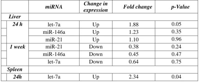
Table I. Results of statistical analysis. Presented fold change values are the gene expression ratios of DMBA treated mice over the untreated controls according to tissue samples, duration of the treatment and miRNAs.
miRNA Change in
expression Fold change p-Value Liver
24 h let-7a Up 1.88 0.05
miR-146a Up 1.23 0.35
miR-21 Up 1.10 0.96
1 week miR-21 Down 0.38 0.24
miR-146a Down 0.45 0.47
let-7a Down 0.64 0.75
Spleen
24h let-7a Up 2.34 0.04
15
miR-21 Up 1.80 0.06
miR-146a Up 1.12 0.71
1 week
miR-146a Down 0.31 0.07
miR-21 Down 0.46 0.44
let-7a Down 0.47 0.46
Lung
24 h let-7a Up 2.90 0.01
miR-21 Up 2.64 0.03
miR-146a Up 2.55 0.17
1 week let-7a Down 0.80 0.63
miR-21 Down 0.82 0.63
miR146a Down 0.83 0.75
Kidney
24 h let-7a Up 2.72 0.03
miR-21 Up 1.44 0.60
miR-146a Up 1.26 0.65
1 week miR-21 Down 0.67 0.10
miR-146 Down 0.70 0.25
let-7a Down 0.98 0.71