Dániel Szűcs, Nóra Judit Béres, Réka Rokonay, Kriszta Boros, Katalin Borka, Zoltán Kiss, András Arató, Attila J Szabó, Ádám Vannay, Erna Sziksz, Csaba Bereczki, Gábor Veres
Dániel Szűcs, Csaba Bereczki, Department of Pediatrics and Pediatric Health Care Center, University of Szeged, H-6725 Szeged, Hungary
Nóra Judit Béres, Zoltán Kiss, Réka Rokonay, Kriszta Boros, András Arató, Attila J Szabó, Gábor Veres,1st Department of Pediatrics, Semmelweis University, H-1083 Budapest, Hungary Katalin Borka, 2nd Department of Pathology, Semmelweis University, H-1091 Budapest, Hungary
Erna Sziksz, Attila J Szabó, Ádám Vannay, 1st Department of Pediatrics, Semmelweis University and MTA-SE Pediatrics and Nephrology Research Group, H-1083 Budapest, Hungary Author contributions: Szűcs D was the gastroenterologist providing the mucosal biopsies for the study conducted by Bereczki C, Szűcs D, Béres NJ and Veres G designed the research; Arató A and Szabó AJ coordinated the research; Béres NJ, Kiss Z, Rokonay R and Boros K performed the research;
Borka K performed the pathological evaluations of the biopsies;
Szűcs D and Béres NJ analyzed the data; Szűcs D, Béres NJ, Sziksz E and Vannay A wrote the paper.
Supported by Hungarian Scientific Research Fund (OTKA), No. K105530, No. K108688, No. K116928, No. PD105361 and No. LP008/2016.
Institutional review board statement: The study was reviewed and approved by the Medical Research Council Scientific and Research Committee Institutional Review Board. This work was approved by TUKEB No. 10408/2012.
Conflict-of-interest statement: None of the authors have any conflict of interest or financial disclosures.
Open-Access: This article is an open-access article which was selected by an in-house editor and fully peer-reviewed by external reviewers. It is distributed in accordance with the Creative Commons Attribution Non Commercial (CC BY-NC 4.0) license, which permits others to distribute, remix, adapt, build upon this work non-commercially, and license their derivative works on
the use is non-commercial. See: http://creativecommons.org/
licenses/by-nc/4.0/
Manuscript source: Invited manuscript
Correspondence to: Dániel Szűcs, MD, Department of Pediatrics and Pediatric Health Care Center, University of Szeged, Hungary, Korányi fasor 14-15., H-6725 Szeged, Hungary. szucs.daniel@med.u-szeged.hu
Telephone: +36-62-545330 Fax: +36-62-545329 Received: March 25, 2016
Peer-review started: March 26, 2016 First decision: May 12, 2016 Revised: June 2, 2016 Accepted: June 15, 2016 Article in press: June 15, 2016 Published online: July 14, 2016
Abstract
AIM: To evaluate the role of microRNA (miR)-146a, -155 and -122 in the duodenal mucosa of pediatric patients with Crohn’s disease (CD) and the effect of transforming growth factor-β (TGF-β) on these miRs in duodenal epithelial and fibroblast cells.
METHODS: Formalin-fixed, paraffin-embedded biopsies derived from the macroscopically inflamed (CD inflamed: n = 10) and intact (CD intact: n = 10) duodenal mucosa of pediatric CD patients and control children (C: n = 10) were examined. Expression of miR-146a, -155 and -122 was determined by real- time polymerase-chain reaction (PCR). The expression of the above miRs was investigated in recombinant human TGF-β (1 nmol/L, 24 h) or vehicle treated small intestinal epithelial cells (CCL-241) and primary duodenal fibroblast cells derived from healthy children
ORIGINAL ARTICLE
DOI: 10.3748/wjg.v22.i26.6027 © 2016 Baishideng Publishing Group Inc. All rights reserved.
Basic Study
Increased duodenal expression of miR-146a and -155 in
pediatric Crohn’s disease
INTRODUCTION
Crohn’s disease (CD) is a chronic immune-mediated disorder, which is frequently characterized by the appearance of lesions in the upper gastrointestinal (GI) tract, primarily in pediatric patients[1,2]. In adults, upper GI lesions in the cardiac area with bamboo joint- like appearance[3] are regarded to be significant risk factors for the progression of the disease from the inflammatory to stenotic or penetration form of CD[4].
In the pathogenesis of CD, epigenetic factors including microRNAs (miRs) have come into focus as potent modulators of the progression of the disease.
An increasing number of studies investigates the role of these short single-stranded RNAs in inflammatory bowel disease (IBD)[5-11]; however, the expression profile of miRs in the upper GI region of CD patients is completely unknown.
The aim of the present study was to investigate the mucosal expression of miR-146a, -155 and -122 in the upper GI tract of children with CD, and to examine the effect of the anti-inflammatory transforming growth factor (TGF)-β on the expression of the investigated miRs. These miRs affect a number of key biological functions involved in the pathomechanism of CD, including inflammatory response, intracellular signaling cascades and response to the presence of bacteria[5].
However, upper GI tract endoscopy is an important tool to diagnose CD in pediatric patients, recently there have been no specific markers to definitely distinguish it from other GI diseases with erosions, ulcerations [Helicobacter pylori (H. pylori) infection, peptic ulcer caused by an infection or medication, or eosinophil gastritis], and disorders with granulomas (sarcoidosis, Mycobacterium tuberculosis). Therefore, we suppose that our results may contribute to the identification of potential novel biomarkers or therapeutic targets of CD.
MATERIALS AND METHODS
Patients
CD was diagnosed according to the Porto criteria[12,13]. The disease activity score was assessed regarding the Pediatric CD Activity Index (PCDAI). Control children were referred to the outpatient clinic due to recurrent abdominal pain and GI symptoms to exclude organic diseases. Esophago-gastro-duodenoscopy was part of their diagnostic procedure showing normal macroscopic appearance and histology. Duodenal biopsy samples were taken from different patients, macroscopically inflamed (CD inflamed: n = 10) and non-inflamed (CD intact: n = 10) regions of the duodenal mucosa from children with CD and controls (C: n = 10). Biopsies were immediately fixed in buffered formaldehyde and embedded into paraffin (PF). Clinical characteristics RESULTS: Expression of miR-146a was significantly
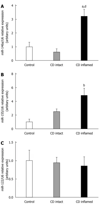
higher in the inflamed duodenal mucosa compared to the intact duodenal mucosa of children with CD (CD inflamed: 3.21 ± 0.50 vs CD intact: 0.62 ± 0.26, p ≤ 0.01) and to the control group (CD inflamed: 3.21 ± 0.50 vs C: 1.00 ± 0.33, p ≤ 0.05). The expression of miR-155 was significantly increased in the inflamed region of the duodenum compared to the control group (CD inflamed: 4.87 ± 1.02 vs Control: 1.00 ± 0.40, p
≤ 0.001). The expression of miR-122 was unchanged in the inflamed or intact mucosa of CD patients compared to controls. TGF-β treatment significantly decreased the expression of miR-155 in small intestinal epithelial cells (TGF-β: 0.7 ± 0.083 vs Control: 1 ± 0.09, p ≤ 0.05) and also the expression of miR-146a (TGF-β: 0.67 ± 0.04 vs Control: 1 ± 0.15, p ≤ 0.01) and miR-155 (TGF-β: 0.72 ± 0.09 vs Control: 1 ± 0.06, p ≤ 0.05) in primary duodenal fibroblasts compared to corresponding vehicle treated controls. TGF-β treatment did not influence the expression of miR-122.
CONCLUSION: The elevated expression of miR- 146a and -155 in the inflamed duodenal mucosa of CD patients suggests the role of these miRs in the pathomechanism of inflammatory bowel disease. Anti- inflammatory TGF-β plays an important role in the regulation of the expression of these miRs.
Key words: Inflammatory bowel disease; Crohn’s disease; Pediatric; MicroRNAs; transforming growth factor-β
© The Author(s) 2016. Published by Baishideng Publishing Group Inc. All rights reserved.
Core tip: Recent evidence suggests that besides the genetic basis, epigenetic factors including microRNAs (miRs) also act as potent inflammatory modulators during the pathogenesis of inflammatory bowel disease. MiR expression in the upper-gastrointestinal tract of pediatric patients with Crohn’s disease (CD) has not yet been analyzed. Moreover, the relation of transforming growth factor-β, playing a prominent role in the pathomechanism of CD, to miRs in this setting is also unknown. The description of precise miR patterns specific for the different segments of the gastrointestinal tract may contribute to the introduction of novel diagnostic markers and to the identification of potential therapeutic targets.
Szűcs D, Béres NJ, Rokonay R, Boros K, Borka K, Kiss Z, Arató A, Szabó AJ, Vannay Á, Sziksz E, Bereczki C, Veres G.
Increased duodenal expression of miR-146a and -155 in pediatric Crohn’s disease. World J Gastroenterol 2016; 22(26): 6027-6035 Available from: URL: http://www.wjgnet.com/1007-9327/full/
v22/i26/6027.htm DOI: http://dx.doi.org/10.3748/wjg.v22.
i26.6027
of patients with CD and controls are shown in Table 1. Written informed consent was obtained from the parents prior to the procedure, and the study was approved by the Semmelweis University Regional and Institutional Research Ethics Committee (TUKEB No.:
10408/2012).
TGF-β treatment of duodenal epithelial cells and primary
fibroblastsNormal small intestinal epithelial cells (CCL-241, American Type Culture Collection, Manassas, VA, United States) were grown in HybriCare medium (American Type Culture Collection, Manassas, VA, United States) supplemented with 10% fetal bovine serum (FBS) (Invitrogen, Carlsbad, CA, United States), 30 ng/mL epidermal growth factor (R&D Systems, Minneapolis, MN, United States), and 1% Penicillin and Streptomycin mixture (Sigma-Aldrich Co., St. Louis, MO, United States). Epithelial cells were grown under standard cell culture conditions (37 ℃, humidified, 5%
CO2/95% air environment).
Based on the previously described method, primary duodenal fibroblast cells were freshly isolated[14] from the duodenal mucosa of healthy children. Briefly the biopsies were washed in phosphate-buffered saline (PBS) and homogenized in 1 mg/mL collagenase content PBS (Sigma-Aldrich Co., St. Louis, MO, United States). Cells were grown in Dulbecco’s Modified Eagle Medium supplemented with 1% FBS (Invitrogen, Carlsbad, CA, United States) and 1% Penicillin and Streptomycin mixture. Cells were grown under standard cell culture conditions (37 ℃, humidified, 5% CO2/95%
air environment) until a confluent monolayer was obtained. During culturing, the unattached cells were removed after every 24 h culture period.
Epithelial and primary fibroblast cells were seeded into 6-well plates at a density of 5 × 10⁵ cells/well and treated for 24 h with recombinant human TGF-β (R&D Systems, Minneapolis, MN, United States) at a concentration of 1 nmol/L or vehicle only for control cells.
RNA isolation
Total RNA was isolated from formalin-fixed, paraffin- embedded biopsies using RNeasy Minikit (Qiagen, Düsseldorf, Germany) after removing paraffin from the samples according to the instructions of the manu- facturer. All contaminants were efficiently washed away, DNase was used to remove DNA from the samples using on-column DNase treatment. Concentrated RNA was filtrated using RNeasy MinElute spin columns (Qiagen, Düsseldorf, Germany). RNA was eluted in 30 µL water.
Total RNA of the intestinal epithelial and fibroblast cells was isolated by the Quick-RNA MiniPrep isolation kit (Zymo Research, Irvine, CA, United States) according to the instructions of the manufacturer.
RNA Lysis Buffer purified the RNA using Zymo-Spin™
Columns. All contaminants were washed away (RNA Prep and Wash Buffer) and DNase was used to remove the DNA from the samples. RNA was eluted in 30 µL water and used further immediately.
Reverse transcription and real-time polymerase-chain- reaction
The total RNA was reversely transcribed by the TaqMan MicroRNA Reverse Transcription kit (Applied Biosystems - ABI, Foster City, CA, United States).
TaqMan MicroRNA Assay (Applied Biosystems - ABI, Foster City, CA, United States) was used to quantify individual microRNA levels with real-time Reverse transcription and real-time polymerase-chain-reaction (RT-PCR) on a LightCycler 480 instrument (Roche Diagnostics, Basel, Switzerland). Reactions were performed in triplicates. Relative expression of miRs was determined by the 2ΔCq method using U6 as an internal control.
Statistical analysis
Statistical analysis was performed by Graphpad statistical software package (Graphpad Software, La Jolla, CA, United States). Normality was tested by the Shapiro-Wilk test. Analysis was based on the Mann-Whitney U-test, Kruskal-Wallis-test, Analysis of variance (ANOVA) and Dunn’s Post-Hoc test, and p ≤ 0.05 was considered as statistically significant. Data were presented as mean ± SE.
RESULTS
Expression of miR-146a in the duodenal mucosa of pediatric patients with CD
The expression of miR-146a was significantly higher in the inflamed duodenal mucosa of children with CD compared to the intact mucosa (CD inflamed: 3.21 ± 0.50 vs CD intact: 0.62 ± 0.26, p ≤ 0.01) and controls (CD inflamed: 3.21 ± 0.50 vs Control: 1.00 ± 0.33, p
≤ 0.05). There was no significant difference between the uninflamed group and the control one (CD intact Table 1 Clinical characteristics of patients
Control CD intact CD inflamed
n 10 10 10
Age 8.75 ± 2.36 12.4 ± 1.52 12.11 ± 1.63
Gender 2f/8m 5f/5m 2f/8m
BMI (kg/m2) 20.07 ± 2.64 14.83 ± 0.8 18.64 ± 1.81
PCDAI 0 21.94 ± 4.37c 16.11 ± 3.51b
Iron (µmol/L) 15.83 ± 2.53 4.89 ± 0.84b 8.33 ± 2.61a TIBC (µmol/L) 60.4 ± 2.71 48.5 ± 2.64 59.71 ± 8.70 Albumin (g/L) 47.33 ± 1.31 38.67 ± 1.62 48.4 ± 5.56 Hemoglobin (g/L) 132.8 ± 6.72 113.1 ± 4.02a 124.4 ± 5.93 Hematocrit (%) 0.38 ± 0.02 0.344 ± 0.01 0.37 ± 0.02 Platelet count (Giga/L) 365.5 ± 30.29 517.1 ± 34.62a 416.7 ± 63.77
aP ≤ 0.05, bP ≤ 0.01, cP ≤ 0.001 vs Control. BMI: Body mass index; CD:
Crohn’s disease; PCDAI: Pediatric Crohn’s disease activity index; TIBC:
Total iron binding capacity.
± 0.38 vs Control: 1.00 ± 0.40, p = N.S.) (Figure 1B).
Expression of miR-122 in the duodenal mucosa of pediatric patients with CD
There was no significant difference in the expression of miR-122 between the CD and control groups (CD inflamed: 0.86 ± 0.25, CD intact: 0.96 ± 0.14, Control: 1.00 ± 0.28, p = N.S.) (Figure 1C).
Expression of miR-146a, -155 and -122 in TGF-β treated duodenal epithelial cells
TGF-β had no effect on the expression of miR-146a (p
= N.S., Figure 2A); however, it decreased significantly the expression of miR-155 in CCL-241 small intestinal epithelial cells (TGF-β: 0.7 ± 0.083 vs Control: 1 ± 0.09, p ≤ 0.05) (Figure 2B). No miR-122 was detected in the small intestinal epithelial cells.
Expression of miR-146a, -155 and -122 in TGF-β treated
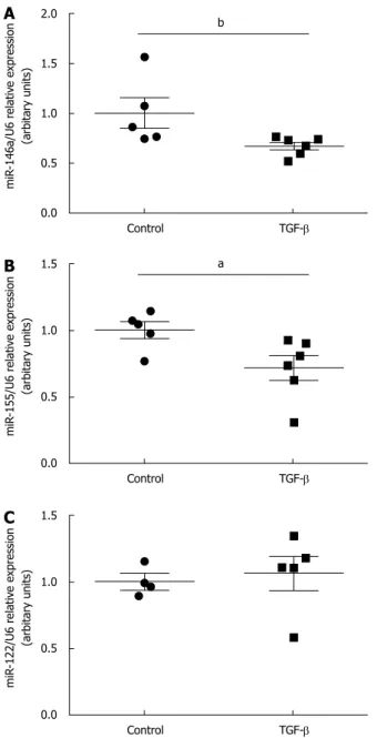
duodenal fibroblastsTGF-β significantly decreased the expression of miR- 146a (TGF-β: 0.67 ± 0.04 vs Control: 1 ± 0.15, p ≤ 0.01) (Figure 3A) and miR-155 (TGF-β: 0.72 ± 0.09 vs Control: 1 ± 0.06, p ≤ 0.05) (Figure 3B) in duodenal fibroblasts compared to vehicle treated control cells.
There was no difference in the expression of miR-122 vs Control: p = N.S.) (Figure 1A).
Expression of miR-155 in the duodenal mucosa of pediatric patients with CD
miR-155 showed significantly elevated expression in the inflamed region of the duodenal mucosa of CD patients compared to the control group (CD inflamed:
4.87 ± 1.02 vs Control: 1.00 ± 0.40, p ≤ 0.001).
There was no significant difference between the uninflamed group and the control one (CD intact: 2.50
4
3
2
1
0
a,d
Control CD intact CD inflamed
miR-146a/U6 relative expression (arbitary units)
8
6
4
2
0
b
Control CD intact CD inflamed
miR-155/U6 relative expression (arbitary units)
1.5
1.0
0.5
0.0
Control CD intact CD inflamed
miR-122/U6 relative expression (arbitary units)
Figure 1 Expression of microRNA-146a (A), microRNA-155 (B) and microRNA-122 (C) in the duodenal mucosa of pediatric patients with Crohn’s disease. Expression of microRNA (miR)-146a was significantly higher in the inflamed duodenal mucosa of children with Crohn’s disease (CD) compared to the intact mucosa and controls. miR-155 showed significantly elevated expression in the inflamed region of the duodenum compared to the control group. There was no significant difference in the expression of miR-122 between the groups. aP ≤ 0.05 vs Control; bP ≤ 0.001 vs Control; dP ≤ 0.01 vs CD intact.
A
B
C
A 2.0
1.5
1.0
0.5
0.0
Control TGF-β
miR-146a/U6 relative expression (arbitary units)
1.5
1.0
0.5
0.0
Control TGF-β
miR-155/U6 relative expression (arbitary units)
B a
Figure 2 Effect of transforming growth factor-β on the expression of microRNA-146a (A), and microRNA-155 (B) in duodenal epithelial cells.
Transforming growth factor (TGF)-β did not have any effect on the expression of microRNA (miR)-146a. TGF-β significantly decreased the expression of miR-155. There was no miR-122 expression detected, aP ≤ 0.05 vs Control.
when TGF-β was administered compared to the control group (p = N.S., Figure 3C).
DISCUSSION
In the present study, we demonstrated the elevated expression of miR-146a and -155 in the macrosco- pically inflamed duodenal mucosa of newly diagnosed, treatment-naive pediatric patients with CD compared to the control group for the first time. These results are in accordance with our previous observations demonstrating the increased expression of miR-146a and -155 in the inflamed colonic region of pediatric IBD patients. However, in contrary to our earlier observations related to the colon, we found unchanged
expression of miR-122 in the duodenal mucosa of children with CD[5].
According to the criteria of the “Porto” IBD Working Group of the European Society for Pediatric Gastro- enterology, Hepatology and Nutrition (ESPGHAN), upper GI tract endoscopy is recommended to be performed to identify lesions in all children irrespective of presence or absence of upper GI symptoms[15]. Based on the Hungarian Pediatric IBD Registry Group (HUPIR), upper GI tract abnormalities were found in 64% and 40% of children with CD and UC, respectively[16]. Characteristic findings (ulcers, erosions, aphthous lesions, and granulomas) were noted in about one third of children with CD; moreover, upper GI tract endoscopy was advantageous in establishing the final diagnosis in 9% of children with CD (diagnostic yield)[16]. However, to the best of our knowledge, there is no specific marker to clearly diagnose CD in the upper GI tract and to distinguish it from H. pylori infection, eosinophilic enteropathy or drug-induced lesions. Moreover, H. pylori-negative chronic active gastritis appears frequently in adult patients with CD, which is hard to differentiate from CD lesions[17]. Therefore, the aim of our present study was to establish a duodenum-specific miR pattern in pediatric patients with CD, which could facilitate the deeper understanding of the pathomechanism of IBD, and it may serve as a diagnostic tool in the future.
Recently miR-146a, -155 and -122 have come into focus as potential regulators of the inflammatory response, intracellular signaling cascades, regulation of cytokine production and response to bacteria, all of which play an important role in IBD. The main findings of the previous studies regarding to the expression of miR-146a, -155 and -122 in the GI tract of adult and pediatric IBD patients also confirm it. Based on these results and our present findings, we can conclude that independently of the localization of the intestinal inflammatory regions (colon, rectum, or duodenum), the expression of miR-146a and -155 was elevated in patients with CD suggesting that these miRs are rather inflammation- than region-specific[5,8,18-26]
. Both in vivo and in vitro studies confirm that miR-146a and -155 have opposite effects during inflammation. While miR-155 is a promoter of inflammation, miR-146a is a mediator of immune suppressive responses[27,28].
In contrary to miR-146a and -155, different expression pattern of miR-122 has been demonstrated throughout the GI tract. Previously we observed elevated expression of miR-122 in the intact colonic region of pediatric CD patients[5], but in the present study we found no difference in the duodenal miR-122 expression between CD patients and the control ones.
Chen et al[29] have found that miR-122 may decrease intestinal injury by suppressing the NOD2-induced NF- κB signaling pathway. On the contrary, overexpression of miR-122 in enterocytes have resulted in the mRNA degradation of occludin leading to disturbed tight- A 2.0
1.5
1.0
0.5
0.0
Control TGF-β
miR-146a/U6 relative expression (arbitary units)
b
1.5
1.0
0.5
0.0
Control TGF-β
miR-155/U6 relative expression (arbitary units)
B a
1.5
1.0
0.5
0.0
Control TGF-β
miR-122/U6 relative expression (arbitary units)
C
Figure 3 Effect of transforming growth factor-β on the expression of microRNA-146a (A), microRNA-155 (B), and microRNA-122 (C) in duodenal fibroblasts. Transforming growth factor (TGF)-β significantly decreased the expression of microRNA (miR)-146a and miR-155. TGF-β had no effect on the expression of miR-122, aP ≤ 0.05 vs Control, bP ≤ 0.01 vs Control.
junction integrity and increased intestinal permeability, which are important processes in the pathogenesis of IBD[30,31].
Inflammatory factors have been shown to modulate the expression of miRs, but the potential regulators of miR-146a, -155 and -122 in IBD are largely unknown.
One of the known key modulators of inflammatory responses is anti-inflammatory TGF-β[32-34].
In case of IBD, increased expression of TGF-β was demonstrated in the inflamed intestinal regions.
However, due to the reduced level of phosphorylated mothers against decapentaplegic homolog (SMAD)3 and diminished complex formation with SMAD4, the TGF-β-mediated signaling is insufficient in IBD patients[35].
Moreover, elevated level of SMAD7, the known endogenous inhibitor of TGF-β signaling, contributes to the enhanced production of pro-inflammatory cytokines resulting in the maintenance of inflammation in IBD[36,37]. This disadvantageous effect of SMAD7 in IBD is proved by its inhibition in a human phase
Ⅱ study. In fact, the oral administration of SMAD7 targeting antisense oligonucleotides has led to clinical remission in 60% of adult CD patients[38]. Moreover, Rho proteins, which are also connected to the TGF-β signaling pathway are involved in the regulation of IBD. Previous studies have reported Ras homolog gene family member A (RhoA) protein activation in CD patients. RhoA is involved in inflammation through the activation of the NF-κB, IL-1β and the TNF-α pathway, regulation of cell adhesion, migration, phagocytosis, and proliferation[39,40] (Figure 4).
Based on these key effects of TGF-β in the patho-
mechanism of IBD, we also aimed to investigate the possible link between TGF-β and miR-146a, -155 and -122. Following TGF-β treatment, we observed downregulation of miR-155 in both small intestinal epithelial and primary duodenal fibroblast cells and reduced expression of miR-146a in duodenal fibroblasts. However, TGF-β had no effect on the expression of miR-122 either in epithelial or in fibroblast cells.
Previous studies have confirmed the effect of these miRs on the TGF-β-mediated signaling pathway[41-49]
(Figure 4). In chondrocytes, fetal femur derived skeletal stem cells and gastric cancer cells miR-146a negatively regulates the expression of SMAD 2/3 and SMAD4, while in monocyte, cervical and prostate cancer cells miR-155 negatively modulates SMAD2/3, the key mediators of TGF-β-induced effects[46,47,49,50]. Moreover, the ectopic expression of miR-155 and -122 has significantly reduced the expression of RhoA indicating the role of these miRs in the TGF-β-induced epithelial-mesenchymal transition, cell migration and invasion, as well[41,43].
In conclusion, increased expression of miR-146a and -155 in the inflamed intestinal mucosa suggests their involvement in the pathomechanism of CD as inflammation-specific markers. Our results suggest that the expression of miR-146a and -155 is independent of and that of miR-122 is dependent on the localization of CD. Moreover, anti-inflammatory TGF-β is a negative regulator of miR-146a and -155 in small intestine epithelial and primary duodenal fibroblast cells. Our recent data have provided a baseline to explore the possible role of these miRs as diagnostic markers or
TGF-β
TGFBRI TGFBRII
SMAD2/3 SMAD2/3
SMAD4 SMAD7
SMAD2/3 SMAD4
RhoA
miR-155[44,45]
miR-146a[46]
miR-155[41]
miR-122[43]
miR-155[47-49]
miR-146a[44]
Cell proliferation Apoptosis Transcription factors (co-activators, co-repressors)
Cell proliferation Cell invasion Cell migration Epithelial-mesenchymal
transition
Figure 4 Schematic representation of the target interactions between transforming growth factor-β - microRNA-146a, -155 and -122. Transforming growth factor (TGF)-β is considered to be a major anti-inflammatory cytokine playing an important role in the pathogenesis of inflammatory bowel disease. MicroRNA (miR)-146a, -155 and -122 act as possible regulators of the TGF-β signal transduction, with capacity to induce apoptosis, cell migration, invasion and proliferation.
Moreover, TGF-β induces and represses the transcription of various genes. Data shown in the figure are based on MiRTarBase Database. SMAD: Mothers against decapentaplegic homolog; RhoA: Ras homolog gene family, member A.
their potential as therapeutic targets.
ACKNOWLEDGMENTS
We are grateful to Maria Bernáth for her skillful tech- nical assistance. The authors are grateful to Csilla Keresztes for the linguistic correction of the manuscript.
COMMENTS
Background
Crohn’s disease (CD) is a chronic immune-mediated disorder frequently characterized by lesions of the upper gastrointestinal (GI) tract, primarily in pediatric patients. Recently, epigenetic factors including microRNAs (miRs) have come into focus as potent modulators of the progression of the disease.
An increasing number of studies investigate the role of these short single- stranded RNAs in inflammatory bowel disease (IBD); however, the expression profile of miRs in the upper GI region of pediatric CD patients is completely unknown.
Research frontiers
In the previous study, the authors observed increased expression of miR-146a, -155 and -122 in the inflamed colonic region of pediatric IBD patients.
Innovations and breakthroughs
This is the first study investigating the mucosal expression of miR-146a, -155 and -122 in the upper GI tract of children with CD and examining the effect of anti-inflammatory transforming growth factor (TGF)-β on their expression of small intestinal epithelial and primary duodenal fibroblast cells.
Applications
Increased expression of miR-146a and -155 in the inflamed intestinal mucosa of pediatric patients with CD suggests their involvement in the pathomechanism of CD as inflammation-specific markers. Anti-inflammatory TGF-β is a negative regulator of miR-146a and -155 in the small intestinal epithelial and primary duodenal fibroblast cells. The recent data provide a baseline to explore the possible use of these miRs as diagnostic markers or their potential role as therapeutic targets.
Terminology
MiRs are 19-24 nucleotide-long single-stranded RNAs involved in the regulation of gene expression at transcriptional and posttranscriptional level. MiRs play a determining regulatory role in innate and adaptive immune processes.
Peer-review
The authors demonstrated an interesting study based on their previous findings connected to the expression of miR-146a, -155 and -122 in the colonic mucosa of pediatric inflammatory bowel disease. It would be interesting to measure the effect of the pro-inflammatory tumor-necrosis-factor-α on the small intestine epithelial and primary duodenal fibroblast cells as a future plan.
REFERENCES
1 Herfarth H, Rogler G. Inflammatory bowel disease. Endoscopy 2005; 37: 42-47 [PMID: 15657857 DOI: 10.1055/s-2004-826083]
2 Oberhuber G, Hirsch M, Stolte M. High incidence of upper gastrointestinal tract involvement in Crohn’s disease. Virchows Arch 1998; 432: 49-52 [PMID: 9463587]
3 Fujiya M, Sakatani A, Dokoshi T, Tanaka K, Ando K, Ueno N, Gotoh T, Kashima S, Tominaga M, Inaba Y, Ito T, Moriichi K, Tanabe H, Ikuta K, Ohtake T, Yokota K, Watari J, Saitoh Y, Kohgo Y. A Bamboo Joint-Like Appearance is a Characteristic Finding in the Upper Gastrointestinal Tract of Crohn’s Disease Patients: A Case-Control Study. Medicine (Baltimore) 2015; 94: e1500 [PMID:
26376393 DOI: 10.1097/MD.0000000000001500]
4 Magro F, Rodrigues-Pinto E, Coelho R, Andrade P, Santos- Antunes J, Lopes S, Camila-Dias C, Macedo G. Is it possible to change phenotype progression in Crohn’s disease in the era of immunomodulators? Predictive factors of phenotype progression.
Am J Gastroenterol 2014; 109: 1026-1036 [PMID: 24796767 DOI:
10.1038/ajg.2014.97]
5 Béres NJ, Szabó D, Kocsis D, Szűcs D, Kiss Z, Müller KE, Lendvai G, Kiss A, Arató A, Sziksz E, Vannay Á, Szabó AJ, Veres G.
Role of Altered Expression of miR-146a, miR-155, and miR-122 in Pediatric Patients with Inflammatory Bowel Disease. Inflamm Bowel Dis 2016; 22: 327-335 [PMID: 26752469 DOI: 10.1097/
MIB.0000000000000687]
6 Iborra M, Bernuzzi F, Correale C, Vetrano S, Fiorino G, Beltrán B, Marabita F, Locati M, Spinelli A, Nos P, Invernizzi P, Danese S.
Identification of serum and tissue micro-RNA expression profiles in different stages of inflammatory bowel disease. Clin Exp Immunol 2013; 173: 250-258 [PMID: 23607522 DOI: 10.1111/cei.12104]
7 Iborra M, Bernuzzi F, Invernizzi P, Danese S. MicroRNAs in autoimmunity and inflammatory bowel disease: crucial regulators in immune response. Autoimmun Rev 2012; 11: 305-314 [PMID:
20627134 DOI: 10.1016/j.autrev.2010.07.002]
8 Schaefer JS, Attumi T, Opekun AR, Abraham B, Hou J, Shelby H, Graham DY, Streckfus C, Klein JR. MicroRNA signatures differentiate Crohn’s disease from ulcerative colitis. BMC Immunol 2015; 16: 5 [PMID: 25886994 DOI: 10.1186/s12865-015-0069-0]
9 Ventham NT, Kennedy NA, Nimmo ER, Satsangi J. Beyond gene discovery in inflammatory bowel disease: the emerging role of epigenetics. Gastroenterology 2013; 145: 293-308 [PMID:
23751777 DOI: 10.1053/j.gastro.2013.05.050]
10 Chen WX, Ren LH, Shi RH. Implication of miRNAs for inflammatory bowel disease treatment: Systematic review. World J Gastrointest Pathophysiol 2014; 5: 63-70 [PMID: 24891977 DOI:
10.4291/wjgp.v5.i2.63]
11 Coskun M, Bjerrum JT, Seidelin JB, Nielsen OH. MicroRNAs in inflammatory bowel disease--pathogenesis, diagnostics and therapeutics. World J Gastroenterol 2012; 18: 4629-4634 [PMID:
23002331 DOI: 10.3748/wjg.v18.i34.4629]
12 Levine A, Griffiths A, Markowitz J, Wilson DC, Turner D, Russell RK, Fell J, Ruemmele FM, Walters T, Sherlock M, Dubinsky M, Hyams JS. Pediatric modification of the Montreal classification for inflammatory bowel disease: the Paris classification. Inflamm Bowel Dis 2011; 17: 1314-1321 [PMID: 21560194 DOI: 10.1002/
ibd.21493]
13 Levine A, Koletzko S, Turner D, Escher JC, Cucchiara S, de Ridder L, Kolho KL, Veres G, Russell RK, Paerregaard A, Buderus S, Greer ML, Dias JA, Veereman-Wauters G, Lionetti P, Sladek M, Martin de Carpi J, Staiano A, Ruemmele FM, Wilson DC.
ESPGHAN revised porto criteria for the diagnosis of inflammatory bowel disease in children and adolescents. J Pediatr Gastroenterol Nutr 2014; 58: 795-806 [PMID: 24231644 DOI: 10.1097/
mpg.0000000000000239]
14 Seymour ML, Binion DG, Compton SJ, Hollenberg MD, MacNaughton WK. Expression of proteinase-activated receptor 2 on human primary gastrointestinal myofibroblasts and stimulation of prostaglandin synthesis. Can J Physiol Pharmacol 2005; 83:
605-616 [PMID: 16091786 DOI: 10.1139/y05-046]
15 Ruemmele FM, Veres G, Kolho KL, Griffiths A, Levine A, Escher JC, Amil Dias J, Barabino A, Braegger CP, Bronsky J, Buderus S, Martín-de-Carpi J, De Ridder L, Fagerberg UL, Hugot JP, Kierkus J, Kolacek S, Koletzko S, Lionetti P, Miele E, Navas López VM, Paerregaard A, Russell RK, Serban DE, Shaoul R, Van Rheenen P, Veereman G, Weiss B, Wilson D, Dignass A, Eliakim A, Winter H, Turner D. Consensus guidelines of ECCO/ESPGHAN on the medical management of pediatric Crohn’s disease. J Crohns Colitis 2014; 8: 1179-1207 [PMID: 24909831 DOI: 10.1016/
j.crohns.2014.04.005]
16 Kovacs M, Muller KE, Arato A, Lakatos PL, Kovacs JB, Varkonyi A, Solyom E, Polgar M, Nemes E, Guthy I, Tokodi I, Toth G, Horvath A, Tarnok A, Tomsits E, Csoszánszky N, Balogh M, Vass N, Bodi P, Dezsofi A, Gardos L, Micskey E, Papp M, Szucs D, Cseh A,
COMMENTS
Molnar K, Szabo D, Veres G. Diagnostic yield of upper endoscopy in paediatric patients with Crohn’s disease and ulcerative colitis.
Subanalysis of the HUPIR registry. J Crohns Colitis 2012; 6: 86-94 [PMID: 22261532 DOI: 10.1016/j.crohns.2011.07.008]
17 So H, Ye BD, Park YS, Kim J, Kim JS, Moon W, Lee KM, Kim YS, Keum B, Kim SE, Kim KO, Kim ES, Lee CK, Hong SP, Im JP, Koo JS, Choi CH, Shin JE, Lee BI, Huh KC, Kim YH, Kim HS, Park YS, Han DS. Gastric lesions in patients with Crohn’s disease in Korea: a multicenter study. Intest Res 2016; 14: 60-68 [PMID:
26884736 DOI: 10.5217/ir.2016.14.1.60]
18 Fasseu M, Tréton X, Guichard C, Pedruzzi E, Cazals-Hatem D, Richard C, Aparicio T, Daniel F, Soulé JC, Moreau R, Bouhnik Y, Laburthe M, Groyer A, Ogier-Denis E. Identification of restricted subsets of mature microRNA abnormally expressed in inactive colonic mucosa of patients with inflammatory bowel disease.
PLoS One 2010; 5: [PMID: 20957151 DOI: 10.1371/journal.
pone.0013160]
19 Günaltay S, Nyhlin N, Kumawat AK, Tysk C, Bohr J, Hultgren O, Hultgren Hörnquist E. Differential expression of interleukin-1/Toll- like receptor signaling regulators in microscopic and ulcerative colitis. World J Gastroenterol 2014; 20: 12249-12259 [PMID:
25232259 DOI: 10.3748/wjg.v20.i34.12249]
20 Kanaan Z, Rai SN, Eichenberger MR, Barnes C, Dworkin AM, Weller C, Cohen E, Roberts H, Keskey B, Petras RE, Crawford NP, Galandiuk S. Differential microRNA expression tracks neoplastic progression in inflammatory bowel disease-associated colorectal cancer. Hum Mutat 2012; 33: 551-560 [PMID: 22241525 DOI:
10.1002/humu.22021]
21 Lin CJ, Gong HY, Tseng HC, Wang WL, Wu JL. miR-122 targets an anti-apoptotic gene, Bcl-w, in human hepatocellular carcinoma cell lines. Biochem Biophys Res Commun 2008; 375: 315-320 [PMID: 18692484 DOI: 10.1016/j.bbrc.2008.07.154]
22 Min M, Peng L, Yang Y, Guo M, Wang W, Sun G. MicroRNA-155 is involved in the pathogenesis of ulcerative colitis by targeting FOXO3a. Inflamm Bowel Dis 2014; 20: 652-659 [PMID: 24583476 DOI: 10.1097/MIB.0000000000000009]
23 Pathak S, Grillo AR, Scarpa M, Brun P, D’Incà R, Nai L, Banerjee A, Cavallo D, Barzon L, Palù G, Sturniolo GC, Buda A, Castagliuolo I. MiR-155 modulates the inflammatory phenotype of intestinal myofibroblasts by targeting SOCS1 in ulcerative colitis.
Exp Mol Med 2015; 47: e164 [PMID: 25998827 DOI: 10.1038/
emm.2015.21]
24 Svrcek M, El-Murr N, Wanherdrick K, Dumont S, Beaugerie L, Cosnes J, Colombel JF, Tiret E, Fléjou JF, Lesuffleur T, Duval A.
Overexpression of microRNAs-155 and 21 targeting mismatch repair proteins in inflammatory bowel diseases. Carcinogenesis 2013; 34: 828-834 [PMID: 23288924 DOI: 10.1093/carcin/bgs408]
25 Takagi T, Naito Y, Mizushima K, Hirata I, Yagi N, Tomatsuri N, Ando T, Oyamada Y, Isozaki Y, Hongo H, Uchiyama K, Handa O, Kokura S, Ichikawa H, Yoshikawa T. Increased expression of microRNA in the inflamed colonic mucosa of patients with active ulcerative colitis. J Gastroenterol Hepatol 2010; 25 Suppl 1: S129-S133 [PMID: 20586854 DOI: 10.1111/
j.1440-1746.2009.06216.x]
26 Zahm AM, Hand NJ, Tsoucas DM, Le Guen CL, Baldassano RN, Friedman JR. Rectal microRNAs are perturbed in pediatric inflammatory bowel disease of the colon. J Crohns Colitis 2014; 8:
1108-1117 [PMID: 24613022 DOI: 10.1016/j.crohns.2014.02.012]
27 Sun W, Sheng Y, Chen J, Xu D, Gu Y. Down-Regulation of miR-146a Expression Induces Allergic Conjunctivitis in Mice by Increasing TSLP Level. Med Sci Monit 2015; 21: 2000-2007 [PMID: 26166175 DOI: 10.12659/MSM.894563]
28 Alexander M, Hu R, Runtsch MC, Kagele DA, Mosbruger TL, Tolmachova T, Seabra MC, Round JL, Ward DM, O’Connell RM. Exosome-delivered microRNAs modulate the inflammatory response to endotoxin. Nat Commun 2015; 6: 7321 [PMID:
26084661 DOI: 10.1038/ncomms8321]
29 Chen Y, Wang C, Liu Y, Tang L, Zheng M, Xu C, Song J, Meng X.
miR-122 targets NOD2 to decrease intestinal epithelial cell injury in Crohn’s disease. Biochem Biophys Res Commun 2013; 438:
133-139 [PMID: 23872065 DOI: 10.1016/j.bbrc.2013.07.040]
30 Ye D, Guo S, Al-Sadi R, Ma TY. MicroRNA regulation of intestinal epithelial tight junction permeability. Gastroenterology 2011; 141:
1323-1333 [PMID: 21763238 DOI: 10.1053/j.gastro.2011.07.005]
31 Landy J, Ronde E, English N, Clark SK, Hart AL, Knight SC, Ciclitira PJ, Al-Hassi HO. Tight junctions in inflammatory bowel diseases and inflammatory bowel disease associated colorectal cancer. World J Gastroenterol 2016; 22: 3117-3126 [PMID:
27003989 DOI: 10.3748/wjg.v22.i11.3117]
32 Biancheri P, Giuffrida P, Docena GH, MacDonald TT, Corazza GR, Di Sabatino A. The role of transforming growth factor (TGF)-β in modulating the immune response and fibrogenesis in the gut.
Cytokine Growth Factor Rev 2014; 25: 45-55 [PMID: 24332927 DOI: 10.1016/j.cytogfr.2013.11.001]
33 Li C, Kuemmerle JF. Mechanisms that mediate the development of fibrosis in patients with Crohn’s disease. Inflamm Bowel Dis 2014; 20: 1250-1258 [PMID: 24831560 DOI: 10.1097/
MIB.0000000000000043]
34 Principi M, Giorgio F, Losurdo G, Neve V, Contaldo A, Di Leo A, Ierardi E. Fibrogenesis and fibrosis in inflammatory bowel diseases: Good and bad side of same coin? World J Gastrointest Pathophysiol 2013; 4: 100-107 [PMID: 24244878 DOI: 10.4291/
wjgp.v4.i4.100]
35 Feagins LA. Role of transforming growth factor-β in inflammatory bowel disease and colitis-associated colon cancer. Inflamm Bowel Dis 2010; 16: 1963-1968 [PMID: 20848467 DOI: 10.1002/
ibd.21281]
36 Monteleone G, Kumberova A, Croft NM, McKenzie C, Steer HW, MacDonald TT. Blocking Smad7 restores TGF-beta1 signaling in chronic inflammatory bowel disease. J Clin Invest 2001; 108:
601-609 [PMID: 11518734 DOI: 10.1172/JCI12821]
37 Monteleone G, Caruso R, Pallone F. Role of Smad7 in inflammatory bowel diseases. World J Gastroenterol 2012; 18:
5664-5668 [PMID: 23155305 DOI: 10.3748/wjg.v18.i40.5664]
38 Monteleone G, Di Sabatino A, Ardizzone S, Pallone F, Usiskin K, Zhan X, Rossiter G, Neurath MF. Impact of patient characteristics on the clinical efficacy of mongersen (GED-0301), an oral Smad7 antisense oligonucleotide, in active Crohn’s disease. Aliment Pharmacol Ther 2016; 43: 717-724 [PMID: 26766141 DOI:
10.1111/apt.13526]
39 Ippolito C, Colucci R, Segnani C, Errede M, Girolamo F, Virgintino D, Dolfi A, Tirotta E, Buccianti P, Di Candio G, Campani D, Castagna M, Bassotti G, Villanacci V, Blandizzi C, Bernardini N. Fibrotic and Vascular Remodelling of Colonic Wall in Patients with Active Ulcerative Colitis. J Crohns Colitis 2016;
Epub ahead of print [PMID: 26995183]
40 Segain JP, Raingeard de la Blétière D, Sauzeau V, Bourreille A, Hilaret G, Cario-Toumaniantz C, Pacaud P, Galmiche JP, Loirand G. Rho kinase blockade prevents inflammation via nuclear factor kappa B inhibition: evidence in Crohn’s disease and experimental colitis. Gastroenterology 2003; 124: 1180-1187 [PMID: 12730857]
41 Kong W, Yang H, He L, Zhao JJ, Coppola D, Dalton WS, Cheng JQ. MicroRNA-155 is regulated by the transforming growth factor beta/Smad pathway and contributes to epithelial cell plasticity by targeting RhoA. Mol Cell Biol 2008; 28: 6773-6784 [PMID:
18794355 DOI: 10.1128/MCB.00941-08]
42 Rai D, Kim SW, McKeller MR, Dahia PL, Aguiar RC. Targeting of SMAD5 links microRNA-155 to the TGF-beta pathway and lymphomagenesis. Proc Natl Acad Sci USA 2010; 107: 3111-3116 [PMID: 20133617 DOI: 10.1073/pnas.0910667107]
43 Wang SC, Lin XL, Li J, Zhang TT, Wang HY, Shi JW, Yang S, Zhao WT, Xie RY, Wei F, Qin YJ, Chen L, Yang J, Yao KT, Xiao D. MicroRNA-122 triggers mesenchymal-epithelial transition and suppresses hepatocellular carcinoma cell motility and invasion by targeting RhoA. PLoS One 2014; 9: e101330 [PMID: 24992599 DOI: 10.1371/journal.pone.0101330]
44 Louafi F, Martinez-Nunez RT, Sanchez-Elsner T. MicroRNA-155 targets SMAD2 and modulates the response of macrophages to transforming growth factor-{beta}. J Biol Chem 2010; 285:
41328-41336 [PMID: 21036908 DOI: 10.1074/jbc.M110.146852]
45 Li CL, Nie H, Wang M, Su LP, Li JF, Yu YY, Yan M, Qu QL, Zhu ZG, Liu BY. microRNA-155 is downregulated in gastric cancer cells and involved in cell metastasis. Oncol Rep 2012; 27:
1960-1966 [PMID: 22426647 DOI: 10.3892/or.2012.1719]
46 Cheung KS, Sposito N, Stumpf PS, Wilson DI, Sanchez-Elsner T, Oreffo RO. MicroRNA-146a regulates human foetal femur derived skeletal stem cell differentiation by down-regulating SMAD2 and SMAD3. PLoS One 2014; 9: e98063 [PMID: 24892945 DOI:
10.1371/journal.pone.0098063]
47 Li J, Huang J, Dai L, Yu D, Chen Q, Zhang X, Dai K. miR-146a, an IL-1β responsive miRNA, induces vascular endothelial growth factor and chondrocyte apoptosis by targeting Smad4. Arthritis Res Ther 2012; 14: R75 [PMID: 22507670 DOI: 10.1186/ar3798]
48 Liu Z, Lu CL, Cui LP, Hu YL, Yu Q, Jiang Y, Ma T, Jiao DK,
Wang D, Jia CY. MicroRNA-146a modulates TGF-β1-induced phenotypic differentiation in human dermal fibroblasts by targeting SMAD4. Arch Dermatol Res 2012; 304: 195-202 [PMID:
21968601 DOI: 10.1007/s00403-011-1178-0]
49 Xiao B, Zhu ED, Li N, Lu DS, Li W, Li BS, Zhao YL, Mao XH, Guo G, Yu PW, Zou QM. Increased miR-146a in gastric cancer directly targets SMAD4 and is involved in modulating cell proliferation and apoptosis. Oncol Rep 2012; 27: 559-566 [PMID:
22020746 DOI: 10.3892/or.2011.1514]
50 Ji H, Li Y, Jiang F, Wang X, Zhang J, Shen J, Yang X. Inhibition of transforming growth factor beta/SMAD signal by MiR-155 is involved in arsenic trioxide-induced anti-angiogenesis in prostate cancer. Cancer Sci 2014; 105: 1541-1549 [PMID: 25283513 DOI:
10.1111/cas.12548]
P- Reviewer: Decorti G S- Editor: Ma YJ L- Editor: A E- Editor: Ma S
8226 Regency Drive, Pleasanton, CA 94588, USA Telephone: +1-925-223-8242
Fax: +1-925-223-8243 E-mail: bpgoffice@wjgnet.com
Help Desk: http://www.wjgnet.com/esps/helpdesk.aspx http://www.wjgnet.com
I S S N 1 0 0 7 - 9 3 2 7
9 7 7 1 0 07 9 3 2 0 45 2 6