AGING IN CELL CULTURE
V. J. CRISTOFALO J. M. RYAN G. L. GROVE
The Wistar Institute of Anatomy and Biology Philadelphia, Pennsylvania
I. INTRODUCTION
Biological aging can be characterized as a process which (1) occurs in all members of a population; (2) is progressive and eventually harmful to the organism; (3) is irreversible under usu- al conditions; (4) is characterized by changes in functional ca- pacity which result in the loss of the organism's ability to re- spond to environmental change. Loss of capacity for adaptive re- sponse appears to occur across a wide range of biological organi- zation within the animal organism. In approaching complex bio- logical problems, model systems have proven useful in elucidating fundamental mechanisms by which cells carry out specific proces- ses or functions. In this discussion, we will be considering an approach to the study of aging using diploid human cells in cul- ture as a model system.
223
224 V. J. C R I S T O F A L O et al.
It is now reasonably well established that populations of normal human fibroblast-like cells can proliferate in culture for only limited periods of time. Typically, after explantation, there is a period of rapid proliferation during which the cul- tures can be subcultivated relatively often. This gives way to a period of declining proliferative capacity during which the cells become granular, debris accumulates, and ultimately the culture is lost. Detailed descriptions of the natural history of these cultures which establish the generality of the phenomenon are con- tained in Swim and Parker (1957), Hayflick and Moorhead (1961), Hayflick (1965), Hay (1967), and Cristofalo (1972).
Initially, the inability of human cell cultures to proliferate indefinitely was ascribed to various technical difficulties. How- ever, Hayflick and Moorhead (1961) showed that when mixtures of young and old populations (male and female), distinguishable by karyotypic markers (Barr Bodies), were co-cultivated in the same culture vessel, the older population was lost after it had under- gone a total of approximately 50 population doublings. The younger population continued to proliferate until the 50 or so population doublings had been completed. These results would seem to rule out any simple direct effect of nutritional defi- ciency, microcontaminants or the presence of toxic substances in the culture.
This limited lifespan phenomenon in terms of proliferative capacity has been well documented for human (Hayflick, 1965;
Hayakawa, 1969), chick (Haff and Swim, 1956), bovine (Stenkvist, 1966), and tortoise (Goldstein, 1974) cells in culture. It is significant that the in vitro lifespan (proliferative capacity) of a given tissue is species^specific and reproducible. These observations suggest that the limited lifespan phenomenon is in- trinsic to the cell and, in part at least, is genetically pro- grammed. In addition, recently Dell1Oreo et al. (1973; 1975) have maintained human cells in a quiescent state for as long as six months in medium in which the serum supplement is too low to
support significant proliferation. When these cells were returned to growth medium, all cultures resumed proliferation and completed the same number of population doublings as control cultures that were continually subcultured. Thus, the cellular mechanism for regulating proliferative lifespan seems to involve the counting of division events.
To one interested in the study of senescence, however, other important questions are: C D Does aging in vitro bear any rela- tionship to aging in the whole organism? and (2) Do studies car- ried out in cell culture have any relevance to aging in the in- tact animal?
These questions have been discussed in detail elsewhere (Cristofalo, 1972; Hayflick, 1972), Clearly, there is no evi- dence to support the notion that animals die because they exhaust the supply of proliferating cells; nor has this been seriously suggested. In addition, there is not, at present, sufficient evidence to relate the time course of this loss of proliferative capacity in vitro to the maximum lifespan of the donor animal.
On the other hand, there is evidence in the literature indicating that in some tissues in vivo there are declines in proliferative capacity with age. Similarly in some serial transplantation studies, a decline in proliferative capacity has been shown (for review see Cristofalo, 1972).
Perhaps the strongest evidence supporting a relationship be- tween aging in vivo and in vitro derives from a variety of work that suggests a cumulative effect of in situ plus in vitro aging by showing a relationship between the age of the cell donor and the proliferative capacity of the cells derived from that donor.
For example, Hayflick C1965) showed a striking difference in pro- liferative lifespan between cultures derived from human embryonic and human adult lung. Goldstein and his co-workers C1969) found that an inverse correlation existed between the age of the donor and the proliferative lifespan for a series of human skin cul- tures.
226 V. J. C R I S T O F A L O et al.
Martin et al. 0-970) studied over 100 mass cultures of fibro- blast-like human skin cells from a variety of donors ranging from newborn to 90 years. They found a significant negative regression of growth potential as a function of age of the donor. More re- cently, these findings have been confirmed by Le Guilly et al.
(1973). These workers used 100 cell lines derived from human liver and found a statistically significant negative correlation between the age of the donor and the proliferative lifespan of the culture and the number of doublings in phase II. Goldstein et al. (1969) and Martin et al. (1970) have shown that cell cul- tures derived from patients with progeroid syndromes or with Werner's syndrome (diseases associated with premature aging) have a reduced proliferative lifespan as compared with cultures de- rived from normal donors of the same ages.
Correlative metabolic findings show that cell cultures de- rived from adult lung have lysosomal enzyme characteristics after only 12-14 passages similar to those of fetal cells after 35-45 passages (Cristofalo et al., 1967). In addition, adult skin cells show cell cycle characteristics after only a few passages similar to those of degenerating fetal cultures (Macieira-Coelho and Pon- ten, 1969; Macieira-Coelho, 1970).
In any case, as we mentioned previously, aging in vivo is characterized, in part, by a decline in functional capacity. In our model system in cell culture, there is also a decline in func- tional capacity viz. proliferative capacity. In the studies to be described below, we are concerned with understanding how the regulation of cell proliferation changes during in vitro aging.
Some years ago (Cristofalo, 1970), we reported that hydro- cortisone (Cortisol), at a concentration of 5 pg/ml, increased the proliferative lifespan of WI-38 cells. Macieira-Coelho (1966) had reported a similar finding for human cells grown with corti- sone (5 yg/ml).
As a result of our initial studies, the principal features of the hydrocortisone effect can be summarized as follows: (1) Repli-
cate experiments based on cell counts have shown that the life- span in terms of actual population doublings was extended 30-40%
by the continuous inclusion of 5 pg/ml hydrocortisone in the medium; (2) This effect seemed to be maximal with 5 yg/ml hydro- cortisone; (3) There is no rejuvenation with the hormone, i.e.
once a culture could no longer achieve confluency, hydrocortisone would not reverse this condition; (4) If hydrocortisone was ad- ded at different periods in the lifespan, the magnitude of the extension of lifespan was in direct proportion to the amount of time the culture was grown in the presence of the hormone; (5) The saturation density of the culture was increased in the presence of the hormone; (6) The overall effect on proliferative capacity was not due to improved attachment of the cells to the glass or plas- tic growth surface.
This hydrocortisone effect on cell lifespan represented the action of a chemically defined modulator of cell division and population lifespan. We have further documented these observa- tions in the hope of using hydrocortisone as a probe for modulat- ing the regulation of cell division and the limitation of pro- liferating capacity of these diploid cell cultures.
II. MATERIALS AND METHODS
Except where noted, all studies were done with human diploid cell lines WI-38 and WI-26 (Hayflick, 1965; Hayflick and Moorhead, 1961). Starter cultures were obtained either from frozen stock maintained here at The Wistar Institute or from Dr. Leonard Hay- flick of Stanford University.
The cells were grown as previously described (Cristofalo and Sharf, 1973). Cell population doublings were calculated by com- paring the cell counts/vessel at seeding and when the cultures reached confluency. All cell counts were done electronically us- ing a Coulter Counter.
228 V. J. C R I S T O F A L O et al
Autoradiography was carried out by our standard procedures (Cristofalo and Sharf, 1973). The autoradiographs were analyzed microscopically by scoring the percentage of cells with labeled nuclei (5 silver grains or more) in random fields throughout the coverslips. At least 400 cells were counted on each coverslip and all coverslips were prepared in duplicate.
Cell cycle analysis was carried out essentially according to the method of Quastler and Sherman (1959) and analysis of the data was done according to the method of Mendelsohn and Takahashi
C 1 9 7 1 ) .
For evaluation of the effect of various steroids on DNA syn- thesis, the hormones were purchased from commercial suppliers at the best grade of purity available. Active hormones were also checked for the presence of impurities by standard thin-layer chromatography. For autoradiographic analysis of their biological activity, the procedures of Cristofalo and Sharf (1973) were used.
All hormones were used at a concentration of 5 yg/ml. Where sol- ubility was limiting, the hormones were dissolved in 100% ethanol.
Subsequent dilution was carried out in medium to give a final con- centration of ethanol of no more than 0.5%. Paired controls were always run with an identical concentration of ethanol in the medi- um. For all active hormones, the change in the labeling index was correlated with direct cell counts.
To determine the effect of hydrocortisone on transcriptional activity, cells between the 29th and 39th population doubling were used. Transcriptional activity of exponentially growing cells or confluent cells stimulated to divide was determined using three different methods: (1) Incubation of isolated chromatin from hy- drocortisone- treated and control cells with Ε coli RNA polymerase, CTP, GTP, ATP and 3H-UMP incorporated into RNA (Ryan and Cristo- falo, 1975); (2) Formation of nuclear monolayers by treating cells with 0.5% NP40 and comparing the rate of 3H-UMP incorporated into RNA (Tsai and Green, 1973; Ryan and Cristofalo, 1975); (3) Isola- tion of nuclei from hormone-treated and control cells by Dounce
homogenization and differential centrifugation followed by incuba- tion of nuclear suspension (3-5 yg DNA) with 3H-UTP and the re- maining nucleotide triphosphates (Marzluff et al., 1973).
III. RESULTS
We have shown previously that aging in human diploid popula- tions is characterized by a gradual decline in the number of cells in the rapidly proliferating pool (Cristofalo and Sharff 1973).
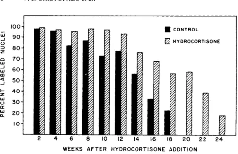
It was of interest to determine the effect of hydrocortisone on the kinetics of this decline. Figure 1 shows the percent labeled nuclei as a function of time in the presence and absence of ex- ogenously added hydrocortisone over a portion of the lifespan of the culture. Initially, nearly 100% of the cells were labeled in both cases. As the lifespan progressed, the decline in the frac- tion of labeled cells was more rapid in the control cultures and they phased out well before the hydrocortisone-treated cultures;
however, the pattern of decline in labeling was the same in both cases. Hydrocortisone seemed to retain the cells in the actively proliferating pool for longer periods.
Finally, it is important to note that the differences between hydrocortisone and control were greater as the culture aged. How- ever, the percentage of cells responding to the stimulus for divi- sion declined with age. The rate of decline was slower in the hydrocortisone-treated culture.
In order to better understand the action of hydrocortisone in extending lifespan, it was of interest to determine the effect during a single growth cycle.
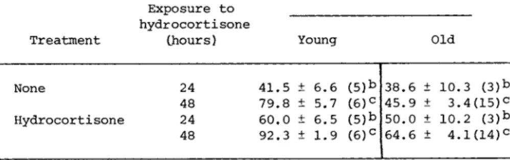
Table I shows the effect of nuclear labeling of previously untreated cultures of older cells in which hydrocortisone was present for either 24 or 48 hours while the 3H - d T was present for 24 hours. There was a statistically significant increase in the percentage of cells in both young and old cultures incorporating
230 V. J. C R I S T O F A L O et al.
\ΟΟΛ • CONTROL
0 HYDROCORTISONE
2 4 6 8 ΙΟ 12
W E E K S A F T E R H Y D R O C O R T I S O N E A D D I T I O N
FIGURE 1 The effect of hydrocortisone on the fraction of cells incorporating ^H—dT during a 30 hour pulse.
above, parallel cultures showed an increase in cell number in the presence of the hormone and the results are consistent with stimu- lation of both DNA synthesis and cell division.
In other experiments reported elsewhere (Cristofalo, 1975)r we have shown by chemical determination that the rate of DNA synthe- sis is higher in hydrocortisone-treated cultures and that % - d T incorporation parallels DNA synthesis in both treated and control cultures. Thus, the hydrocortisone-mediated increase in DNA syn- thesis seems to be due, in part at least, to an increase in the fraction of cells in the rapidly proliferating pool. Hydrocorti- sone appears to amplify the stimulus for proliferation.
To evaluate the effect of hydrocortisone on DNA synthesis, further experiments were designed to determine if the biological activity was specific for this molecular structure or whether it was simply a non-specific effect. To test these alternatives, various steroids were assayed for their biological activity with
^H-dT in the presence of hydrocortisone after 48 hours. As shown
TABLE I The Effect of Hydrocortisone for 24 or 30 Hours on the Fraction of Cells Incorporating
3H - d T During a 24 Hour Pulse
Percent labeled nucleia Exposure to
hydrocortisone
Treatment (hours) Young Old
None 24 41.5 + 6.6 (5)*> 38.6 + 10.3 ( 3 )b 48 79.8 + 5.7 ( 6 )c 45.9 ± 3.4(15)c Hydrocortisone 24 60.0 + 6.5 (5)b 50.0 ± 10.2 ( 3 )b
48 92.3 ± 1.9 ( 6 )c 64.6 ± 4.1(14)c
aMean ± Standard error of mean (number of samples).
bp < .005
cp < .001
WI-38 cells as determined by the increase in the percentage of labeled nuclei in the culture.
Three groups of compounds could be distinguished: those that were very active, hydrocortisone being the most active in stimu- lating DNA synthesis; those that were clearly inhibitory to DNA synthesis, testosterone being the most inhibitory; and a third group in which there may have been moderate borderline effects but which were not significantly different from the control.
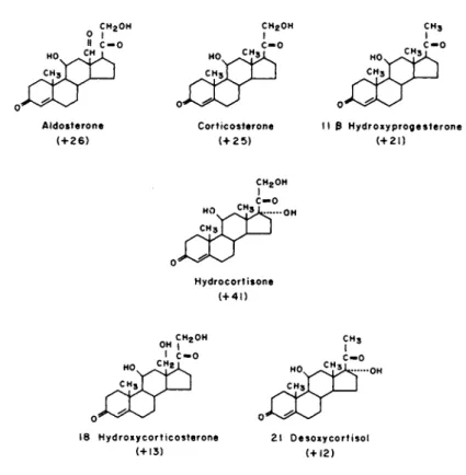
Figure 2 shows a comparison of the molecular structures of hydrocortisone and the other five stimulatory steroids. Note that all have the 3 keto, Δ 4 configuration in Ring A, and llß-hydroxy group and a keto group at C-20. Other functional groups such as the 17a and the 21-hydroxy groups, present in hydrocortisone but not in some of the others, or the C-18 methyl group present in all but aldosterone and 19-hydroxycorticosterone seemed of minor and variable significance to the action of the hormone. In gen- eral, the specificity observed here is very similar to that re- ported for stimulation of cell division in confluent 3T3 mouse
fibroblasts by Thrash et al. (1974).
232 V. J. C R I S T O F A L O et al.
C H2O H C H2O H C H3
A l d o s t e r o n e C o r t i c o s t e r o n e 11 3 H y d r o x y p r o g e s t e r o n e
( + 2 6 ) ( + 2 5 ) ( + 2 1 )
C H2O H
H y d r o c o r t i s o n e ( + 4 1 )
18 H y d r o x y c o r t i c o s t e r o n e 2 1 O e s o x y c o r t i s o l
( + 1 3 ) ( + 1 2 )
FIGURE 2 Molecular structure of steroids which stimulate cell division in WI-38 cells.
Initially, the increase in proliferative capacity seemed en- igmatic. Typically, hydrocortisone has been reported to inhibit cell proliferation in tissue culture (Ruhmann and Berliner, 1965) and we were interested in determining whether our observations were specific for this cell type and whether under identical con- ditions we could duplicate the inhibitory effects reported by others for other cell types.
Table II shows a partial listing of the results of experiments which are representative of a larger number of cell cultures that we have screened for their response to hydrocortisone. In the upper portion of the table are seven human fibroblast-like popula- tions. These include human fetal lung cell cultures from four
r f i
•Η -Ρ
£ CU Φ Ο fi en W
rfi M
ϋ Ο ϋ
-Ρ Ο
α u Φ Ό
ϋ >1 U Λ φ
ω
•Ρ Ο)
ϋ *Η φ
-Ρ ο ϋ
CO Ο Η Ο Ο Is CO 00 LO CM CM CM Μ1
+ + + + + + +
ο ι +
rH ^ CO CM
+ +
ο VO
+
o c o m ^ r v o i n o o O O O O M H H ( N H f T Î i n
1 ( 1 1 1 1 1 1
ι η θ ι Η Η ^ ρ Η ^ τ σ » ο θ Γ ^ O C N O O C O O O i n v O O O I CM CM VX)
I I I 1 ι I I I I I
υ φ
φ CT»
(ϋ m w rd E U
ο oo σ> ο σ» vo m
^J* CO CO LT) CO in ι ι ι ι O CN co i n
CO ^ rH
ο
- Η
•Ρ (d
c Cn
•H
co φ Q
co vo m co CO CN l 1
ι ι q A
CO rH
<
CN> >
rH 1 ι >
vo CO 00 vO I rH 1 1 co CN
l 00 rH La CN S H H 1 φ
O ÎU w o. S
O 2
•H fi
•H en Jh O
Φ
w w
•H E H
M œ fi Cn en Cn Cn φ · Η
3 o w
rH M H
-P
rH rH fi rd fd fd
•P -P M H
Φ Φ Φ fi Τ3 C M C M C M fd fd
•P -P
C M C M φ φ
00 VO td w co CN 0
^ 1 1 ü
— rfi H H
u -P £ B Φ -H
c ft T3 TJ u Φ φ Φ φ φ ρ g ρ B
> Ί 0 0 0 5
Φ ΜΗ MH MH
fi y » fi CO CO m
•H Φ Ό fi fi C
Λί B •Η fd rd (d CO 0 M U u *H
u -Ρ -P -P
-ρ rH rH fi -P fd O o Vf O
w φ > C M Ul en > > Ul
(d φ u
u >i fd
•H Φ Φ rH
> fi rH O M Ό Ο Φ Φ · Η . g ÎH
u fc4 S <
Φ Φ Φ -P
rH rH rH H _ - 0 0 0 2 ϋ α s ϋ s <
•H ϋ φ ft w
C fi fi fi fi fi fi fd fd fd fd fd fd fd
> 1 Φ fi fi fi fi fi Φ -P
rd cd fd fd fd M œ
Β Β Β B g fi -
te s s s S
Φ M m o 3 -H Ο X!
S U
SH fd en ä n o w
• H }H - H H ! C M C M
233
234 V. J. C R I S T O F A L O et al.
individuals and at comparable passage levels, and cultures of fe^
tal, infant and adult human skin. The adult skin was received from Dr. George M. Martin of the University of Washington and was obtained at autopsy from a patient with Werner's syndrome, a dis- ease of premature aging. For all of these human cells, both DNA synthesis and cell division were stimulated by hydrocortisone.
In more recent studies, we have assayed a series of human skin cell lines from various aged individuals including embryonic, neonatal, and adult cell cultures and have found that, based on the percent labeled nuclei assay, the response to hydrocortisone was quite variable. Effects ranged from stimulation to inhibition of DNA synthesis. We are currently extending these studies to other diploid human skin cell cultures to determine the relation- ship, if any, between hydrocortisone response and age or tissue of origin.
Human fetal kidney cultures of epithelial-like cells, ob- tained from Dr. Warren Nichols of the Institute for Medical Re- search, Camden, New Jersey, and studied at passage 5 were unre- sponsive to hydrocortisone.
In contrast to their normal progenitor strains, the 3 SV40- transformed cultures (WI-38-VA13A, WI-26-VA4, W-18-VA2) showed an inhibition of cell division of about 30%, but no reduction in the percentage of cells synthesizing DNA. HeLa cells, on the other hand, showed an inhibition of both DNA synthesis and cell divi-
sion, thus suggesting the possibility of separate effects of the hormone on mitosis and DNA synthesis.
Finally, in the lower portion of the table, we show the ef- fect of Cortisol on cell lines derived from species representing all the vertebrate classes: three mammalian lines and representa- tives of lines derived from birds, reptiles, amphibians, and fish.
For all of these, both DNA synthesis and cell division were in- hibited by hydrocortisone.
By employing the percent labeled mitosis method (Quastler and Sherman, 1959) we have been able to characterize the cell cycle of
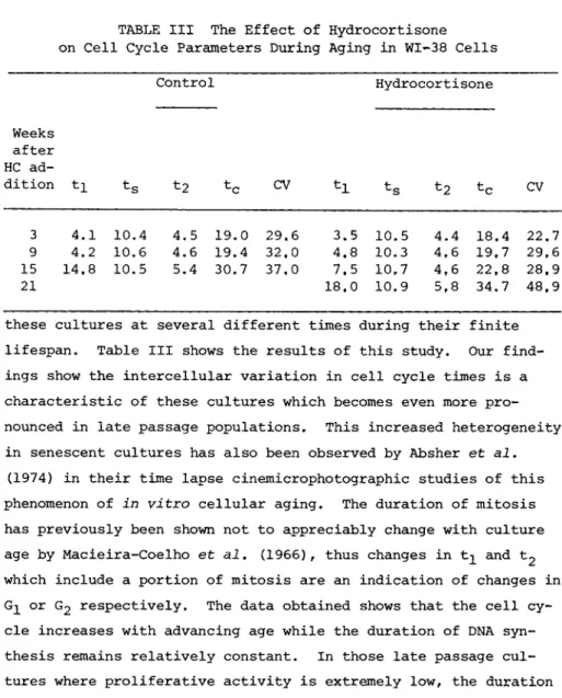
TABLE III The Effect of Hydrocortisone on Cell Cycle Parameters During Aging in WI-38 Cells
Control Hydrocorti sone
Weeks after HC ad-
dition ti ts t2 tc CV tc CV
3 4.1 10.4 4.5 19.0 29.6 3.5 10.5 4.4 18.4 22,7 9 4.2 10.6 4.6 19.4 32.0 4.8 10.3 4,6 19,7 29,6 15 14.8 10.5 5.4 30.7 37,0 7.5 10,7 4,6 22,8 28,9
21 18.0 10,9 5,8 34,7 48.9
these cultures at several different times during their finite lifespan. Table III shows the results of this study. Our find- ings show the intercellular variation in cell cycle times is a characteristic of these cultures which becomes even more pro- nounced in late passage populations. This increased heterogeneity in senescent cultures has also been observed by Absher et al.
(1974) in their time lapse cinemicrophotographic studies of this phenomenon of in vitro cellular aging. The duration of mitosis has previously been shown not to appreciably change with culture age by Macieira-Coelho et al. (1966), thus changes in and t2 which include a portion of mitosis are an indication of changes in
or G2 respectively. The data obtained shows that the cell cy- cle increases with advancing age while the duration of DNA syn- thesis remains relatively constant. In those late passage cul- tures where proliferative activity is extremely low, the duration of G 2 is also prolonged. However, it is the changes in the dura- tion of G^ which account for almost all the differences in cell cycle times. These data are in agreement with the previous studies of Macieira-Coelho et al. (1966) and Macieira-Coelho
(1973). Hydrocortisone-treated cultures show the same increases.
However, we have found that in the presence of the hormone these changes are significantly delayed.
236 V. J. C R I S T O F A L O et al.
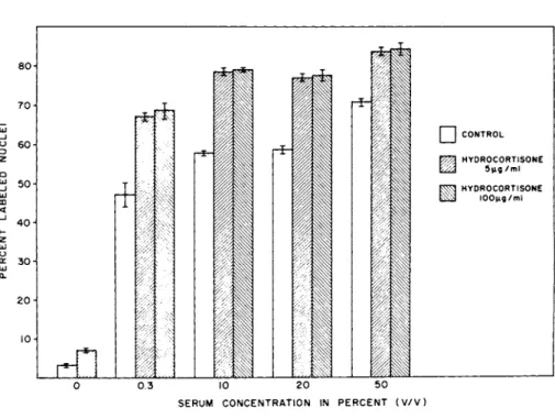
CONTROL HYDROCORTISONE
5 μ ς / m l HYDROCORTISONE
ΐΟΟμς/σιΙ
Ο 0 .3 10 20 50 S E R U M C O N C E N T R A T I O N IN P E R C E N T ( V / V )
FIGURE 3 The effect of serum and hydrocortisone on the frac- tion of cells incorporating 3H-dT during a 30 hour pulse.
The changes in proliferative patterns obtained in our study are consistent with the notion that cellular senescence is due to a transition from a rapidly cycling state to one or a series of more slowly cycling states and finally to a state in which the cells are arrested or cycling so slowly as to be unable to repopu- late the culture vessel (Cristofalo, 1975; Macieira-Coelho et al., 1975). One simple interpretation of the hydrocortisone extension of culture lifespan is that the steroid delays these transitions.
In considering this effect, a question of major interest was whether hydrocortisone stimulated division directly or functioned as an amplifier of the serum mitogen. Figure 3 shows that in the absence of serum there was essentially no division; the primary signal for division was clearly serum. With graded increases in serum there was a graded response in the fraction of cells in- corporating labeled precursor. Note that in the presence of hy-
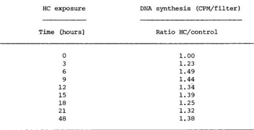
HC exposure DNA synthesis (CPM/filter)
Time (hours) Ratio HC/control
0 1.00
3 1.23
6 1.49
9 1.44
12 1.34
15 1.39
18 1.25
21 1.32
48 1.38
drocortisone, 0.3% serum gave a response higher than 10% serum without hydrocortisone, and 10% serum plus hydrocortisone was equivalent to or greater than 50% serum. Thus, the hormone func- tions as an amplifier of the serum signal.
Confluent monolayers of cells can be stimulated to divide by the addition of fresh serum or complete medium. Upon stimulation, there is a parasynchronous wave of DNA synthesis and cell division and thus the effects of various agents and their time course of action can be resolved. When various combinations of serum and hydrocortisone were added to confluent monolayers of WI-38 cells, amplifying effects of hydrocortisone similar to those described above were observed.
In order to approach the mechanism of action of the hormone, we first designed experiments to determine the time course of the hormone effect. For these experiments, confluent monolayers were stimulated by the addition of serum or serum plus hydrocortisone.
At the times indicated in Table IV, the hydrocortisone-containing media was removed and replaced with similarly conditioned media
TABLE IV The Effect of the Time of Exposure to Hydrocortisone on Cell Proliferation
238 V. J. C R I S T O F A L O et al.
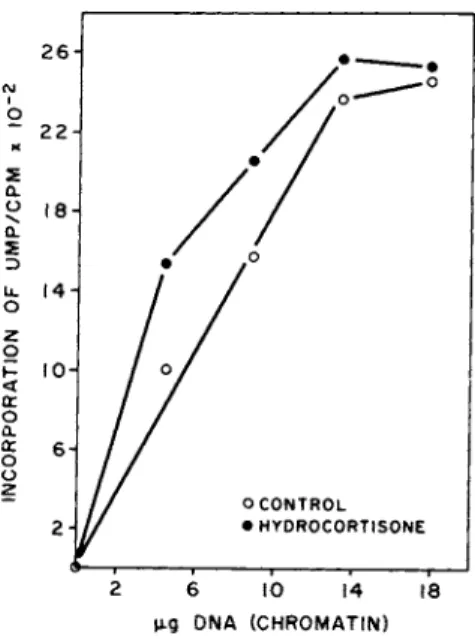
μ-g DNA (CHROMATIN)
FIGURE 4 The effect of hydrocortisone on transcriptional activity in WI-38 cells.
from dummy flasks and then the cells were fixed after a total incubation of 48 hours. At each time point, controls were handled in exactly the same way. The results when the hormone is present for possibly three but certainly the first six hours after stimulation, are as if the hormone had been present continually for the entire 48 hour period. Furthermore, we have found that if we add the hormone after six hours no enhance- ment of proliferation is seen indicating that the hormonal effect occurs somewhere during these first six hours of the prereplica- tive phase.
The effects of Hydrocortisone can also be resolved at the mo- lecular level. Figure 4 shows that exponentially growing WI-38 cultures in the presence of 14 μΜ hydrocortisone for 48 hr have an increased transcriptional activity over control cells. To further evaluate the kinetics of hormone-induced alterations in chromatin activity and to identify when in the cell cycle increased tran-
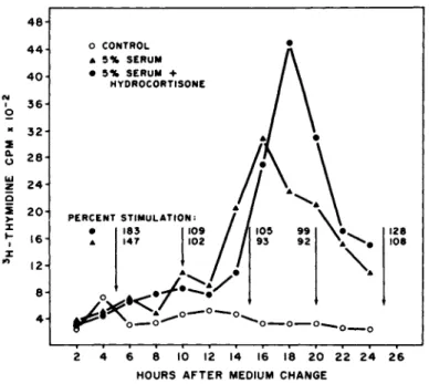
FIGURE 5 Time course of DNA synthesis and transcriptional activity in cultures stimulated to divide in the presence and ab- sence of exogenous hydrocortisone.
scriptional activity occurred, chromatin was isolated and assayed for transcriptional activity at five hr intervals following stimu- lation of confluent cultures as described above. Results, based on H-thymidine incorporation, (Fig. 5 ) showed that the pre-DNA synthesis period of control and hormone-treated cultures was about 1 2 hours and the period of active DNA synthesis lasted 1 0 - 1 2 hours in both cultures, consistent with the cell cycle data described above. There was essentially no difference in the time required for hormone~treated and control cells to enter DNA synthesis, or in the time required to complete DNA synthesis, although the mag- nitude of 3H - d T incorporation was greater in hormone-treated cul- tures. The transcriptional activity (arrows) of serum-stimulated and serum-hydrocortisoneT-stimulated cultures increased early in the pre-DNA synthesis period, however, serum-hydrocortisoner treated cells had an increased activity over serum-treated cells
240 V. J. C R I S T O F A L O et al.
( 1 8 3 vs 1 4 7 % ), As cells from both groups approached and entered
the S phase of the cell cycle, the increased transcriptional acti- vity of stimulated cultures was abolished ( 9 9 and 92%) but reap- peared to a slight extent as cells entered the post-DNA synthesis period ( 1 2 8 and 1 0 8 % ) .
It is important to note here that the increase in tran- scriptional activity in the presence of hydrocortisone occurs dur- ing the first five hour period following stimulation and this time course is consistent with the time course shown above for the pe- riod when hydrocortisone is active in increasing cell prolifera- tion.
One possible mechanism by which hydrocortisone increases tran- scriptional activity is by permitting a greater quantity of cells to initiate preparations for DNA synthesis and cell division.
This would result in a greater fraction of active chromatin which would be reflected in "increased" chromatin transcriptional acti- vity. In addition, there may be possible quantitative and/or qualitative differences in gene products of the hormone-treated cells.
IV. DISCUSSION
The data presented above show that ( 1 ) hydrocortisone in- creases the fraction of cells in the rapidly proliferating pool;
( 2 ) There is a delay in the lengthening of the G± phase of the cell cycle that occurs during aging; ( 3 ) Hydrocortisone is not a primary mitogen but acts by amplifying the serum signal for divi- sion; ( 4 ) Certain critical events take place within six hours following stimulation for division; ( 5 ) Chromatin template acti- vity is increased in the presence of hydrocortisone; ( 6 ) The most significant increase in template activity occurs within the first five hours.
In the past, hydrocortisone, cortisone and other corticos- teroids have been reported to prolong the in vitro survival time of several cell types (Arpels et al., 1964; Yuan and Chang, 1969;
Yuan et al., 1967). These studies have been concerned with post- mitotic maintenance of the cultures. It was Macieira-Coelho
(1966) who first reported the increase in proliferative lifespan of diploid, fibroblast-like cells in culture with glucocorti- coids. Division stimulating effects with hydrocortisone have also been reported by Castor and Prince (1964) for cartilage cells and by Smith et al. (1973) for human fetal lung cells. Re- cently, Thrash and Cunningham (1973) have shown the stimulation of division by hydrocortisone in density-inhibited 3T3 cells and Armelin (1973) and Gospodarowicz (1974) have shown that hydro- cortisone amplifies the activity of the pituitary and brain-de- rived polypeptides that stimulate cell division. Thus, division- stimulating effects for hydrocortisone seem now to be well docu- mented in the literature.
Perhaps the best documented effects of glucocorticoids in- volves hormone-dependent alterations in transcriptional activity.
The data described above indicate that there is an increase in transcriptional activity following stimulation to divide and that the magnitude of increase is enhanced when exogenous hydrocorti- sone is present in the medium.
On the other hand, we must consider the possibility that, al- though changes in transcriptional activity represent the best de- fined action of glucocorticoids, there is a wide range of other, less well understood actions of steriods which are termed per- missive effects (Thompson and Lipmann, 1974). The increased tran-
scriptional activity, described above, need not reflect a direct action of the hormone on transcription but could reflect a per- missive effect allowing more cells to respond to the serum mito- gen and enter the rapidly proliferating pool.
If we assume that aging of the population is reflected at the cellular level by a transition from a rapidly cycling state to
242 V. J. C R I S T O F A L O et al
one or a series of more slowly cycling states, and finally, to a sterile state, i.e., a state in which the cells are arrested or are cycling so slowly as to be unable to repopulate the culture vessel (Cristofalo, 1975; Macieira-Coelho, 1975), then, somehow, the steroid appears to delay these transitions.
Several explanations can be offered for this effect. One in- terpretation is that the cells change responsiveness to the mito- genic stimuli. Possibly young cells are responsive to 10% serum initially, but eventually lose their responsiveness to this con- centration of serum and undergo a transition to a second state where 10% serum plus hydrocortisone are required to elicit a di- vision response. There may be a second population in the culture which succeeds the first and which will only proliferate in the presence of serum plus hydrocortisone. We might also postulate that slowly cycling or arrested cells inhibit the growth of those cells still capable of division, either simply by the space they occupy or by eliciting an inhibitor or chalone into the environ- ment. In these rapidly dividing cells hydrocortisone has no ef- fect. In the slowly cycling cells, however, hydrocortisone could work either by some inactivation of the hypothetical inhibitor or by killing the cells responsible for its secretion. Thus, the young cells in the population are not inhibited to the same ex- tent. Our current work is designed to clarify these possibili- ties.
ACKNOWLEDGMENTS
The expert technical assistance of Barbara Sharf, Joan Kabakjian, E. Donald Kress, and Joanne Cochran in various parts of these studies is gratefully acknowledged.
The work reported above was supported by grants HD02721 and HD06323 from the National Institute of Child Health and Human De-
velopment and a Young Investigator Pulmonary Research Award, HD17224, to J. M. Ryan.
REFERENCES
Absher, P. Μ., Absher, R. G., and Barnes, W. D. (1974). Exptl.
Cell Res. 88, 95-104.
Armelin, H. A. C1973). Proc, Nat. Acad. Sei. U,S,A, 70, 2702-2706.
Arpels, C., Babcock, V. I. and Southam, C. M. (1964). Proc. Soc.
Exptl. Biol. Med. 115, 102-106.
Castor, C. W. and Prince, R. K. (1964). Biochim. Biophys. Acta 83, 165-177.
Cristofalo, V. J. (1970). In "Aging in Cell and Tissue Culture"
(E. Holeckovâ and V. J. Cristofalo, eds.), pp. 83-119. Plenum Press, New York.
Cristofalo, V. J. (1972). Adv. Gerontol. Res. 4, 45-79.
Cristofalo, V. J. (1975). In "Cell Impairment in Aging and De- velopment" (V. J. Cristofalo and E. Holeckovâ, eds.), pp. 7-22.
Plenum Press, New York.
Cristofalo, V. J. and Sharf, Β, B. (1973). Exptl. Cell Res. 76, 419-427.
Cristofalo, V. J., Parris, N., and Kritchevsky, D. (1967). J.
Cell Physiol. 69, 263-272.
Dell'Orco, R. T. (1975). In "Cell Impairment in Aging and Develop- ment" (V. J. Cristofalo and E. Holeckovâ, eds.), pp. 41-49.
Plenum Press, New York.
Dell'Orco, R. T., Mertens, J. G. and Kruse, P. F., Jr. (1973).
Exptl. Cell Res. 77, 356-360.
Goldstein, S. (1974). Exptl. Cell Res. 83, 297-302.
Goldstein, S., Littlefield, J. W. and Soeldner, J. S. (1969).
Proc. Nat. Acad. Sei. U.S.A. 64, 155-160.
Gospodarowicz, D. C 1 9 7 4 ) . Nature 249, 123-127.
Haff, R. F. and Swim, H. E. (1956). Proc. Soc. Exptl.
244 V . J . C R I S T O F A L O et al.
Biol. Med. 93, 200-204.
Hay, R. J . (1967). Adv. Gerontol. Res. 2, 121-158.
Hayakawa, M. (1969). J. Exp. Med. 48, 171-179.
Hayflick, L. (1965). Exptl. Cell Res. 37, 614-636.
Hayflick. L. (1972). In "Aging and Development" (H. Bredt and J . W. Rohen, eds.). pp. 1-15. Mainz Academy of Science and Litera- ture, Mainz, Germany.
Hayflick, L. and Moorhead, P. S. (1961). Exptl. Cell Res. 25, 585- 621.
Le Guilly, Υ., Simon, Μ., Lenoir, P., and Bourel, M. (1973).
Gerontologia 19, 303-313.
Macieira-Coelho, A. (1966). Experientia 22, 390-391.
Macieira-Coelho, A. (1970). In "Aging in Cell and Tissue Culture"
(E. Holeckovâ and V . J . Cristofalo, eds.), pp. 121-132. Plenum Press, New York.
Macieira-Coelho, A. (1973). In "Frontiers of Matrix Biology" (L.
Robert and B. Robert, eds.), Vol. II, pp. 46-77. S. Karger, Basel.
Macieira-Coelho, A. and Ponten, J . (1969). J. Cell Biol. 43, 374- 377.
Macieira-Coelho, Α., Loria, E. and Berumen, L. (1975). In "Cell Impairment in Aging and Development" ( V . J . Cristofalo and E.
Holeckovâ, eds.), pp. 51-65. Plenum Press, New York.
Martin, G. M., Sprague, C. Α., and Epstein, C. J . (1970). LaJb.
Invest. 23, 86-92.
Marzluff, W. F., Murphy, E. C. and Huang, R. C. C. (1973). Bio- chemistry 12, 3440-3446.
Mendelsohn, M. L. and Takahashi, M. (1971). In "The Cell Cycle and Cancer" (R. Baserga, e d . ) , pp. 58-95. Marcel Dekker, Inc., New York.
Quastler, Η. and Sherman, F. G. (1959). Exptl. Cell Res. 17, 420- 438.
Ruhmann, A. G. and Berliner, D. L. (1965). Endocrinology 76, 916- 927.
Ryan, J. M. and Cristofalo, V. J, (1975). Exptl. Cell Res. 90, 456-458.
Smith, B. T., Torday, J. S. and Giroud, C. J. P. (1973). Steroids 22 515-524.
Stenkvist, B. (1966). Acta Path, et Microbiol. Scand. 67, 180-190.
Swim, H. E. and Parker, R. F. (1957). Am. J. Hyg. 66, 235-243.
Thompson, Ε. B. and Lipmann, M. E. (1974). Metabolism 23 159- 202.
Thrash, C. R. and Cunningham, D. D. (1973). Nature 242, 399-401.
Thrash, C. R., Ho, T., and Cunningham, D. D. (1974). J. Biol.
Chem. 249 6099-6103.
Tsai, R. L. and Green H. (1973). Nature New Biol. 243, 168-170.
Yuan, G. C. and Chang, R. S. (1969). Proc. Soc. Exptl. Biol. Med.
130, 934-936.
Yuan, G. C., Chang, R. S., Little, J. Β., and Cornil, G. (1967).
J. Gerontol. 22, 174-179.