Dear Author,
Here are the proofs of your article.
• You can submit your corrections online, via e-mail or by fax.
• For online submission please insert your corrections in the online correction form. Always indicate the line number to which the correction refers.
• You can also insert your corrections in the proof PDF and email the annotated PDF.
• For fax submission, please ensure that your corrections are clearly legible. Use a fine black pen and write the correction in the margin, not too close to the edge of the page.
• Remember to note the journal title, article number, and your name when sending your response via e-mail or fax.
•
Check the metadata sheet to make sure that the header information, especially author namesand the corresponding affiliations are correctly shown.
•
Check the questions that may have arisen during copy editing and insert your answers/corrections.
•
Check that the text is complete and that all figures, tables and their legends are included. Alsocheck the accuracy of special characters, equations, and electronic supplementary material if applicable. If necessary refer to the Edited manuscript.
• The publication of inaccurate data such as dosages and units can have serious consequences.
Please take particular care that all such details are correct.
• Please do not make changes that involve only matters of style. We have generally introduced forms that follow the journal’s style.
Substantial changes in content, e.g., new results, corrected values, title and authorship are not allowed without the approval of the responsible editor. In such a case, please contact the Editorial Office and return his/her consent together with the proof.
• If we do not receive your corrections within 48 hours, we will send you a reminder.
• Your article will be published Online First approximately one week after receipt of your corrected proofs. This is the official first publication citable with the DOI. Further changes
are, therefore, not possible.• The printed version will follow in a forthcoming issue.
Please note
After online publication, subscribers (personal/institutional) to this journal will have access to the complete article via the DOI using the URL: http://dx.doi.org/[DOI].
If you would like to know when your article has been published online, take advantage of our free
alert service. For registration and further information go to: http://www.link.springer.com.
Metadata of the article that will be visualized in OnlineFirst
ArticleTitle ATP-Evoked Intracellular Ca2+ Signaling of Different Supporting Cells in the Hearing Mouse Hemicochlea
Article Sub-Title
Article CopyRight Springer Science+Business Media New York (This will be the copyright line in the final PDF) Journal Name Neurochemical Research
Corresponding Author Family Name Zelles
Particle
Given Name T.
Suffix
Division Department of Pharmacology and Pharmacotherapy Organization Semmelweis University
Address Nagyvárad tér 4., Budapest, 1089, Hungary Division Institute of Experimental Medicine Organization Hungarian Academy of Sciences
Address Budapest, Hungary
Email zelles.tibor@med.semmelweis-univ.hu
Author Family Name Horváth
Particle
Given Name T.
Suffix
Division Department of Pharmacology and Pharmacotherapy Organization Semmelweis University
Address Nagyvárad tér 4., Budapest, 1089, Hungary
Division Department of Otorhinolaryngology, Head and Neck Surgery Organization Bajcsy-Zsilinszky Hospital
Address Budapest, Hungary
Author Family Name Polony
Particle
Given Name G.
Suffix
Division Department of Otorhinolaryngology, Head and Neck Surgery Organization Semmelweis University
Address Budapest, Hungary
Author Family Name Fekete
Particle
Given Name Á.
Suffix
Division Program in Neurosciences and Mental Health Organization The Hospital for Sick Children
Address Toronto, ON, Canada
Author Family Name Aller
Particle
Given Name M.
Suffix
Division Department of Pharmacology and Pharmacotherapy Organization Semmelweis University
Address Nagyvárad tér 4., Budapest, 1089, Hungary
Division Computational Cognitive Neuroimaging Laboratory, Computational Neuroscience and Cognitive Robotics Centre
Organization University of Birmingham
Address Birmingham, UK
Author Family Name Halmos
Particle
Given Name G.
Suffix
Division Department of Otolaryngology, Head and Neck Surgery, University Medical Center Groningen
Organization University of Groningen
Address Groningen, The Netherlands
Author Family Name Lendvai
Particle
Given Name B.
Suffix Division
Organization Pharmacological and Drug Safety Research Address Gedeon Richter Plc., Budapest, Hungary Email
Author Family Name Heinrich
Particle
Given Name A.
Suffix
Division Institute of Experimental Medicine Organization Hungarian Academy of Sciences
Address Budapest, Hungary
Author Family Name Sperlágh
Organization Hungarian Academy of Sciences
Address Budapest, Hungary
Author Family Name Vizi
Particle
Given Name E. S.
Suffix
Division Institute of Experimental Medicine Organization Hungarian Academy of Sciences
Address Budapest, Hungary
Schedule
Received 13 September 2015
Revised 23 December 2015
Accepted 25 December 2015
Abstract Hearing and its protection is regulated by ATP-evoked Ca2+ signaling in the supporting cells of the organ of Corti, however, the unique anatomy of the cochlea hampers observing these mechanisms. For the first time, we have performed functional ratiometric Ca2+ imaging (fura-2) in three different supporting cell types in the hemicochlea preparation of hearing mice to measure purinergic receptor-mediated Ca2+
signaling in pillar, Deiters’ and Hensen’s cells. Their resting [Ca2+]i was determined and compared in the same type of preparation. ATP evoked reversible, repeatable and dose-dependent Ca2+ transients in all three cell types, showing desensitization. Inhibiting the Ca2+ signaling of the ionotropic P2X (omission of extracellular Ca2+) and metabotropic P2Y purinergic receptors (depletion of intracellular Ca2+ stores) revealed the involvement of both receptor types. Detection of P2X2,3,4,6,7 and P2Y1,2,6,12,14 receptor mRNAs by RT-PCR supported this finding and antagonism by PPADS suggested different functional purinergic receptor population in pillar versus Deiters’ and Hensen’s cells. The sum of the extra- and intracellular Ca2+-dependent components of the response was about equal with the control ATP response (linear additivity) in pillar cells, and showed supralinearity in Deiters’ and Hensen’s cells. Calcium-induced calcium release might explain this synergistic interaction. The more pronounced Ca2+ leak from the endoplasmic reticulum in Deiters’ and Hensen’s cells, unmasked by cyclopiazonic acid, may also suggests the higher activity of the internal stores in Ca2+ signaling in these cells. Differences in Ca2+ homeostasis and ATP-induced Ca2+ signaling might reflect the distinct roles these cells play in cochlear function and pathophysiology.
Keywords (separated by '-') Hemicochlea - Ca2+ imaging - ATP - Pillar cells - Deiters’ cells - Hensen’s cells Footnote Information
UNCORRECT
ED
PROOF
O R I G I N A L P A P E R 1
2
ATP-Evoked Intracellular Ca
2+Signaling of Different Supporting
3
Cells in the Hearing Mouse Hemicochlea
4 T. Horva´th1,2• G. Polony3•A´ . Fekete4•M. Aller1,8•G. Halmos5• 5 B. Lendvai6•A. Heinrich7•B. Sperla´gh7•E. S. Vizi7•T. Zelles1,7
6 Received: 13 September 2015 / Revised: 23 December 2015 / Accepted: 25 December 2015 7 ÓSpringer Science+Business Media New York 2016
8 Abstract Hearing and its protection is regulated by ATP- 9 evoked Ca2?signaling in the supporting cells of the organ 10 of Corti, however, the unique anatomy of the cochlea 11 hampers observing these mechanisms. For the first time, we 12 have performed functional ratiometric Ca2?imaging (fura- 13 2) in three different supporting cell types in the hemic- 14 ochlea preparation of hearing mice to measure purinergic 15 receptor-mediated Ca2? signaling in pillar, Deiters’ and 16 Hensen’s cells. Their resting [Ca2?]i was determined and 17 compared in the same type of preparation. ATP evoked 18 reversible, repeatable and dose-dependent Ca2?transients 19 in all three cell types, showing desensitization. Inhibiting
the Ca2? signaling of the ionotropic P2X (omission of 20 extracellular Ca2?) and metabotropic P2Y purinergic 21 receptors (depletion of intracellular Ca2? stores) revealed 22 the involvement of both receptor types. Detection of 23 P2X2,3,4,6,7and P2Y1,2,6,12,14receptor mRNAs by RT-PCR 24 supported this finding and antagonism by PPADS sug- 25 gested different functional purinergic receptor population 26 in pillar versus Deiters’ and Hensen’s cells. The sum of the 27 extra- and intracellular Ca2?-dependent components of the 28 response was about equal with the control ATP response 29 (linear additivity) in pillar cells, and showed supralinearity 30 in Deiters’ and Hensen’s cells. Calcium-induced calcium 31 release might explain this synergistic interaction. The more 32 pronounced Ca2?leak from the endoplasmic reticulum in 33 Deiters’ and Hensen’s cells, unmasked by cyclopiazonic 34 acid, may also suggests the higher activity of the internal 35 stores in Ca2?signaling in these cells. Differences in Ca2? 36 homeostasis and ATP-induced Ca2?signaling might reflect 37 the distinct roles these cells play in cochlear function and 38
pathophysiology. 3940
Keywords HemicochleaCa2?imaging ATPPillar 41 cellsDeiters’ cellsHensen’s cells 42
Abbreviations 43
AM Acetoxymethyl 44 ATP Adenosine triphosphate 45
[Ca2?]i Intracellular Ca2?concentration 46 CICR Calcium-induced calcium release 47 CCD Charge-coupled device 48
CPA Cyclopiazonic acid 49
EC50 Half maximal effective concentration 50 EGTA Ethylene glycol-bis(2-aminoethylether)- 51
N,N,N0,N0-tetraacetic acid ER Endoplasmic reticulum 52
A1 & T. Zelles
A2 zelles.tibor@med.semmelweis-univ.hu
A3 1 Department of Pharmacology and Pharmacotherapy, A4 Semmelweis University, Nagyva´rad te´r 4., Budapest 1089,
A5 Hungary
A6 2 Department of Otorhinolaryngology, Head and Neck A7 Surgery, Bajcsy-Zsilinszky Hospital, Budapest, Hungary A8 3 Department of Otorhinolaryngology, Head and Neck
A9 Surgery, Semmelweis University, Budapest, Hungary A10 4 Program in Neurosciences and Mental Health, The Hospital
A11 for Sick Children, Toronto, ON, Canada
A12 5 Department of Otolaryngology, Head and Neck Surgery, A13 University Medical Center Groningen, University of A14 Groningen, Groningen, The Netherlands
A15 6 Pharmacological and Drug Safety Research, Gedeon Richter A16 Plc., Budapest, Hungary
A17 7 Institute of Experimental Medicine, Hungarian Academy of A18 Sciences, Budapest, Hungary
A19 8 Present Address: Computational Cognitive Neuroimaging A20 Laboratory, Computational Neuroscience and Cognitive A21 Robotics Centre, University of Birmingham, Birmingham,
UK
AQ1
AQ2
Neurochem Res
DOI 10.1007/s11064-015-1818-4
Author Proof
UNCORRECT
ED
PROOF
53 PPADS Pyridoxal-5-phosphate-6-azophenyl-20,40- disulphonic acid
54 RT-PCR Real-time polymerase chain reaction 55 SERCA Sarco/endoplasmic reticulum Ca2?-ATPase 56
57
58 Introduction
59 Hair cells, the sensory receptors in the organ of Corti are 60 surrounded by a glia-like network of supporting cells 61 including pillar, Deiters’ and Hensen’s cells. After a long 62 inferior role, supporting cells are emerging as central 63 players in the inner ear [1]. They are supposed to help 64 maintaining cochlear homeostasis and also play an 65 important active role in normal functions and pathological 66 processes in hearing like cochlear amplification [2,3] and 67 protection against excessive noise exposure [4]. However, 68 the specific physiological and pathophysiological role of 69 the different supporting cells and their regulation have not 70 been well explored.
71 ATP signaling has a central role in sensory transduction.
72 By stimulating its seven ionotropic P2X (P2X1–7) and eight 73 metabotropic P2Y (P2Y1–2, P2Y4, P2Y6 and P2Y11–14) 74 receptors, it regulates diverse functions in auditory physi- 75 ology and pathophysiology [5, 6]. Although it is not 76 investigated systematically based on species, age and 77 receptor subtype, there are several lines of evidence 78 showing the presence of P2X and P2Y receptors in the 79 cochlea, including supporting cells of the organ of Corti 80 [5]. Intracellular Ca2?seems to be the main second mes- 81 senger in ATP-mediated signaling [7–9].
82 ATP is widely distributed in the inner ear [5]. It can be 83 released to the endolymph by the stria vascularis [10,11]
84 but cells of the organ of Corti also use ATP as a paracrine 85 mediator [12, 13]. Both hemichannel-mediated [13] and 86 Ca2?-dependent vesicular release were suggested [12] but 87 ATP can also escape from injured hair cells and transfer the 88 information of damage to the surrounding supporting cells 89 [4,14].
90 Extracellular ATP controls the intercellular Ca2?waves, 91 which travel through supporting cells and are suggested to 92 take an important part in the regulation of the K?recycling 93 and repair mechanism in noise trauma [15–17]. Altering 94 the function of this ATP-mediated connexin-based network 95 of the supporting cells results in hearing impairment [2,18, 96 19].
97 The purinergic transmitter ATP can modify hearing 98 sensitivity through other actions on the supporting cells, as 99 well. ATP may influence active cochlear micromechanics 100 and the cochlear amplifier via inducing the movement of 101 the stalks, shown on isolated Deiters’ cells [20]. Increase of
intracellular Ca2?concentration ([Ca2?]i) also caused the 102 immediate movement of the head of the Deiters’ cell’s 103 phalangeal process, as it was shown by photorelease of 104 caged-Ca2?[21]. 105
Although the phenomenon of ATP-evoked intracellular 106 Ca2? response have been shown in different types of 107 supporting cells, including Deiters’, Hensen’s and pillar 108 cells, these studies did not explore the precise role and 109 interplay of the P2X and P2Y receptors and were largely 110 performed on isolated cells [21–25] or in neonatal tissue [4, 111 16, 26]. Functional Ca2? imaging studies, which were 112 performed on supporting cells in in situ young adult or 113 adult preparations, did not show any ATP-evoked Ca2? 114 transient in pillar, Deiters’ or Hensen’s cells [27] or were 115 focusing solely on one of the cell types [28, 29], 116 respectively. 117
A study which investigates both P2X and P2Y receptor- 118 mediated purinergic signaling in all three types of cells in 119 the same preparation was missing. Functional Ca2?imag- 120 ing measurements in supporting cells of the organ of Corti 121 were performed in the in situ hemicochlea preparation from 122 hearing mice for the first time. The hemicochlea technique 123 [30–33] provides a radial perspective for observation of the 124 cochlear material that retains the delicate cytoarchitecture 125 of the organ of Corti and ensures an advantage over 126 experiments on isolated cochlear cells or on tissue prepared 127 from mice with immature hearing. Here we measured and 128 compared the basal [Ca2?]i, the ATP-evoked Ca2? tran- 129 sients and the involvement of the ionotropic, extracellular 130 Ca2?-dependent P2X and the metabotropic, intracellular 131 store-dependent P2Y signaling of the three supporting cell 132 types, in the same experimental model. The results sug- 133 gested the role of both P2X and P2Y receptor-mediated 134 ATP signaling in all three cell types, but a higher leak of 135 Ca2?from the sarco/endoplasmic reticulum Ca2?-ATPase 136 (SERCA)-dependent Ca2? stores, a possible involvement 137 of calcium-induced calcium release (CICR) and a pyri- 138 doxal-5-phosphate-6-azophenyl-20,40-disulphonic acid 139 (PPADS) insensitivity in Deiters’ and Hensen’s cells, 140 compared to the pillar ones. 141
Materials and Methods 142 Tissue Preparation 143
All animal care and experimental procedures were in 144 accordance with the National Institute of Health Guide for 145 the Care and Use of Laboratory Animals. Procedures were 146 approved by the Animal Use Committee of Semmelweis 147 University, Budapest and the Institute of Experimental 148 Medicine, Hungarian Academy of Sciences. Hemicochlea 149 preparations were carried out as described by the Dallos’ 150
AQ3
Neurochem Res
123
Journal :Large 11064 Dispatch : 2-1-2016 Pages : 12
Article No. : 1818 h LE h TYPESET
MS Code : NERE-D-15-00592 h4CP h4DISK
Author Proof
UNCORRECT
ED
PROOF
151 lab [30–33]. Acutely dissected cochleae of CD-1 mice from 152 postnatal day 15 (P15) to P21 were used. The onset of 153 hearing in mice is around P10-14 [34]. Majority of physi- 154 ological and structural bases of mice hearing over this age 155 are considered mature [35–38].
156 Following decapitation, the head was divided in the 157 medial plane and the bullae were removed. The bullae were 158 placed in standard experimental solution (composition in 159 mM: NaCl 150; KCl 3.5; CaCl21; MgCl21; Hepes 7.75;
160 Tris 2.25; glucose 5.55; pH 7.4; 320 mOsm/l), that was 161 continuously gassed with O2. One of the bullae was opened 162 under a stereomicroscope (Olympus SZ2-ST, Olympus 163 Corporation, Philippines) and the cochlea was exposed.
164 The cochlea was dissected from its surrounding bony 165 structures with two forceps, leaving the semicircular canals 166 in place. The medial surface of the cochlea was dried with 167 a small piece of filter paper, and glued (Loctite 404, 168 Hartford, CT) onto a plastic plate with the diameter of 169 7 mm. Then the cochlea was placed into the cutting 170 chamber of a vibratome (Vibratome Series 1000, Technical 171 Products International Inc., St. Louis, Mo, USA) bathed 172 again into the experimental solution, and cut into two 173 halves through the middle of the modiolus with half of a 174 double-edged razor blade (Wilkinson Sword GmbH, Ger- 175 many). Only the half, glued to the plastic plate was used for 176 imaging. By means of the plastic plate the preparation 177 could be easily handled and mounted to the micromanip- 178 ulator holder.
179 Calcium Imaging
180 The whole procedure was performed at room temperature 181 (22–24°C). First, hemicochleae were incubated with the 182 membrane-permeable AM ester derivative of fura-2 183 (10lM) in the presence of pluronic F-127 (0.05 %, w/v) 184 for 30 min, then deesterified in standard experimental 185 solution for 15 min before recording, i.e., rinsed three 186 times in the loading chamber and perfused in the imaging 187 chamber on the microscope stage. Proper positioning of the 188 preparation in the imaging chamber under the microscope 189 objective was ensured by a micromanipulator. The perfu- 190 sion speed was 3.5 ml/min and the fura-2 loaded hemic- 191 ochlea was alternately illuminated by 340±5 nm and 192 380±5 nm excitation light (Polychrome II monochro- 193 mator, TILL Photonics, Germany) during imaging. The 194 emitted light was monitored after passage through a 195 510-nm cut-off filter (20 nm band-pass). Fluorescent ima- 196 ges were obtained with an Olympus BX50WI fluorescence 197 microscope (Olympus, Japan) with a LUMPlanFl 40x/
198 0.80w water immersion objective (Olympus, Japan), 199 equipped with a Photometrics Quantix cooled CCD camera 200 (Photometrics, USA). The system was controlled with the 201 Imaging Workbench 4.0 software (INDEC BioSystems,
USA). The image frame rate was 1–2/sec during the ATP- 202 and CPA-evoked responses and 0.03–0.1/sec otherwise to 203 reduce UV illumination of the dye and the tissue. 204
The use of a 409objective allowed the visualization of 205 the organ of Corti in only one cochlear turn in the prepa- 206 ration. It is well known that many properties of cells in the 207 organ of Corti is determined by their position along the 208 cochlear spiral (Fig.1). We imaged supporting cells in the 209 basal turn of the cochlea throughout this study. 210
The loading efficiency varied between cells, similarly to 211 what is generally experienced with bulk loading of AM 212 dyes, e.g., with fura-2 AM in brain slice preparations [39, 213 40]. Cells in 1–3 layers down the cut surface of the 214 hemicochlea were used for fluorescence imaging because 215 the signal detection of the fluorescent light was efficient 216 from this depth. The focal plane of the experiments was 217 chosen to include the utmost pillar, Deiters’ and Hensen’s 218 cell with sufficient loading. Regions of interest surrounding 219 the whole cells were used to measure average signal 220 intensity and calculate [Ca2?]i (see ‘‘Data Analysis’’ 221 section). 222
Integrity of the preparations was assessed by the gross 223 anatomy, the shape and location of the cells, the basal-, 224 tectorial- and the Reissner’s membranes and only the intact 225 hemicochleae were used for functional imaging measure- 226 ments [30, 32]. Dallos et al. [32] showed that various 227 cellular structures in the preparation appeared to be viable 228 within 1.5-2 h after dissection. Our measurements were 229 typically performed within 1.5–1.9 h. In some experiments, 230 where four different ATP concentrations were tested in the 231 same cells (Fig. 2a) the recordings lasted up to 2.2 h. In 232 addition to the morphological criteria, functional properties 233 as reversibility, repeatability, dose dependency and recov- 234 ery of the ATP response (see the respective sections of the 235
‘‘Results’’ section) also supported the viability of the P15- 236 21 mouse hemicochlea preparation and its applicability for 237 functional Ca2?imaging in this time window. 238
Drug Delivery 239
ATP was added to the perfusion for 30 s, which caused a 240 characteristic, reversible and repeatable response. The 241 volume of buffer in the hemicochlea chamber was about 242 1.9 ml. The estimated ATP concentration at the site of the 243 preparation in the chamber was about six times lower than 244 in the perfusion buffer of ATP as estimated by dilution of 245 phenol red. 246
Cyclopiazonic acid (CPA) and PPADS were present in 247 the perfusion during the 2nd ATP administration (started to 248 be perfused 15 min before) in the appropriate experiments. 249 Ca2?-free condition was achieved by the omission of Ca2? 250 from the buffer (?1 mM EGTA) with timing of application 251 similar to CPA and PPADS application. 252
AQ4
Neurochem Res
Author Proof
UNCORRECT
ED
PROOF
253 Data Analysis
254 The ratio of emitted fluorescence intensity (F340/F380) was 255 calculated and converted into absolute values of [Ca2?]i. 256 Cell image intensities were background-corrected using a 257 nearby area devoid of loaded cells. Values of [Ca2?]iin the 258 cells were calculated off-line using the following equation:
259 [Ca2?]i=Kd9Fmax380/Fmin380 x (R-Rmin)/(Rmax-- 260 R), where R is the actual ratio of emission intensity at 261 340 nm excitation to emission intensity at 380 nm excita- 262 tion, Rminand Rmax are the same ratios at 0 mM or satu- 263 rating [Ca2?], respectively and Fmax380and Fmin380are the 264 fluorescence intensities for 0 mM or saturating [Ca2?] at 265 380 nm excitation, respectively [41]. The parameters Kd, 266 Fmax380/Fmin380, Rmin, and Rmax, which characterize the 267 system, were determined empirically by means of the 268 Calcium Calibration Buffer Kit with Magnesium #2. Ca2?
269 transients were measured as the peak amplitude of ATP- 270 evoked elevation of intracellular Ca2? concentration 271 (D[Ca2?]iin nM; peak–basal; basal means average baseline 272 [Ca2?]i obtained during a 30–60 s period prior to the 273 respective ATP stimulation). Effect of drugs (and Ca2? 274 withdrawal) were expressed as the ratio of ATP response in 275 the presence (D[Ca2?]i,2) over the absence (D[Ca2?]i,1) of 276 the drug (D[Ca2?]i,2/D[Ca2?]i,1). Desensitization was 277 characterized similarly, i.e., the 2nd ATP transient was 278 related to the 1st one. Normalizing the effect to the 1st 279 response decreases the cellular variability (internal
standard arrangement). Absorption of PPADS solution 280 decreased the emitted light intensity by*20 and*30 % 281 at 340 nm and 380 nm excitation, respectively. To avoid 282 the consequent distortion in [Ca2?]i, we have corrected the 283 emitted light intensities for the decrease at both wavelength 284 in every cell individually before its calculation. Data are 285 presented as mean ±standard error of the mean (SEM). 286 Number of experiments (n) shows the number of individual 287 cells. Every treatment group had cells from at least four 288 mice. One-way ANOVA with Bonferroni post hoc test 289 were used to determine the significance of data. In the 290 experiments analysing the effect of both repetition time of 291 ATP application and cell type on desensitization two-way 292 ANOVA followed by Bonferroni post hoc test was used. 293
*p\0.05, **p\0.01 or ***p\0.001. 294 RT-PCR Detection 295
Twenty CD-1 mice (P15-19) were decapitated, and the 296 bullae were removed from the skull. After opening the 297 cochlea, the whole organ of Corti was removed from the 298 bony modiolus under the stereomicroscope. The stria vas- 299 cularis was peeled off, as well. The tissue was immediately 300 collected into Eppendorf tubes cooled on dry ice, then 301 stored at -80°C till analysis. In order to decrease the 302 preparation time, only the organs of Corti of one side per 303 mouse were collected. Total RNA from mouse cochlea 304 samples was isolated with Trizol isolation reagent 305
0 50 100
Ex 380 nm, 40x
Oblique illumination, 40x DCs
OHCs
IHC
BM
PCs TM
A
HCs
Ex 340 nm, 40x
***
B
[Ca2+]i(nM)
Pillar cell Deiters’ cell Hensen’s cell
**
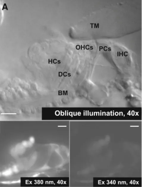
Fig. 1 Calcium imaging of the supporting cells in the hemicochlea preparation of hearing mice.aTheupper imageshows the organ of Corti in the basal turn of the cochlea by oblique illumination. The lower fluorescent imageswere taken in the same preparation at 340 and 380 nm excitation after bulk loading by fura-2 AM.TMTectorial membrane,BMbasilar membrane,IHCinner hair cell,PCpillar cell,
OHCouter hair cell,DCDeiters’ cell,HCHensen’s cell.Scale bars represent 20lm.bBasal [Ca2?]iin different supporting cell types of the organ of Corti. Note the higher resting intracellular Ca2? concentration in the Hensen’s cells (n=53) compared to the pillar (n=41; **p\0.01) and Deiters’ cells (n=65; ***p\0.001)
Neurochem Res
123
Journal :Large 11064 Dispatch : 2-1-2016 Pages : 12
Article No. : 1818 h LE h TYPESET
MS Code : NERE-D-15-00592 h4CP h4DISK
Author Proof
UNCORRECT
ED
PROOF
306 according to the protocol provided by the supplier (Invit- 307 rogen Life Technologies, Rockville, MD USA). RNA 308 (2ll) was reverse transcribed with RevertAid First Stand 309 cDNA Synthesis Kit (Invitrogen Life Technologies) as 310 described in previous studies [42,43]. Primers for ampli- 311 fication of P2X and P2Y receptor cDNAs were the
following: for P2X1 (Fwd) 50-CCT TGG CTA TGT GGT 312 GCG AGA GTC, (Rev) 30-AGG CAG GAT GTG GAG 313 CAA TAA GAG; P2X2 50-ATG GTG CAG CTG CTC 314 ATT, 30-AAA CGT GCA GTG CTT CAG; P2X3 50-ATC 315 AAG AAC AGC ATC CGT TTC CCT, 30-AGT GTT GTC 316 TCA GTC ACC TCC TCA; P2X4 50-ATC GTC ACC GTG 317 B
500 s 100 nM
[Ca2+] •= ATP, 50 µM
0 50 100
∆[Ca2+]i,2/ ∆[Ca2+]i,1
1
0.5
20 10 5 0
50 100
(min, interval) 1
0.5
20 10 5
**
0 50 1001
0.5
20 10 5
*
• • • • • • • • •
• • • • • • • • •
1 10 50 100 1 10 50 100 1 10 50 100
ATP (µM) 500 s
100 nM [Ca2+]
ATP (µM)
A
∆[Ca2+]i(nM)
Pillar cells Deiters’ cells Hensen’s cells
1 10 50 100 1 10 50 100 1 10 50 100
0 100 200 300
0 100 200 300
0 100 200 300
• • • • • • • • • • • •
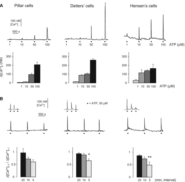
Fig. 2 ATP evoked reversible and repeatable intracellular Ca2? transients in pillar, Deiters’ and Hensen’s cells.aUpper traces: ATP evoked intracellular Ca2?transients in each type of supporting cells in a dose-dependent and repeatable manner. Representative traces show the responses for increasing doses of ATP (1, 10, 50 and 100lM;
30 s perfusion;black dots), applied with 20 min intervals in the same cell. Thescale bars indicate the change of [Ca2?]i and the time.
Lower bar graphs: Mean?SEM of the Ca2? transients evoked by different concentrations of ATP in the three supporting cell types.
1lM (light gray bars), 10lM (gray bars), 50lM (dark gray bars) and 100lM (black bars) of ATP. Pillar cells, n=4, 3, 41, 2; Deiters’
cells, n=9, 9, 65, 8; Hensen’s cells, n=2, 2, 53, 3.bUpper traces:
representative traces of intracellular Ca2? transients evoked by consecutive perfusion (30 s) of 50lM ATP (black dots). The ATP
responses were reversible and repeatable in all three cell types, but repeating the application of ATP in 5 min resulted in the reduction of the transients, while leaving 20 min before the next application allowed the response to recover. Thescale barsindicate the change of [Ca2?]iand the time.Lower bar graphs: Reduction of the 2nd ATP- evoked (50lM) Ca2?transients was dependent on the time intervals between repetitions in all three cell types (20, 10 and 5 min;black, darkandlight gray bars, respectively). The respective 5 min-values differed significantly from the 20-min-ones in Deiters’ and Hensen’s cells and pillar cells also showed a clear tendency of desensitization.
Barsrepresent the mean±SEM of the ratio of the 2nd to the 1st ATP-evoked Ca2? transients (D[Ca2?]i,2/D[Ca2?]i,1). Pillar cells, n=9, 6, 11; Deiters’ cells, n=14, 9, 20 and Hensen’s cells, n=20, 4, 8. *p\0.05; **p\0.01
Neurochem Res
Author Proof
UNCORRECT
ED
PROOF
318 AAC CAG ACA CA, 30-CCA CGA TTG TGC CAA GAC 319 GGA AT, P2X5 50-TTT CTT CGT GGT CAC CAA CCT 320 GAT, 30-ATT TGT GGA GCT GAA GTG ACA GGT;
321 P2X6 50-CTG TGG GAT GTG GCT GAC TT, 30-TCA 322 AAG TCC CCT CCA GTC AT, P2X7 50-CCA CAA CTA 323 CAC CAC GAG AAA C, 30-ACT TCT TGG CCC TTG 324 ACA TCT T, P2Y1 50-AAG ACC GGT TTC CAG TTC 325 TAC TAC, 30-CAC ATT TCT GGG GTC TGG AAA 326 TCC; P2Y2 50-TGC TGG TGC TGG CCT GCC AGG 327 CAC, 30-GCC CTG CCA GGA AGT AGA GTA CCG;
328 P2Y4 50-ATG AGG ATT TCA AGT TCA TCC TGC, 30- 329 TAG ACC ACG TTG ACA ATG TTC AGT; P2Y6 50- 330 CTG CGT CTA CCG TGA GGA TT, 30-GCT ATG AAG 331 GGC AGC AAG AA; P2Y12 50-CAG GTT CTC TTC 332 CCA TTG CT, 50-CAG CAA TGA TGA TGA AAA CC;
333 P2Y13 50-ATC TTG AAC AAG GAG GCA A, 50-TCT 334 TTT TAC GAA CCC TGT T; P2Y14 50-TAG AGG CCA 335 TAA ACT GTG CTT, 50-AAT TCT TCC TGG ACT TGA 336 GGT; b-actin 50-AGC TGA GAG GGAAATCGTGC-30, 337 50-GAT GGA GGG GCC GGA CTC AT-30.
338 The conditions for amplification were as follows: initial 339 denaturation at 95°C for 5 min, hot start at 80°C, then 340 94°C for 1 min, 59°C for 1 min, and 72°C for 1 min for 341 40 cycles, with a final extension at 72°C for 5 min. PCR 342 products were analyzed by agarose gel electrophoresis.
343 Materials
344 Fura-2 AM, Pluronic F-127 and Calcium Calibration Buf- 345 fer Kit with Magnesium #2 was obtained from Molecular 346 Probes, USA, cyclopiazonic acid from Alomone Labs, 347 Israel. All other chemicals were purchased from Sigma- 348 Aldrich, USA.
349 Results
350 Ca21Imaging of Pillar, Deiters’, and Hensen’s Cells 351 in Hearing Mouse Hemicochlea
352 To investigate the mechanism of Ca2? signaling and the 353 regulatory role of ATP in different supporting cell types of 354 the organ of Corti we developed a reliable method for 355 labeling individual cells and measuring [Ca2?]i in the 356 unique hemicochlea preparation of P15-21 mice [30–33].
357 The hemicochleae were bulk loaded with fura-2 AM, a 358 ratiometric, high-affinity Ca2? indicator (Fig. 1a). Only 359 preparations with intact morphology were used (see ‘‘Ma- 360 terials and Methods’’ section). Cells were identified based 361 on their anatomical location and shape under a 409 362 objective with red light oblique illumination.
We determined the basal, resting [Ca2?]i of the sup- 363 porting cells in hearing mice at the beginning of every 364 recording. Figure1b shows that the average resting [Ca2?]i 365 in the pillar (61±4 nM, n=41) and Deiters’ cells 366 (58±5 nM, n=65) was significantly lower than in the 367 Hensen’s cells (98 ±10 nM, n=53). Notably, we found 368 nearly 2 folds higher resting [Ca2?]iin the Hensen’s cells, 369 compared with the other two cell types suggesting a cell- 370 type specific intracellular Ca2?handling mechanism. 371 ATP Evoked Reversible and Repeatable Ca21 372 Signals in the Supporting Cells in a Dose-Dependent 373 Manner 374
In order to test whether ATP, an important regulator of 375 Ca2?signaling in the cochlea, is able to evoke changes in 376 the [Ca2?]iin supporting cells of the organ of Corti in the 377 mature hemicochlea, we applied ATP for 30 s in bath 378 perfusion. 379
ATP evoked characteristic, intracellular Ca2?transients 380 in a dose-dependent manner in the tested 1–100lM range 381 in all three types of supporting cells. The ATP responses 382 were reversible and repeatable (Fig.2a, upper traces). 383 Pillar cells showed the lowest sensitivity for ATP, as 1 and 384 10lM of the nucleotide evoked the smallest transients in 385 this supporting cell type (vs. the Deiters’ and Hensen’s 386 cells; Fig.2a, bar graphs). Application of 50lM ATP 387 induced a fast rising, uniformly shaped Ca2? transient 388 reliably in all three types of supporting cells (D[Ca2?]i in 389 nM; pillar cells: 96±14 nM, n=41; Deiters’ cells: 390 104 ±9 nM, n =65; Hensen’s cells: 140±10 nM, 391 n =53), therefore we used this concentration of ATP in 392 further experiments. We did not observe any contraction 393 based movement in the preparation after ATP application 394 (not even in Deiters’ cells). 395
Upon repeated application, the ATP response showed a 396 reduction, in inverse correlation with the time interval 397 between ATP administrations. There was no difference 398 between the cell types in this respect (Fig.2b, bar graphs). 399 The reduction was negligible when the ATP applications 400 followed each other by 20 min (pillar cells: 3 ±16 %, 401 n =9; Deiters’ cells: 6±4 %, n=14; Hensen’s cells: 402 13±4 %, n=20). Compared to that, a significant 403 reduction in the transients were seen with 5 min intervals 404 in Deiters’ and Hensen’s cells (34±8 %, n=20 and 405 51±12 %, n=8, respectively). The tendency of reduc- 406 tion in the transients was also evident in pillar cells 407 (40±9 %, n=11), although the difference between the 408 20-min and the 5-min-responses was not statistically sig- 409 nificant (Fig.2b, bar graphs). The 3rd applications of ATP 410 has confirmed (Fig.2b, representative traces), that while 411 5 min repetition of ATP resulted in pronounced desensiti- 412 zation of the ATP-evoked Ca2? response, 20 min was 413 Neurochem Res
123
Journal :Large 11064 Dispatch : 2-1-2016 Pages : 12
Article No. : 1818 h LE h TYPESET
MS Code : NERE-D-15-00592 h4CP h4DISK
Author Proof
UNCORRECT
ED
PROOF
414 enough for the transient to be recovered. In further 415 experiments, we repeated the ATP stimuli with 20 min 416 intervals.
417 ATP-Evoked Ca21Transients were Mediated 418 by Ca21Influx and Release of Ca21from Internal 419 Stores in a Cell-Type Specific Manner
420 The calcium ions, building up the ATP-evoked transients, 421 may originate from both extra- and intracellular sources.
422 To explore their involvement we tested the effect of ATP in 423 Ca2?-free buffer and after depletion of the SERCA-de- 424 pendent intracellular Ca2? stores in an internal standard 425 type of experimental design (2nd ATP stimulus presented 426 during perturbation of extra- or intracellular Ca2?sources;
427 see Fig.3a, b and in ‘‘Materials and Methods’’ section).
428 Ca2?-free medium (?1 mM EGTA) suppressed the 429 ATP-evoked intracellular Ca2?signals significantly in all 430 three types of cells (Fig.3a, c), i.e., the D[Ca2?]i,2/ 431 D[Ca2?]i,1ratios were decreased. The inhibition was more
pronounced in the Deiters’ and the Hensen’s cells (22±8 432 and 22±4 % of the 1st ATP response, respectively) 433 compared to the pillar cells (38±14 % of the 1st ATP 434 response; Fig.3c). Recovery of the Ca2?transients for the 435 3rd ATP stimulus after readministration of the normal 436 solution (data not shown) indicated that the cells preserved 437 their integrity and responsiveness. 438
The perfusion of the Ca2?-free medium caused a modest 439 decrease in basal [Ca2?]iof 6 out of 7 (86 %) pillar, 3 out 440 of 12 Deiters’ (25 %) and 2 out of 14 Hensen’s (14 %) 441 cells. 442
The intracellular Ca2? stores were depleted by the 443 specific SERCA inhibitor CPA (10lM), which inhibits 444 store refilling (Fig.3b, c). Empty stores hampered the 445 ATP-evoked transients significantly in all three cell types, 446 i.e., theD[Ca2?]i,2/D[Ca2?]i,1ratios were decreased. Again, 447 the effect was more robust in the Deiters’ and Hensen’s 448 cells (18±4 and 8±3 % of the 1st ATP response, 449 respectively) than in the pillar ones (33±8 % of the 1st 450 ATP response; Fig.3c). Recovery of the Ca2? transients 451
A B
D C
50 nM [Ca2+]
500 s
Ca2+-free Ca2+-free Ca2+-free
CPA 10 µM CPA 10 µM CPA 10 µM
Pillar cell Deiters’ cell Hensen’s cell Pillar cell Deiters’ cell Hensen’s cell
•= ATP, 50 µM
• • • • • • • • • • • •
0 50 100 150
Pillar cells
Deiters’
cells
Hensen’s cells CPA evoked ∆[Ca2+]i
∆[Ca2+]i(nM) Pillar cells Deiters’ cells Hensen’s cells
∆[Ca2+]i,2/ ∆[Ca2+]i,1
**
*
*** *** ***
***
*
1
0.5
0
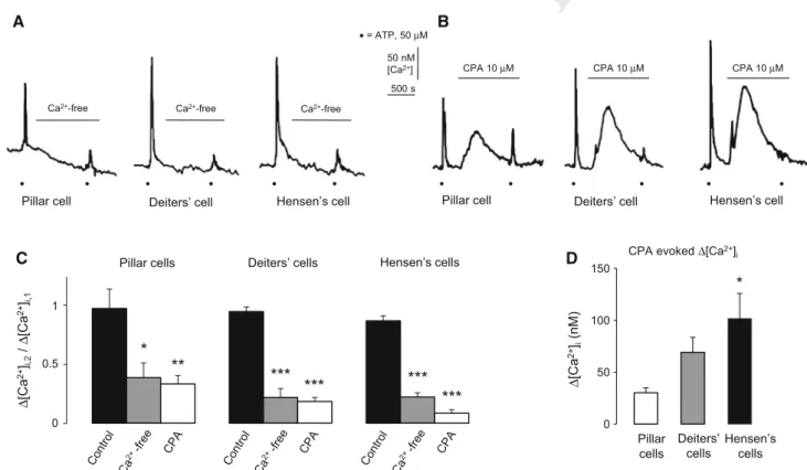
Fig. 3 ATP-evoked intracellular Ca2? transients are extracellular Ca2?and intracellular Ca2?store dependent in the supporting cells of the organ of Corti.a,bThe representative traces show the effect of the omission of extracellular Ca2?(?1 mM EGTA) and perfusion of 10lM CPA on the ATP-evoked Ca2? transients in the different supporting cells.Black dotsindicate the application of ATP (50lM).
Ca2?-free and CPA were administered as indicated by the horizontal lines.cEffect of the withdrawal of Ca2?from the buffer (?1 mM EGTA; Ca2?-free) and 10lM CPA on the transients evoked by 50lM ATP. The interval between the ATP application was 20 min.
Barsrepresent the mean±SEM of the ratio of the Ca2?transients in the presence (2nd ATP response) and in the absence (1st ATP response) of Ca2?omission/CPA (D[Ca2?]i,2/D[Ca2?]i,1). Pillar cells, n=9, 7, 8; Deiters’ cells, n=14, 12, 10 and Hensen’s cells, n=20, 14, 7. *p\0.05; **p\0.01, ***p\0.001. d CPA (10lM) increased the basal [Ca2?]iin all three supporting cell types, slightly in pillar (n=8) and more in Deiters’ (n=10) and Hensen’s cells (n=7; *p\0.05 compared to pillar cells). Bars represent the mean?SEM of the peak responses in nM
Neurochem Res
Author Proof
UNCORRECT
ED
PROOF
452 for the 3rd ATP stimulus after readministration of the 453 normal (no CPA) solution (data not shown) indicated that 454 the cells preserved their integrity and responsiveness.
455 CPA may also be used for characterization of SERCA- 456 dependent intracellular Ca2? stores by revealing their 457 leakage in the absence of refilling. Indeed, CPA itself, 458 before the 2nd ATP application, increased the [Ca2?]iin all 459 three cell types (Fig.3b). There was a modest effect in 460 pillar cells and more pronounced in Deiters’ and Hensen’s 461 cells, suggesting a difference in the regulation of [Ca2?]iin 462 these supporting cell types, as well (Fig.3d).
463 Both P2X and P2Y Receptor Subtype mRNAs were 464 Detected in the Organ of Corti: PPADS Revealed 465 Difference in the Functional Purinergic Receptor 466 Population of Pillar versus Deiters’ and Hensen’s 467 Cells
468 In order to determine the possible subtypes of P2 receptors 469 that may be involved in the action of ATP, we measured 470 the mRNA expression of P2X and P2Y receptor subunits in 471 the excised organ of Corti of P15-19 CD-1 mice. The RT- 472 PCR analysis showed the presence of the mRNA of P2X2, 473 P2X3, P2X4, P2X6, P2X7 and P2Y1, P2Y2, P2Y6, P2Y12, 474 P2Y14 receptors in the whole organ of Corti (Fig.4a). The 475 widely used, broad-spectrum purinergic receptor antagonist 476 PPADS (30lM) inhibited the 50 lM ATP-evoked Ca2? 477 transients in the pillar cells, but did not influence them 478 significantly in the Deiters’ and Hensen’s cells (Fig.4b).
479 Discussion
480 Intracellular Ca2? signals universally serve as second 481 messengers [44], regulating a variety of intra- and inter- 482 cellular processes also in the organ of Corti [8]. The 483 intracellular Ca2?signaling of the cochlear cells are con- 484 trolled or affected by ATP through purinergic receptors 485 during maturation, physiological sound transduction and 486 under pathological conditions, as well [4,5,35,45,46].
487 Besides a hormone-like tonic regulation, based on sound 488 exposure-induced release of ATP from the stria vascularis 489 [11], locally released ATP, as an auto- and paracrine regulator, 490 can modulate purinergic activity in the organ of Corti. ATP, 491 escaped from injured hair cells induces intercellular Ca2? 492 signaling among the supporting cells [4,16]. Furthermore, the 493 supporting cells themselves are able to release ATP into the 494 extracellular space through connexin hemichannels [13]. This 495 kind of ATP-mediated paracrine signaling was previously 496 observed in glia and glia-like tissue as well [47–49].
p2x1 p2x2 p2x3 p2x4 p2x5 p2x6 p2x7 β-act st
β-act p2y1 p2y2 p2y4 p2y6p2y11p2y12p2y13p2y14 st A
B Pillar
cells
Deiters’
cells
Hensen’s cells
∆[Ca2+]i,2/ ∆[Ca2+]i,1 1 0.5
0
***
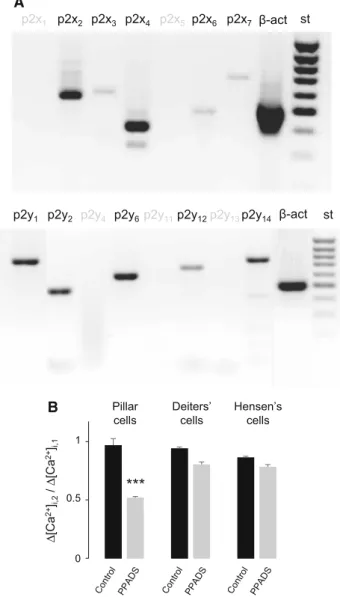
Fig. 4 RT-PCR analysis reveals the expression of multiple P2X and P2Y receptor subtypes in the organ of Corti of hearing mice. PPADS effect indicates different functional purinergic receptor population on pillar versus Deiters’ and Hensen’s cells.aTotal RNA samples from organs of Corti of one side of twenty P15-19 CD-1 mice each were reverse transcribed and amplified by PCR using primers specific to P2X and P2Y receptor transcripts. Amplification of b-actin (b-act) was used as an internal control. The identity of the amplified PCR products has previously been verified by sequencing [42]. A 100-bp DNA ladder (Fermantas, Vilnius, Lithuania) was used to identify PCR fragment sizes (st). The gel shown is representative of three independent analysis. mRNAs encoding P2X2, P2X3, P2X4, P2X6, P2X7, and P2Y1, P2Y2, P2Y6, P2Y12, P2Y14 receptors (black letters) were present in the organ of Corti.bThe widely used, broad- spectrum purinergic receptor antagonist PPADS (30lM) inhibited the ATP (50lM) response in the pillar cells, while it failed to cause significant effect in the Deiters’ and Hensen’s cells.Barsrepresent the mean±SEM of the ratio of the Ca2?transients in the presence (2nd ATP response) and in the absence (1st ATP response) of PPADS (D[Ca2?]i,2/D[Ca2?]i,1). Pillar cells, n=9, 9; Deiters’ cells, n=14, 14 and Hensen’s cells, n=20, 7. ***p\0.001
Neurochem Res
123
Journal :Large 11064 Dispatch : 2-1-2016 Pages : 12
Article No. : 1818 h LE h TYPESET
MS Code : NERE-D-15-00592 h4CP h4DISK
Author Proof
UNCORRECT
ED
PROOF
497 Investigation of the ATP-regulated Ca2? signaling is 498 predominantly performed in isolated cells [21–25] or 499 cochlear explants from embrionic or newborn murines [4, 500 16, 26], experimental models which lacks normal tissue 501 organization or contaminated by developmental biological 502 factors. The advantage of our approach is that it allows the 503 comparison of calibrated [Ca2?]i values of three different 504 supporting cell types (pillar, Deiters’ and Hensen’s) 505 investigated in the same in situ preparation from mature 506 hearing mice [34].
507 The real physiological concentration of ATP directly at 508 the release site and nearby the receptors is only predicted, 509 because of unresolved methodological challenges [50].
510 That is also the case in the organ of Corti [37]. In tissue- 511 cultured supporting cells of newborn animals, locally 512 applied ATP in a nanomolar range elicited repeatable intra- 513 and intercellular Ca2? oscillation that turned to a slowly 514 declining Ca2?response above few lM concentrations of 515 ATP [4, 16]. A broad concentration range of ATP was 516 tested (0.01–1000lM) and used (predominantly 100 and 517 10lM) in different studies in dissociated supporting cells 518 isolated from mature cochleae [22–25]. The ATP induced 519 intracellular Ca2? transients were also shown in Deiters’
520 [29] and Hensen’s [28] cells in in situ preparation from 521 adult guinea-pigs, where EC50 for ATP was *50lM in 522 Hensen’s cells and properties of the 100lM (puff from 523 pipette) and 1 mM (caged) ATP evoked transients were 524 investigated further. Direct comparison of ATP sensitivity 525 of the supporting cells in different studies is encumbered 526 by the different preparations used and the different ways of 527 ATP applications (different puffs, perfusions and caged 528 release). In our in situ hemicochlea preparation from 529 hearing mice, ATP evoked reversible and repeatable Ca2? 530 transients in a dose dependent manner in the 1–100lM 531 range in all the investigated supporting cell types (pillar, 532 Deiters’ and Hensen’s cells). Considering the method of 533 our ATP application (in short perfusion, see ‘‘Materials and 534 Methods’’ section) there is some overestimation of ATP 535 concentration that really reached the receptors on the cells, 536 i.e., the sensitivity of the cells for ATP is supposed to be 537 somewhat higher. However, our data, as they were col- 538 lected from the three different supporting cells under 539 identical conditions, may show reliably the bit lower sen- 540 sitivity of pillar cells, which produced an insignificant 541 Ca2? response for 10lM ATP, contrary to Deiters’ and 542 Hensen’s cells.
543 The amplitudes of the evoked transients and basal Ca2? 544 concentrations we measured in absolute values (nM) are in 545 the same magnitude published for dissociated Deiters’ and 546 Hensen’s cells of adult guinea-pigs [22,24], but thorough 547 comparisons are halted by the fact that other studies rather 548 used uncalibrated ratio orDF/F values of single wavelength 549 dyes.
Repeating the stimulus in 5 and 10 min showed desen- 550 sitization of the ATP response in all three cell types what 551 have already been observed in isolated Deiters’ and Hen- 552 sen’s cells [22,24]. We have not investigated the mecha- 553 nism underlying desensitization in this study. Decrease in 554 the amplitude of subsequent responses disappeared at 20 555 min stimulation interval, providing the opportunity for 556 internal standard experimental arrangement (see ‘‘Materi- 557 als and Methods’’ section). 558
A straightforward way of separating the ionotropic P2X 559 and the metabotropic P2Y receptor-mediated components 560 of ATP-evoked Ca2? responses from each other is with- 561 drawing Ca2? from the extracellular buffer and depleting 562 intracellular Ca2?stores by blocking their SERCA pump, 563 respectively. In our experiments both intervention, omis- 564 sion of Ca2? and application of CPA, inhibited the 565 response, suggesting the involvement of both the iono- 566 tropic- and the metabotropic ATP receptors, in all three cell 567 types. This conclusion, although without cell specificity 568 and not on the protein level, was supported by the presence 569 of the mRNA of P2X2,3,4,6,7and P2Y1,2,6,12,14 receptors in 570 the organ of Corti of the same preparation. 571
Imaging experiments with a broad-spectrum purinergic 572 antagonist provided further data regarding the functional 573 expression of purinergic receptors in supporting cells. 574 PPADS antagonizes several P2X and also some P2Y 575 receptors [51,52]. Its effect in pillar cells and the lack of its 576 significant effect in Deiters’ and Hensen’s cells suggests 577 involvement of distinct functional purinergic receptor 578 populations in the ATP response in these cells. 579
The inhibitory effect of Ca2?withdrawal on the ATP- 580 evoked Ca2? transients in pillar, Deiters’ and Hensen’s 581 cells was shown in different experimental models of lar- 582 gely isolated cells [22–25] and/or in situ preparation [28, 583 29], but neither of these studies investigated all three types 584 of supporting cells in the same preparation synchronously. 585 Probably this is the reason of the relatively broad published 586 range of efficiency of Ca2?omission in inhibiting the effect 587 of ATP, including even the total abolishment of the 588 response in Hensen’s cells [22]. The effect of depletion of 589 endoplasmic reticulum (ER) Ca2?stores was investigated 590 much sparsely and in P1-3 rat organ culture preparation [4, 591 16]. The measurement of the effect of both interventions in 592 the same study, especially in all three cell types in the same 593 preparation, was not performed hitherto, according to our 594 best knowledge. 595
We found that, besides the contribution of extracellular 596 Ca2?, the ATP-evoked Ca2? transients were also depen- 597 dent on the intracellular Ca2?stores, but more strongly in 598 the Deiters’ and Hensen’s cells than in the pillar ones. It 599 has been shown previously that SERCA pump inhibition by 600 thapsigargin or CPA unmasks the leak of Ca2?from the ER 601 in different cell types [53, 54] including glial cells [55]. 602 Neurochem Res