Extracellular vesicles in diagnostics and therapy of the ischaemic heart: Position Paper from the
Working Group on Cellular Biology of the Heart of the European Society of Cardiology
Joost Petrus Gerardus Sluijter
1*
†, Sean Michael Davidson
2†,
Chantal M. Boulanger
3, Edit Iren Buza´s
4,5, Dominique Paschalis Victor de Kleijn
6,7, Felix Benedikt Engel
8, Zolta´n Giricz
9, Derek J. Hausenloy
10,11,12,13,14,15, Raj Kishore
16, Sandrine Lecour
17, Jonathan Leor
18, Rosalinda Madonna
19,20,21, Cinzia Perrino
22, Fabrice Prunier
23, Susmita Sahoo
24, Ray Michel Schiffelers
25, Rainer Schulz
26, Linda Wilhelmina Van Laake
27, Kirsti Ytrehus
28, and Pe´ter Ferdinandy
29,30*
1Experimental Cardiology Laboratory, UMC Utrecht Regenerative Medicine Center, University Medical Center Utrecht, University Utrecht, 3508GA Utrecht, The Netherlands;2The Hatter Cardiovascular Institute, University College London, London, UK;3INSERM UMR-S 970, Paris Cardiovascular Research Center–PARCC, Paris, France;4Department of Genetics, Cell- and Immunobiology, Semmelweis University, Budapest, Hungary;5MTA-SE Immunoproteogenomics Research Group, Budapest, Hungary;6Department of Vascular Surgery, UMC Utrecht, Utrecht University, Utrecht, the Netherlands; 7Netherlands Heart Institute, Utrecht, the Netherlands;8Experimental Renal and Cardiovascular Research, Department of Nephropathology, Institute of Pathology, Friedrich-Alexander-Universita¨t Erlangen-Nu¨rnberg, Erlangen, Germany;9Department of Pharmacology and Pharmacotherapy, Semmelweis University, Budapest, Hungary;10Cardiovascular & Metabolic Disorders Program, Duke-National University of Singapore Medical School, 8 College Road, Singapore 169857;11National Heart Research Institute Singapore, National Heart Centre Singapore, 5 Hospital Drive, Singapore 169609;12Yong Loo Lin School of Medicine, National University Singapore, 1E Kent Ridge Road, Singapore 119228;13The Hatter Cardiovascular Institute, University College London, 67 Chenies Mews, London WC1E 6HX, UK;14The National Institute of Health Research University College London Hospitals Biomedical Research Centre, Research & Development, Maple House 1st floor, 149 Tottenham Court Road, London W1T 7DN, UK;
15Department of Cardiology, Barts Heart Centre, St Bartholomew’s Hospital, W Smithfield, London EC1A 7BE, UK;16Department of Pharmacology, Center for Translational Medicine, Lewis Katz School of Medicine, Temple University, Philadelphia, PA, USA;17Hatter Institute for Cardiovascular Research in Africa and Lionel Opie Preclinical Imaging Core Facility, Faculty of Health Sciences, University of Cape Town, South Africa;18Neufeld Cardiac Research Institute, Sackler Faculty of Medicine, Tel-Aviv University, Tel Hashomer, Israel; Tamman Cardiovascular Research Institute, Heart Center, Sheba Medical Center, Tel Hashomer, Israel;19Center of Aging Science and Regenerative Medicine, CESI-Met and Institute of Cardiology, “G. D’Annunzio” University, Chieti-Pescara, Chieti, Italy;20Department of Internal Medicine, University of Texas Medical School in Houston, TX, USA;21Texas Heart Institute, Houston, TX, USA;22Department of Advanced Biomedical Sciences, Federico II University, Naples, Italy;23Institut Mitovasc, CHU d’Angers, Universite´ d’Angers, Angers, France;
24Cardiovascular Research Institute, Icahn School of Medicine at Mount Sinai, New York, NY, USA;25Laboratory Clinical Chemistry and Hematology Division, University Medical Center Utrecht, Utrecht, The Netherlands;26Institute of Physiology, Justus-Liebig University of Giessen, Aulweg 129, 35392, Giessen, Germany;27Division Heart and Lungs, and Hubrecht Institute, University Medical Center Utrecht, Utrecht, The Netherlands;28Cardiovascular Research Group, Department of Medical Biology, UiT The Arctic University of Norway, Tromsø, Norway;29Department of Pharmacology and Pharmacotherapy, Semmelweis University, Nagyva´rad te´r 4, Budapest 1089, Hungary and30Pharmahungary Group, Szeged, Hungary;
Received 13 July 2017; revised 2 October 2017; editorial decision 29 October 2017; accepted 1 November 2017; online publish-ahead-of-print 2 November 2017
Abstract Extracellular vesicles (EVs)—particularly exosomes and microvesicles (MVs)—are attracting considerable interest in the cardiovascular field as the wide range of their functions is recognized. These capabilities include transporting regulatory molecules including different RNA species, lipids, and proteins through the extracellular space including blood and delivering these cargos to recipient cells to modify cellular activity. EVs powerfully stimulate angiogenesis, and can protect the heart against myocardial infarction. They also appear to mediate some of the paracrine effects of cells, and have therefore been proposed as a potential alternative to cell-based regenerative therapies.
Moreover, EVs of different sources may be useful biomarkers of cardiovascular disease identities. However, the methods used for the detection and isolation of EVs have several limitations and vary widely between studies, lead- ing to uncertainties regarding the exact population of EVs studied and how to interpret the data. The number of publications in the exosome and MV field has been increasing exponentially in recent years and, therefore, in this ESC Working Group Position Paper, the overall objective is to provide a set of recommendations for the analysis
* Corresponding authors. Tel:þ31 88 75 575 67; fax:þ31 30 25 226 93, E-mail: j.sluijter@umcutrecht.nl (J.P.G.S.); Tel:þ36 1 2104416; fax:þ36 1 2104412, E-mail: peter.ferdinandy@pharmahungary.com (P.F.)
†The first two authors contributed equally to the study.
VCThe Author 2017. Published by Oxford University Press on behalf of the European Society of Cardiology.
This is an Open Access article distributed under the terms of the Creative Commons Attribution License (http://creativecommons.org/licenses/by/4.0/), which permits unrestricted reuse, distribution, and reproduction in any medium, provided the original work is properly cited.
doi:10.1093/cvr/cvx211
.. ..
.. ..
.. ..
.. ..
.. ..
.. ..
.. ..
.. ..
.. ..
.. ..
.. ..
.. ..
.. ..
.. ..
.. ..
.. ..
.. ..
.. ..
.. ..
.. ..
.
and translational application of EVs focussing on the diagnosis and therapy of the ischaemic heart. This should help to ensure that the data from emerging studies are robust and repeatable, and optimize the pathway towards the diagnostic and therapeutic use of EVs in clinical studies for patient benefit.
䊏 䊏 䊏 䊏 䊏 䊏 䊏 䊏 䊏 䊏 䊏 䊏 䊏 䊏 䊏 䊏 䊏 䊏 䊏 䊏 䊏 䊏 䊏 䊏 䊏 䊏 䊏 䊏 䊏 䊏 䊏 䊏 䊏 䊏 䊏 䊏 䊏 䊏 䊏 䊏 䊏 䊏 䊏 䊏 䊏 䊏 䊏 䊏 䊏 䊏 䊏 䊏 䊏 䊏 䊏 䊏 䊏 䊏 䊏 䊏 䊏 䊏 䊏 䊏 䊏 䊏 䊏 䊏 䊏 䊏 䊏 䊏 䊏 䊏 䊏 䊏 䊏 䊏 䊏 䊏 䊏 䊏 䊏 䊏 䊏 䊏 䊏 䊏 䊏 䊏 䊏 䊏 䊏 䊏 䊏 䊏 䊏 䊏 䊏 䊏 䊏 䊏 䊏 䊏 䊏 䊏 䊏 䊏 䊏 䊏 䊏 䊏 䊏 䊏 䊏 䊏 䊏 䊏 䊏 䊏 䊏 䊏 䊏 䊏 䊏 䊏 䊏 䊏 䊏 䊏 䊏 䊏 䊏 䊏 䊏 䊏 䊏 䊏 䊏 䊏 䊏 䊏 䊏 䊏 䊏 䊏 䊏 䊏 䊏 䊏 䊏 䊏 䊏 䊏 䊏 䊏 䊏 䊏 䊏 䊏 䊏 䊏 䊏 䊏 䊏 䊏 䊏 䊏 䊏 䊏 䊏 䊏 䊏 䊏 䊏 䊏 䊏 䊏 䊏 䊏 䊏 䊏 䊏 䊏 䊏 䊏 䊏 䊏 䊏 䊏 䊏 䊏 䊏 䊏 䊏 䊏 䊏 䊏 䊏 䊏 䊏 䊏 䊏 䊏 䊏 䊏 䊏 䊏 䊏 䊏 䊏 䊏
Keywords Exosomes
•
Microvesicles•
Extracellular vesicles•
Ischaemia•
Reperfusion•
Cardioprotection•
Heartfailure
•
Remote conditioning•
Preconditioning•
Postconditioning•
Co-morbidities•
Regenerative medicine1. Introduction
1.1 Cellular secretion for communication;
extracellular vesicles
Cells in multicellular organisms must communicate efficiently with each other in order to propagate signals and co-ordinate function. In addition to distinct chemical signals (paracrine and endocrine) and direct cell-cell contact, a growing body of evidence shows that cells communicate via a variety of small, membrane-enclosed vesicles, collectively termed
‘extracellular vesicles’ (EVs). EVs ranging from 40 nm to several microns in size (Figure1), are released into all extracellular fluids includ- ing blood. They transport a cell-specific cargo of proteins, lipids, metabo- lites, and nucleic acids that can affect target cells. This process occurs during normal cellular physiology as well as during stress and disease. In this rapidly evolving area of research, with its associated technical chal- lenges, it is vital to establish standardized techniques for their isolation and criteria for their identification. EVs are released by all cardiac, endo- thelial and inflammatory cell types, suggesting they have an important role in the cardiovascular system, including the ischaemic heart.1–6
Therefore, the overall objective of this position paper is to provide a set of recommendations for the isolation, characterization, analysis, and translational application of EVs focussing mainly on the ischaemic heart, i.e. acute myocardial infarction and post-ischaemic heart failure.
Coronary atherosclerosis is not discussed in this position paper in detail (see recent reviews and position papers of the European Society of Cardiology that covers this topic extensively7–9).
2. Isolation and characterization of EVs
2.1 Definition of EVs
Eukaryotic EVs include (i) exosomes released by exocytosis of multivesicu- lar bodies (usually 50–150 nm), (ii) Microvesicles (MVs, also called micro- particles or ectosomes), vesicles around 0.1–1lm in diameter shed from the plasma membrane,10–12and (iii) apoptotic vesicles released by blebbing of apoptotic cells some of which are > 1lm (apoptotic bodies),13,14and (iv) large oncosomes released by migratory tumour cells (> 1mm)15,16 (Figure2). However, since current isolation protocols only result in relative enrichment of vesicle subpopulations rather than their complete purification,17and that no specific markers are available for the subpopula- tions, it is preferable to refer to purified vesicles as ‘EVs’ and accurately report the purification method used and characteristics present. An opera- tional definition of small EVs (sEVs) is appropriate for the exosome- enriched population pelleting at high speeds.17
2.2 Methods for the isolation and characterization of EVs
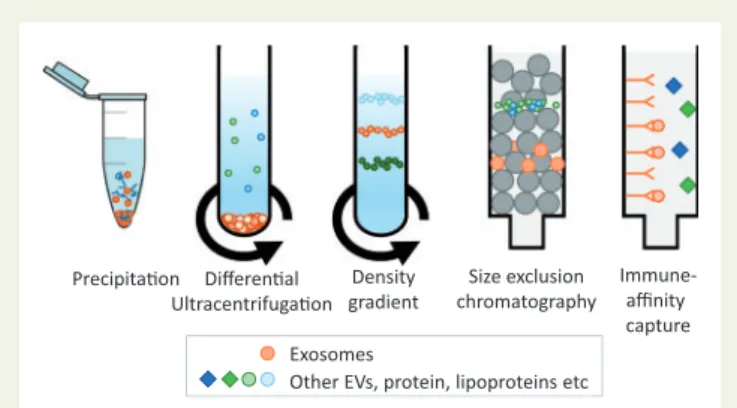
Several methods of isolating EVs have been developed (Figure3). The optimal EV isolation procedure will depend on the source biofluid, the
EV subpopulation of interest, and their intended end-use, whether that is to be diagnostic biomarker studies, mechanistic studies, or for in vivo administration. However, no EV isolation method yet exists that can be considered as a gold standard, since residual proteins and/or lipoproteins remains problematic.18Complete removal of lipoproteins (present in both blood and tissue culture serum) remains challenging due to over- lapping size and/or densities between EVs and different lipoprotein par- ticles (Figures1and2).12,19,20Moreover, low density lipoprotein (LDL) and exosomes may associate, rendering their complete separation from blood samples impossible using any technique.12This, however, might be used in isolating a subset of EVs co-precipitated with LDL or high density lipoprotein (HDL) particles.21,22
In the simplest technique based on precipitation (e.g. ExoQuickTM), the biofluid sample is mixed and incubated with a hydrophilic polymer prior to low speed centrifugation. The polymers attract water molecules away from the solvation layer around the EVs causing their precipitation.
Several manufacturers now market products based on this technique.
However, they result in high levels of contaminants including serum pro- teins and lipoproteins as well as residual matrix that can affect EV biologi- cal functions.23,24
Differential centrifugation has long been regarded as the gold standard technique.19In the most commonly used protocol, cells are removed by centrifugation at 300 x g for 10 min; the supernatant is then cleared of apoptotic bodies by centrifugation at 2000 x g for 10 min followed by 10 000 x g for 30 min to preferentially pellet MVs; exosome-enriched sEVs are then purified from the supernatant by ultracentrifugation at
20 40 60 80 100 120 1000
Parcle diameter (nm)
HDL LDL
IDL VLDL
1.20 1.10 1.06 1.02 1.006 0.95
Density (g/ml)
Chylomicrons
Exosomes Microvesicles
Figure 1Exosomes and MVs overlap in size with VLDL and chylomi- crons, and in density with HDL/LDL particles. Exosome density is typi- cally 1.06–1.20 g/mL. MV density is not well defined but they have been found between1.03–1.08 g/mL.10–12
.. ..
.. ..
.. ..
.. ..
.. ..
.. ..
.. ..
.. ..
.. ..
.. ..
.. ..
.. ..
.. ..
.. ..
.. ..
.. ..
.. ..
.. ..
..
.. ..
.. ..
.. ..
.. ..
.. ..
.. ..
.. ..
.. ..
.. ..
.. ..
.. ..
.. ..
.. ..
.. ..
.. ..
.. ..
.. ..
.. ..
.. ..
.. ..
.. ..
.. ..
.. ..
.. ..
.. ..
.. ..
.. ..
.. .
100 000 x g for 70 min.19 However, optimal parameters are highly dependent on the type of centrifuge rotor used.25For further purifica- tion of EVs from co-pelleted protein complexes26and lipoproteins, a density gradient is recommended (sucrose or preferably iodixanol).19 Because of the number of steps involved and the length of the proce- dure, it is clearly unsuited for the analysis of large numbers of samples.
More recently, concerns have been raised that ultracentrifugation can cause damage, fusion, and/or aggregation of vesicles.27
There is increasing enthusiasm for the use of size-exclusion chromatography.28Suitable matrices include Sepharose 2B, Sepharose CL-4B, or Sephacryl S-400.29 The resolution of size separation is dependent on column length. This technique is effective at separating EVs from proteins and some lipoproteins, but samples usually become considerably diluted and efficient separation of lipoproteins remain challenging.12,29,30
Other techniques are less well established. Filtration (0.2lm–
0.8lm) aids in removal of larger vesicles, but under high pressure it is possible that it could cause the fragmentation of larger EVs into
smaller vesicles. Immuno-affinity can be an effective means to isolate specific EV populations, using columns or magnetic beads for exam- ple, but for functional follow-up studies this is still challenging. Flow cytometry-based analysis of individual EVs is a valuable tool for EV characterization and quantification, although it remains challenging due to size and sensitivity limitations.31Progress has been made in developing these technologies for specific EV sorting, but some pre- requisite steps need to be taken.32 Furthermore, this approach requires certainty as to the specificity of epitopes expressed by the desired population of EVs–which may not be the case for exo- somes.33Other isolation techniques such as microfluidics are under development but have not yet been rigorously tested. For pre-clinical or clinical studies of the ischaemic heart, measurement of EVs can be performed fromin vivoblood, lymphatic or pericardial fluid samples, ex vivoheart perfusate samples, and tissue culture media samples that may require different isolation techniques.
2.2.1 Isolation from blood
Pre-analytical procedures can have a large impact on blood EV measure- ments. For example, since clotting may increase the number of EVs in blood by 10-fold,34it is usually preferable to use plasma. On the other hand, serum may be useful when overall yield of platelet MVs is more important than accurate quantification of particle number. A crucial con- cern is the minimization of platelet activation and EV release.
Standardized procedures to minimize platelet activation during plasma isolation should be followed.35,36Fasting before blood sampling can help to minimize chylomicron contamination.12Blood should be collected in citrated or acid-citrate-dextrose anticoagulant tubes,23,35,37such as vacu- tainers, and the first tube of blood should be discarded.23,35It is recom- mended to dilute blood plasma or serum at least 2x in Ca2þ-free phosphate buffered saline (PBS) prior to centrifugation in order to reduce the viscosity.19However, if annexin V binding will be assessed (which requires Ca2þ), PBS should be avoided in order to prevent for- mation of calcium-phosphate micro-precipitates. The plasma or serum should be centrifuged within 2 h, and agitation avoided.35,38After centri- fugation at 2500 x g for 15 min at room temperature without application Exosomes Microvesicles Apoptoc bodies Large oncosomes
Diameter range
50-150 nm 0.1-1 μm > 1μm 1-10 μm
buoyant densies
1.06-1.20 g/ml 1.03-1.18 g/ml 1.24-1.28 g/ml ~1.15 g/ml
possible contaminants /co-isolates
HDL, LDL aggregates protein aggregates
AGO-2-RNA complexes
chylomicrons, LDL aggregates;
protein aggregates,
chylomicrons platelets
platelets Extracellular
vesicles
Figure 2The major classes of EV. Typical size and density of EV classes and some of the contaminants that may be co-isolated, depending on biofluid.
Density gradient
Immune- affinity capture Size exclusion
chromatography Differenal
Ultracentrifugaon Precipitaon
Exosomes
Other EVs, protein, lipoproteins etc
Figure 3 Standard techniques used for isolating exosomes from other EVs, protein, and lipoproteins present in blood and cell-culture medium.
.. ..
.. ..
.. ..
.. ..
.. ..
.. ..
.. ..
.. ..
.. ..
.. ..
.. ..
.. ..
.. ..
.. ..
.. ..
.. ..
.. ..
.. ..
.. ..
.. ..
.. ..
.. ..
.. ..
.. ..
.. ..
.. ..
.. ..
.. ..
.. ..
.. ..
.. ..
.. ..
.. ..
.. ..
.. ..
.. ..
.. ..
.. ..
.. ..
.. ..
.. ..
.. ..
.. ..
.. ..
.. .
of the centrifuge brake, plasma can be carefully collected, and re- centrifuged under identical conditions. This platelet-free-plasma may be snap frozen and stored at –80C prior to analysis.
Even when using the same protocol, inter-laboratory variability in plasma EV counts can vary by an order of magnitude.35Given these problems of irreproducibility, The International Society on Thrombosis, and Haemostasis has advised that further refinements are required before flow cytometric enumeration of platelet MV numbers is ready for clinical use.35
2.2.2 Isolation from pericardial fluid
Pericardial fluid contains EVs that may provide useful biomarker informa- tion about cardiac health.39,40As yet there is no consensus as to the ideal method for isolation of EVs from pericardial fluid.
2.2.3 Isolation from conditioned media of cultured cells For the isolation of vesicles produced by cells in tissue culture the impor- tant considerations are quite different. The main potential source of con- tamination is typically from foetal calf serum (FCS) added to the culture medium.41FCS contains large number of vesicles including exosomes as well as lipoproteins. Exosomes can be largely removed by pre-treating FCS by 18 h ultracentrifugation at 100 000g,41 and removal is enhanced by diluting FCS five-fold in culture medium to reduce viscos- ity.23 Several companies market FCS which has been processed to remove exosomes, though the method used is not specified. However, some caution should be taken for FBS-associated RNA which might be co-isolated with cell-culture derived extracellular RNA (exRNA), thereby interfering with the downstream RNA analysis.42Alternatively, pre-defined serum or serum-free conditions can be used, and indeed is essential if preparing EVs for clinical use.43However, cells may undergo apoptosis or autophagy and release apoptotic bodies after extended periods in the absence of serum. Conditioned medium is usually col- lected after 24–48 h culture. Although sequential filtration offers the advantage of using large volumes of culture media,44its effect on biologi- cal activity of the isolated EVs has not been well characterized. HPLC has been successfully used to purify exosomes.45
2.2.4 Isolation from isolated heart perfusate
EVs can be isolated from hearts perfused with buffer such as those mounted on a Langendorff apparatus.46Pre-concentration of the perfu- sate by ultrafiltration may be necessary for a sufficient yield, but subse- quently any of the techniques described above may be used. It is important to be aware that exosome-sized, calcium-phosphate nanopar- ticles form spontaneously in Ca2þ-containing bicarbonate buffer, which can interfere with some analyses such as nanoparticle tracking analysis.47
2.2.5 Storage of EVs
EVs appear to be relatively stable when stored at –80C or less,48but repeated freeze-thaw cycles should be avoided and cryo-preservatives such as glycerol and DMSO should not be added as they may lyse EVs.48 Trehalose has recently been proposed to preserve exosomes.49
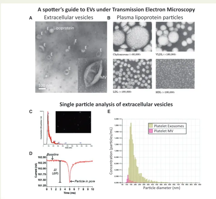
2.2.6 Visualization and quantitation of EVs
In most studies, it is informative to quantify EVs using either nanoparticle tracking analysis, tuneable resistive pulse sensing, or dynamic light scat- tering (Figure4). Importantly, these methods usually do not discriminate EVs from non-vesicular events such as LDL particles.12The ratio of par- ticle number or lipid content to protein content of the preparations can
give an indication of relative EV purity, as compared to protein concen- tration only.50,51Exosome morphology should be confirmed by trans- mission electron microscopy,19cryo-electron microscopy,52or atomic force microscopy.53In addition, presented images should contain multi- ple EVs per field (Figure4A). Using flow cytometry, the lower size-limit of detection is steadily decreasing, but it remains technically challenging to obtain comprehensive analyses of MVs let alone exosome-sized particles.32
2.2.7 Characterization of EVs
Further characterization of EVs should include the detection of specific markers. It is generally recommended to demonstrate presence of tetra- spanin proteins such as CD9, CD81, CD63, and the intra-vesicular pro- tein Alix, which are involved in exosome biogenesis, in addition to other typical marker proteins such as HSP70, flotillin-1, or major histocompati- bility complex (MHC) class I and class II. However, recent studies indi- cate some of these are not as specific for exosomes as thought.17,54 Co-enrichment of CD63, CD9, and CD81 tetraspanins and endosome markers such as syntenin-1 and TSG101 may be seen as indicative of exosome presence.17MV surface marker expression is a useful index of cell-type of origin, and can be quantified by flow cytometry in order to analyse sub-populations. In addition, MVs may be characterized as
‘calcium-dependent annexin V binding’ or ‘calcium-independent lactad- herin (MFGE8) binding’. Gating strategies for flow cytometry involve the use of fluorescent reference beads of known sizes. Preferred beads are the silica beads because their refractive index is close to the one of bio- logical particles.55Of note, not all MVs are detectable this way. To quan- titatively detect exosome proteins, a nano-plasmonic exosome sensor was developed that comprises arrays of periodic nanoholes patterned in a metal film. The arrays are functionalized with affinity ligands for differ- ent exosomal protein markers and offers highly sensitive and label-free exosome analyses by continuous and real-time monitoring of molecular binding.56
In order to demonstrate EV functionality it may be useful to demon- strate their interaction with, fusion, or uptake into recipient cells. To this end, EVs can be fluorescently labelled with lipophilic dyes, or by transfec- tion of parent cells with GFP-tagged proteins packaged in EVs. Control experiments using ‘dye-only’ samples prepared in parallel the same way as EVs are essential to confirm the involvement of EVs57and that free dye has been removed, which can otherwise form micelles. Density gra- dient ultracentrifugation is a frequently used approach to remove this non-EV-associated dye. Co-incubation with receptor antagonists or uptake inhibitors will provide insights in mechanisms of uptake although these compounds are notorious for their toxicity and limited specificity.58
2.2.8 Analysis of nucleic acid, protein, and lipid contents of EVs
Proteomics can provide a more comprehensive description of EV pro- tein content, but the isolation protocol must be carefully optimized in order to remove plasma protein contamination in blood EV samples.59,60 Similar concerns arise with transcriptomic analyses of miRNA, mRNA, or DNA contents, particularly with regard to potentially contaminating HDL particles and argonaute 2-RNA complexes, which are known to transport miRNA.61Care must be taken to avoid haemolysis which can affect circulating miRNA levels.62
EV proteomics has been studies in large clinical cohorts,63 but undoubtedly the greatest interest is currently focused at the RNA
.. ..
.. ..
.. ..
.. ..
.. ..
.. ..
.. ..
.. ..
.. ..
content of EVs as potential biomarkers.63EV lipidomics is an interesting
.
but under-explored area.64Nucleic acid, protein and lipid entries from EV studies are available in EV databases (http://www.exocarta.org/, http://student4.postech.ac.kr/evpedia2_xe/xe/ and http://microvesicles.
org/ (10 November 2017, date last accessed)). However, because of the diversity of EV isolation protocols used, these database entries should be treated with caution.
2.2.9 Technical control experiments
RNase or protease may be applied to remove extra-vesicular nucleic acids and proteins and confirm intra-vesicular localization. Detergent control during flow cytometry or sizing performed by tuneable resistive
pulse sensing or dynamic light scattering enables distinguishing EVs from protein aggregates (the latter being more resistant to detergents lysis than EVs).26,65,66Of note, small and large EVs have different detergent sensitivities67and lipoproteins also show a limited sensitivity to deter- gent lysis.12
2.3 Recommendations for isolation and purification of EVs
At present, there is no universally agreed protocol for isolation of pure populations of EVs or subpopulations of EVs. Even their precise nomen- clature is in flux, and will presumably remain so until clear surface protein signatures of individual EV subtypes will be established. Given the rapid
A spoer’s guide to EVs under Transmission Electron Microscopy Plasma lipoprotein parcles
Single parcle analysis of extracellular vesicles
100 nm
Extracellular vesicles
E E
E
E
E E
E E Lipoprotein
MV E
A
C E
D
B
Platelet MV Platelet Exosomes
Parcle diameter (nm)
Concentraon (parcles/mL)
Figure 4Standard methods of characterizing EVs. (A) Transmission electron microscopy (TEM) of negative-stained EVs reveals the ‘cup-shaped’ appear- ance of exosomes (E) and MVs once they have been dried for TEM (they are spherical in solution). (B) The spherical appearance of lipoprotein particles by TEM is quite distinct (image courtesy of Robert L. Hamilton and the Arteriosclerosis Specialized Center of Research, University of California, San Francisco).
(C) Nanoparticle tracking analysis (NTA) provides a size distribution of particles based on calculating their size by their random Brownian motion. (D,E) Tuneable resistance pulse sensing (TRP) determines size distribution by the change in resistance as the particle crosses a small pore in a membrane (which is selected according to the size range examined).
.. ..
.. ..
.. ..
.. ..
.. ..
.. ..
.. ..
.. ..
.. ..
.. ..
.. ..
.. ..
.. ..
.. ..
.. ..
.. ..
.. ..
.. ..
.. ..
.. ..
developments in this area it is important to remain cognizant of experi-
.
mental limitations and caveats in order to avoid overstating conclusions.
At present, it is challenging to isolate EVs from tissue homogenates. The likely contamination of EV preparations with proteins and lipoproteins renders many results suspect unless crucial control experiments are per- formed. Differential detergent lysis is a simple, inexpensive EV control for flow cytometry.26,65Furthermore, to avoid swarm detection at high particle concentrations, analysis of serial dilution of samples is recom- mended during flow cytometry.32 A characterization of EV proteins will provide a basis on purity without excluding a potential biological role.Table1provides a check-list of criteria for the successful isolation and characterization of EVs, as well as more specific criteria for exosomes.
3. Mechanism of EV actions in the heart
EVs appear to be released from all major cell types found in the heart.
For example, exosomes have been shown to be released by primary adult cardiomyocytes,68,69 primary cardiac endothelial cells,70 primary cardiac fibroblasts,71 and vascular smooth muscle cells.72 Most MVs present in the plasma of healthy individuals are derived from platelets and erythrocytes, but plasma MVs also originate from leucocytes, endo- thelial cells, monocytes neutrophils, and lymphocytes.73,74Most mecha- nistic experiments to date use EVs isolated from cultured cells, since isolation of cell-type specific EVsin vivois not currently feasible. Recently,
a clear overview was provided on the role of EVs in coronary artery dis- ease.7This section will discuss the most relevant examples of the func- tion of EVs released from cells of the heart tissue and their postulated mechanisms of action.
3.1 Cardiomyocyte-derived EVs
Cardiomyocytes are potentially an important source of diagnostic EVs particularly in situations of stress such as myocardial ischaemia and fail- ure.1 However, few studies have examined exosomes from adult as opposed to neonatal cardiomyocytes, and even here it is unclear to what extent this relates to the situationin vivo.In vitro, hypoxia and re- oxygenation leads to the release of heat shock proteins (HSPs) HSP70 and HSP90, as well as HSP60, a ‘danger signal’, via exosomes in primary adult rat cardiomyocytes.68Cardiomyocytes also release EVs containing tumour necrosis factor (TNF)-a,75potentially participating in the propa- gation of an inflammatory response. Glucose deprivation induced the loading of neonatal rat cardiomyocyte exosomes with functional glucose transporters and glycolytic enzymes. When internalized by endothelial cells, these exosomes increased glucose uptake, glycolytic activity, and pyruvate production in recipient cells.76The transfer of exosomes from glucose-deprived H9C2 cardiac myoblasts to endothelial cells also induced changes in transcriptional activity of pro-angiogenic genes.77 Hyperglycaemia altered cardiomyocyte-derived exosomes in a model of diabetes-associated cardiomyopathy,78as well as from the hypoxic myo- cardium which can activatein vitrocultured endothelial cells.79,80Finally, external stretch caused cardiomyocytes to release exosomes enriched Table 1Recommendations for the isolation and characterization of EVs [adapted from Ref. 18]
General recommendations for EVs
(1) EVs can be isolated from either tissue culture supernatant or extracellular fluids. Reliable methods for EV isolation from tissue homogenates remain to be established.
(2) Ensure consistency of pre-analytical procedures.
(3) Report complete experimental details, including pre-analytical and isolation procedure and details of all antibodies used.190Also include all negative data sets.
(4) If EV function is analysed, include:
a. a dose-response curve.
b. systematic negative (‘EV-depleted’) controls.
c. demonstrate an association between a protein/miRNA and EVs in support of any function ascribed to them, e.g. using immuno-EM, or co-purification on a density gradient.
Specific recommendations for exosomes (1) Avoid precipitation methods of isolation,
(2) Tissue culture of cells for exosome isolation must be cultured with exosome-free FCS or under FCS-free conditions, (3) Confirm the presence of at least 3 ‘marker’ proteins typically enriched in exosomes, e.g.
a. Tetraspanins: CD9, CD63a, and CD81 b. Endosomal markers: Syntenin-1 and TSG101
(4) Assess levels of contaminating proteins, e.g. serum albumin, extracellular matrix, mitochondrial, nuclear protein, argonaute, lipoproteins. It is not currently possible to state a ‘minimum acceptable level’ but protein contamination can form an important internal quality control.
(5) If electron microscope images are shown, they should include more than 1 exosome per field.
(6) Determine the size distribution using two orthogonal techniques, e.g.: nanoparticle tracking analysis, electron microscopy, tuneable resistive pulse sensing or dynamic light scattering.
Specific recommendations for MVs
(1) Establish rigorous guidelines for consistency of isolation methods,
(2) Determine the accuracy and precision (coefficient of variation) of the quantification methods used.
aSome markers such as CD63 may not be completely specific for exosomes.17
.. ..
.. ..
.. ..
.. ..
.. ..
.. ..
.. ..
.. ..
.. ..
.. ..
.. ..
.. ..
.. ..
.. ..
.. ..
.. ..
.. ..
.. ..
.. ..
.. ..
.. ..
.. ..
.. ..
.. ..
.. ..
.. ..
.. ..
.. ..
.. ..
.. ..
.. ..
.. ..
.. ..
.. ..
.. ..
.. ..
.. ..
.. ..
.. ..
.. ..
.. ..
.. ..
.. ..
.. ..
.. .
in functional angiotensin II type-1 receptors (AT1Rs).69Administration of AT1R-enriched exosomes restored responsiveness to angiotensin II in the vessels of AT1R-KO mice. Together, these data indicates that car- diomyocytes respond to environmental changes by releasing specific EVs that specifically modulate neighbour-cell function.
3.2 Cardiac progenitor cells-derived EVs
A variety of cells with the capacity to proliferate and differentiate into cardiac cells have been termed cardiac progenitor cells (CPCs).
Exosomes released from these cell types appear to recapitulate the car- dioprotective and regenerative benefits of their parent cells.81,82 Hypoxia stimulated exosome release from CPCs and upregulated their expression of pro-angiogenic genes, anti-fibrotic genes, and a cluster of miRs, and increased their ability to improved cardiac function after IR injury in rats.83,84Vrijsenet al.reported that human CPCs release exo- somes into their environment, and that exosomes from CPCs are able to stimulate the migration of endothelial cells in anin vitroscratch wound assay.85They also showed that CPC-exosomes contain matrix metallo- proteinases (MMPs) and the extracellular matrix metalloproteinase inducer (EMPRINN), which mediate their angiogenic potential.86
3.3 Endothelial cell-derived EVs
Endothelial cells are an important source of EVs. Vascular endothelial cells secrete exosomes and MVs that exchange biological messages with other cell types of the heart.4,7There is extensive literature demonstrat- ing the effects of endothelial MV in promoting angiogenesis (reviewed in Ref.87). Endothelial exosomes have also been shown to stimulate angio- genesis via a mechanism that is believed to involve transfer of miR- 146a.88Hypoxia alters mRNA and protein composition of exosomes released by cultured endothelial cells.89TNF-a-treated endothelial cells release exosomes expressing increased levels of intercellular adhesion protein (ICAM)-1.89These findings exemplify the potential function of endothelial cell-derived exosomes and MV that may also make them use- ful as biomarkers of cardiac stress and disease.4,9
3.4 Vascular progenitor cell-derived EVs
While their exact differentiation potential has been debated, it is evident that CD34þcells from bone marrow secrete exosomes that possess angiogenic characteristics, enhance tube formation of endothelial cells, and increase neovascularization in vivo.90 Further analysis revealed enrichment of several pro-angiogenic miRs (miR-126 and 130a) in CD34þcell-derived exosomes. MVs derived from human vascular pro- genitor cells carry several markers similar to receptors expressed on their membranes.91In various disease models, these MVs were shown to enhance expression of angiogenic miRs (miR-126 and 296), and pro- mote neovascularization of pancreatic islets92and ischaemic hind limb.93
3.5 EVs from fibroblasts, smooth muscle cells, and mesenchymal stromal cells
Ischaemia, pressure, and volume overload induce hypertrophic cellular responses mediated by cross-talk among fibroblast cardiomyocytes, endothelial cells, and inflammatory cells via EVs. In response to angioten- sin II, cardiac fibroblasts secreted exosomes that stimulated angiotensin II production and its receptor expression in cardiomyocytes, and stimu- lated myocyte hypertrophy.94 Exosomes released from cardiac fibro- blasts contained high levels of miR-21-3p/miR-21, which induced cardiomyocyte hypertrophy.94Smooth muscle cells also release exo- somes and are implicated in vessel calcification and atherosclerosis.72,95
Mesenchymal stromal/stem cells (MSCs) are resident in almost all tis- sues, including the heart, and play a major role in tissue repair and regen- eration.96 Characterization of the MSC exosome proteome has revealed many cytokines, growth factors, inflammatory molecules, com- ponents of the extracellular matrix, and proteases. Analysis of protein content revealed >400 different proteins.97Many signalling molecules related to MSC self-renewal, differentiation, and signalling pathways were found to be enriched in MSC exosomes, potentially affecting a diverse range of cellular processes, including cell cycle, proliferation, cell adhesion, cell migration, and cell morphogenesis. Similarly, miRNAs shut- tle within MSC exosomes but mostly in precursor form, driving down- stream signalling pathways. Additionally, MSC exosomes possess immunologic properties, including secretion of anti-inflammatory cyto- kines, such as interleukin-10, tumour growth factor (TGF)-b, and pro- mote inhibition of lymphocyte proliferation.98
3.6 Immune cell-derived EVs
Immune system cells, such as B cells and dendritic cells, mediate MHC- dependent immune responses upon EV secretion.99,100For this purpose, vesicles express particular adhesion molecules for specific targeting of recipient cells.101Other immune cells release MVs with immune func- tions, for example, NK-derived exosomes enclose perforin and gran- zyme B and mediate anti-tumour activities eitherin vitro orin vivo.102 Furthermore, peptides expressed in exosomes released by mast cells are presented by DCs and induce specific immune responsesin vivo.103It has also been reported that macrophages release IL-1b on inflamma- some activation, suggesting that these MVs play a role in pro- inflammatory activity and innate immune response.104
3.7 Platelet-derived EVs
Platelets release both exosomes and MVs,105and release is strikingly enhanced by many stimuli, including physicochemical stresses and apop- tosis.106Platelet-derived exosomes are able to regulate the coagulation response,105and mediate platelet atherogenic interactions with endo- thelial cells and monocytes.107,108Platelet MV stimulate angiogenesis and intramyocardial injection improved post-ischaemic revascularization.109 Platelet exosomal cargoes include diverse cytokines, chemokines, growth factors, coagulation factors, lipoproteins, and other lipids, as well as several types of RNA.105,106,110,111
Platelet exosomal membrane pro- teins also reflect their platelet source, including the constitutively expressed glycoprotein GPIb, as well as GPVI,aIIbb3, CD40 ligand, and P-selectin from activated platelets.105,106
It has been suggested that platelet activation in some vascular diseases will elevate loading of cyto-adhesive, thrombogenic, and inflammatory factors into platelet exosomal cargo to promote their delivery to endo- thelial cells and macrophages at sites of vascular lesions. Augmented delivery of platelet exosomal atherogenic cargo to lesional endothelial cells and macrophages, may consequently accelerate development of vascular plaques, clots, and atherosclerosis.107,108,112
3.8 Technical control experiments for mechanistic studies
Given that current methods of purification are imperfect, the inclusion of appropriate control experiments is crucial (Table1). First and fore- most, any biological effects observed using purified EVs should be absent in ‘sham’ control samples depleted of EVs. Furthermore, when using EVs isolated from tissue culture medium, inhibition of exosome release may be used to confirm EV involvement in a process (e.g. Rab27a and
.. ..
.. ..
.. ..
.. ..
.. ..
.. ..
.. ..
.. ..
.. ..
.. ..
.. ..
.. ..
.. ..
.. ..
.. ..
.. ..
.. ..
.. ..
.. ..
.. ..
.. ..
.. ..
.. ..
.. ..
.. ..
.. ..
.. ..
.. ..
.. ..
.. ..
.. ..
.. ..
.. ..
.. ..
.. ..
.. ..
.. ..
.. ..
.. ..
.. ..
.. ..
.. ..
.. ..
.. ..
.. .
Rab27b silencing).113Current chemical inhibitors inhibiting sphingomye- linase (e.g. GW4869) are not specific to exosome release.57
3.9 Recommendations for mechanistic studies
EVs and exosomes appear to mediate a regulated process of intercellular communication, which could be capitalized on in order to obtain infor- mation more easily and less invasively than from in vivoexperiments.
However, for studies of exosomal communication, advanced cell models are needed that can compare the use of single cell types with multiple cell types in a 2D or 3D structure. Once identified, exosomal signalling molecules could potentially be used as biomarkers that reflect the cellu- lar state during cardiovascular disease.
Mechanistic experiments should include ‘sham’ EV-depleted control groups. A dose-response curve should be performed to relate the mech- anism of action to the EV concentration. Because of the hundreds of dif- ferent bioactive molecular species in any EV preparation it is often difficult to ascribe function to a specific EV component. At minimum, an association between a protein/miRNA and EVs in support of any func- tion ascribed to them, e.g. using immuno-EM, or co-purification on a den- sity gradient should be performed. It is important to consider the likelihood that a single miRNA or EV component mediates all of its effects, as opposed to there being a network effect of multiple miRNAs, and other mediators.83
4. EVs as biomarkers for the ischaemic heart
Given their easily-accessible presence in bodily extracellular fluids, EVs have been investigated as potential biomarkers for many diseases, and their presence in blood and pericardial or lymphatic fluid lends promise to their use in ischaemic heart disease. Proteins, lipids, coding and non- coding RNAs, and even DNA specific to their cells of origin are incorpo- rated into EVs and released into bio-fluids. They have therefore been described as a form of ‘liquid biopsy’ of the cell of origin, and its patho- physiological status, and are attractive sources of biomarkers for clinical diagnostic applications in ischaemic heart disease.114 Their power derives from the enrichment of potential protein markers which other- wise constitute only a very small proportion (less than 0.01%) of the total proteome of body fluids.115Their sequestration within membranes might also protect the cargo from degradation. Similarly, many miRNAs detectable in serum and saliva are concentrated in exosomes,116where they are protected against RNases.117The RNA content of EVs, espe- cially the miRNA, has provoked great interest as diagnostic biomarkers in exosomal RNA research.118Technical issues related to analysis of EV RNA and proteins contents are described in Section 2.2.8.
Several potential applications for novel biomarkers can be identified.
Blood-borne biomarkers of persistent myocardial ischaemia or vascular injury without concomitant cell death are lacking. Subclinical or silent myocardial ischaemia without infarction, different variants of angina and especially microvascular angina86 would benefit from additional non- invasive diagnostic options in addition to ECG.119Furthermore, in popu- lation studies, biomarkers of persistent low grade myocardial ischaemia without cell death would help to determine the prevalence of such con- ditions. Similarly, diagnosis of acute coronary syndrome (ACS) need to be improved, especially at early time points after coronary occlusion,120 for microvascular angina and in the case of non-ST segment elevation
ACS.121Given the wide range of cardiac cell types that are capable of releasing EVs upon exposure towards stress, it is clear that circulating EVs offer great potential for the identification of biomarkers to aid diag- nosis. Indeed, circulating MV signature (characterized by e.g. CD66bþ/ CD62Eþ/CD142þ, or CD235aþ) in coronary and/or peripheral has been shown to reflect the formation of coronary thrombotic occlusions in STEMI-patients.122,123Moreover, EVs of lymphatic origin have been recently shown in mice as promising biomarkers for lymphatic dysfunc- tion or inflammatory disease progression in atherosclerosis.124
4.1 EV number as biomarker
The number of EVs including exosomes seem to be associated with the presence of cardiac ischaemia and correlates with the severity of cardiac injury. The number of procoagulant EVs, particularly MVs, is elevated in the peripheral blood of patients with ACS and chronic ischaemic heart disease.3,5The quantity of endothelium-derived MVs even allowed dis- crimination between patients with stable angina, first time-, and recurring myocardial infarction.125Both systemic and intracoronary endothelial and platelet MV correlated with the degree of thrombosis and ischaemic area at risk.126,127MV protein levels are associated with increased risk for future vascular events and mortality in patients with clinically manifest vascular disease.128In atherosclerosis, increases in EVs of various cell ori- gins have been demonstrated, predicting cardiovascular morbidity and mortality.129 However, since reports on both pro- and anti- atherosclerotic effects of different EV populations have been published (see for review: Ref.130), their usefulness as diagnostics needs further exploration. Given the caveats mentioned previously with regards the accuracy of current methods of EV quantification, it is important to establish rigorous guidelines for consistency, and to determine the accu- racy and precision (coefficient of variation) of the methods used.
4.2 EV content as a source of biomarkers
Since EV content is altered by pathology, diagnostics based on the analy- sis of exosomal content of body fluids may provide further benefit for diagnosing ischaemic heart diseases. The concentration of certain miRNAs in the blood, such as miR-208a, miR-133a, and miR-499 is ele- vated after ACS.80,131Circulating miRNA have therefore been proposed as diagnostic biomarkers in cardiovascular diseases.118,119,132–134
Some of these may be transported by EVs, especially under pathological condi- tions80,116,135,136
or after coronary-artery by-pass graft surgery.137MVs containing miR-126 and miR-199a, but not freely circulating miRNA expression, predict the occurrence of CV events in patients with stable coronary artery disease.138 Assessing miRNAs carried specifically by exosomes can even improve diagnostic sensitivity and precision: exoso- mal miR-208a content correlated well with cTn-I levels after CABG sur- gery, while the whole-blood miR-208a levels did not.137Similar results were recently reported in ACS patients, where sensitivity of exosomal miR-208a measurement was superior to that of the serum, which even showed a certain degree of prognostic value regarding 1-year survival rate.139Exosomal miR-192, miR-194, and miR-34a have been identified as prognostic biomarkers predicting the development of heart failure and pathological remodelling after myocardial infarction.140
The diagnostic value of EV protein content has been little studied. De Hooget al.found that the proteome of ExoQuickTM-precipitated EVs from ACS patients differed from those from non-ACS patients: elevated vesicular levels of polygenic immunoglobulin receptor, cystatin C, and complement factor C5a was associated with ACS diagnosis.141Zhang
.. ..
.. ..
.. ..
.. ..
.. ..
.. ..
.. ..
.. ..
.. ..
.. ..
.. ..
.. ..
.. ..
.. ..
.. ..
.. ..
.. ..
.. ..
.. ..
.. ..
.. ..
.. ..
.. ..
.. ..
.. ..
.. ..
.. ..
.. ..
.. ..
.. ..
.. ..
.. ..
.. ..
.. ..
..
et al.showed that protein levels in EV sub-fractions are associated with heart failure in dyspnea cohort.22
4.3 The effect of co-morbidities on EVs
It is well known that cardiovascular risk factors and co-morbidities (espe- cially aging, gender, and metabolic diseases such as hyperlipidaemia and diabetes), and their medications alter the phenotype of the normal and pathological myocardium including its response to ischaemia/reperfusion and cardioprotective or regenerative therapies (see for extensive reviews).142–145The presence of these confounding factors may inter- fere with diagnostic opportunities by means of MVs, however, very little is known in this field. Some recent reviews on the diagnosis of diabetes and dyslipidaemia concluded that since EVs may be involved in these pathologies, analysis of EVs might reveal additional biomarkers.146–148 For example, the quantity of MVs/microparticles is elevated in the blood plasma of patients with metabolic syndrome,149,150and elevated levels of certain miRNAs derived from ExoQuick-precipitated EVs have been shown to be associated with metabolic syndrome or diabetes,151but the practical relevance of these reports is yet to be assessed. Since the car- diac transcriptome including miRNA expression profile has been shown to be dramatically changed in the rat myocardium by hyperlipidemia,152 analysis of transcriptome including non-coding RNA content of EVs may provide specific diagnostic markers of the heart affected by this and other co-morbidities.153In a study on over 1000 patients, EV-associated cystatin C content was found to be significantly elevated (a marginal, 9%
difference), in metabolic syndrome patients with cardiovascular disease, and a proteomics study on EVs derived from adipocytes of obese rats revealed 200 proteins with differential expression.154Moreover, it has been shown that at similar plasma cholesterol levels, patients on statin treatment had a significant lower number of circulating MVs carrying markers of activated cells.155These studies suggest that EVs have the potential to evolve into useful biomarkers of various metabolic diseases in the future, however, a substantial amount of research has to be done before their clinical use might be realized.
4.4 Recommendations for study of EVs as biomarkers
Since miRNAs are transferred by either protein complexes, HDL, or EVs,134and since protein contamination of isolated EVs is difficult to con- trol, adequate isolation techniques are of the utmost importance if exo- somal miRNAs or proteins are to be assessed as biomarkers. Other recommendations relevant to plasma miRNA biomarker studies in gen- eral apply equally to EV miRNA studies.156It should be noted that although detectable amounts of relatively clean EVs can be isolated, e.g.
with size exclusion chromatography,29methods for the isolation of EVs need to be improved to allow clinical utilization of EV-based diagnostics.
Current technical limitations for EV isolation do not allow definitive guidelines for the use of EVs as biomarkers. Several factors that are lack- ing include: the lack of standardized pre-analytical and isolation proce- dures; lack of gold standard for processing, characterization and purity;
an unknown influence of comorbidities, co-medication and other con- founding factors; lack of disease specificity; lack of tissue-specific markers (Table2). As with any biomarker study, pilot observations from small cohorts should be validated in larger patient datasets, and the reproduci- bility of isolation procedures, including markers of EVs and normal range levels in the healthy population should be reported. Moreover, evidence should be provided of the additional value of EVs over current gold- standard biomarkers.
5. Therapeutic potential of EVs for the ischaemic heart
EVs purified from defined cell types have been suggested as novel thera- peutic options for various cardiac diseases including ischaemic heart dis- ease, myocardial infarction, and heart failure, as well as for pathogen vaccination, immune-modulatory and regenerative therapies and drug delivery. However, the first clinical steps have been made using exo- somes as an anti-tumour therapy: an effective way to destroy non-small cell lung cancer (NSCLC) is to activate tumour-specific cytotoxic T cells (CTL), and for this autologous dendritic cell (DC)-derived exosomes (DEX) loaded with tumour antigens have been tested not only in phase I157 but also in a phase II Trial (ClinicalTrials.gov Identifier:
NCT01159288). Although this did not induce detectable effector T cell responses, a positive effect on natural killer (NK) cells could be observed in some patients.158This first exosome Phase I trial highlighted the feasi- bility of large scale exosomes production and the safety of exosome administration.159 Several other Phase I studies have been initiated, exploring the use of EVs/exosomes as a therapy, including the effect of MSC-derived exosomes on b-cell mass in Type 1 diabetes mellitus (T1DM) Patients (ClinicalTrials.gov Identifier: NCT02138331).
Little is known about whether EVs derived from animals might be fea- sible for human therapy. However, EV fractions derived from human umbilical cord MSCs and administered to different healthy and diseased animal species (including rats with myocardial infarction) were found to be well tolerated.160After further study, these results demonstrating a lack of adverse immunological reactions may open the possibility of pro- ducing therapeutic EVs from non-human sources. To develop the appli- cation of exosome-based therapeutics, a clear understanding of Table 2Current technical limitations for clinical translation of EV biomarker
(1) Lack of standardized pre-analytical and isolation procedures.
(2) Currently no gold standard.
(3) Need to establish methods of purifying specific subpopulations of EVs originating from the heart, vasculature, or blood cells, no golden standard for process- ing, characterization, and purity.
(4) Unknown influence of confounding factors of EV quality, including disease specificity and presence of comorbidities and their medications.
(5) Small yields of EV subpopulations obtained for content analysis: transcriptomics, lipidomics, and proteomics.
(6) Validation of novel biomarkers in large patient cohorts, including normal range levels in the healthy population.
(7) Determine additional value of EV markers over current golden standard clinical biomarkers, or its use as a combinatory marker.