p190RhoGAP has cellular RacGAP activity regulated by a polybasic region
Magdolna Lévay, Balázs Bartos, Erzsébet Ligeti
*Department of Physiology, Semmelweis University, Budapest, Hungary
*Correspondence: Erzsébet Ligeti M.D., Ph.D.
Department of Physiology, Semmelweis University H-1444 Budapest, P.O.Box 259., Hungary
phone: 361 266 7426, fax: 361 266 7480 email: ligeti@puskin.sote.hu
RUNNING HEAD
Cellular RacGAP activity of p190RhoGAP
ABBREVIATIONS
GAP, GTPase activating protein; EGF, epidermal growth factor; LPA, lysophosphatidic acid;
MEF, mouse embryonic fibroblast; PBR, polybasic region; PAK, p21-activated kinase
ABSTRACT
p190RhoGAP is a GTPase-activating protein (GAP) known to regulate actin cytoskeleton dynamics by decreasing RhoGTP levels through activation the intrinsic GTPase activity of Rho.
Although the GAP domain of p190RhoGAP stimulates the intrinsic GTPase activity of several Rho family members (Rho, Rac, Cdc42) under in vitro conditions, p190RhoGAP is generally regarded as a GAP for RhoA in the cell. The cellular RacGAP activity of the protein has not been proved directly. We have previously shown that the in vitro RacGAP and RhoGAP activity of p190RhoGAP was inversely regulated through a polybasic region of the protein. Here we provide evidence that p190RhoGAP shows remarkable GAP activity toward Rac also in the cell.
The cellular RacGAP activity of p190RhoGAP requires an intact polybasic region adjacent to the GAP domain whereas the RhoGAP activity is inhibited by the same domain. Our data indicate that through its alternating RacGAP and RhoGAP activity, p190RhoGAP probably plays a more complex role in the Rac-Rho antagonism than it was realized earlier.
INTRODUCTION
Rho family GTPases (Rac, Rho, Cdc42) are crucial regulators of the actin cytoskeleton and they are involved in the control of many different cellular processes, including adhesion, migration, and polarity (Bishop and Hall, 2000). Rho family GTPases are regulated by GTPase-activating proteins (GAPs), guanine nucleotide exchange factors (GEFs), and guanine nucleotide dissociation inhibitors (GDIs) (Burridge and Wennerberg, 2004). GAPs promote inactivation of GTPases by stimulating their relatively weak intrinsic GTP hydrolyzing activity thereby converting them to an inactive GDP bound form. One small G-protein can be regulated through many GAPs and one GAP can regulate several G-proteins. The number of potential GAPs of the Rho family is especially numerous (Bernards, 2003; Tcherkezian and Lamarche-Vane, 2007;
Ligeti et al., 2012). p190RhoGAP (p190-A) and the closely related p190-B are among the most important regulators of RhoA and the actin cytoskeleton (Chang et al., 1995; Arthur and Burridge, 2001).
p190RhoGAP is a 190 kDa multidomain protein that includes an amino-terminal GTP- binding domain (GBD), a large middle domain with multiple protein-protein interaction motifs - for example several diphenylalanine (FF) motifs that bind RNA-binding proteins - and a carboxy-terminal GAP domain (Settleman et al., 1992b; Foster et al., 1994; Jiang et al., 2005).
In vitro, p190RhoGAP is able to inactivate several Rho family GTPases, the GAP domain functions in a hierarchical fashion to activate the intrinsic GTPase activity of Rho, Rac and Cdc42 (Settleman et al., 1992a; Ridley et al., 1993). Ridley et al. showed that microinjected p190RhoGAP catalytic domain preferentially targeted Rho, and impaired Rho-dependent cytoskeleton re-arrangements (Ridley et al., 1993). These microinjection experiments have suggested that p190RhoGAP was specific for the Rho small G protein.
Accumulating evidence underlines the importance of „regulation of regulators” (Bernards and Settleman, 2004; Ligeti and Settleman, 2006; Ligeti et al., 2012). In our previous study we have shown that different phospholipids can regulate the in vitro RacGAP and RhoGAP activity of p190RhoGAP in opposite direction. A polybasic region (PBR, amino acids 1213-1236) in which 11 out of 24 amino acids are of basic character (7 arginines and 4 lysines) allows binding to phospholipids through electrostatic interactions and plays a key role in the switch of substrate preference of p190RhoGAP (Ligeti et al., 2004; Ligeti and Settleman, 2006; Levay et al., 2009).
The aim of the present investigation was to decide whether p190RhoGAP possessed any RacGAP activity and the PBR had any regulatory role under cellular conditions.
RESULTS
p190RhoGAP has PBR-dependent RacGAP activity in the cell
To investigate the potential GAP activity of p190RhoGAP towards Rac1 in the cell we performed Rac GTPase activation assay. COS7 cells were transfected transiently with control EGFP-C1 vector or GFP-fused full length wild-type or mutant p190RhoGAP constructs (Figure 1A). Cells transfected with wild type p190RhoGAP (p190-WT-GFP) showed a significant decrease in Rac1 activity. In the case of the GAP-deficient p190RhoGAP mutant (p190-RA- GFP) there was no decrease in the amount of active Rac1 (Figure 1B,C). Thus, p190RhoGAP requires GAP catalytic domain to inhibit Rac1 small G protein. Interestingly p190RhoGAP with intact GAP domain but lacking the PBR (p190-∆PBR-GFP) was also unable to inhibit Rac1,
which suggests that the PBR is essential for the RacGAP activity of p190RhoGAP (Figure 1B,C). Next we investigated the effect of the endogenous p190RhoGAP on the Rac activity and performed PAK precipitation experiments in p190RhoGAP +/+ and p190RhoGAP -/- mouse embryonic fibroblasts (MEFs) (Brouns et al., 2000). We have found that the amount of active Rac was significantly higher in p190RhoGAP -/- MEFs as compared to the p190RhoGAP +/+ MEF cells (Figure 1D,E). Taken together these results strongly suggest that p190RhoGAP is able to inhibit Rac1 in the cell and intact GAP domain and PBR are required for this function.
p190RhoGAP inhibits membrane ruffling, a Rac-dependent cytoskeletal rearrangement Rac proteins are critical elements in the extracellular growth factor induced signalling pathway that leads eventually to membrane ruffling (Ridley et al., 1992). To confirm the RacGTPase activating effect of p190RhoGAP in the cell, we investigated the effect of wild type or mutant p190RhoGAP on the EGF-induced membrane ruffling in COS7 cells. We transiently transfected cells with GFP-tagged full-length wild type, GAP mutant and PBR mutant p190RhoGAP or control vector. As shown in Figure 2G-I and P, over-expression of wild type p190RhoGAP (p190-WT-GFP) abolished the EGF-induced ruffling whereas ruffling was observed on the cells transfected with the control vector (Figure 2D-F and P) or the GAP-deficient mutant (p190-RA- GFP) (Figure 2J-L, and P). Interestingly, ruffling was not abolished in PBR mutant p190RhoGAP (p190-∆PBR-GFP) transfected cells (Figure 2, M-P) similar to the GAP-deficient mutant. Thus PBR seems to be important for inhibitory effect of p190RhoGAP on Rac- dependent membrane ruffling.
The PBR is not essential for the RhoGAP activity of p190RhoGAP
In our previous work we have found that PBR played a key role in the switch between the RacGAP and RhoGAP activity of p190RhoGAP in vitro (Levay et al., 2009). To study the role of the PBR on the cellular RhoGAP activity of p190RhoGAP, we performed Rho GTPase activation assay in COS7 cells transfected transiently with control vector or p190RhoGAP constructs. As expected, wild type p190RhoGAP (p190-WT-GFP) decreased the amount of active Rho in the transfected cell to ~20% of the value detected in the control cells (Figure 3A- B). There was no change in the amount of active Rho, when the cells were transfected with GAP-deficient p190RhoGAP mutant (p190-RA-GFP). The p190RhoGAP mutant lacking the PBR (p190-∆PBR-GFP) has shown an even higher RhoGAP activity than the wild type protein (remaining RhoGTP was ~5 resp. ~20% of the control value), however this change was statistically not significant (Figure 3B).
Rho activity is known to correlate with stress fiber formation in the cell and p190RhoGAP is able to inhibit the Rho dependent stress fiber formation (Ridley and Hall, 1992;
Chang et al., 1995). We investigated whether the abscence of the PBR of the protein can influence this function. COS7 cells were transfected transiently with control vector or p190RhoGAP constructs. LPA induced stress fiber formation in ~60% of the cells transfected with the control vector (Figure 4D-F and P). Cells transfected with wild type p190RhoGAP (p190-WT-GFP) showed reduced (9%) stress fiber formation, whereas in the cells transfected with the GAP mutant p190RhoGAP (p190-RA-GFP) the stress fiber formation was intact (Figure 4J-L and P). The PBR mutant (p190-∆PBR-GFP) transfected cells showed reduced stress fiber formation similarly to the wild type p190RhoGAP transfected cells (Figure 4M-P). We can conclude that the PBR is not essential for the RhoGAP activity of p190RhoGAP.
The PBR inhibits the p190RhoGAP-induced multinucleation
Overexpression of full length wild type p190RhoGAP almost completely decreased the amount of active Rho and abolished the stress fiber formation, so that a potential increase of RhoGAP activity could not be detected in these systems. Overexpression of p190RhoGAP causes multinucleation of the cells and it was shown that the RhoGAP activity of the protein is both necessary and sufficient to induce this phenotype (Manchinelly et al.; Su et al., 2003). We investigated whether overexpression of the PBR mutant p190RhoGAP has similar or different effect on multinucleation than the wild type protein. COS7 cells were transiently transfected with control vector or p190RhoGAP constructs. Quantification of the multinucleation phenotype showed that over-expression of the wild type p190RhoGAP (p190-WT-GFP) caused a higher multinucleation percentage (14%) than occurred in cells transfected with the control vector (8%), or with GAP-deficient p190RhoGAP (p190-RA-GFP) (7%). Interestingly, the PBR mutant (p190-∆PBR-GFP) was even more efficient than the wild type protein in causing multinucleation phenotype. The percentage of the multinucleated cells was significantly higher in cells overexpressing the PBR mutant (p190-∆PBR-GFP) (26%) as compared to the wild type p190RhoGAP transfected cells (Figure 5). This result strongly supports the hypothesis, that the PBR negatively regulates the RhoGAP activity of p190RhoGAP.
DISCUSSION
In our previous experiments we found that different acidic phospholipids were able to regulate the in vitro RacGAP and RhoGAP activity of p190RhoGAP reciprocally. The PBR domain (amino acids 1213-1236), located on the N-terminal side of the GAP domain played a key role in the substrate preference switch (Ligeti et al., 2004; Ligeti and Settleman, 2006; Levay et al., 2009). Our in vitro results apparently contradict to earlier data obtained in transfected cells (Ridley et al. 1993).
Ridley et al. showed that the microinjected GAP domain (amino acids 1260-1469) of p190RhoGAP inhibited the Rho dependent stress fibres, but not the Rac dependent membrane ruffles (Ridley et al., 1993). This experiment suggested that p190RhoGAP is a Rho specific GAP in vivo. There are indications that p190RhoGAP is also able to inhibit Rac function in the cell.
Zhang et al. suggest that although p190RhoGAP has higher affinity for Rho than Rac, the catalytic efficiency of p190RhoGAP for Rac is not substantially different from that of other known Rho family GAPs and thereby p190RhoGAP is capable of acting as a negative regulator for Rac-mediated signaling (Zhang et al., 1998). Tatsis et al. showed that the morphological changes caused by overexpression of p190RhoGAP could be suppressed not only by co- expression of a gain-of-function Rho mutant, RhoA(G14V), but also by the gain-of-function Rac mutant, Rac1(G12V) (Tatsis et al., 1998). There are also other sporadic data suggesting that p190RhoGAP could be able to inhibit the Rac signaling pathway in different cell types (Heyworth et al., 1993; Coso et al., 1995; Threadgill et al., 1997) but no direct proof has been presented hitherto.
In the present paper we demonstrate that overexpression of full length p190RhoGAP reduces the amount of GTP-bound Rac in COS7 cells. We also show that MEF cells lacking endogenous p190RhoGAP have higher Rac activity than the WT MEF cells. Overexpression of full length WT p190RhoGAP but not the GAP defective mutant was able to inhibit EGF induced membrane ruffling, a Rac dependent function in COS7 cells. These results support that p190RhoGAP possesses RacGAP activity also within the cell.
In the presented experiments we show that the RacGAP activity of p190RhoGAP is abolished in the mutant protein lacking the PBR, similar to the GAP-deficient mutant. Thus PBR is essential for the cellular RacGAP activity of p190RhoGAP. It is important to note that Ridley et al. used in the microinjected experiments the isolated GAP domain of p190RhoGAP which lacked the PBR. Therefore our present data are not inconsistent with the previous results of Ridley et al., but provide evidence that the full length p190RhoGAP is able to inhibit Rac small G-protein in the cell.
In contrast to the RacGAP activity, the PBR is not essential for the RhoGAP activity of p190RhoGAP. The lack of the PBR even slightly enhanced the RhoGAP activity of p190RhoGAP in the Rho precipitation assay indicating an inhibitory effect of the PBR. This inhibitory effect was more prominent when we investigated the multinucleation phenotype caused by p190RhoGAP. Mitotic cell rounding is accompanied by changes in the actin cytoskeleton. High RhoGTP and low p190RhoGAP activity is required for cortical retraction during mitosis and for the successful completion of cytokinesis (Manchinelly et al.; Maddox and Burridge, 2003). Su et al. showed that p190RhoGAP is cell cycle regulated and overexpression of the protein resulted in a multinucleated phenotype that was dependent on the GAP domain (Su et al., 2003). Manchinelly et al. showed that the percentage of multinucleation phenotype was higher if the cells expressed the GAP domain of p190RhoGAP than if they expressed the full length protein (Manchinelly et al.). The possible explanation is an inhibitory region within the p190RhoGAP molecule. In our experiments, the percentage of multinucleation was significantly higher in the cells transfected with the PBR deficient p190RhoGAP, than in the cells transfected with wild type construct. Our results indicate an inhibitory action of PBR on the RhoGAP activity of p190RhoGAP.
Antagonism of Rac and Rho activity is required for several vital biological functions such as neurite outgrowth, directed cell migration or cell-cell adhesion and p190RhoGAPs seem to play a significant role in these processes (Tatsis et al., 1998; Brouns et al., 2000; Nimnual et al., 2003; Noren et al., 2003; Bradley et al., 2006; Wildenberg et al., 2006; Grande-Garcia et al., 2007; Bass et al., 2008; Bustos et al., 2008). In many cell types, Rho activity is negatively regulated by Rac via generation of ROS, which inhibits the low molecular weight protein tyrosine phosphatase (LMWPTP) ensueing tyrosine phosphorylation of p190RhoGAP (Nimnual et al., 2003). Cadherin-mediated adhesion inhibits Rho activation via tyrosine phosphorylation and specific localization of p190RhoGAP too (Noren et al., 2003; Wildenberg et al., 2006).
p190RhoGAP is also the convergence point of adhesion signals from integrin-α5β1 and syndecan-4 in Rac activation and Rho inhibition (Bass et al., 2008) and plays a role in the caveolin-1 mediated Rac-Rho antagonism (Grande-Garcia et al., 2007). Our results presume that the role of p190RhoGAP in the Rac-Rho antagonism is not only due to its RhoGAP function as known previously but also the altered RacGAP function of p190RhoGAP. Thus due to its variable substrate specificity, p190RhoGAP seems to play a more complex role in the Rac-Rho antagonism in the cell than it was previously described.
In summary, we provide the first direct evidence that p190RhoGAP is able to decrease the activity of Rac in the cell. Furthermore, we demonstrate the regulatory role of the PBR that is required for the RacGAP activity but inhibits the RhoGAP activity of p190RhoGAP in the cellular environment.
MATERIALS AND METHODS
Materials
PfuTurbo DNA polymerase was purcased from Stratagene. Paraformaldehyde was from Sigma (St Louis, MO, USA). EGF was purchased from R&D Systems (Wiesbaden, Germany), LPA was from Cayman Chemical (Ann Arbor, MI, USA). Anti-p190A antibody and anti-Rac1 antibody were from BD Transduction Laboratories (San Jose, CA, USA), anti-RhoA antibody was from Santa Cruz Biotechnology (Santa Cruz, CA, USA), anti-β-actin mAb was from Sigma.
Horseradish peroxidase (HRP)-labeled anti-mouse IgG antibody was purchased from GE Healthcare BioScience (Uppsala, Sweden). Alexa Fluor 568–labeled phalloidin and 4',6- Diamino-2-phenylindole dihydrochloride (DAPI) was obtained was from Invitrogen (Carlsbad, CA, USA).
Plasmids for Transfection
p190-WT-GFP and p190-∆PBR-GFP were constructed by first performing PCR on RcHAp190 RhoGAP or RcHAp190 RhoGAP∆PBR (Hu and Settleman, 1997) and then subcloning the BspEI and EcoRI digested PCR product into the EGFP-C1 vector. Mutation of the critical arginine of the GAP domain for p190-RA-GFP construct was performed using QuickChange site-directed mutagenesis kit following the manufacturer’s instructions (Stratagene, La Jolla, CA,
USA), the mutagenic primer pair was 5’-
CAGAAGGCATCTACGCGGTGAGCGGAAACAAG-3’, and 5’-
CTTGTTTCCGCTCACCGCGTAGATGCCTTCTG-3’. The mutant clones were verified by restriction enzyme mapping and DNA sequencing.
Cell culture and transfection
COS-7 cells, p190RhoGAP -/- mouse embryonic fibroblasts (MEFs) and p190RhoGAP +/+ MEFs were grown in Dulbecco’s modified Eagle’s medium (DMEM, Invitrogen) supplemented with 10% (w/v) fetal bovine serum, 50 units/mL penicillin and 50 µg/mL streptomycin in a humidified atmosphere of 5% CO2 at 37°C. Transient overnight transfection of COS7 cells with GFP-fused full length wild-type or mutant p190RhoGAP costructs or control EGFP-C1 vector, was performed using FugeneHD (Roche Applied Science, Penzberg, Germany) according to the manufacturer’s instructions. Cells were investigated 24-72 hours after transfection.
Rho and Rac GTPase activation assay
The cellular levels of GTP loaded RhoA and Rac1 were determined with pull-down assay using GST fusion proteins containing the Rho GTPase-binding domain of rhotekin (GST-RBD) or p21-activated kinase (PAK) (GST-PBD) as described (Ren and Schwartz, 2000; Benard and Bokoch, 2002). GST-RBD and GST-PBD have been expressed in Escherichia coli. For pull- down, COS-7 cells were plated on 60 mm diameter culture dish, transiently transfected with GFP-fused full length wild-type or mutant p190RhoGAP and cultured for 48 h. The growth medium was replaced with medium containing 0,5% FCS 24 hours before the pull-down assay.
The cells were activated with 10 ng/ml EGF for Rac activation or 5 µM LPA for Rho activation in serum free DMEM at 37°C for 20 min (EGF) or 5 min (LPA). The activation medium was removed carefully, then the cells were scraped in 500µl ice-cold pull-down lysis buffer (50 mM Tris-HCl, pH 7,4, 100 mM NaCl, 2 mM MgCl2, 10% (w/v) Glycerol, 1% (w/v) Nonidet P). Then the samples were collected and centrifuged at 16000 g for 5 min at 4 °C. From the supernatant 50
µl were taken out to check the total amount of the GTPase. The rest of the supernatant was transferred into tubes containing the RBD-GST-Glutathion-Sepharose or the PBD-GST- Glutathion-Sepharose beads. The samples were shaked at 4°C for 1 h. After centrifugation at 16000 g for 2 min, beads were washed twice with ice-cold pull-down lysis buffer. Bound proteins and lysates in Laemmli sample buffer were boiled at 95 °C for 5 min and run on 15%
(w/v) polyacrylamide gels followed by transfer to nitrocellulose membranes and Western blotting with anti-RhoA or anti-Rac1 antibodies. NIH Image J software was used for densitometry analysis.
Western Blot Experiments
Nitrocellulose membranes were blocked in PBS containing 5% milk powder and 0.1% (w/v) Tween 20 for 1 h at room temperature. After incubating the membranes with the first antibody (anti p190-A antibody, anti-Rac1 antibody, anti Rho-A antibody in 1:1000 dilution, anti-β-actin mAb in 1:10000 dilution) for 1 h at room temperature, membranes were washed 5× in PBS containing 0.1% (w/v) Tween 20. Horseradish peroxidase-conjugated anti-mouse-Ig secondary antibody was used in 1:2000 dilution and signals were detected on FUJI Super RX films using the enhanced chemiluminescence method.
Immunofluorescent experiments
COS7 cells grown on glass coverslips in six-well plates were transiently transfected with GFP- fused full length wild-type or mutant p190RhoGAP constructsor control vector and cultured for 24-72 h. Stimulation was carried out with 10 ng/ml EGF for 20 min, or 5 µM LPA for 5 min.
Cells were fixed with 4% (w/v) paraformaldehyde and permeabilized with 0.5% Triton X-100 for 3 min. Actin was visualized by staining with Alexa 568-conjugated phalloidin for 30 min.
Nuclei were stained for 5 min with DAPI. Mowiol antifade reagent was used before investigating the cells in confocal microscope.
Confocal laser microscopy
Cell images were captured with an LSM 510 Zeiss confocal laser scanning microscope using 40×
1.3 numerical aperture plan Neofluar objective (Carl Zeiss). Excitation was with 25-milliwatt argon laser emitting 488 nm and a 1.0-milliwatt helium/neon laser emitting at 543 nm. Emissions were collected using a 500–530-nm band pass filter for GFP and a 560-nm long pass filter for Alexa568.
Statistical analysis
P values were determined by unpaired student’s t test and significance is indicated as follows:
*** p ˂ 0.001, ** p ˂ 0.01, * p ˂ 0.05, and n.s. is p > 0.05.
ACKNOWLEDGEMENTS
The authors thank Prof. J. Settleman for providing RcHAp190 RhoGAP and RcHAp190 RhoGAP∆PBR plasmids and mouse embryonic fibroblasts, Prof. T. Wieland for rhotekin (GST- RBD) and PAK (GST-PBD) expressing E. coli bacteria, L. Fülöp for help with confocal microscopy, R. Csépányi-Kömi and Á. Lőrincz for help in some experiments, E. Fedina and E.
Horváthné Seres for technical assistance, Prof. T. Wieland, Dr. M. Geiszt and R. Csépányi-Kömi
for advises and critical reading of the manuscript. Experimental work has been financially supported by the Hungarian Research Fund (OTKA K81277 and K75084), and TÁMOP (grants 4.2.1/B-09/1/KMR-2010-0001 and 4.2.2/B10/1-2010-0013).
REFERENCES
Arthur, W.T., and Burridge, K. (2001). RhoA inactivation by p190RhoGAP regulates cell spreading and migration by promoting membrane protrusion and polarity. Mol Biol Cell 12, 2711-2720.
Bass, M.D., Morgan, M.R., Roach, K.A., Settleman, J., Goryachev, A.B., and Humphries, M.J.
(2008). p190RhoGAP is the convergence point of adhesion signals from alpha 5 beta 1 integrin and syndecan-4. J Cell Biol 181, 1013-1026.
Benard, V., and Bokoch, G.M. (2002). Assay of Cdc42, Rac, and Rho GTPase activation by affinity methods. Methods Enzymol 345, 349-359.
Bernards, A. (2003). GAPs galore! A survey of putative Ras superfamily GTPase activating proteins in man and Drosophila. Biochim Biophys Acta 1603, 47-82.
Bernards, A., and Settleman, J. (2004). GAP control: regulating the regulators of small GTPases.
Trends Cell Biol 14, 377-385.
Bishop, A.L., and Hall, A. (2000). Rho GTPases and their effector proteins. Biochem J 348 Pt 2, 241-255.
Bradley, W.D., Hernandez, S.E., Settleman, J., and Koleske, A.J. (2006). Integrin signaling through Arg activates p190RhoGAP by promoting its binding to p120RasGAP and recruitment to the membrane. Mol Biol Cell 17, 4827-4836.
Brouns, M.R., Matheson, S.F., Hu, K.Q., Delalle, I., Caviness, V.S., Silver, J., Bronson, R.T., and Settleman, J. (2000). The adhesion signaling molecule p190 RhoGAP is required for morphogenetic processes in neural development. Development 127, 4891-4903.
Burridge, K., and Wennerberg, K. (2004). Rho and Rac take center stage. Cell 116, 167-179.
Bustos, R.I., Forget, M.A., Settleman, J.E., and Hansen, S.H. (2008). Coordination of Rho and Rac GTPase function via p190B RhoGAP. Curr Biol 18, 1606-1611.
Chang, J.H., Gill, S., Settleman, J., and Parsons, S.J. (1995). c-Src regulates the simultaneous rearrangement of actin cytoskeleton, p190RhoGAP, and p120RasGAP following epidermal growth factor stimulation. J Cell Biol 130, 355-368.
Coso, O.A., Chiariello, M., Yu, J.C., Teramoto, H., Crespo, P., Xu, N., Miki, T., and Gutkind, J.S. (1995). The small GTP-binding proteins Rac1 and Cdc42 regulate the activity of the JNK/SAPK signaling pathway. Cell 81, 1137-1146.
Foster, R., Hu, K.Q., Shaywitz, D.A., and Settleman, J. (1994). p190 RhoGAP, the major RasGAP-associated protein, binds GTP directly. Mol Cell Biol 14, 7173-7181.
Grande-Garcia, A., Echarri, A., de Rooij, J., Alderson, N.B., Waterman-Storer, C.M., Valdivielso, J.M., and del Pozo, M.A. (2007). Caveolin-1 regulates cell polarization and directional migration through Src kinase and Rho GTPases. J Cell Biol 177, 683-694.
Heyworth, P.G., Knaus, U.G., Settleman, J., Curnutte, J.T., and Bokoch, G.M. (1993).
Regulation of NADPH oxidase activity by Rac GTPase activating protein(s). Mol Biol Cell 4, 1217-1223.
Hu, K.Q., and Settleman, J. (1997). Tandem SH2 binding sites mediate the RasGAP-RhoGAP interaction: a conformational mechanism for SH3 domain regulation. Embo J 16, 473-483.
Jiang, W., Sordella, R., Chen, G.C., Hakre, S., Roy, A.L., and Settleman, J. (2005). An FF domain-dependent protein interaction mediates a signaling pathway for growth factor- induced gene expression. Mol Cell 17, 23-35.
Levay, M., Settleman, J., and Ligeti, E. (2009). Regulation of the substrate preference of p190RhoGAP by protein kinase C-mediated phosphorylation of a phospholipid binding site.
Biochemistry 48, 8615-8623.
Ligeti, E., Dagher, M.C., Hernandez, S.E., Koleske, A.J., and Settleman, J. (2004).
Phospholipids can switch the GTPase substrate preference of a GTPase-activating protein. J Biol Chem 279, 5055-5058.
Ligeti, E., and Settleman, J. (2006). Regulation of RhoGAP specificity by phospholipids and prenylation. Methods Enzymol 406, 104-117.
Ligeti, E., Welti, S., and Scheffzek, K. (2012). Inhibition and termination of physiological responses by GTPase activating proteins. Physiol Rev 92, 237-272.
Maddox, A.S., and Burridge, K. (2003). RhoA is required for cortical retraction and rigidity during mitotic cell rounding. J Cell Biol 160, 255-265.
Manchinelly, S.A., Miller, J.A., Su, L., Miyake, T., Palmer, L., Mikawa, M., and Parsons, S.J.
Mitotic down-regulation of p190RhoGAP is required for the successful completion of cytokinesis. J Biol Chem 285, 26923-26932.
Nimnual, A.S., Taylor, L.J., and Bar-Sagi, D. (2003). Redox-dependent downregulation of Rho by Rac. Nat Cell Biol 5, 236-241.
Noren, N.K., Arthur, W.T., and Burridge, K. (2003). Cadherin engagement inhibits RhoA via p190RhoGAP. J Biol Chem 278, 13615-13618.
Ren, X.D., and Schwartz, M.A. (2000). Determination of GTP loading on Rho. Methods Enzymol 325, 264-272.
Ridley, A.J., and Hall, A. (1992). The small GTP-binding protein rho regulates the assembly of focal adhesions and actin stress fibers in response to growth factors. Cell 70, 389-399.
Ridley, A.J., Paterson, H.F., Johnston, C.L., Diekmann, D., and Hall, A. (1992). The small GTP- binding protein rac regulates growth factor-induced membrane ruffling. Cell 70, 401-410.
Ridley, A.J., Self, A.J., Kasmi, F., Paterson, H.F., Hall, A., Marshall, C.J., and Ellis, C. (1993).
rho family GTPase activating proteins p190, bcr and rhoGAP show distinct specificities in vitro and in vivo. Embo J 12, 5151-5160.
Settleman, J., Albright, C.F., Foster, L.C., and Weinberg, R.A. (1992a). Association between GTPase activators for Rho and Ras families. Nature 359, 153-154.
Settleman, J., Narasimhan, V., Foster, L.C., and Weinberg, R.A. (1992b). Molecular cloning of cDNAs encoding the GAP-associated protein p190: implications for a signaling pathway from ras to the nucleus. Cell 69, 539-549.
Su, L., Agati, J.M., and Parsons, S.J. (2003). p190RhoGAP is cell cycle regulated and affects cytokinesis. J Cell Biol 163, 571-582.
Tatsis, N., Lannigan, D.A., and Macara, I.G. (1998). The function of the p190 Rho GTPase- activating protein is controlled by its N-terminal GTP binding domain. J Biol Chem 273, 34631-34638.
Tcherkezian, J., and Lamarche-Vane, N. (2007). Current knowledge of the large RhoGAP family of proteins. Biol Cell 99, 67-86.
Threadgill, R., Bobb, K., and Ghosh, A. (1997). Regulation of dendritic growth and remodeling by Rho, Rac, and Cdc42. Neuron 19, 625-634.
Wildenberg, G.A., Dohn, M.R., Carnahan, R.H., Davis, M.A., Lobdell, N.A., Settleman, J., and Reynolds, A.B. (2006). p120-catenin and p190RhoGAP regulate cell-cell adhesion by coordinating antagonism between Rac and Rho. Cell 127, 1027-1039.
Zhang, B., Chernoff, J., and Zheng, Y. (1998). Interaction of Rac1 with GTPase-activating proteins and putative effectors. A comparison with Cdc42 and RhoA. J Biol Chem 273, 8776-8782.
FIGURE LEGENDS
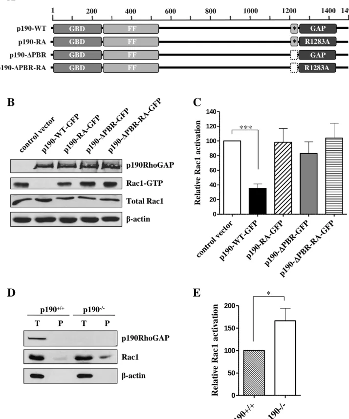
Figure 1.
p190RhoGAP inhibits Rac small G-protein in RacGTPase activation assay, the RacGAP-activity requires PBR of p190RhoGAP.
(A) Domain structure of wild type (p190-WT) and mutant (p190-RA, p190-∆PBR, p190-∆PBR- RA) p190RhoGAP. GBD = GTP-binding domain, FF = FF domain, GAP = GAP domain, * = PBR (polybasic region, amino acids 1213-1236) (B) COS7 cells transiently expressing either GFP or GFP-tagged wild type or mutant p190RhoGAP constructs were treated with 10 ng/ml EGF for 20 min, lysed and mixed with PAK (GST-PBD) bound to glutathione-agarose beads to precipitate the active form of Rac1 small GTPase (Rac1-GTP). Bound active Rac1 was resolved on SDS-PAGE and detected by immunoblotting with anti Rac1 antibody as well as total Rac1.
p190RhoGAP expression was analyzed by immunoblot with anti-p190A antibody, β-actin is shown as loading control. (C) Quantitation of Rac1-activation (Rac1-GTP/Total Rac1) in COS7 cells. 100% corresponds to Rac1-GTP detected in cells transfected with control vector. Values are means +SEM (n=11,11,5,10,3), ***p<0.001. (D) Rac GTPase activation assay in p190RhoGAP +/+ (p190+/+) and p190RhoGAP -/- (p190-/-) mouse embryonal fibroblasts (MEFs). T
= total Rac1 in cell lysates, P = precipitate of active Rac1 (Rac1-GTP). Presence of p190RhoGAP and β-actin as loading control were detected in cell lysates (T) by the antibodies described above. (E) Quantification of Rac1-activation in MEFs. 100% corresponds to Rac1- GTP detected in p190RhoGAP +/+ cells. Values are means +SEM (n=5)
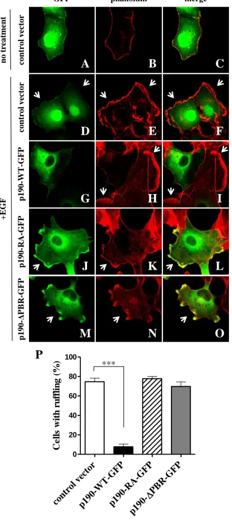
Figure 2.
p190RhoGAP inhibits EGF induced membrane ruffling, which requires GAP activity and intact PBR.
(Panel A-O) COS7 cells were transfected with control vector, GFP-tagged wild type or mutant p190RhoGAP (green). Actin was stained with Alexa-568-labelled phalloidin (red). Ruffling is indicated by arrows. Stimulation was carried out with 10 ng/ml EGF for 20 min. (P) Quantitation of EGF-induced ruffling in COS7 cells. Percentage of ruffled cells from hundred transfected cells was calculated.Mean +SEM of n=7 (control vector, p190-WT-GFP, p190-∆PBR-GFP) or n=6 (p190-RA-GFP) experiments are shown. ***p<0.001.
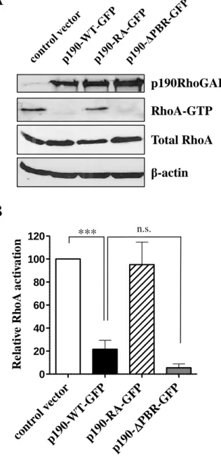
Figure 3.
Effect of WT and mutant p190RhoGAP on the Rho activity in RhoGTPase activation assay.
(A) Level of RhoA-GTP was analysed by RhoGTPase activation assay in transfected COS7 cells treated with 5 µM LPA for 5 min. The precipitates, and the control lysates were separated by SDS-PAGE and analyzed by Western blotting. Active RhoA (RhoA-GTP) and total RhoA were detected by immunoblotting with anti RhoA antibody, p190RhoGAP expression was analyzed by immunoblot with anti-p190A antibody, β-actin was used as loading control. (B) Quantitation of
RhoA-activation (RhoA-GTP/Total RhoA). 100% corresponds to RhoA-GTP detected in cells transfected with control vector. Values are means +SEM (n=3,4,3,5), ***p<0.001.
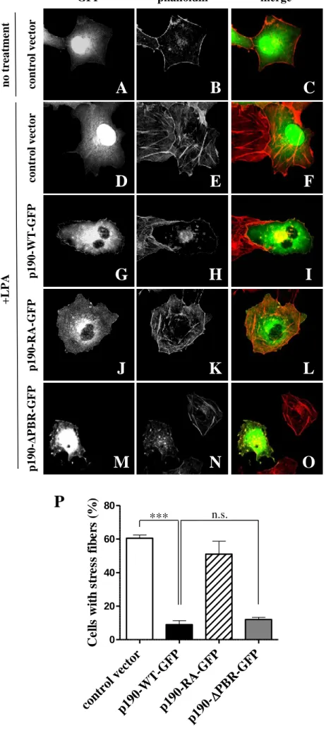
Figure 4.
Inhibition of LPA-induced stress fiber formation by p190RhoGAP in COS7 cells.
(Panel A-O) COS7 cells were transfected with control vector, wild type or mutant p190RhoGAP (green). Actin was stained with Alexa-568-labelled phalloidin (red). Stimulation was carried out with 5 µM LPA for 5 min. (P) Quantitation of LPA-induced stress fiber formation. Percentage of cells with stress fibers from hundred transfected cells was calculated. Mean +SEM of n=6 (control vector, p190-WT-GFP, p190-RA-GFP) or n=4 (p190-∆PBR-GFP) experiments are shown. ***p<0.001.
Figure 5.
p190RhoGAP overexpression results in the multinucleation in COS7 cells. (Panel A-L) Multinucleation in control cells, in a p190-WT-GFP, p190-RA-GFP or p190-∆PBR-GFP transfected cells (green). Nuclei were stained blue with DAPI. (M) Quantitation of the percentage of multinucleation (nuclei ≥2) in 100 transfected cells. Values are means +SEM (n=5,8,5,8), *p<0.05, ***p<0.001.
/+
p190- /-
0 50 100 150 200
Relative Rac1 activation
con
trol vector p190-
WT- GFP
p190- RA-
GFP
PBR-GFP
p190- PBR-RA-GFP
p190-
0 20 40 60 80 100 120 140
Relative Rac1 activation
Figure 1.
A
B C
***
1 200 400 600 800 1000 1200 1400 1499
GBD FF GAP
p190-WT
GBD FF GAP
p190-ΔPBR
GBD FF R1283A
p190-RA
GBD FF R1283A
p190-ΔPBR-RA
Total Rac1 Rac1-GTP
β-actin
*
*
D E
p190RhoGAP
p190RhoGAP
β-actin Rac1 p190+/+ p190-/-
T P T P
*
ol vectorWT -GFP
RA -GFP
PBR- GFP
0 20 40 60 80 100
Cells with ruffling (%)
Figure 2.
P
p190-ΔPBR-GFPp190-WT-GFPp190-RA-GFPcontrol vector
phalloidin merge
***
D E F
G H I
J K L
M N O
GFP
control vector
A B C
no treatment+EGF
con
trol vector p190-
WT -GFP
p190- RA
-GFP PBR-
GFP
p190- 0
20 40 60 80 100 120
Relative RhoA activation
Figure 3.
Total RhoA RhoA-GTP
β-actin
A
B
***
p190RhoGAP
n.s.
Figure 4.
P
ol vectorWT -GFP
RA -GFP
PBR- GFP
0 20 40 60 80
Cells with stress fibers (%)
*** n.s.
GFP phalloidin merge
p190-WT-GFPp190-RA-GFPp190-ΔPBR-GFP
D E F
G H I
J K L
M N O
control vectorcontrol vector
no treatment+LPA
A B C
con
trol vector p190-
WT -GFP
p190- RA
-GFP PBR-
GFP
0 10 20 30
Multinucleation (%)
Figure 5.
control vector
M
***DAPI merge
p190-ΔPBR-GFPp190-WT-GFP
A B C
p190-RA-GFP
J K L
D E F
G H I
GFP
*