Hypervulnerability to sound-exposure through impaired adaptive proliferation of peroxisomes
Sedigheh Delmaghani1,2,3, Jean Defourny1,2,3, Asadollah Aghaie2,3,4, Maryline Beurg5, Didier Dulon5, Nicolas Thelen6, Isabelle Perfettini1,2,3, Tibor Zelles7,8, Mate Aller7, Anaïs Meyer1,2,3, Alice Emptoz1,2,3, Fabrice Giraudet9,10,11, Michel Leibovici12, Sylvie Dartevelle13, Marc Thiry6, E. Sylvester Vizi7, Saaid Safieddine1,2,3, Jean-Pierre Hardelin1,2,3, Paul Avan9,10,11,15 and Christine Petit1,2,3,4,14,15,*
1Unité de Génétique et Physiologie de l’Audition, Institut Pasteur, 75015 Paris, France
2UMRS 1120, Institut National de la Santé et de la Recherche Médicale (INSERM), 75015 Paris, France
3Sorbonne Universités, UPMC Université Paris 06, Complexité du Vivant, 75005 Paris, France
4Syndrome de Usher et Autres Atteintes Rétino-Cochléaires, Institut de la Vision, 75012 Paris, France
5Equipe Neurophysiologie de la Synapse Auditive, Université Victor Segalen and Bordeaux Neurosciences Institute, CHU Pellegrin, 33076 Bordeaux, France
6Unit of Cell and Tissue Biology, GIGA-Neurosciences, University of Liege, CHU Sart- Tilman, B36, 4000 Liege, Belgium
7Institute of Experimental Medicine, Hungarian Academy of Sciences, Budapest, Hungary
8Department of Pharmacology and Pharmacotherapy, Semmelweis University, Budapest, Hungary
9Laboratoire de Biophysique Sensorielle, Université d’Auvergne, 63000 Clermont-Ferrand, France
10UMR 1107, Institut National de la Santé et de la Recherche Médicale (INSERM), Clermont-Ferrand, France
11Centre Jean Perrin, 63000 Clermont-Ferrand, France
12UMR 8104, Institut Cochin, 75014 Paris, France
13Plateforme d’Ingénierie des Anticorps, Institut Pasteur, 75015 Paris, France
14Collège de France, 75005 Paris, France
1 15Co-senior author 2
3
*Correspondence: christine.petit@pasteur.fr 4
5
SUMMARY : 6
A deficiency of pejvakin, a protein of unknown function, causes a strikingly 7
heterogeneous form of deafness. Pejvakin-deficient (Pjvk-/-) mice also exhibited variable 8
phenotypes. Correlation between their hearing thresholds and the number of pups per 9
cage suggested a possible harmful effect of pup vocalizations. Direct sound or electrical 10
stimulation showed that the cochlear sensory hair cells and auditory pathway neurons 11
of Pjvk-/- mice and patients were exceptionally vulnerable to sound. Pjvk-/- cochleas 12
displayed features of marked oxidative stress and impaired anti-oxidant defenses. We 13
showed that pejvakin is associated with peroxisomes, and is required for the oxidative 14
stress-induced proliferation of these organelles. In Pjvk-/- hair cells, peroxisomes 15
displayed structural abnormalities after the onset of hearing. Noise-exposure of wild- 16
type mice rapidly upregulated Pjvk cochlear transcription, and triggered peroxisome 17
proliferation in hair cells and primary auditory neurons. Our results reveal that the 18
anti-oxidant activity of peroxisomes protects the auditory system against noise-induced 19
damage.
20 21
HIGHLIGHTS 22
23
Pejvakin-deficient mice and humans are hypervulnerable to sound exposure.
24 25
Oxidative stress induces a pejvakin-dependent proliferation of peroxisomes.
26
Peroxisome proliferation contributes to the physiological response to sound exposure.
27
Pjvk gene transfer can rescue Pjvk-/- mice from auditory dysfunction.
28 29
INTRODUCTION 1
Mutations of PJVK, which encodes pejvakin, a protein of unknown function present 2
only in vertebrates, cause the DFNB59 recessive form of sensorineural hearing impairment.
3
In the first patients described (Delmaghani et al., 2006), the impairment was restricted to 4
neurons of the auditory pathway, as demonstrated by the combination of abnormal auditory 5
brainstem responses (ABRs) with decreased wave amplitudes and increased inter-wave 6
latencies (Starr & Rance, 2015). ABRs monitor the electrical response of auditory pathways 7
to brief sound stimuli, from the primary auditory neurons synapsing with the sensory cells of 8
the cochlea, the inner hair cells (IHCs), to the colliculus in the midbrain (Møller & Jannetta, 9
1983). However, some DFNB59 patients were found to have a cochlear dysfunction, as 10
shown by an absence of the otoacoustic emissions (OAEs) that are produced by the outer 11
hair cells (OHCs), frequency-tuned cells endowed with electromotility that mechanically 12
amplify the sound-stimulation of neighboring IHCs (Ashmore, 2008). These patients had 13
truncating mutations of PJVK, whereas the previously identified patients had missense 14
mutations (p.T54I or p.R183W) (Ebermann et al., 2007; Schwander et al., 2007; Borck et 15
al., 2011). However, the identification of patients also carrying the p.R183W missense 16
mutation but lacking OAEs (Collin et al., 2007) refuted any straightforward connection 17
between the nature of the PJVK mutation and the hearing phenotype. The severity of 18
deafness in DFNB59 patients varies from moderate to profound, and may even be 19
progressive in some patients, suggesting that extrinsic factors may influence the hearing 20
phenotype.
21
We investigated the role of pejvakin, with the aim of determining the origin of the 22
phenotypic variability of the DFNB59 form of deafness. Our study of Pjvk knockout mouse 23
models and of patients revealed an unprecedented hypervulnerability of auditory hair cells 24
and neurons to sound-exposure, accounting for phenotypic variability. We found that 25
pejvakin is a peroxisome-associated protein involved in the oxidative stress-induced 1
proliferation of this organelle. Pejvakin-deficient mice revealed the key role of peroxisomes 2
in the redox homeostasis of the auditory system and in the protection against noise-induced 3
hearing loss.
4 5
RESULTS 6
Heterogeneity in the hearing sensitivity of Pjvk-/- mice 7
We generated pejvakin-null (Pjvk-/-) mice carrying a deletion of Pjvk exon 2 resulting 8
in a frameshift at codon position 71 (p.Gly71fs*9) (Figure S1 and see Extended 9
Experimental Procedures). ABRs were recorded on postnatal day 30 (P30), to assess hearing 10
sensitivity. ABR thresholds at 10 kHz ranged from 35 to 110 dB SPL in these mice (n = 48), 11
but never exceeded 30 dB SPL in their Pjvk+/+ littermates (n = 26) (Figure 1A). This broad 12
range of hearing sensitivity in Pjvk-/- mice, from near-normal hearing to almost complete 13
deafness, extended across the whole frequency spectrum. The thresholds of distortion- 14
product OAEs (DPOAEs) at 10 kHz (i.e. the minimum stimulus required for DPOAEs 15
production by OHCs) also fell within an abnormally large range of values, from 30 to 75 dB 16
SPL, in 28 Pjvk-/- mice, corresponding to OHC dysfunction, and DPOAEs were undetectable 17
in another 20 Pjvk-/- mice, indicating a complete OHC defect (Figure 1B). The absence of 18
pejvakin in mice thus results in a puzzlingly large degree of hearing phenotype variability.
19 20
Hypervulnerability to the natural acoustic environment in Pjvk-/- mice 21
We investigated the variability of Pjvk-/- auditory phenotypes, by first determining 22
the ABR thresholds of Pjvk-/- littermates from different crosses. Large differences were 23
observed between crosses, with much lesser differences between the Pjvk-/- littermates of 24
individual crosses. Litters with larger numbers of pups (6 to 12) had higher ABR thresholds, 25
suggesting that the natural acoustic environment, with the calls of larger numbers of pups, 1
might be deleterious in Pjvk-/- mice. Pups are vocally very active from birth to about P20.
2
We manipulated the level of exposure to pup calls by randomly splitting large litters of Pjvk- 3
/- pups into groups of 2, 4, 6 and 10 pups per cage, with foster mothers, before the onset of 4
hearing (P10). The ABR thresholds at P21 were significantly correlated with the number of 5
pups raised together (p < 0.001, r2 = 0.51, i.e. 51% of the variation in ABR threshold is 6
determined by the number of pups per cage) (Figure 1C).
7
We then evaluated the effect on hearing of a controlled sound-stimulation, by 8
presenting 1000 tone bursts at 10 kHz, 105 dB SPL (2-ms plateau stimulations separated by 9
60-ms intervals of silence), energetically equivalent to a 3-minute stay in the natural 10
environment of a 12-pup litter, while monitoring the ABRs during sound-exposure. These 11
conditions are referred to hereafter as "controlled sound-exposure". We probed the effect of 12
sound exposure by ABR tests which, limited to 50 repetitions of tone bursts, did not 13
influence the hearing thresholds of Pjvk-/- mice. In a sample of P30 Pjvk-/- mice with initial 14
ABR threshold elevation below 35 dB SPL, controlled sound-exposure affected ABR 15
thresholds in the 12-20 kHz frequency interval (corresponding to the cochlear zones where 16
hair-cell stimulation was strongest (Cody & Johnstone, 1981)), with an immediate increase 17
of 21.7 ± 10.3 dB (n = 8; p < 0.001), not observed in Pjvk+/+ mice (2.2 ± 2.4 dB, n = 12; p = 18
0.3) (Figure 1D). Pjvk-/- mice transferred to a silent environment after exposure displayed a 19
further increase of 33.7 ± 16.0 dB (n = 8) two days after exposure. The threshold shift 20
decreased to 23.7 ± 18.0 dB at seven days, and disappeared entirely by 14 days. When 21
exposed mice were returned to the box with their littermates, their ABR continued to 22
increase, at a rate of 15 dB per week. Pejvakin deficiency thus results in particularly high 23
levels of vulnerability to low levels of acoustic energy, and the increase in ABR thresholds 24
is reversible but only slowly and in a quiet environment.
25
1
Hair cells and auditory pathway neurons are affected by pejvakin deficiency 2
To identify the cellular targets of the pejvakin deficiency, we specifically probed the 3
function of auditory hair cells and neurons in Pjvk-/-, hair cell-conditional Pjvk knockout 4
(Pjvkfl/flMyo15-cre-/-), and Pjvk+/+ mice, at the age of three weeks, before and after controlled 5
sound-exposure or controlled electrical simulation. The responses of the IHCs to sound- 6
induced vibrations amplified by OHCs trigger action potentials in the distal part of primary 7
auditory neurons, at the origin of ABR wave-I. In Pjvkfl/flMyo15-cre+/- mice, which lack 8
pejvakin only in the hair cells, ABR wave-I amplitude and latency at 105 dB SPL 9
specifically probed IHC function, because IHC responses to such loud sounds are 10
independent of OHC activity (Robles & Ruggero, 2001). The larger wave-I latency (1.58 ms 11
in Pjvkfl/flMyo15-cre+/- mice (n = 20)vs. 1.32 ms in Pjvk+/+ littermates (n = 30); p < 0.001) 12
and lower wave-I amplitude (37% of the amplitude in Pjvk+/+ littermates; p < 0.001) 13
suggested a dysfunction of the IHCs in the absence of pejvakin. Controlled sound exposure 14
induced further decreases in ABR wave-I amplitude in Pjvk-/- and Pjvkfl/flMyo15-cre+/- mice 15
(48% and 55% of pre-exposure amplitude, respectively) with respect to Pjvk+/+ mice (108%;
16
p < 0.001 for both comparisons) (Figure 2A), demonstrating that IHCs lacking pejvakin are 17
hypervulnerable to sound. As shown above, OHCs are also affected by the pejvakin 18
deficiency. Controlled sound-exposure triggered a mean increase in the DPOAE threshold of 19
17.1 ± 6.7 dB in the 12 to 20 kHz frequency interval (p < 0.0001) in Pjvk-/- mice with 20
persistent DPOAEs (n = 8), but had no effect on the DPOAEs of Pjvk+/+ mice (n = 9; p = 21
0.51) (Figure 2B). OHCs lacking pejvakin are, thus, also hypervulnerable to sound.
22
We investigated the effect of the absence of pejvakin on the auditory pathway by 23
comparing electrically evoked brainstem responses (EEBR) in Pjvk-/- and Pjvkfl/flMyo15- 24
cre+/- mice (see Extended Experimental Procedures). The amplitudes of the most distinctive 25
EEBR waves, E II and E IV, did not differ between the two types of mice (e.g., for wave E 1
IV: 2.6 ± 1.8 µV in Pjvk-/- mice (n = 18) and 2.2 ± 1.2 µV in Pjvkfl/flMyo15-cre+/- mice (n = 2
11); t-test, p = 0.13). However, following controlled electrical exposure at 200 impulses / s 3
for 1 minute, as opposed to electric-impulse stimulation with 16 impulses / s for 10 s for pre- 4
and post-exposure EEBR tests, the amplitudes of the E II and E IV EEBR waves were 41%
5
and 47% smaller, respectively, for at least 3 minutes, in Pjvk-/- mice (n = 5; p = 0.02 and p = 6
0.01, respectively), but were unaffected in Pjvkfl/flMyo15-cre+/- mice (n = 10) (Figures 2D, 7
2G-2I). The E II-E IV interwave interval was 0.41 ms longer in Pjvk-/- mice (n = 5) than in 8
Pjvkfl/flMyo15-cre+/- mice (n = 10; t-test; p = 0.003) and controlled electrical exposure 9
extended this interval by a further 0.15 ms in Pjvk-/- mice only (paired t-test, p = 0.001) 10
(Figures 2H and 2I). Likewise, the latency interval between ABR wave I and wave IV (the 11
counterpart of wave E IV) was abnormal in one third of the Pjvk-/- mice tested (n = 12) 12
(Figures 2C and 2E), and was further increased by controlled sound-exposure in all Pjvk-/- 13
mice (n = 12 ears with an ABR threshold < 95 dB SPL, a mean increase of 0.16 ms relative 14
to the pre-exposure value; paired t-test, p < 0.001). By contrast, it remained normal in 15
Pjvkfl/flMyo15-cre+/- mice (n = 10 ears; p = 0.73) (Figures 2C and 2F). We conclude that the
16
absence of pejvakin affects the propagation of action potentials in the auditory pathway after 17
both controlled electrical and sound-exposure, as demonstrated by the reduced amplitude of 18
the E II wave and the increased E II-E IV and ABR I-IV interwave intervals in Pjvk-/- mice.
19
We tested whether these abnormalities were of neuronal or glial origin by performing 20
a rescue experiment in Pjvk-/- mice, using adeno-associated virus 8 (AAV8) vector-mediated 21
transfer of the murine pejvakin cDNA. AAV8 injected into the cochlea transduces the 22
primary auditory neurons and neurons of the cochlear nucleus (Figure S2A). All Pjvk-/- mice 23
(n = 7) injected on P3 and tested on P21 had normal ABR interwave I-IV latencies (Figure 24
2J), and their EEBR wave-E IV amplitude was insensitive to controlled electrical 25
stimulation (1.91 ± 0.97 µV before and 1.87 µV ± 1.07 after stimulation; paired t-test, p = 1
0.59) (Figures 2K and 2L). The absence of pejvakin thus renders auditory pathway neurons 2
hypervulnerable to exposure to mild, short stimuli.
3 4
Hypervulnerability to sound in DFNB59 patients 5
We then investigated whether the hearing of DFNB59 patients was also 6
hypervulnerable to sound-exposure. We tested five patients carrying the p.T54I mutation 7
from the series reported by Delmaghani et al. (2006). Transient-evoked OAEs (TEOAEs) 8
assessing OHC function over a broad range of frequencies were detected for all ears, despite 9
the severe hearing impairment (hearing threshold increasing from 66 dB HL at 250 Hz to 84 10
dB at 8 kHz). Following minimal exposure to impulse stimuli (clicks at 99 dB nHL), ABR 11
waves were clearly identified in response to 250 clicks. When exposure was prolonged to 12
1000 clicks (the standard procedure),wave V, the equivalent of mouse ABR-wave IV, which 13
was initially conspicuous, displayed a marked decrease in amplitude (to 39 ± 30% of its 14
initial amplitude) and an increase in latency (of 0.30 ± 0.15 ms) (Figure 3A, 3C, and 3D). In 15
parallel, the I-V interwave interval increased by 0.30 ± 0.15 ms. Wave-V amplitude and 16
latency recovered fully after 10 minutes of silence (Figure 3B). In control patients with 17
sensorineural hearing impairment of cochlear origin matched for ABR thresholds, similar 18
sound stimulation did not affect ABR wave-V amplitude (105 ± 14% of the initial amplitude 19
after exposure; n = 13 patients) or latency (-0.02 ± 0.07 ms change after exposure) (Figures 20
3C and 3D). Exposure of the DFNB59 patients to 1000 clicks also affected TEOAEs (6.1 ± 21
5.2 dB nHL decrease in amplitude; paired t-test, p = 0.02). Therefore, as in pejvakin- 22
deficient mice, the cochlear and neuronal responses of DFNB59 patients were affected by 23
exposure to low-energy sound.
24 25
Redox status abnormalities and ROS-induced cell damage in the cochlea of Pjvk-/- mice 1
We investigated the impact of pejvakin deficiency on cochlear structure in Pjvk-/- mice, 2
by light microscopy on semithin sections, and electron microscopy. On P15 and P21, both 3
OHCs and IHCs were normal in number and shape. Their hair bundles (the 4
mechanoreceptive structures responding to sound), the ribbon synapses of the IHCs and their 5
primary auditory neurons were unmodified (data not shown). On P30, we observed the loss 6
of a few OHCs (16 %), restricted to the basal region of the cochlea (tuned to high-frequency 7
sounds). From P30 onwards, numerous OHCs and cochlear ganglion neurons disappeared 8
and the sensory epithelium (organ of Corti) progressively degenerated (Figure S3A).
9
We investigated possible changes in gene expression in the organ of Corti (which 10
includes not only hair cells, but also various types of supporting cells) in young Pjvk-/- mice, 11
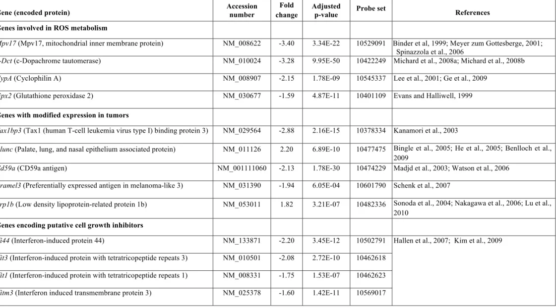
by microarray studies on P15 (see Extended Experimental Procedures). Eighteen genes had 12
expression levels at least 1.5-fold higher or lower in Pjvk-/- mice than in Pjvk+/+ mice.
13
Marked differences were observed for four genes involved in the redox balance — CypA, 14
Gpx2, c-Dct, and Mpv17 — encoding cyclophilin A, glutathione peroxidase 2, c- 15
dopachrome tautomerase, and Mpv17, respectively (Table S1). All these genes were 16
downregulated in Pjvk-/- mice, a result confirmed by quantitative RT-PCR (Figure S4A), and 17
all encode antioxidant proteins, suggesting an impairment of antioxidant defenses in Pjvk-/- 18
mice (Table S1).
19
We thus assessed anti-oxidant defenses in the cochlea of P21 Pjvk-/- mice, by 20
determining the ratio of reduced to oxidized glutathione (GSH:GSSG). The GSSG content 21
was about three times larger than in Pjvk+/+ mice, whereas the GSH content was 23%
22
smaller, resulting in a GSH:GSSG ratio in Pjvk-/- cochleas lower than that in Pjvk+/+ cochleas 23
by a factor of 3.4 (Figure 4A). No significant differences in GSSG and GSH levels or in the 24
GSH:GSSG ratio in the liver were detected between Pjvk+/+ and Pjvk-/- mice (data not 25
shown). Increase in GSSG level and decrease in the GSH:GSSG ratio are markers of the 1
cochlear oxidative stress accompanying the pejvakin deficit.
2
We assessed lipid peroxidation by reactive oxygen species (ROS) in Pjvk-/- mice, by 3
immunofluorescence-based detection of the by-product 4-hydroxy-2-nonenal (4-HNE).
4
Strong immunoreactivity was observed in P60 Pjvk-/- hair cells and cochlear ganglion 5
neurons (Figure S3B). Quantification of lipid peroxidation in microdissected organs of Corti 6
from P30 Pjvk-/- and Pjvk+/+ mice, showed a moderate (16%) but statistically significant 7
increase of the malondialdehyde content in the absence of pejvakin (2.15 ± 0.14 µM in Pjvk- 8
/- vs. 1.84 ± 0.11 µM in Pjvk+/+ mice, means ± SD; t-test, p = 0.04). Thus, pejvakin 9
deficiency led to impaired antioxidant defenses in the cochlea, resulting in ROS-induced cell 10
damage.
11
We then studied electrophysiological features of IHCs and OHCs in the mature 12
cochlea of P19-P21 Pjvk-/- mice. In IHCs, the number of synaptic ribbons, Ca2+ currents, and 13
synaptic exocytosis (assessed by monitoring the increase of the cell capacitance following 14
cell depolarization) were unaffected (Figure S7A). As IHC functions also depend on 15
potassium ion channels known to be affected by ROS, we investigated whether Pjvk-/- mice 16
display the main K+ currents found in mature IHCs, specifically IK,f, which plays a major 17
part in IHC repolarization and is involved in the high temporal precision of action potentials 18
in postsynaptic nerve fibers, IK,s, and IK,n (Oliver et al., 2006). In Pjvk-/-mice, IK,f flowing 19
through the large conductance voltage- and Ca2+-activated potassium (BK) channels, a 20
known target of ROS (Tang et al., 2004), was detected in only 4 out of 11 Pjvk-/- IHCs, and 21
the mean number of spots immunolabeled for the BK α-subunit per IHC was much lower in 22
Pjvk-/- mice (5.0 ± 1.4, n = 283 IHCs from 7 mice) than in Pjvk+/+ mice (13.9 ± 2.6, n = 204 23
IHCs from 9 mice; t-test, p < 0.001). By contrast, the IK,s and IK,n currents were not affected 24
(Figure 4B and Figure S7B). The electromotility of OHCs was also impaired in P19-P21 25
Pjvk-/- mice, as shown by the reduction of the non-linear cell capacitance, the electrical 1
correlate of electromotility (Ashmore, 2008) (Figure S7C). Moreover, the total loss of 2
DPOAE in a large majority of Pjvk-/- mice from P15 on, even at the highest possible stimulus 3
level of 75 dB SPL, pinpointed a major additional defect of the mechanoelectrical 4
transduction in OHCs, the main determinant of DPOAEs at high stimulus levels when OHC 5
electromotility, which amplifies cochlear vibrations at low levels, bears minimal influence 6
on DPOAE production (Avan et al., 2013). Measurements of the cochlear microphonic 7
potential at the round window, a far-field electric potential oscillating at the stimulus 8
frequency that reflects mechanoelectrical transduction currents through OHCs of the basal- 9
most cochlear region, indeed corroborated the DPOAE measurements: this potential, 10
recorded for a 5 kHz sound stimulus at 95 dB SPL, was always larger than 10 µV in Pjvk+/+
11
mice (n = 8), but fell between 3 µV and 5 µV in the P21 Pjvk-/- mice that displayed residual 12
DPOAEs (n = 2), and was below 1 µV, indistinguishable from electric artefacts, in the 13
mutants that had lost their DPOAEs (n = 6). Taken together, these results allowed us to 14
conclude that the impaired antioxidant defenses in the Pjvk-/- cochlea have an impact on 15
various electrophysiological properties of the hair cells, particularly mechanotransduction 16
and K+ current through BK channels.
17
Even though mitochondrial defects are a common cause of ROS overproduction, we 18
didn’t find evidence that mitochondria were damaged, as vulnerability of the mitochondrial 19
membrane potential, ∆ψm, to the uncoupler carbonyl cyanide 4- 20
(trifluoromethoxy)phenylhydrazone (FCCP) in the organ of Corti and cochlear ganglion was 21
similar in P17-P30 Pjvk-/- and Pjvk+/+ mice, and transmission electron microscopy of Pjvk-/- 22
hair cells revealed no mitochondrial abnormalities (Figure S7D and data not shown).
23 24
Pejvakin is a peroxisome-associated protein 25
By using Pjvk-/- cochlea as control, we found that neither the commercially available 1
antibodies nor our initial polyclonal antibody (Delmaghani et al., 2006) specifically 2
recognized pejvakin (data not shown). Given the limited divergence of the pejvakin amino- 3
acid sequence among vertebrates (97% sequence identity between mice and humans), we 4
tried to elicit an immune response in Pjvk-/- mice (see Experimental Procedures). The 5
monoclonal antibody obtained, Pjvk-G21, yielded punctate immunostaining throughout the 6
cytoplasm of transfected HeLa cells expressing pejvakin, whereas no such staining was 7
observed in non-transfected cells. Double staining for peroxisome membrane protein 70 8
(PMP70) demonstrated the colocalization of pejvakin and peroxisomes (Figure S5A). In the 9
human HepG2 hepatoblastoma cell line, which is particularly rich in peroxisomes, strong 10
immunolabeling for endogenous pejvakin was associated with the peroxisomes (Figure 5A).
11
Finally, the specificity of the Pjvk-G21 antibody was demonstrated by the immunoreactivity 12
of peroxisomes in the hair cells of Pjvk+/+, but not Pjvk-/- mice (Figure 5B and S5B).
13
Peroxisomal matrix proteins are imported into the peroxisome by interaction with 14
peroxin 5 or peroxin 7, via the PTS1 or PTS2 targeting signals (Smith and Aitchison, 2013).
15
Prediction programs identified no PTS1 or PTS2 motifs in the pejvakin sequence (Mizuno 16
et al., 2008), suggesting that pejvakin is a peroxisomal membrane or membrane-associated 17
protein.
18 19
Ultrastructural abnormalities of peroxisomes in the hair cells of Pjvk-/- mice 20
We investigated the distribution and morphology of peroxisomes in the Pjvk-/- 21
cochlea by transmission electron microscopy. Peroxisomes were identified on the basis of 22
catalase activity detection using 3,3'-diaminobenzidine as substrate. We focused on OHCs, 23
the first to display a dysfunction in the mutant mice. On P30 but not P15, both the 24
distribution and shape of peroxisomes differed between Pjvk-/- and Pjvk+/+ mice (Figure 5E).
25
In Pjvk+/+ mice, the peroxisomes were restricted to an area immediately below the cuticular 1
plate, a meshwork of actin filaments under the apical surface of the hair cells. In Pjvk-/- mice, 2
the peroxisomes located just below the cuticular plate were slightly larger than those in 3
Pjvk+/+ mice. Strikingly, irregular catalase-containing structures, some of which were 4
juxtaposed, were present in the perinuclear region, at the immediate vicinity of the nuclear 5
membrane, of all Pjvk-/- OHCs, but not of Pjvk+/+ OHCs, which never contained any 6
peroxisome either at this emplacement (Figure 5E). The lack of pejvakin thus results in 7
peroxisome abnormalities in cochlear OHCs after the onset of hearing.
8 9
Pejvakin is involved in oxidative stress-induced peroxisome proliferation 10
In HepG2 cells, protrusions emerging from some peroxisomes, the first step of 11
peroxisome biogenesis from pre-existing peroxisomes, were immunoreactive for pejvakin.
12
String-of-beads structures corresponding to elongated and constricted peroxisomes, 13
preceding final fission (Smith and Aitchison, 2013), were also pejvakin-immunoreactive, 14
suggesting a role of this protein in peroxisome proliferation (Figure S5C). Peroxisomes 15
actively contribute to cellular redox balance, by producing and scavenging/degrading H2O2
16
through a broad spectrum of oxidases and peroxidases (especially catalase), respectively 17
(Schrader and Fahimi, 2006). Because Pjvk-/- mice displayed features of marked oxidative 18
stress in the cochlea, we investigated the possible role of pejvakin in peroxisome 19
proliferation in response to oxidative stress induced by H2O2, which upregulates the 20
expression of peroxisome biogenesis genes (Lopez-Huertas et al., 2000). Embryonic 21
fibroblasts derived from Pjvk+/+ and Pjvk-/- mice were exposed to 0.5 mM H2O2 for 4 hours, 22
and cells were analyzed after 18 hours of incubation in H2O2-free culture medium (see 23
Experimental Procedures). In unexposed cells, the number of peroxisomes was similar 24
between the two genotypes (t-test, p = 0.82). After H2O2 treatment, the number of 25
peroxisomes was increased by 46% in Pjvk+/+ fibroblasts (p = 0.004) but remained 1
unchanged in Pjvk-/- fibroblasts (p = 0.83), resulting in a statistically significant difference 2
between the two genotypes (p < 0.001) (Figures 5C and S6A).
3
We then asked whether mutations reported in DFNB9 patients also affect peroxisome 4
proliferation. To this purpose, we assessed the number of peroxisomes in transfected HeLa 5
cells producing EGFP alone, EGFP and murine pejvakin, or EGFP and one of the mutated 6
forms of murine pejvakin carrying the mutations responsible for DFNB59 (p.T54I, 7
p.R183W, p.C343S, or p.V330Lfs*7). Cells producing the non-mutated pejvakin had larger 8
numbers of peroxisomes than cells producing EGFP alone, whereas cells producing any of 9
the mutated forms of pejvakin (mutPjvk-IRES-EGFP) had smaller peroxisome numbers. In 10
addition, many of these cells contained enlarged peroxisomes, a feature typical of 11
peroxisome proliferation disorders (Ebberink et al., 2012) (Figure 5D and S6B). Together, 12
these results strongly suggest that pejvakin is directly involved in the production of new 13
peroxisomes from pre-existing peroxisomes.
14 15
Upregulation of Pjvk cochlear transcription and peroxisome proliferation in response 16
to sound 17
We then asked whether pejvakin is involved in the physiological response to sound.
18
We first assessed the transcription of Pjvk, and of CypA, Gpx2, c-Dct, and Mpv17 that were 19
downregulated in Pjvk-/- mice, in microdissected organs of Corti from P21 wild-type mice, 20
with or without prior sound-stimulation (5-20 kHz, 105 dB SPL for 1 hour; see Extended 21
Experimental Procedures). Transcript levels were analyzed by quantitative RT-PCR at 22
various times (1, 3, 6, and 18 hours) after sound-exposure (Figure 6A). Pjvk transcript levels 23
had increased by factors of 1.9 ± 0.1 and 3.5 ± 0.7 after 1 hour and 6 hours, respectively.
24
CypA, c-Dct and Mpv17 were also upregulated after 6 hours (by factors of 6.6 ± 1.2, 4.3 ± 25
0.6 and 1.5 ± 0.1, respectively), as were c-Fos and Hsp70, used as a positive control, but 1
Gpx2 was not. Thus, noise-exposure elicits an upregulation of the transcription of Pjvk and 2
of genes downregulated in Pjvk-/- mice and this effect is dependent on acoustic energy level 3
(Figure S4B).
4
Based on this result, we predicted that sound-exposure would lead to peroxisome 5
proliferation in the auditory system of wild-type mice. Six hours after exposure (5-20 kHz, 6
105 dB SPL for 1 hour), the numbers of peroxisomes were unchanged (34.5 ± 0.8 and 35.9 7
± 1.0 per IHC from unexposed and sound-exposed mice, respectively, n = 75 cells from 6 8
mice, respectively; t-test, p = 0.25). However, at 48 hours, they had markedly increased, by 9
a factor of 2.3, in both IHCs and OHCs (84.7 ± 5.0 per IHC and 16.5 ± 1.0 per OHC, n = 90 10
cells and n = 150 cells from 6 mice, respectively) compared to unexposed mice (36.8 ± 3.0 11
per IHC and 7.3 ± 0.4 per OHC, n = 90 cells and n = 150 cells from 6 mice, respectively; t- 12
test, p < 0.0001 for both comparisons). The number of peroxisomes had also increased, by 13
35%, in the dendrites of primary auditory neurons (1.7 ± 0.1 and 2.3 ± 0.2 peroxisomes per 14
µm of neurite length, n = 40 neurites from 5 unexposed and 5 sound-exposed Pjvk+/+mice,
15
respectively; t-test, p = 0.003).
16 17
Therapeutic approaches in Pjvk-/- mice 18
We used the classical anti-oxidant drug N-acetyl cysteine (either alone or associated 19
with α-lipoic acid and α-tocopherol; see Experimental procedures), which was administered 20
to Pjvk-/- pups. The ABR thresholds of P21 Pjvk-/- pups treated with N-acetyl cysteine alone 21
(n = 21) were about 10 dB lower than those of untreated Pjvk-/- pups (n = 24) for all 22
frequencies tested (10, 15, 20, and 32 kHz; t-test, p < 0.001 for all comparisons) (Figure 23
7A). N-acetyl cysteine principally affected the amplitude of the ABR wave I elicited at 105 24
dB SPL, which was similar to that in age-matched Pjvk+/+ mice and greater than that in 25
untreated Pjvk-/- mice (4.35 ± 1.16 µV, n = 21; 4.36 ± 1.15 µV, n = 18; and 1.88 ± 1.07 µV, n 1
= 24, respectively; ANOVA, p < 0.001) (Figure 7B). EEBRs were more resistant to the 2
high-rate electrical stimulation in treated than in untreated mutant mice (Figure 7C).
3
Conversely, N-acetyl cysteine had no beneficial effect on OHCs (data not shown). The 4
association of N-acetyl cysteine with α-lipoic acid and α-tocopherol did not perform any 5
better (data not shown).
6
Full recovery of the neuronal phenotype was achieved by the intracochlear injection 7
of AAV8-mediated pejvakin cDNA (see above). This viral vector does not transduce hair 8
cells, so OHCs remained non-functional (no detectable DPOAE, n = 9), and ABR wave-I 9
amplitude, which is controlled by IHC function, did not differ significantly between Pjvk-/- 10
mice receiving AAV8 containing the pejvakin cDNA (3.3 ± 1.8 µV, n = 9), the EGFP cDNA 11
only (3.2 ± 1.3 µV, n = 5), or with no injection (2.7 ± 1.4 µV, n = 11) (ANOVA, p = 0.54).
12
We thus investigated whether AAV2/8, which transduces hair cells only (Figure S2B), could 13
rescue the hair-cell phenotype in Pjvk-/- mice. The auditory function of Pjvk-/- mice (n = 7, 14
four pups per cage in every experiment) receiving intracochlear injections of AAV2/8- 15
pejvakin cDNA (Pjvk-IRES-EGFP) on P3 was assessed on P21, and the percentage of 16
transduced IHCs and OHCs was evaluated in each injected and contralateral cochlea, on the 17
basis of EGFP fluorescence. Improvements in ABR thresholds of 20 to 30 dB SPL with 18
respect to untreated mice were observed for frequencies between 10 and 20 kHz (t-test, p <
19
0.001 for all comparisons; Figure 7D). With the same vector expressing the EGFP cDNA 20
only (n = 6), ABR thresholds were similar to those of untreated Pjvk-/- mice (n = 10) (81.6 21
vs. 78.6 dB SPL for 10 kHz, p = 0.64). A partial reversion of the OHC dysfunction was 22
obtained, with detectable DPOAEs in pejvakin cDNA-treated cochleas (threshold 54.0 ± 23
10.7 dB), but not in contralateral, untreated cochleas (Figure 7F). DPOAE thresholds were 24
linearly correlated (r2 = 0.74, p < 0.001) with the number of EGFP-tagged OHCs (Figure 25
7G), suggesting that the normalization of DPOAE thresholds may be possible if all OHCs 1
could be transduced. The latency of the ABR wave I in response to a 105 dB SPL 2
stimulation decreased significantly (1.38 ± 0.11 ms vs. 1.53 ± 0.10 ms; t-test, p = 0.026) 3
(Figure 7E), and its amplitude increased into the normal range (7.34 ± 0.80 µV for the 4
treated ears, n = 6, vs. 2.93 ± 0.92 µV for the contralateral, untreated ears; paired t-test, p <
5
0.001) (Figure 7H), in relation to the number of EGFP-tagged IHCs (r2 = 0.89 for wave I 6
amplitude, p < 0.001; Figure 7I). No correction of the interwave I-IV latency was observed, 7
as expected (data not shown).
8
Finally, we investigated the effect of the transduction of Pjvk-/- IHCs by AAV2/8- 9
pejvakin cDNA on their peroxisomes. Before sound-exposure, the numbers of peroxisomes 10
in IHCs of Pjvk-/- and AAV2/8-Pjvk Pjvk-/- mice did not differ from that of Pjvk+/+ mice 11
(30.5 ± 1.9, 32.3 ± 2.1, and 36.8 ± 3.0 peroxisomes per IHC, n = 60 cells from 4 Pjvk-/- and 12
4 AAV2/8-Pjvk Pjvk-/- mice, and n = 90 cells from 6 Pjvk-+/+ mice, respectively; respective t- 13
test, p = 0.11 and p = 0.30). By contrast, 48 hours after sound-exposure (5-20 kHz) at 105 14
dB SPL for 1 hour, the number of peroxisomes had decreased by 63% in Pjvk-/- IHCs (30.5 ± 15
1.9 and 11.2 ± 1.3 peroxisomes per IHC, n = 75 cells from 5 unexposed and 5 sound- 16
exposed Pjvk-/- mice, respectively; t-test, p < 0.0001), and enlarged PMP70-labeled 17
structures were present close to the nucleus (Figure 7J). In response to the same sound but of 18
a lower intensity, i.e. 97 dB SPL for 1 hour, the number of peroxisomes was unchanged in 19
Pjvk-/- IHCs (30.5 ± 1.9 and 34.6 ± 2.3 peroxisomes per IHC, n = 60 cells from 4 unexposed 20
and 4 sound-exposed Pjvk-/- mice, respectively; t-test, p = 0.17), and no enlarged PMP70- 21
stained structures were detected (data not shown). Thus, depending on the acoustic energy of 22
the sound stimulation, in the absence of pejvakin, peroxisomes failed to proliferate in IHCs 23
(both at 105 dB SPL and 97 dB SPL), and even degenerated (at 105 dB SPL). In AAV2/8- 24
Pjvk injected Pjvk-/- mice exposed to 105 dB SPL for 1 hour, enlarged PMP70-labeled 25
structures were no longer detected in transduced IHCs and the number of peroxisomes 1
increased by 35% (32.3 ± 2.1 and 43.7 ± 3.0 peroxisomes per IHC, n = 60 cells from 2
unexposed and exposed transduced Pjvk-/- IHCs, respectively, t-test, p = 0.002) (Figure 7J).
3
We conclude that pejvakin re-expression fully protects Pjvk-/- IHCs from peroxisome 4
degenerescence and partially restores their impaired adaptive proliferation.
5 6
DISCUSSION 7
Noise overexposure, a major threat to hearing, affects 15% of people between the 8
ages of 20 and 69 years. There are currently no efficient methods for curing noise-induced 9
hearing loss (NIHL), and we still know little about the underlying pathogenic processes. We 10
describe here a genetic form of NIHL, by showing that pejvakin deficiency in mice and 11
DFNB59 patients leads to hypervulnerability to sound, due to a peroxisomal deficiency.
12
This is the first reported peroxisomal cause of an isolated (non-syndromic) form of inherited 13
deafness. The peroxisome emerges as a key organelle in the redox homeostasis of the 14
auditory system, for coping with the overproduction of ROS induced by high levels of 15
acoustic energy.
16
Acoustic energy is the main determinant of NIHL. The LEX,8 hour (for level of 17
exposure over an 8-hour workshift) index has been defined such that an LEX,8 hour of X dB 18
delivers the same energy as a stable sound of X dB played over a period of eight hours.
19
Chronic occupational exposures to less than 85 dB (or 80 dB, depending on the country) are 20
deemed safe. In Pjvk-/- mice, a single exposure to 63 dB LEX,8 hour increased hearing 21
thresholds by 30 dB, with full recovery occurring after about two weeks. By contrast, a ten- 22
times more energetic exposure to a LEX,8 hour of 73 dB in wild-type mice of the same strain 23
produces only an 18 dB shift in threshold, with a recovery time of 12 hours (Housley et al., 24
2013). This hypersensitivity of Pjvk-/- mice to noise suggests that the LEX,8 hour, of about 83 25
dB for a cage of 10 pups, is sufficient to account for permanent hearing loss in these Pjvk-/- 1
pups, whilst some of those housed in small numbers in quiet rooms can display near-normal 2
hearing thresholds (see Figure 1C). Likewise, the auditory function of DFNB59 patients was 3
transiently affected by a 57 dB LEX,8 hour exposure, routinely used in ABR tests.
4
NIHL involves the excessive production of ROS, overwhelming the anti-oxidant defense 5
system and causing irreversible oxidative damage to DNA, proteins, and lipids within the 6
cell (Henderson et al., 2006). Noise-induced oxidative stress results in the production of 7
H2O2 and other ROS as by-products, thought to derive from the intense sollicitation of 8
mitochondrial activity. Several mouse mutants with mitochondrial defects are prone to 9
NIHL (Ohlemiller et al., 1999), including mice lacking sirtuin 3, a mitochondrial NAD+- 10
dependent deacetylase (Brown et al., 2014). Our studies of pejvakin-deficient mouse 11
mutants and rescue experiments targeting the hair cells and auditory neurons unambiguously 12
show that IHCs, OHCs, primary auditory neurons and neurons of the cochlear nucleus are 13
hypervulnerable to sound in the absence of pejvakin, which is consistent with previous 14
results showing that hair cells and neurons of the auditory system are targets of NIHL 15
(Wang et al., 2002; Kujawa & Liberman, 2009; Imig and Durham, 2005). However, our 16
study goes one step further, by implicating a possible common mechanism: peroxisomal 17
failure, the importance of which is demonstrated by the impairment of the redox 18
homeostasis caused by pejvakin deficiency. It also reveals a major cause of the unusually 19
high level of phenotypic variability observed in pejvakin-deficient mice and humans: the 20
difference in sound-exposure and the inability of the defective peroxisomes to cope with the 21
resulting activity-dependent oxidative stress. Incidentally, this can account for the apparent 22
paradox that mice carrying the R183W mutation in pejvakin displayed a much more severe 23
neural pathway defect than the Pjvk-/- mice (Delmaghani et al., 2006). Due to the 24
preservation of hair cell functions, the auditory neurons of R183W mutant mice should be 25
strongly stimulated, whereas the early permanent damage to cochlear hair cells in Pjvk-/- 1
mice acts as a protective "muffler" of the neuronal pathway.
2
In mammals, the number and metabolic functions of peroxisomes differ between cell 3
types. However, all cell types are able to adapt rapidly to modifications in physiological 4
conditions by changing the number, shape, size, and molecular content of peroxisomes, 5
resulting in considerable functional plasticity of these organelles (Schrader et al., 2012;
6
Smith and Aitchison, 2013). Our experiments on Pjvk-/- and Pjvk+/+ mouse embryonic 7
fibroblasts stressed with H2O2 showed that pejvakin is critically involved in the oxidative 8
stress-induced proliferation of peroxisomes through growth and fission of pre-existing 9
peroxisomes. Although the molecular machinery underlying this adaptive process is still 10
poorly understood, the specific involvement of Pex11α that recruits COP I has been reported 11
(Li et al., 2002; Passreiter et al., 1998). Of note, the absence of pejvakin only affects the 12
proliferation of peroxisomes from pre-existing peroxisomes, but not the constitutive 13
biogenesis of this organelle. Accordingly, structural abnormalities of peroxisomes in Pjvk-/- 14
mice became apparent only after hearing onset, in the context of the oxidative stress 15
produced by noise-exposure. By contrast, the PEX gene defects causing Zellweger syndrome 16
spectrum (ZSS) disorders (Waterham & Ebberink, 2012) and rhizomelic chondrodysplasia 17
punctata affect the constitutive biogenesis of peroxisomes. Hearing impairment in ZSS 18
disorders involves a severe impairment of neuronal conduction, and has been attributed to 19
defects in the synthesis of two essential myelin sheath components — plasmalogens and 20
docosahexaenoic acid —, which is critically dependent on peroxisomes. Our results suggest 21
that ZSS also includes a defective redox balance in the hair cells and neurons of the auditory 22
system.
23
In the context of noise-exposure, the upregulation of Pjvk transcription in the cochlea 24
and the subsequent peroxisome proliferation in the hair cells and auditory neurons of wild- 25
type mice suggest that pejvakin-dependent peroxisome proliferation in the auditory system 1
is part of the physiological response to high levels of acoustic energy that result in increased 2
amounts of ROS. This and the marked oxidative stress detected in the Pjvk-/- cochlea imply 3
that the proliferation of peroxisomes plays an anti-oxidant role, similar to that reported in 4
other cell types (Santos et al., 2005; Diano et al., 2011). The rapid elevation of the hearing 5
threshold in Pjvk-/- mice in response to low-energy sounds and the increase in interwave I-IV 6
latency observed in DFNB59 patients within a few seconds are consistent with an activity- 7
dependent H2O2 production that, due to impaired cellular redox homeostasis, results in 8
concentrations of H2O2 high enough to impact on the activity of various target proteins 9
including ion channels and transporters (Rice, 2011). The worsening of hearing sensitivity, 10
two days later, in the mutant mice lacking pejvakin, exacerbated by putting back the mice in 11
a noisy environment, fits the picture of the absence of sound-induced biogenesis of 12
peroxisomes (with their degeneration occurring in a high acoustic energy environment). We 13
thus conclude that the hypervulnerability of Pjvk-/- mice and DFNB59 patients to sound does 14
not result simply from an exacerbation, by sound, of a pre-existing redox balance defect, but 15
is the consequence of the impaired adaptive proliferation of peroxisomes in the absence of 16
pejvakin. Both the defective peroxisome proliferation in IHCs of Pjvk-/- mice in response to 17
sound-exposure and its partial recovery by pejvakin cDNA transfer support this conclusion.
18
A full recovery of the adaptive peroxisome proliferation produced by sound-exposure may 19
require higher concentrations of pejvakin or the sound-induced dynamic modulation of Pjvk 20
expression (see Figure 6A), which is missing in our rescue experiments (the expression of 21
the pejvakin cDNA being driven by a constitutive promoter).
22
In patients with hearing impairment, the amplification of sound by hearing aids or 23
direct electrical stimulation of the auditory nerve by a cochlear implant delivers a stimulus 24
with an energy level similar to that shown here to worsen the hearing impairment of Pjvk-/- 25
mice within one minute of sound exposure. Therefore, in cases of peroxisomal deficiency, as 1
in DFNB59, specific protection against redox homeostasis failure is essential, and patients 2
with such conditions should avoid noisy environments. N-acetyl cysteine was the only 3
antioxidant drug tested here to display some, albeit limited, efficacy. By contrast AAV- 4
mediated gene therapy, could potentially provide full protection. Finally, deciphering the 5
sound stress-induced protective signaling pathway involving pejvakin, and possibly 6
cyclophilin A, c-dopachrome tautomerase, and Mpv17, might lead to the discovery of 7
therapeutic agents for NIHL.
8 9
EXPERIMENTAL PROCEDURES 10
11
Audiological studies in mice 12
Auditory tests were performed in an anechoic room, on anesthetized animals for which core 13
temperature was maintained at 37°C (see Extended Experimental Procedures).
14 15
Audiological tests in patients 16
Informed consent was obtained from all the subjects included in the study. Pure-tone 17
audiometry was performed, with air- and bone-transmitted tones. Hearing impairment was 18
assessed objectively, by measuring ABRs and transient-evoked otoacoustic emissions 19
(TEOAEs). The nonlinear TEOAE recording procedure was used (derived from the ILO88 20
system), making it possible to extract TEOAEs from linear reflection artifacts from the 21
middle ear, and to evaluate background noise. TEOAE responses were analyzed in 1 kHz- 22
wide bands centered on 1, 2, 3 and 4 kHz.
23 24
Generation of an anti-pejvakin monoclonal antibody 25
The 3’-end of the coding sequence of the Pjvk cDNA (accession number NM_001080711.2) 1
was inserted into a pGST-parallel-2 vector (derived from pGEX-4T-1; Amersham). The 2
resulting construct, encoding the C-terminal region of pejvakin (residues 290-352; accession 3
number NP_001074180.1) fused to an N-terminal glutathione S-transferase tag, was 4
introduced into E. coli BL21-Gold (DE3) competent cells (Stratagene). The pejvakin protein 5
fragment was purified on a glutathione-Sepharose 4B column, then subjected to size- 6
exclusion chromatography and used as the antigen for immunization. Antibodies were 7
produced by immunizing Pjvk-/- mice. An IgG monoclonal antibody (KD of 6 x 10⁻⁸ M), 8
Pjvk-G21, was selected by ELISA on immunogen-coated plates.
9 10
Determination of total and oxidized glutathione 11
Total glutathione (GSH + GSSG) and oxidized glutathione (GSSG) levels were determined 12
as described by Rahman et al. (2006). Total glutathione and GSSG levels were evaluated by 13
spectrometry at 405 nm. GSH concentration was calculated by subtracting the concentration 14
of GSSG from the total glutathione concentration. Three independent experiments were 15
performed, in P21 Pjvk+/+ and Pjvk-/- mice.
16 17
Statistical analyses 18
Statistical analyses were performed using GraphPad. Data were analyzed by either paired or 19
unpaired Student’s t-tests, and, for more than two groups, we used one-way or two-way 20
ANOVA analyses of variance. Statistical significance is defined as p < 0.05 and indicated 21
by asterisks.
22 23
SUPPLEMENTAL INFORMATION 24
The supplemental information includes Extended Experimental Procedures, seven figures, 1
and one table.
2 3
AUTHOR CONTRIBUTIONS 4
C.P. and P.A. designed study. P.A. and S.D. analyzed auditory tests in patients. P.A., S.D., 5
and F.G. performed audiological tests in mice. S.D. performed recombination experiments 6
on embryonic stem cells to produce knockout mice, and transcriptomic and biochemical 7
studies. A.A. produced recombinant proteins. S.Da. produced the monoclonal antibody. I.P., 8
J.D., and S.D. performed cell transfections, immunohistolabeling experiments. I.P. and S.D.
9
produced mouse embryonic fibroblasts. N.T., M.T., M.L., S.D., and S.S. performed 10
ultrastructural studies. S.D., A.M., and A.E. supervised by S.S., performed rescue 11
experiments in mice. M.B. and D.D. performed electrophysiological experiments in mice.
12
T.Z., M.A., and E.S.V. performed mitochondrial physiological analysis. C.P., P.A., S.D., 13
J.D., and J.-P.H. wrote the manuscript.
14 15
ACKNOWLEDGMENTS 16
We thank M. Aghaie for assistance with the collection of clinical data, M. Mobasheri for 17
audiological tests in the patients, F. Langa-Vives and the Centre d’ingénierie génétique 18
murine platform for producing Pjvk-/- recombinant mice, the Transcriptome et épigénome 19
platform for microarray experiments, P. Aubourg for fruitful discussions, and M. Ricchetti 20
and J. Boutet de Monvel for critical reading of the manuscript. This study was supported by 21
grants from Louis-Jeantet Foundation, ANR – NKTH “HearDeafTreat” 2010-INTB-1402- 22
01, European Research Council (ERC)-Hair Bundle (ERC-2011-ADG-294570), Humanis 23
Novalis-Taitbout, Réunica-Prévoyance, BNP Paribas, and French state program 24
‘‘Investissements d’Avenir’’ (ANR-10-LABX-65) to C.P.
25 26
REFERENCES 27
Ashmore, J. (2008). Cochlear outer hair cell motility. Physiol. Rev. 88, 173-210.
28
Avan, P., Buki, B., and Petit, C. (2013). Auditory distortions: origins and functions.
29
Physiological reviews 93, 1563-1619.
30
Borck, G., Rainshtein, L., Hellman-Aharony, S., Volk, A.E., Friedrich, K., Taub, E., Magal, 1
N., Kanaan, M., Kubisch, C., Shohat, M., et al. (2012). High frequency of autosomal- 2
recessive DFNB59 hearing loss in an isolated Arab population in Israel. Clin. Genet. 82, 3
271-276.
4
Brown, K.D., Maqsood, S., Huang, J.Y., Pan, Y., Harkcom, W., Li, W., Sauve, A., Verdin, 5
E., and Jaffrey, S.R. (2014). Activation of SIRT3 by the NAD(+) precursor nicotinamide 6
riboside protects from noise-induced hearing loss. Cell Metab. 20, 1059-1068.
7
Cody, A.R., and Johnstone, B.M. (1981). Acoustic trauma: single neuron basis for the "half- 8
octave shift". J. Acoust. Soc. Am. 70, 707-711.
9
Collin, R.W., Kalay, E., Oostrik, J., Caylan, R., Wollnik, B., Arslan, S., den Hollander, A.I., 10
Birinci, Y., Lichtner, P., Strom, T.M., et al. (2007). Involvement of DFNB59 mutations in 11
autosomal recessive nonsyndromic hearing impairment. Hum. Mutat. 28, 718-723.
12
Delmaghani, S., del Castillo, F.J., Michel, V., Leibovici, M., Aghaie, A., Ron, U., Van Laer, 13
L., Ben-Tal, N., Van Camp, G., Weil, D., et al. (2006). Mutations in the gene encoding 14
pejvakin, a newly identified protein of the afferent auditory pathway, cause DFNB59 15
auditory neuropathy. Nat. Genet. 38, 770-778.
16
Diano, S., Liu, Z.W., Jeong, J.K., Dietrich, M.O., Ruan, H.B., Kim, E., Suyama, S., Kelly, 17
K., Gyengesi, E., Arbiser, J.L., et al. (2011). Peroxisome proliferation-associated control of 18
reactive oxygen species sets melanocortin tone and feeding in diet-induced obesity. Nat.
19
Med. 17, 1121-1127.
20
Ebberink, M.S., Koster, J., Visser, G., Spronsen, F., Stolte-Dijkstra, I., Smit, G.P., Fock, 21
J.M., Kemp, S., Wanders, R.J., and Waterham, H.R. (2012). A novel defect of peroxisome 22
division due to a homozygous non-sense mutation in the PEX11beta gene. J. Med. Genet.
23
49, 307-313.
24
Ebermann, I., Walger, M., Scholl, H.P., Charbel Issa, P., Luke, C., Nurnberg, G., Lang- 25
Roth, R., Becker, C., Nurnberg, P., and Bolz, H.J. (2007). Truncating mutation of the 26
DFNB59 gene causes cochlear hearing impairment and central vestibular dysfunction. Hum.
27
Mutat. 28, 571-577.
28
Henderson, D., Bielefeld, E.C., Harris, K.C., and Hu, B.H. (2006). The role of oxidative 29
stress in noise-induced hearing loss. Ear Hearing 27, 1-19.
30
Housley, G.D., Morton-Jones, R., Vlajkovic, S.M., Telang, R.S., Paramananthasivam, V., 31
Tadros, S.F., Wong, A.C., Froud, K.E., Cederholm, J.M., Sivakumaran, Y., et al. (2013).
32
ATP-gated ion channels mediate adaptation to elevated sound levels. Proc. Natl. Acad. Sci.
33
Imig, T.J., and Durham, D. (2005). Effect of unilateral noise exposure on the tonotopic 1
distribution of spontaneous activity in the cochlear nucleus and inferior colliculus in the 2
cortically intact and decorticate rat. J. Comp. Neurol. 490, 391-413.
3
Kujawa, S.G., and Liberman, M.C. (2009). Adding insult to injury: cochlear nerve 4
degeneration after "temporary" noise-induced hearing loss. J. Neurosci. 29, 14077-14085.
5
Li, X., Baumgart, E., Dong, G.X., Morrell, J.C., Jimenez-Sanchez, G., Valle, D., Smith, K.D.
6
and Gould, S.J. (2002). PEX11alpha is required for peroxisome proliferation in 7
response to 4-‐phenylbutyrate but is dispensable for peroxisome proliferator-‐activated 8
receptor alpha-‐mediated peroxisome proliferation. Mol. Cell. Biol. 22, 8226-‐8240.
9
Lopez-Huertas, E., Charlton, W.L., Johnson, B., Graham, I.A., and Baker, A. (2000). Stress 10
induces peroxisome biogenesis genes. EMBO J. 19, 6770-6777.
11
Mizuno, Y., Kurochkin, I.V., Herberth, M., Okazaki, Y., and Schonbach, C. (2008).
12
Predicted mouse peroxisome-targeted proteins and their actual subcellular locations. BMC 13
Bioinform. 9 Suppl 12, S16.
14
Møller, A.R., Jannetta, P.J. (1983). Interpretation of brainstem auditory evoked potentials:
15
results from intracranial recordings in humans, Scand Audiol. 12, 125-33.
16
Ohlemiller, K.K., Wright, J.S., and Dugan, L.L. (1999). Early elevation of cochlear reactive 17
oxygen species following noise exposure. Audiol. Neurootol. 4, 229-236.
18
Oliver, D., Taberner, A.M., Thurm, H., Sausbier, M., Arntz, C., Ruth, P., Fakler, B., and 19
Liberman, M.C. (2006). The role of BKCa channels in electrical signal encoding in the 20
mammalian auditory periphery. J. Neurosci. 26, 6181-6189.
21
Passreiter, M., Anton, M., Lay, D., Frank, R., Harter, C., Wieland, F.T., Gorgas, K., and 22
Just, W.W. (1998). Peroxisome biogenesis: involvement of ARF and coatomer. J. Cell Biol.
23
141, 373-383.
24
Rahman, I., Kode, A., and Biswas, S.K. (2006). Assay for quantitative determination of 25
glutathione and glutathione disulfide levels using enzymatic recycling method. Nat. Protoc.
26
1, 3159-3165.
27
Rice, M.E. (2011). H2O2: a dynamic neuromodulator. Neuroscientist 17, 389-406.
28
Robles, L., and Ruggero, M.A. (2001). Mechanics of the mammalian cochlea. Physiol. Rev.
29
81, 1305-1352.
30
Santos, M.J., Quintanilla, R.A., Toro, A., Grandy, R., Dinamarca, M.C., Godoy, J.A., and 31
Inestrosa, N.C. (2005). Peroxisomal proliferation protects from beta-amyloid 32
neurodegeneration. J. Biol. Chem. 280, 41057-41068.
33
Schrader, M., Bonekamp, N.A. and Islinger, M. (2012) Fission and proliferation of 1
peroxisomes. Biochim. Biophys. Acta 1822, 1343-1357.
2
Schrader, M., and Fahimi, H.D. (2006). Peroxisomes and oxidative stress. Biochim.
3
Biophys. Acta 1763, 1755-1766.
4
Schwander, M., Sczaniecka, A., Grillet, N., Bailey, J.S., Avenarius, M., Najmabadi, H., 5
Steffy, B.M., Federe, G.C., Lagler, E.A., Banan, R., et al. (2007) A forward genetics screen 6
in mice identifies recessive deafness traits and reveals that pejvakin is essential for outer hair 7
cell function. J. Neurosci. 27, 2163-2175.
8
Smith, J.J., and Aitchison, J.D. (2013). Peroxisomes take shape. Nat. Rev. Mol. Cell Biol.
9
14, 803-817.
10
Starr, A. & Rance, G. (2015). Auditory neuropathy. Handb Clin Neurol. 129, 495-508.
11
Tang, X.D., Garcia, M.L., Heinemann, S.H., and Hoshi, T. (2004). Reactive oxygen species 12
impair Slo1 BK channel function by altering cysteine-mediated calcium sensing. Nat. Struct.
13
Mol. Biol. 11, 171-178.
14
Wang, Y., Hirose, K., and Liberman, M.C. (2002). Dynamics of noise-induced cellular 15
injury and repair in the mouse cochlea. J. Assoc. Res. Otolaryngol. 3, 248-268.
16
Waterham, H.R., and Ebberink, M.S. (2012). Genetics and molecular basis of human 17
peroxisome biogenesis disorders. Biochim. Biophys. Acta 1822, 1430-1441.
18 19 20 21
FIGURE LEGENDS 22
Figure 1. Hearing loss variability and greater sensitivity to controlled sound-exposure 23
in Pjvk-/- mice. (A) ABR thresholds at 10 kHz in P30 Pjvk+/+ (n = 26 mice) and Pjvk-/- (n = 24
48 mice) littermates. (B) DPOAE thresholds at 10 kHz in P30 Pjvk+/+ (n = 14 mice) and 25
Pjvk-/- (n = 48 mice) littermates. In ears with no DPOAE, even at 75 dB SPL (the highest 26
sound intensity tested), DPOAE thresholds were arbitrarily set at 80 dB SPL. (C) 27
Relationship between the number of pups raised together (determining sound levels in the 28
immediate environment) and ABR thresholds at 10 kHz in P21 Pjvk-/- pups. Inset: Time- 29
frequency analysis of a mouse pup’s vocalization. Pup calls from P0 to P21 form harmonic 30
series of about 5 kHz, with the most energetic harmonic at about 10 kHz. In a 12-pup litter, 31