Finding bioactive compounds in plant extracts by HPLC-coupled assays: novel approaches to natural
product-based drug discovery
Ph.D. Thesis
Árpád Könczöl Semmelweis University
Doctoral School of Pharmaceutical Sciences
Supervisor: Dr. György Tibor Balogh, Ph.D.
Consultant: Dr. Ágnes Kéry, Ph.D.
Official Reviewers: Dr. Huba Kalász, D.Sc.
Dr. Dezső Csupor, Ph.D.
Chair of Exam Committee: Dr. Kornélia Tekes, Ph.D.
Exam Committee: Dr. Éva Lemberkovics, Ph.D.
Dr. József Balla, Ph.D.
Budapest, 2013
Contents
CONTENTS ... 2
ABBREVIATIONS AND SYMBOLS ... 5
1. INTRODUCTION ... 7
1.1. Paradigm and challenges of the current drug discovery ... 7
1.2. Status and relevance of natural products in drug discovery ... 8
1.3. Novel approaches to natural product lead finding: profiling by coupled techniques ... 11
1.4. Review of targets and methods used in the screening and profiling of the plant extract library ... 15
1.4.1. Free radicals, oxidative stress and antioxidants ... 16
1.4.1.1. The DPPH (2,2-diphenyl-1-picrylhydrazyl) method ... 18
1.4.1.2. The peroxynitrite anion (ONOO–): biochemistry and methodologies for measurement of ONOO– scavenging activity ... 21
1.4.2. The blood-brain barrier (BBB) ... 23
1.4.2.1. Methodologies for measurement of blood-brain barrier transport ... 25
1.4.2.2. The parallel artificial membrane permeability assay for blood-brain barrier (PAMPA-BBB) ... 26
1.4.2.3. Rationale of studying natural products and plant extracts by PAMPA- BBB ... 27
2. OBJECTIVES ... 28
3. MATERIALS AND METHODS ... 30
3.1. Chemicals and reference compounds ... 30
3.2. Plant material ... 30
3.2.1. Plant extract library ... 30
3.2.2. Artemisia gmelinii Webb. ex Stechm. (Asteraceae) ... 31
3.2.3. Salvia miltiorrhiza Bunge (Lamiaceae) ... 32
3.2.4. Tanacetum parthenium (L.) Sch. Bip. (Asteraceae) ... 32
3.2.5. Corydalis cava (L.) Schweig. & Kört. (Papaveraceae) ... 33
3.2.6. Salvia officinalis L. (Lamiaceae) ... 33
3.2.7. Vinca major L. (Apocynaceae) ... 34
3.3. Instrumentation ... 34
3.3.1. High performance liquid chromatography – mass spectrometry ... 34
3.3.2. Nuclear magnetic resonance spectroscopy ... 35
3.4. Cytotoxicity screening campaign ... 35
3.4.1. Cell line ... 35
3.4.2. Procedure of screening ... 36
3.4.3. Data handling ... 36
3.5. Antioxidant activity screening campaign ... 37
3.5.1. Procedure of screening ... 37
3.5.2. Data handling and IC50 measurement ... 38
3.6. Antioxidant activity-guided phytochemical investigation of Artemisia gmelinii 39 3.6.1. HPLC-based DPPH scavenging assay ... 39
3.6.2. Isolation of compounds 7a and 8a with preparative HPLC ... 39
3.6.3. High resolution mass spectrometry analysis ... 40
3.6.4. NMR spectroscopy ... 40
3.7. HPLC-based peroxynitrite scavenging assay ... 41
3.7.1. Synthesis of the peroxynitrite anion ... 41
3.7.2. Preparation of the Salvia specific model mixture ... 41
3.7.3. HPLC conditions ... 41
3.7.4. Peroxynitrite scavenging activity measurement in 96-well plate ... 42
3.8. Blood-brain barrier permeability screening campaign and related methods ... 43
3.8.1. PAMPA-BBB procedures ... 43
3.8.2. HPLC-MS analysis ... 44
3.8.3. Gas chromatography – flame ionization detector analysis of PAMPA-BBB co-solvents ... 46
3.8.4. NMR spectroscopy ... 46
3.9. Statistical analysis... 47
4. RESULTS ... 48
4.1. Cytotoxicity screening ... 48
4.2. Antioxidant activity screening ... 49
4.3. HPLC-based antioxidant activity profiling of the methanolic extract of Artemisia gmelinii ... 51
4.3.1. Introduction ... 51
4.3.2. Phytochemical and antioxidant characterization ... 52
4.4. HPLC-based peroxynitrite scavenging activity profiling of Salvia spp. ... 56
4.4.1. Introduction ... 56
4.4.2. Optimization of the chromatographic conditions ... 59
4.4.3. Calibration for pyrogallol red ... 60
4.4.4. Validation of the assay with a Salvia specific model mixture ... 61
4.4.4.1. Comparison of degradation kinetics in mixture to individual scavenging activities ... 63
4.4.4.2. Validation of the assay parameters with structure – activity relationships ... 65
4.4.5. Demonstration of the assay performance on the methanolic extract of Salvia miltiorrhiza ... 65
4.5. Blood-brain barrier (BBB) permeability screening and HPLC-based hit profiling
... 67
4.5.1. Introduction ... 67
4.5.2. Validation of the PAMPA-BBB assay for natural products ... 68
4.5.3. Characterization of the effective BBB-permeability potential of major phytochemical compound classes ... 69
4.5.4. Co-solvent retention profile of the PAMPA-BBB assay ... 71
4.5.5. Screening of the plant extract library and tentative physicochemical characterization of BBB+ and BBB- plant extracts by LC-MS ... 72
4.5.6. Application of the PAMPA-BBB/LC-MS/NMR procedure to four BBB+ plant extracts ... 74
4.5.7. Evaluating the CNS-activity of the identified BBB+ compounds... 80
5. DISCUSSION ... 81
5.1. HPLC-based antioxidant activity profiling of the methanolic extract of Artemisia gmelinii ... 81
5.2. HPLC-based peroxynitrite scavenging activity profiling of Salvia spp. ... 81
5.3. Blood-brain barrier (BBB) permeability screening and HPLC-based hit profiling ... 82
6. CONCLUSIONS ... 84
7. SUMMARY ... 87
8. ÖSSZEFOGLALÁS ... 88
9. REFERENCES ... 89
10. LIST OF PUBLICATIONS ... 108
10.1. Publications related to the thesis ... 108
10.2. Further scientific publications ... 109
11. ACKNOWLEDGEMENTS ... 110
12. APPENDIX ... 111
Abbreviations and Symbols
Abs Absorbance
BBB Blood-brain barrier
Caco-2 Human epithelial colorectal adenocarcinoma cell line CHO Chinese hamster ovarian cell line
CNS Central nervous system
DAD Diode array detector
DCQA Dicaffeoylquinic acid
DMSO Dimethylsulfoxide
DPPH 2,2-diphenyl-1-picrylhydrazyl radical
ESI Electrospray ionization
ESR Electron spin resonance spectroscopy
FCCP Carbonyl cyanide-p-(trifluoromethoxy)phenylhydrazone
FID Flame ionization detector
GCOSY Gradient correlation spectroscopy
GHMBCAD Gradient heteronuclear multiple bond coherence spectroscopy (adiabatic version)
GHSQCAD Gradient heteronuclear single quantum coherence spectroscopy (adiabatic pulse version)
HPLC High-performance liquid chromatography HRMS High-resolution mass spectrometry
HSCCC High-speed counter current chromatography HTS High-throughput screening
I% Inhibition percentage
IC50/EC50 Half maximal inhibitory/effective concentration
LC Liquid chromatography
LOD Limit of detection
log BB Logarithm value of brain tissue to plasma concentration ratio of a given drug
log D Logarithm value of distribution coefficient
log k Logarithm value of chromatographic retention factor
log P Logarithm value of partition coefficient (measure of lipophilicity) LOQ Limit of quantitation
MS Mass spectrometry
MDCK Madin-Darby canine kidney epithelial cell line MW Molecular weight (dalton)
NCE New chemical entity
NMR Nuclear magnetic resonance (spectroscopy)
NP Natural product
ONOO– Peroxynitrite anion
PAMPA-BBB Parallel artificial membrane permeability assay for blood-brain barrier
PBL Porcine brain lipid extract
PBS Phosphate buffered saline
Pe Effective permeability (cm/s)
PR Pyrogallol red
PTFE Polytetrafluoroethylene (teflon) QQQ Triple-quadruple mass analyzer
ROS Reactive oxygen species
RP Reversed phase
RSD Relative standard deviation
SE Standard error
SIM Single ion mode
SPE Solid phase extraction
TFA Trifluoroacetic acid
TLC Thin layer chromatography
TMS Tetramethylsilane
tR Retention time (min)
UHPLC Ultra-high pressure liquid chromatography
UV Ultraviolet light
Note that bolded compound numbering refers to chromatographic retention order and indexes within are plant specific.
1. Introduction
1.1. Paradigm and challenges of the current drug discovery
Drug discovery is defined as the multidisciplinary process by which new medications for human diseases are discovered or designed. The complex process of modern drug discovery involves several distinct phases, such as the target identification (e.g., enzyme, receptor or ion channel, presumably involved in the pathological phenomenon of interest), the hit generation, and finally the lead generation and optimization. Hit generation can be based on a number of strategies, however since the advent of large chemical libraries produced by combinatorial synthesis in the 1990s, the high- throughput screening (HTS) became the mainstream (primary) approach to identify chemical starting points for drug discovery programs. In parallel with the expansion of these two technologies, the practice of the “olden” natural product (NP)-based drug discovery has been increasingly de-emphasized by the pharmaceutical industry. Causes and consequences of this partly controversial situation have been extensively discussed and analyzed in the recent literature [1-5], and have been briefly summarized in the next chapter of this thesis.
Nevertheless, it must be pointed out, that despite the significant advances in molecular biology, genomics, medicinal and analytical chemistry, the attrition rate and costs of drug development have reached an extremely high level: out of the 10 000 compounds evaluated in discovery efforts, only 250 (2.5%) enter preclinical testing, 5 (0.05%) move forward into clinical trials, and only 1 (0.01%) is granted approval by the Food and Drug Administration at a cost that is estimated between US$ 1.3-1.6 billion [6]. Combining this fact with the lengthening of the overall R&D time at around 12-15 years, it has been concluded that the pharmaceutical industry is currently in a productivity crisis [7]. In such a difficult scenario, new and alternative methodological approaches and concepts that endeavor to accelerate and improve the drug discovery process are particularly needed. It is noteworthy that some of these innovative efforts have been clearly directed to harmonize and reintegrate the “conventional” NP-based lead generation within today’s fast-paced, HTS-utilizing drug discovery paradigm.
1.2. Status and relevance of natural products in drug discovery
Chemical substances derived from animals, plants and microbes (i.e., natural products) have been used by mankind to treat diseases since the dawn of medicine: fossil records date use of plants as medicines at least to the Middle Paleolithic age some 60 000 BC [8], while the oldest record on the use of NPs in medicine was written in cuneiform in Mesopotamia on clay tablets approx. 2600 BC [9]. Chemical and pharmacological investigation of traditional medicines in the 19th century, which were derived predominantly from plants, led to the discovery of most early drugs such as aspirin, digitoxin, codeine, morphine, quinine, and pilocarpine [1]. Turning to present times, as Fig. 1 clearly shows, NPs have served as an important source and inspiration for a significant fraction (approx. 40%) of the current pharmacopeia. In certain therapeutic areas the contribution is even higher: approx. 60% of anticancer remedies and 75% of drugs for infectious diseases are NPs or derivatives thereof [11].
Figure 1. Sources of small molecule new chemical entities (NCEs) by source/year from 1981 to 2010. N: Natural product; NB: Natural product “Botanical” (defined mixtures); ND: Derived from a natural product and is usually a semisynthetic modification; S: Totally synthetic drug;
S*: Made by total synthesis, but the pharmacophore is/was from a natural product; NM: Natural product mimic. (Adapted from [10] without modification).
Nevertheless, it has also been demonstrated on Fig. 1, that the number of NCEs reaching the pharmaceutical market has shown a downward trend over most of the past two decades. It was supposed that beside many other commercial factors, the coincided decreasing emphasis in the mainstream pharmaceutical industry on NPs as the source of novel lead compounds has contributed to this decline [12, 13]. What were the underlying reasons for this trend in the past, and what changes are taking place today?
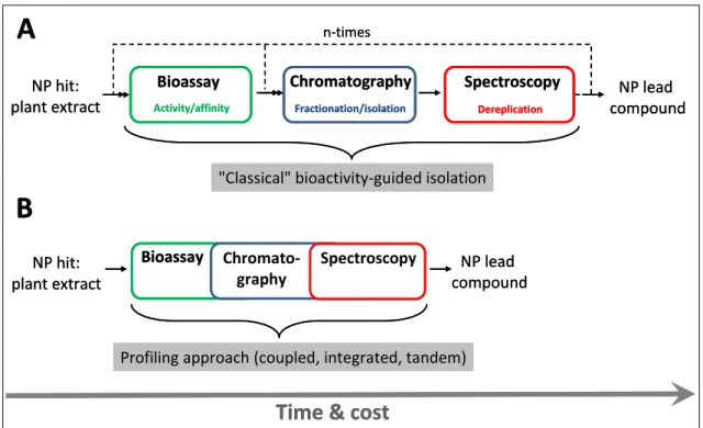
The advent of the HTS approach, followed by the introduction of combinatorial chemistry has fundamentally shifted the drug discovery paradigm in the 1990s. Practice of the NP-based lead generation has become incompatible, and thus uncompetitive with the HTS of large and pure synthetic compound libraries. First, NP extracts tend to be complex mixtures of secondary metabolites, containing hundreds or thousands of compounds, often including constituents (so called “nuisance compounds”) such as tannins, fatty acids, colored or autofluorescent chemicals, which are specifically interfering with routinely applied bioassays [14]. Thus, crude NP extracts cannot be screened directly in HTS campaigns. Second, once a NP extract has been identified as active, the principle responsible for the bioactivity must be isolated by a time- consuming and laborious bioactivity-guided fractionation. This step is further burdened and lengthened by the identification and elimination of already known compounds (dereplication), to avoid the duplication of effort. Third, after a single biologically active compound has been obtained from an extract, the de novo structure determination of the novel NP lead must be carried out (Fig. 2).
Figure 2. General scheme of HTS-based drug discovery and development. Unique processes of the NP-based approach within the hit-to-lead phase are enlarged and highlighted.
Furthermore, the complex nature of some NP chemical structures may present an obstacle for synthetic medchem modifications during the lead optimization phase and for total- or semi-synthesis in case of limited supply of the lead compound [1-3]. One can conclude, reviewing the above summarized drawbacks and concerns, that the NP- based drug research has not kept pace with the modern HTS approach and thus is not worthy of being practiced in today’s drug discovery environment.
However, the success (hit rate) of any HTS campaign is inherently dependent on the quality of the screened library and the quality is determined by three factors:
chemical diversity, lead-likeness, and biological relevance [15, 16]. Without a more detailed discussion, in terms of these features, NPs are considered as superior to any synthetic chemical collection: NPs offer high and unique chemical diversity [17, 18], combined with high probability of lead-likeness (or even drug-likeness) [19], and of affinity to biological macromolecules [20].
Moreover, recent advancements in separation and structure elucidation technologies, and particularly in the hyphenation thereof, have greatly improved the processes of isolation, dereplication, and structure elucidation. Owing to reliable and robust ionization interfaces (e.g., electrospray ionization), directly coupled high performance liquid chromatography (HPLC) - mass spectrometer (MS) systems have assumed a pivotal role in this area [21]. In the field of nuclear magnetic resonance (NMR) spectroscopy, the advent of multidimensional pulse methods and sensitivity improvements (e.g., micro-probe technologies) have dramatically lowered the amount of material needed (in the sub-milligram range) for structural analysis [22-24].
Combining the above mentioned techniques with purification (e.g., solid phase extraction) and liquid handling solutions has led to the development of automated, high- throughput fractionation systems, which were also capable of generating pure NP libraries in an efficient way [25-27]. Last but not least, innovative approaches, coupling of bioassays of interest to analytical procedures (typically to LC-MS) permitting the rapid and simultaneous separation and dereplication of active constituent(s) in NP extracts (i.e., activity profiling) have recently gained popularity as well. As the work presented in this thesis has focused on the same topic, detailed review of these profiling- like concepts and technologies is provided in the next chapter.
1.3. Novel approaches to natural product lead finding: profiling by coupled techniques
The major bottleneck (i.e., the rate limiting step) in the NP-based drug discovery is still the procedure of isolation and purification of the active principle (defined as the lead compound in this context) from an exceptionally complex matrix [2]. As can be seen in Fig. 3A, progression in the conventional bioactivity-guided route depends on the number of “cycles” of subsequent fractionation and bioassay steps required. As a result, the overall time needed to obtain a pure enough NP lead is the sum of the turnaround times of the three single steps multiplied by the number of cycles. In contrast, Fig. 3B depicts an alternative way: through simultaneous assessment and rapid correlation of the biological responses (e.g., on-line) to each constituent of the mixture, the synergistic melding of the bioassay and the analytical cascade is capable of shortening significantly the time needed to identify and/or isolate the bioactive agent(s).
Figure 3. Comparison of the working schemes of the sequential (iterative) (A), and the more rapid and cost effective profiling approaches (B).
In addition, coupling of such techniques/assays to biological screens can improve the quality and information content of the assay result. Interaction (e.g., synergism) between mixture constituents or lack thereof could also be revealed by such
Bioassay Chromatography Spectroscopy
Fractionation/isolation Dereplication
NP hit:
plant extract
NP lead compound
Time & cost
NP hit:
plant extract
Bioassay Chromato- graphy
Spectroscopy
Activity/affinity
n-times
NP lead compound
"Classical" bioactivity-guided isolation
Profiling approach (coupled, integrated, tandem)
A
B
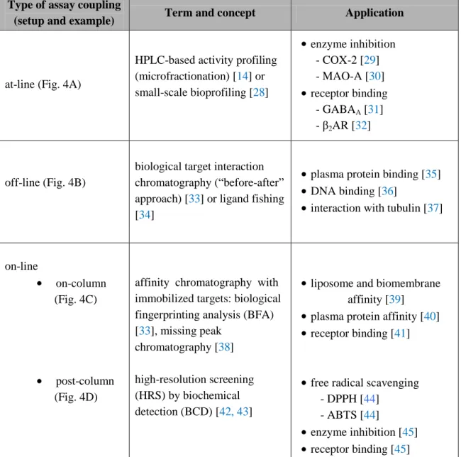
methods. It must be noted, however, that this type of coupling may represent a significant challenge in practice and must be carefully validated from several aspects before it is used. Depending on the studied biological phenomenon, the separation and detection technique, a number of different terms have been created in the literature for these types of approaches. In practice, the coupling follows basically three major strategies (Table 1 and Fig. 4).
Table 1. Summary of different types and setups of approaches applied for the profiling of complex mixtures (e.g., plant extracts) in NP-based drug discovery.
Type of assay coupling
(setup and example) Term and concept Application
at-line (Fig. 4A)
HPLC-based activity profiling (microfractionation) [14] or small-scale bioprofiling [28]
enzyme inhibition - COX-2 [29]
- MAO-A [30]
receptor binding - GABAA [31]
- β2AR [32]
off-line (Fig. 4B)
biological target interaction chromatography (“before-after”
approach) [33] or ligand fishing [34]
plasma protein binding [35]
DNA binding [36]
interaction with tubulin [37]
on-line
on-column (Fig. 4C)
post-column (Fig. 4D)
affinity chromatography with immobilized targets: biological fingerprinting analysis (BFA) [33], missing peak
chromatography [38]
high-resolution screening (HRS) by biochemical detection (BCD) [42, 43]
liposome and biomembrane affinity [39]
plasma protein affinity [40]
receptor binding [41]
free radical scavenging - DPPH [44]
- ABTS [44]
enzyme inhibition [45]
receptor binding [45]
COX-2: cyclooxygenase-2; MAO-A: monoamine oxidase-A; GABAA: γ-aminobutyric acid; β2AR: β2 adrenergic receptor; DPPH: 2,2-diphenyl-1-picrylhydrazyl; ABTS: 2,2'-azino-bis(3-ethylbenzothiazoline-6-sulphonic acid).
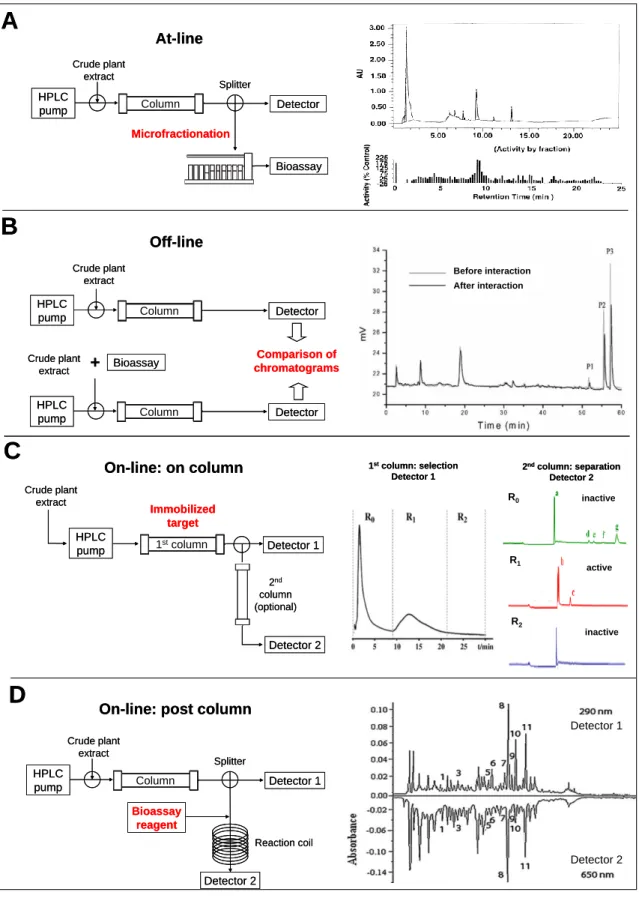
Figure 4. Instrumental configuration and one example from the literature of each profiling approach. (A) HPLC-based activity profiling [21], (B) Biological target interaction chromatography [36], (C) Affinity chromatography [41], (D) High resolution screening by post- column biochemical detection [53].
D C B A
At-line
Off-line
On-line: on column
On-line: post column
HPLC pump
Crude plant extract
Column Detector
Splitter
Microfractionation
Bioassay
HPLC
pump Column Detector
Crude plant extract
HPLC
pump Column Detector
Crude plant
extract + Bioassay Comparison of
chromatograms
HPLC pump Crude plant
extract
1stcolumn Immobilized
target
Detector 1
HPLC pump
Crude plant extract
Column Detector 1
Splitter
Detector 2 Bioassay
reagent
Reaction coil 2nd column (optional)
Detector 2
R0
R1
R2 1stcolumn: selection
Detector 1
inactive
inactive active 2ndcolumn: separation
Detector 2
Detector 1
Detector 2 Before interaction
After interaction
According to the first and simplest one, crude extracts are subjected to analytical or semi-preparative HPLC, and a portion of the effluent is collected into microplates (Fig. 4A). These microfractions are dried under vacuum, redissolved and assayed separately for bioactivity. The chromatogram and the activity profile are matched to identify active peaks [14]. Thus, this approach represents the miniaturized format of the conventional bioactivity-guided fractionation.
In the second approach, the multi-component NP extracts are allowed to interact with the targeted biomacromolecule prior to the chromatographic step (Fig. 4B). Next, comparison of chromatograms of the sample before and after being treated with the target indicates clearly the “biointeractive” constituents of the extract [33].
The most integrated and complex solution has been achieved by the development of the so called on-line assay configurations. In some of these systems the targets are immobilized on the stationary phase of a column and the mixture is continuously infused through of this (Fig. 4C). Compounds with the highest affinity for the target will have the longest breakthrough times (frontal affinity chromatography) [2, 33, 46].
Another group of on-line assays represents the high-resolution screening (HRS) methods (Fig. 4D), which utilizes continuous-flow biochemical detection in a so called post-chromatographic way: activity assessment of HPLC eluate is carried out in a post- column reaction chamber [42-45].
Moving to techniques used for the steps of separation and dereplication in these setups, literature reflects on the unequivocal dominance of HPLC-coupled spectroscopic systems, such as LC-DAD-MS/MS [44], and LC-NMR [47]. It must be noted, however, that most profiling/fingerprint analysis have been developed with Reversed Phase-LC using a simple UV detector. Furthermore, the recent advent of ultra-high performance liquid chromatography (UHPLC) [48], and the spreading of analytical columns packed with fully porous sub-2 µm and superficially porous particles have created ideal conditions for conducting such studies in terms of superior separation efficiency and sensitivity [49]. Finally, NP-specific, comprehensive chemical and biological databases, such as the NAPRALERT [50], the Dictionary of Natural Products (DNP) [51], and the Plant Profiler of Sigma-Aldrich [52] are supporting even more efficiently the critical step of dereplication in these workflows. For example, the database of DNP documents
and organizes every, virtually known natural product in a searchable form up to date (approx. 230 000 compounds).
Detailed analysis of strengths and limitations of the discussed approaches is beyond the scope of this thesis. Anyway, the following has been concluded in a very recent in-depth review: “Fully integrated systems represent technically impressive achievements, their implementation in other laboratories could be somewhat intricate.
As a consequence many on-line assays have not been adopted outside of the lab where they were originally conceived. Compared to on-line assays, at-line and off-line approaches are more versatile, robust and probably have higher potential for broad implementation in NP-based drug discovery programs” [54]. In conclusion, the overall performance of a coupled system relies basically on two factors: on the resolving power/selectivity of the chromatographic separation as well as on the robustness/compatibility of the coupled bioassay.
1.4. Review of targets and methods used in the screening and profiling of the plant extract library
In this chapter, the biological background, the therapeutic impact and the methodological aspects of the two distinctive areas investigated in our screening and hit profiling studies are discussed:
First, general biochemistry of free radicals and antioxidants is shortly summarized (Chapter 1.4.1.). It is followed by the detailed bibliographic presentation of the in vitro radical scavenging assay (2,2-diphenyl-1- picrylhydrazyl (DPPH)) used in our primary antioxidant screen. Next, an other reactive species (the peroxynitrite anion (ONOO–)), and the related methodologies used in a secondary/focused antioxidant screen are introduced.
Second, a biological barrier (the blood-brain barrier, (BBB)), with exceptional impact on the CNS drug discovery, and methodologies (in vitro mainly) investigating the permeation of molecules (e.g., drugs) through this barrier are reviewed (Chapter 1.4.2.). Finally, the last part of this chapter is devoted to the evaluation of the studied and applied permeability assay (parallel artificial membrane permeability assay for blood-brain barrier (PAMPA-BBB)).
Since our entire work focused on the screening of the plant extract library of Gedeon Richter Plc., much emphasis was placed on the potential role and relevance of plant metabolites in both of the above mentioned two topics. Moreover, where it was available from the literature, particular case studies related to the coupling of the bioassays used by us to analytical processes (typically to HPLC or LC-MS) are summarized as well.
1.4.1. Free radicals, oxidative stress and antioxidants
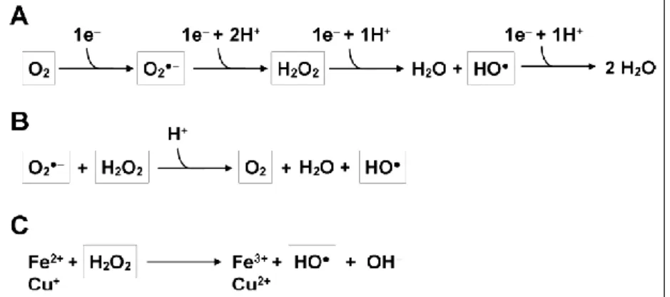
Free radicals (species with one or more unpaired electrons), such as reactive oxygen species (ROS) are generated by normal metabolic processes in all aerobic organisms due to the incomplete reduction of molecular oxygen in the electron flow chain (Fig. 5).
Figure 5. Production of ROS. (A) The stepwise transfer of electrons to O2 leading to the generation of superoxide anion (O2•−
), then hydrogen peroxide (H2O2), and finally the highly toxic hydroxyl radical (HO•). (B) The Haber-Weiss reaction, and (C) the Fenton reaction for the formation of HO•. (Adapted from [55] without modification).
As the so formed ROS are highly reactive and deleterious towards other substances within cells (e.g., lipid membranes, proteins, and nucleic acids), endogenous antioxidant defense systems have been evolved during the course of evolution to minimize and repair free radical-induced damages [56]. These defensive mechanisms and molecular networks include enzymatic as well as non-enzymatic components, which are acting in concert to maintain the redox homeostasis of cells. For instance, superoxide dismutase, catalase, glutathione peroxidase, and glutathione reductase are enzymatic antioxidants in human plasma and erythrocytes, while bilirubin, melatonin, uric acid, glutathione, and ubiquinol belong to the group of small molecule antioxidant agents (i.e., radical scavengers) [56]. The efficiency, however, of this endogenous defence is considered as
incomplete, particularly under some pathophysiological conditions such as UV- irradiation, heavy metal exposure, inflammation, and ischemia/reperfusion [56]. As a result, oxidative stress develops, in which free radicals are produced in excess and at the wrong time and place. This leads to the oxidative damaging of cellular macromolecules, promoting ultimately cell death.
It has been shown and confirmed recently that oxidative stress is involved in aging [57] and several degenerative diseases, including cancer [58], cognitive dysfunction [59], diabetes mellitus [60], and atherosclerosis [61]. Therefore, considerable efforts have been paid to the quest for effective, exogenous antioxidants in the pharmaceutical industry: it was found that more than 300 000 papers associated with antioxidants were published from 1980 to 2008 [62]. Some of these works are devoted to synthesize antioxidants with novel structures [63-65], and characterize the radical scavenging properties of existing drugs to enlarge the therapeutic application of these drugs as antioxidants [66, 67]. Other works focus on the screening and extraction of antioxidant compounds from natural products to identify and isolate the valid and most potent scavengers in these mixtures [68-70]. It must be noted at this point, that the research for radical scavenger agents in the pharmaceutical and food industry has been partially merged into each other (see nutraceuticals), since many dietary compounds, such as vitamins A, C, and E, carotenoids, and plant phenoloids play important role in the maintenance of human health as well as in the prevention and treatment of diseases [56, 71].
Nevertheless, as phytotherapy and medicinal plant research are mainly focused on the areas of cancer and inflammatory diseases, huge number of studies have been published which are dealing with the screening and evaluation of the antioxidant/anti- inflammatory potential of plant extract collections/libraries [68, 72-77]. First, a common feature of these investigations was the use of robust in vitro bioassays (photometric- based) in the screening experiments. Second, it was revealed by mean of bioactivity- guided isolation that phenoloid compounds, such as flavonoids, simple phenols and caffeic acid derivatives were basically responsible for the measured radical scavenging activities of the extracts. Third, it can be concluded that the IC50 values of crude or fractionated plant extracts which were considered as actives/hits in these screens fell in the range of 1-10 µg/mL.
1.4.1.1. The DPPH (2,2-diphenyl-1-picrylhydrazyl) method
DPPH is a stable N-centered radical discovered by Goldsmith and Renn in 1922 [78], and later utilized by Blois as a colorimetric reagent to evaluate the direct radical scavenging properties of small molecule antioxidants such as ascorbic acid, cysteine, and hydroquinone [79]. It is a paramagnetic compound with an odd electron and exhibits a strong absorption band at 517 nm, resulting a deep violet color in its alcoholic solution. Upon reduction by an antioxidant, the odd electron of DPPH becomes paired off (forming the stable, corresponding hydrazine): the violet color changes to yellow and the absorption of the solution decreases or vanishes (i.e., bleached out) (Fig. 6).
This reaction is intended to mimic reactions taking place in oxidizing systems in vivo, such as the peroxidation of membrane lipids.
Figure 6. Reaction scheme for scavenging the DPPH radical by an antioxidant (AH) through hydrogen atom transfer.
Because of the relative simplicity and robustness of the colorimetric assay based on this reaction, now it has gained widespread use in the free radical scavenging activity assessment: it has become a primary method to measure the total antioxidant potential (hydrogen donating ability) of natural compounds, plant extracts, foodstuffs or other biological sources [80]. Moreover, it is also widely implemented to elucidate the redox properties of newly synthesized compounds [81, 82]. As a result, several modification, extension, improvement, and hyphenation of the original assay setup have been published [80]. Developments of the DPPH method, with particular focus on the antioxidant screening of plant extracts by chromatographic techniques are reviewed briefly in the following.
Combination of thin layer chromatography (TLC) with the DPPH method was reported first by Glavind and Holmer in 1967: after TLC separation of the analyte,
radical scavengers (tocopherols) were detected visually by spraying the TLC plates with DPPH solution [83]. Owing to its cost-effectiveness, this approach became very popular: Cieśla et al. have optimized and standardized it recently [84]. Since the spectrophotometric assay suffers from the interference with colored compounds frequently found in foodstuffs and plant extracts, HPLC-based quantitative methods have been proposed as alternatives to the original colorimetric assay to study such complex mixtures: Yamaguchi et al. developed a reversed phase HPLC method to selectively detect at 517 nm the remaining DPPH radical after off-line reaction of standard antioxidants with the probe [85]; while Boudier et al. optimized HPLC conditions to simultaneously quantify the reagent (DPPH) and the reaction product (DPPH-H) of the assay at 330 nm [86].
In addition, off-line LC-MS approaches have been developed to rapidly identify the most potent radical scavenger constituents of plant extracts by simply monitoring the reduction or disappearance of the corresponding peaks after addition of the active radical to the samples (see Fig. 4B for the corresponding instrumental setup): by evaluating the radical scavenger properties of 14 phenolic compounds in the extract of Lonicerae japonica, Tang et al. demonstrated that compounds with peak areas significantly decreasing were natural antioxidants, whereas those with peak areas not changing presented no activities [87]; Helmja et al. applied the same “spiking” test to the analysis of a Solanum melongena extract, and revealed the radical scavenging ability each of the identified single 11 compounds as a contribution to the total activity of the extract [88]. For this purpose, an opposite calculation of EC50 was performed:
compounds were characterized by the concentration of DPPH radical (mM) necessary to oxidize 50% of a compound in competition with all other oxidizable compounds in the mixture (Fig. 7). It was found that cinnamic acid derivatives corresponding to peaks 5-9 in Fig. 7 possessed low EC50 values, thus, were mainly responsible for the antioxidative activity of the extract.
Figure 7. The HPLC–UV chromatograms of Solanum melongena extract and reaction products after treated with increasing concentrations of DPPH recorded at 280 nm. (Adapted from [88]
without modification).
In contrast, van Beek and coworkers have created an on-line setup using the DPPH assay: a solution of the radical is added post-column and a second chromatogram is recorded paralelly at 517 nm. Thus, active compounds appeared as negative peaks (see Fig. 4D for the corresponding instrumental setup) [89]. Since then, a lot of successful examples of application have been reported [42, 90-93]. However, Zhang et al. have compared the performance of the on-line setup, to the pre-column off-line (spiking) method or to the at-line approach (microfractionation), and concluded that the reaction with DPPH prior to the separation gave the best resolution [94].
Off-line and on-line coupling of the prosperous high speed counter current chromatography (HSCCC) to the DPPH assay have also been solved recently by Chinese research groups. Numerous antioxidants have been rapidly identified and isolated from complex plant extracts by the guidance of DPPH-HPLC assays [95-98]. It must also be noted that the DPPH method has been adopted to microplate format, thus it became convenient to HTS [99].
1.4.1.2. The peroxynitrite anion (ONOO–): biochemistry and methodologies for measurement of ONOO– scavenging activity
The peroxynitrite anion (ONOO–), typically produced by the rapid diffusion-controlled reaction of nitric oxide (•NO) with superoxide radical (O2•–) in vivo, is implicated in the pathogenesis of a wide variety of human diseases, such as atherosclerosis, obesity, diabetes mellitus, and Alzheimer-disease [56, 100]. Although not a free radical by chemical nature (it is an unstable structural isomer of the nitrate anion), ONOO– is considered as a powerful oxidizing and nitrating species. Fig. 8 summarizes the major biochemical reactions (direct as well as indirect ones), which are leading to the complex and deleterious/cytotoxic effects of ONOO– [101]. It must be pointed out, however, that only minor part of ONOO– is transformed into radicals: it is mostly trapped in vivo by thiols and metalloproteins (Fig. 8A and B) [101].
Se-GPx: selenium-containing glutathion peroxidase, DNA: deoxyribonucleic acid.
Figure 8. Biochemical reaction pathways of peroxynitrite. Peroxynitrite anion (ONOO–) is in equilibrium with peroxynitrous acid (ONOOH; pKa=6.8) and either one can undergo direct reactions with biomolecules as indicated (A and B). A fundamental reaction of ONOO– in biological systems is its fast reaction with carbon dioxide (C), which leads to the formation of carbonate (CO3•–) and nitrogen dioxide (•NO2) radicals, which are good one-electron oxidants (D) that can readily oxidize amino acids such as cysteine and tyrosine to yield the corresponding cysteinyl and tyrosyl radicals. In addition, •NO2 can undergo diffusion-controlled termination
reactions with biomolecule-derived radicals, resulting in nitrated compounds (D). Alternatively, ONOOH can undergo homolytic fission to generate one-electron oxidants hydroxyl (•OH) and
•NO2 radicals (E). However, this reaction is slow in biological systems compared with the other reactions of ONOO– and ONOOH and therefore is a modest component of the in vivo reactivity of peroxynitrite in aqueous compartments. However, ONOOH readily crosses lipid bilayers (F) and its decomposition to •OH and •NO2 radicals seems to become relevant in hydrophobic phases to initiate lipid peroxidation and lipid and protein nitration processes (G). Moreover, ONOOH in the membranes may undergo direct reactions with metal centres such as hemin or membrane associated thiols. (Adapted from [101] without modification).
Since its half-life is less than a second under physiological conditions, direct detection of ONOO– in biological systems is really difficult. Beside electron spin resonance (ESR) assays [102], fluorescent and luminescent spectroscopic probes have been used frequently for this purpose [103-105]. In addition, indirect approaches have focused on the detection of endogenous biomolecules modified by a ONOO–-dependent reaction, e.g., quantification of 3-nitro-tyrosine as a marker product of ONOO–-specific aromatic nitration of tyrosine by HPLC has gained popularity [106, 107]. Anyway, the specificity of these techniques is considered controversial due to several interfering factors.
In contrast, due to the relatively simple generation procedure and stability of ONOO– at laboratory conditions [108], numerous in vitro assays (with lower biological significance obviously) assessing ONOO– scavenging activity of single compounds or biological samples have also been developed. Among those, the most accepted method is based on the oxidation of dihydrorhodamine by ONOO– to fluorescent rhodamine [109]. Being less sensitive, however, colorimetric bleaching assays have also been published: Balavoine and Geletii have developed a simple and robust colorimetric assay based on pyrogallol red dye bleaching for screening of ONOO– scavenging activity of plant extracts and foodstuffs [110, 111].
Turning to the analyses of naturally occurring peroxynitrite scavengers, it can be concluded that a great number of biological samples and natural compounds have been reported as effective ONOO– scavengers in vitro, however the number of comprehensive screening studies on this topic is rather limited. It was found that representative compounds of flavonoids (e.g., quercetin, kaempferol, luteolin, epicatechin) [112, 113], phenylpropanoids and derivatives thereof (e.g., caffeic acid, sinapinic acid, chlorogenic acid, and curcumin) [107, 114, 115], carotenoids [116], and
tocopherols [116] possessed significant ONOO– eliminating activity. In the case of phenoloids, structure activity relationships have been revealed in further mechanistic studies (Fig. 9): it has been shown that the ortho-hydroxyl structure, especially the catechol group in the ring B seems essential for ONOO– scavenging activity, and the 2,3-double bond also plays an important role, while O-glycosilation reduces the radical scavenging activity (reviewed extensively in [117]). As a conclusion, two possible mechanisms for phenoloid-mediated ONOO– scavenging have been proposed:
monohydroxylated structures act as alternative substrates for nitration, whereas catechol moieties are oxidized to o-quinones (electron donation).
Figure 9. Structural features of quercetin responsible for ONOO– scavenging activity.
Although significant efforts have been made in medicinal plant research to identify and isolate compounds with ONOO– scavenging potential from plant extracts, little or no attention has been paid to the effective coupling of antioxidant activity assays with advanced separation techniques (HPLC). Recent studies are still reporting the practice of the conventional bioassay-guided (sequential) isolation approach [118- 120], thus giving up the chance to characterize the contribution of the single components to the total activity.
1.4.2. The blood-brain barrier (BBB)
The BBB is a unique physical and metabolic barrier formed by brain capillary endothelial cells joined by tight junctions. It serves to isolate the cerebral parenchyma from the systematic circulation and helps to maintain the homeostasis of the brain microenvironment by allowing the entry of selected nutrients and macromolecules, while restricting the penetration of polar molecules [121, 122]. Features that distinguish the brain endothelium from that of other organs include complex tight junctions,
O
O
OH OH
OH
OH O
H
A C
B
2 3
3’
4’
5
7 O
O
OH OH
OH
OH O
H
A C
B
2 3
3’
4’
5 7
relatively low pinocytotic activity, and the expression of a number of specific uptake and efflux transport systems and metabolic enzymes (such as P-glycoprotein and cytochrome P450 enzymes) [123]. These properties greatly limit the transcellular and paracellular movement of drugs and xenobiotics to the CNS and make the BBB a highly selective regulatory interface: only 2% of the possible CNS therapeutic compounds can pass the BBB and reach their therapeutic targets [124]. It means that the BBB poses a major challenge for today’s CNS drug discovery, since CNS drugs must permeate the barrier, whereas compounds targeting peripheral tissues should be impaired in the passage.
The main molecular transport mechanisms, such as passive and active pathways across the BBB are summarized and illustrated in Fig. 10.
Figure 10. The main routes for molecular traffic across the BBB. Note that most CNS drugs enter the brain by transcellular passive diffusion (also called the “pharma route”, highlighted in red bracket). (Adapted from [125] with minor modification).
Since paracellular permeation is practically restricted by tight junctions in the BBB, and uptake transporters are basically intended to enhance the transport of nutrients and cofactors, most small molecule drugs enter the brain by transcellular passive diffusion. This process is driven by a concentration gradient between the blood and the brain, and is inherently affected by the physicochemical properties of discovery compounds, such as molecular size, lipophilicity (log P or log D), flexibility, and total
polar surface area (TPSA). The brain exposure of an individual drug, however, always needs to be considered as a resultant of multiple permeation and distribution mechanisms, including efflux transport, metabolism, and plasma protein binding [126].
As a consequence, the implemented methodology used for the assessment of brain penetration must appropriately reflect on these processes.
1.4.2.1. Methodologies for measurement of blood-brain barrier transport
Owing to the prominent role of BBB transport in CNS drug research, a wealth of new approaches and assays have been established over the years to measure and predict the brain penetration of drugs and discovery compounds (comprehensively reviewed in [126-129]). Among these, the cost-effective in silico models, based on correlations between compound permeation and physicochemical descriptors, have gained popularity in the early phase of drug discovery. However, their scope and predictive power are limited only to aid the design of synthetic libraries and to classify compounds with high and low brain penetration potency. The other end of the BBB-assay spectrum in terms of reliability and cost represents in vivo techniques. These involve traditionally low-throughput and labor-intensive measurements, such as brain microdialysis and brain perfusion studies preformed in rodents. They are designed to assess specific parameters of brain penetration, namely rate, extent and unbound drug concentration.
Since only a limited number of compounds can be evaluated by these in vivo techniques, robust and high-throughput in vitro approaches have emerged in the pharmaceutical industry. In vitro BBB methods could be classified into cell-based and noncell-based assays. Cell-based assays are utilizing either brain-derived (e.g., isolated brain capillaries, bovine brain microvessel endothelial cell culture) or non-brain-derived (e.g., Caco-2, MDCK) cells and are intended to indicate efflux/uptake potential and/or metabolic liabilities. In contrast, the principle of noncell-based models is strictly physicochemical by nature. Thus, they tend to mimic and predict exclusively the transcellular passive diffusion component of the whole brain disposition process. The immobilized artificial membrane chromatography (IAM), and the parallel artificial membrane permeability assay (PAMPA) form this latter group of methods [126-129].
1.4.2.2. The parallel artificial membrane permeability assay for blood-brain barrier (PAMPA-BBB)
The parallel artificial membrane permeability assay was first introduced by Kansy et al.
in 1998 to model oral absorption processes [130]. Di and coworkers have modified the PAMPA system specifically for BBB application: they applied porcine brain lipid extract (PBL) dissolved in n-dodecane as PAMPA membrane, and demonstrated that using this method discovery compounds can be binned into CNS+ and CNS– classes [131]. It has also been reported that the PAMPA-BBB derived values display good correlation to cell-based models and to in situ brain perfusion measurements [132].
Since then, the PAMPA technique has become one of the most powerful and versatile physicochemical screening tool in early stage CNS-targeted drug discovery practice [133].
The system consists of two multiwell microtiter plates, a donor and an acceptor compartment in a “sandwich” like configuration, separated by an artificial lipid impregnated filter membrane (Fig. 11).
Figure 11. General scheme of the PAMPA-BBB system: setup of the “sandwich” plate (left), and schematic representation of passive diffusion in a well (right).
Initially, the test drug is added in the donor plate and allowed to diffuse across the membrane. After incubation (typically for 4 hrs at 37 °C), PAMPA sandwich plates are separated and drug concentrations in donor and acceptor solutions are determined by UV spectroscopy or LC-MS. Thus, the rate of transcellular passive diffusion can be predicted by calculating the effective permeability (Pe, cm/s) of the tested drugs.
Porcine brain lipids 96-well donor plate » blood
96-well acceptor plate » brain
Donor well
Microporous filter membrane Acceptor well Porcine brain lipids 96-well donor plate » blood
96-well acceptor plate » brain
Donor well
Microporous filter membrane Acceptor well
1.4.2.3. Rationale of studying natural products and plant extracts by PAMPA-BBB Plant extracts with traditionally known or ethnomedically proven neurobiological activity are attractive lead sources for CNS drug discovery [134-138]. However, for the vast majority of CNS-active herbal remedies the active principles and the exact molecular mechanism of action have not yet been elucidated [139]. Nevertheless, it can be assumed that certain constituents of such plant extracts are capable of crossing the BBB.
In silico calculations have been developed and validated mainly using large datasets of drugs [140, 141], and, as the NP chemical space significantly differs from that of therapeutic agents and synthetic compounds [142, 143], the predictive accuracy and reliability of these models for plant metabolites are questionable, whereas costly in vivo BBB techniques are considered as impractical for complex mixtures like plant extracts.
In contrast, the PAMPA-BBB assay is a single mechanism-based measurement, and when the complexity of a typical plant extract is considered, this feature has great importance, because evaluation and interpretation of multimechanism-type (i.e., active transport, metabolic transformation) assay results may be quite challenging. Moreover, PAMPA-BBB studies, conducted in cassette dosing (i.e., sample pooling, wherein the number of mixed components varies between three and 32) have indicated no significant interference or difference on effective permeabilities whether assessed with single compounds or mixtures thereof [144-148]. Tarragó et al. have evaluated earlier permeability values in a crude plant extract for baicalin and baicalein using a PAMPA- BBB assay [149]. These features of the PAMPA-BBB system stimulated us to investigate its potential use on crude and pre-fractionated plant extracts in terms of screening and identifying compounds with high brain penetration propensity.
2. Objectives
“These materials have already been prepared
for humans, we should merely reach them.”
Ivan Petrovich Pavlov
The primary aim of our work was to design, adopt and/or validate, and perform screenings of a plant extract library. The screening assays included a cytotoxicity, an antioxidant, and a BBB permeability screening campaign. As the plant extract hits emerged from the latter two screenings proved to be complex mixtures of secondary plant metabolites, we attempted to couple the bioassays of particular screens to HPLC- based analytical procedures. While doing this, our motivation was to shorten the bioassay-guided isolation route of the active principle(s), particularly in the lead compound identification and dereplication step. Therefore, much emphasis was placed on the methodological aspects of the assay couplings: analytical features such as resolution, throughput and applicability were studied and optimized in detail. Moreover, by mean of the case studies presented in this thesis, we have endeavored to contribute to the phytochemical and pharmacological characterization of the investigated plant species.
The specific aims were the followings:
To screen the plant extract library for cytotoxic activity, and to analyze the dependence of the cytotoxic activity of plant extracts on the type (polarity) of the solvent extraction procedure.
To screen the plant extract library for antioxidant activity and to analyze the dependence of the antioxidant activity of plant extracts on the type (polarity) of the solvent extraction procedure.
To analyze and prioritize (dereplicate) the resulted plant extract hits by LC- MS, and to develop a LC-MS method that can be coupled with the DPPH assay in order to effectively identify the radical scavenger constituents in one antioxidant plant extract hit, namely in the methanolic extract of Artemisia gmelinii.
To isolate and elucidate the chemical structure of the most active radical scavenger compounds in the methanolic extract of Artemisia gmelinii.
To adopt and validate the pyrogallol red bleaching test for HPLC in order to screen and characterize chemical constituents with peroxynitrite (ONOO–) scavenging activity in alcoholic extracts of Salvia species, since Salvia extracts proved to be predominant among the antioxidant hits.
To demonstrate the performance of the developed HPLC-based ONOO– scavenging assay on the methanolic extract of Salvia miltiorrhiza Bunge.
To investigate the applicability of the PAMPA-BBB assay for NPs and plant metabolites, and to screen the plant extract library for NP compounds with high brain penetration propensity.
To couple the PAMPA-BBB assay to NMR experiments and to demonstrate the feasibility of this type of coupling on BBB+ plant extract hits (exemplified by the extracts of Tanacetum parthenium, Vinca major, Salvia officinalis, and Corydalis cava).
In conclusion, our entire workflow focused on the acceleration of the lead generation phase in the NP-based drug discovery by coupling (integrating) bioassays with advanced separation and spectroscopic techniques in a valid and efficient way.
3. Materials and Methods
3.1. Chemicals and reference compounds
All solvents used were LC-grade. Acetonitrile (MeCN), ethanol (EtOH), methanol (MeOH), chloroform (CHCl3), dimethyl sulfoxide (DMSO), trifluoroacetic acid (TFA), acetic acid, glycine, sodium nitrite, hydrochloride were purchased from Merck (Darmstadt, Germany). All other reference compounds and reagents were analytical grade and purchased from Sigma-Aldrich (St Louis, MO, USA), except for adhyperforin, α-solanine, apigenin-7-O-glucoside, aucubin, betulin, chamazulene, catalpol, (+)-catechin, cynarin, galantamine, harpagoside, hyperforin, lupeol, luteolin-7- O-glucoside, nicotinic acid, parthenolide, protopine, salvianolic acid A, scopolamine, sinapic acid, solasodine, and stigmasterol, which were obtained from PhytoLab (Vestenbergsgreuth, Germany). BBB specific porcine brain lipid (PBL) extract was purchased from Avanti Polar Lipids (Alabaster, AL, USA). Purified water (18 MΩ·cm) was obtained from a Millipore (Bedford, MA, USA) Milli-Q water-purification system and used for all aqueous solutions and eluents.
The reference compound collection used in the hit characterization and dereplication studies (LC-MS for antioxidant hits, LC-MS/MS for PAMPA-BBB hits) consisted of ubiquitous representatives of carboxylic acids, flavonoids, alkaloids, and terpenes and was identical with the list of Table A2 (Appendix). In addition to that list, gallic acid, caftaric acid, 4-O-caffeoylquinic acid (cryptochlorogenic acid), salvianolic acid B, cynarin, 5-hydroxyflavone, myricetin, carnosol, curcumin, and ursolic acid were included in the used reference substance collection (N=82).
3.2. Plant material
3.2.1. Plant extract library
The plant extract library of Gedeon Richter Plc. was assembled mainly between 1999 and 2001, in the course of a contractual cooperation with (i) the Institute of Ecology and Botany of the Hungarian Academy of Sciences, Vácrátót, Hungary; (ii) the Department of Pharmacognosy, Semmelweis University, Budapest, Hungary; (iii) the Department of Pharmacognosy, University of Szeged, Szeged, Hungary; and (iv) the Medicinal Plant
Research Institute, Budakalász, Hungary. This cooperation resulted in 4400 randomly collected individual extracts, originated from ca. 500 drugs of 300 plant species endemic or to-grow in the Carpathian Basin. The taxonomic composition of the collection shows a highly diverse profile, however, the representatives of the Lamiaceae and Asteraceae families are predominant.
The preparation of the extracts followed basically a general scheme: first, the dried and ground drug was extracted with CHCl3 or in some case with petroleum ether (this procedure yielded the apolar crude extracts). After filtration the residual plant material was dried and extracted successively with aqueous MeOH (this procedure yielded the polar crude extracts). Finally, the crude extracts were evaporated to dryness in vacuo, and were fractionated by open column chromatography according to standard protocols. Stock solutions of the resulted fractions were prepared uniformly in DMSO at 40 mg/mL, filtered through 0.45 µm Millipore (Billerica, MA, USA) filters and stored at -19 °C in polypropylene deep-well plates (80 samples per plate, A2-H11) until required for screening experiments.
3.2.2. Artemisia gmelinii Webb. ex Stechm. (Asteraceae)
Aerial parts of A. gmelinii were collected before full blooming from the experimental field of the Institute of Ecology and Botany of the Hungarian Academy of Sciences, Vácrátót, Hungary. The plant material was identified by Dr. Vilmos Miklósi V. A voucher specimen (no. L8275) has been deposited in the Herbarium of the Institute. Dried and ground aerial parts of A. gmelinii (50 g) were first extracted with 1×200 mL and 1×100 mL of CHCl3/MeOH 90:10 (v/v) using an ultrasonic bath for 2×15 min (this procedure yielded the CHCl3 extract). After filtration the residual plant material was dried and extracted with 1×200 and 1×100 mL of 70% (v/v) aqueous MeOH at room temperature in an ultrasonic bath for 2×15 min. The filtered and combined aqueous methanolic extracts were evaporated to dryness in vacuo to yield 3.7 g of brown oily material. This extract was fractionated by open column chromatography on silica gel (Kieselgel 60, 0.063-0.200 mm, 1.07734.100 Merck, Germany) (sorbent:
Figure 12. Artemisia gmelinii [150].

![Figure 18. Principle of the cytotoxicity assay [156], and a representative profile of a 384-well screening plate](https://thumb-eu.123doks.com/thumbv2/9dokorg/1366239.111608/35.892.132.763.622.820/figure-principle-cytotoxicity-assay-representative-profile-screening-plate.webp)
