Recovery of new diarylheptanoid sources in Betulaceae
Characterisation of the phenolic profile of Corylus species by HPLC-ESI-MS methods
Ph.D. Dissertation
Eszter Riethmüller
Semmelweis University
Doctoral School of Pharmaceutical Sciences
Supervisor: Dr. Ágnes Kéry, Ph.D.
Reviewers: Dr. Gergely Völgyi Ph.D.
Dr. Attila Hunyadi Ph.D.
Chair of final examination committee: Dr. Éva Lemberkovics Ph.D.
Members of final examination committee: Dr. Huba Kalász D.Sc.
Dr. Imre Máthé D.Sc.
Budapest
2016
2
T
ABLE OF CONTENTSTABLE OF CONTENTS ... 2
1. LIST OF ABBREVIATIONS ... 6
2. INTRODUCTION ... 8
2.1. Structural features and biosynthesis of diarylheptanoids ... 10
2.2. Distribution of diarylheptanoids in the plant kingdom... 12
2.3. Biological activities of diarylheptanoids ... 14
2.3.1. Curcuma genus (Zingiberaceae)... 14
2.3.1.1. Structural features and bioavailability of curcumin ... 16
2.3.1.2. Antioxidant activity of curcumin... 16
2.3.1.3. Anti- inflammatory effect of curcumin ... 17
2.3.1.4. Anti-tumoural properties of curcumin ... 19
2.3.1.5. The positive effects of curcumin in neurodegenerative diseases ... 20
2.3.1.6. The dark side of curcumin ... 21
2.3.2. Alpinia genus (Zingiberaceae)... 22
2.3.2.1. Effect on neuronal differentiation ... 23
2.3.2.2. Selective cytotoxic effect ... 23
2.3.2.3. Antibacterial effect ... 24
2.3.2.4. Platelet-activating factor receptor binding inhibitory activity... 24
2.3.2.5. Anti-angiogenic activity ... 24
2.3.2.6. Anti- inflammatory activity ... 25
2.3.3. Morella and Myrica genera (Myricaceae) ... 25
2.3.3.1. Antioxidant activity ... 25
2.3.3.2. Anti- inflammatory activity ... 26
2.3.3.3. Anticancer activity... 26
2.3.3.4. Positive effect in neurodegenerative diseases ... 27
2.3.4. Acer genus (Aceraceae) ... 27
2.3.4.1. Anti- inflammatory effect ... 27
2.3.4.2. Effect on osteoblast differentiation ... 27
3
2.3.4.3. Anti-diabetic effect ... 28
2.3.5. Juglans genus (Juglandaceae) ... 28
2.3.5.1. Neuroprotective and antioxidant effect ... 28
2.3.6. Alnus genus (Betulaceae) ... 29
2.3.6.1. Antioxidant activity ... 29
2.3.6.2. Anticancer activity... 30
2.3.6.3. Anti- inflammatory activity ... 31
2.3.6.4. Immunosuppressive activity ... 31
2.3.6.5. Other effects ... 32
2.3.7. Betula genus (Betulaceae) ... 32
2.3.7.1. Antifibrotic activity ... 32
2.3.7.2. Selective cytotoxic activity... 33
2.4. Corylus avellana L. (Common hazel, Betulaceae)... 33
2.4.1. Ethimology ... 33
2.4.2. Taxonomic classification ... 33
2.4.3. Morphology ... 34
2.4.4. Traditional use ... 34
2.4.5. Pharmacological effects... 35
2.4.6. Phytochemical characterisation ... 36
2.5. Corylus colurna L. (Turkish hazel, Betulaceae) ... 37
2.5.1. Morphology ... 37
2.5.2. Traditional use ... 38
2.5.3. Pharmacological effects... 38
2.5.4. Phytochemical characterisation ... 39
2.6. Corylus maxima Mill. (Filbert, Betulaceae) ... 39
2.6.1. Ethymology ... 39
2.6.2. Morphology ... 39
2.6.3. Traditional use ... 40
2.6.4. Phytochemical characterisation ... 40
3. OBJECTIVES ... 41
4. MATERIALS AND METHODS ... 43
4.1. Plant material ... 43
4
4.2. Solvents and chemicals... 43
4.3. Extraction and sample preparation ... 44
4.4. Quantitative phytochemical analyses ... 44
4.5. Antioxidant activity assays ... 44
4.6. Characterisation of phenolics in the Corylus extracts by HPLC-DAD-MS . 45 4.6.1. HPLC-DAD-QMS conditions ... 45
4.6.2. HPLC-DAD-TOF-MS conditions ... 46
4.6.3. HPLC-DAD-MS/MS conditions ... 48
4.7. Quantitative analyses by HPLC-MS/MS... 49
4.7.1. Determination of diarylheptanoids ... 49
4.7.2. Determination of flavonoids ... 49
4.7.3. Method validation – Calibration, precision, accuracy and quality control .. 50
4.8. HPLC-based DPPH scavenging assay... 51
4.8.1. Sample preparation ... 51
5. RESULTS ... 52
5.1. Quantitative phytochemical analyses ... 52
5.2. Antioxidant activity assays ... 52
5.2.1. Scavenging activity on DPPH free radical ... 53
5.2.2. Scavenging activity on ABTS free radical ... 54
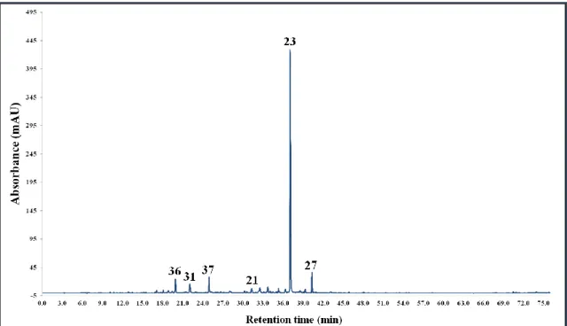
5.3. Characterisation of phenolics in the Corylus extracts by HPLC-MS ... 55
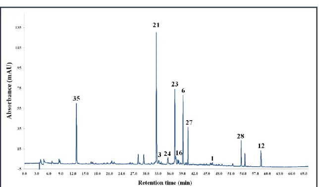
5.3.1. Corylus avellana L. ... 56
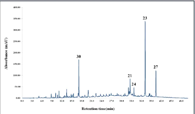
5.3.2. Corylus colurna L... 60
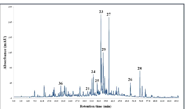
5.3.3. Corylus maxima Mill. ... 64
5.4. Quantitative analyses by HPLC-MS/MS... 68
5.4.1. Method validation... 68
5.4.2. Quantitative results ... 70
5.5. HPLC-based DPPH scavenging assay... 71
6. DISCUSSION ... 73
6.1. Quantitative phytochemical analyses ... 73
6.2. Antioxidant activity assays ... 74
6.3. Characterisation of phenolics in the Corylus extracts by HPLC-MS ... 77
6.3.1. Diarylheptanoids... 77
5
6.3.2. Flavonoids ... 85
6.3.3. Other compounds... 89
6.3.4. Comparison of the phenolic profile of the Corylus extracts... 92
6.4. Quantitative analyses by HPLC-MS/MS... 94
6.5. HPLC-based DPPH scavenging assay... 97
7. CONCLUSIONS ... 106
8. SUMMARY ... 109
9. ÖSSZEFOGLALÁS ... 110
10. BIBLIOGRAPHY ... 111
11.BIBLIOGRAPHY OF THE CANDIDATE’S PUBLICATIONS... 133
11.1. Publications related to the thesis... 133
11.2. Further scientific publications ... 134
12. ACKNOWLEDGEMENTS ... 135
13.APPENDIX ... 136
13.1. Diseases of different body systems taking effects from curcumin ... 136
13.2. Results of the HPLC-based DPPH assay... 142
6
1. L
IST OF ABBREVIATIONS4CL 4-coumarate-CoA ligase
ABTS 2,2'-azino-bis-(3-ethylbenzothiazoline-6-sulfonic acid) AD Alzheimer’s disease
AIC apoptotic proteinase-activating factor amu atomic mass unit
BDE bond dissociation energy C3H cinnamate-3-hydroxylase C4H cinnamate-4-hydroxylase
CABE Corylus avellana bark ethyl acetate extract CABM Corylus avellana bark methanolic extract CALE Corylus avellana leaves ethyl acetate extract CALM Corylus avellana leaves methanolic extract CCBE Corylus colurna bark ethyl acetate extract CCBM Corylus colurna bark methanolic extract CCLE Corylus colurna leaves ethyl acetate extract CCLM Corylus colurna leaves methanolic extract CE collision energy
CID collision- induced dissociation
CMBE Corylus maxima bark ethyl acetate extract CMBM Corylus maxima bark methanolic extract CMLE Corylus maxima leaves ethyl acetate extract CMLM Corylus maxima leaves methanolic extract
CoA coenzyme A
COX cyclooxygenase
CURS curcumin synthase
DAD diode array detector/detection DCS diketide-CoA synthase DMSO dimethyl sulfoxide DNA deoxyribonucleic acid
DPPH 2,2-diphenyl-1-picrylhydrazyl ERK extracellular signal-regulated kinase ESI electrospray ionisation
EtOAc ethyl acetate
GIT gastrointestinal tract HAT hydrogen atom transfer
HCT hydroxycinnamoyl transferase HDL high density lipoprotein HIF1 Hypoxia-inducible factor 1 HIV Human Immunodeficiency Virus
HPLC high-performance liquid chromatography
HPLC-MS high- performance liquid chromatography coupled with mass spectrometry
7 IC50 half maximal inhibitory concentration ICAM intercellular adhesion molecule IκB the inhibitor of κB kinase
IL interleukin
iNOS inducible nitric oxide synthase IP ionisation potential
LDL low density lipoprotein LOD limit of detection LOQ limit of quantitation
LOX lipoxygenase
LPS lipopolysaccharide
MeOH methanol
MIC minimum inhibitory concentration mRNA messenger ribonucleic acid MRM multiple reaction monitoring
MS mass spectrometry
MS/MS tandem mass spectrometry m/z mass-to-charge-ratio
NFAT nuclear factor of activated T-cells NF-κB nuclear factor-κB
NO nitric oxide
OMT O-methyltransferase PAF platelet-activating factor PAL phenylalanine ammonia- lyase Ph.Hg. Pharmacopoea Hungarica PI3K phosphoinositide 3-kinase ROS reactive oxygen species RNA ribonucleic acid
RNS reactive nitrogen species
RP-HPLC reversed-phase high-performance liquid chromatography SD standard deviation
SET single electron transfer SEPP selenoprotein P
Smac second mitochondria-derived activator of caspase Sp-1 specificity protein-1
TNF α tumour necrosis factor α TNF β tumour necrosis factor β TOF time-of-flight mass analyser UV ultra violet
VCAM vascular cell adhesion molecule VEGF vascular endothelial growth factor VLDL very-low-density lipoprotein [Y0-H]-• radical aglycone ion
Y0- aglycone ion
8
2. I
NTRODUCTIONNatural products have been used to treat human diseases since ancient times. In recent years compounds of plant origin still play an important role in therapeutic drug discovery by providing large chemical diversity and covering an alternative chemical space compared with synthetic derivatives. Consequently, nowadays a significant part of phytochemical researches focuses on screening for potential lead molecules among herbal extracts. Orientation of these researches is mainly based on ethnopharmacology:
identification of the major and minor bioactive constituents of traditionally used medicinal plants is a clearly promising way of discovering novel lead compounds. In addition, screening for structural analogues of natural lead molecules in different plants based on taxonomic relatedness also forms an important part of phytochemical studies.
However, the cost- and time-consuming procedures of isolation and identification of natural compounds with beneficial biological activity have limited their use in the pharmaceutical industry in the past two decades.
On the other hand, the popularity of herbal remedies is increasing nowadays which comes together with the upsurge of necessity to assure quality, efficacy and safety of these products. Since plant extracts are complex matrices with numerous different constituents, the development of sophisticated analytical methods is a crucial point in the quality control of phytotherapeutics.
Diode-array detection (DAD) and mass spectrometry (MS) together with high- performance liquid chromatography (HPLC) separation offers great selectivity and sensitivity for the qualitative and quantitative analysis of complex plant samples. In addition, coupling bioassays to these analytical procedures (mainly HPLC-MS) allows the rapid and efficient identification of the bioactive constituents of plant extracts.
The aim of our work was the phytochemical evaluation of the Corylus (Betulaceae) species native to Hungary: Corylus avellana L, Corylus colurna L. and Corylus maxima Mill. Despite the long-term use of these plants in traditional medicine, their phytochemical exploration is still incomplete. Previous studies on the phenolic constituents of C. avellana kernels and leaves focused on the main flavonoid and caffeic acid derivatives, while there is no report concerning the detailed phytochemical composition of C. colurna and C. maxima. Numerous studies of Betulaceae species
9
revealed that besides other phenolic constituents, diarylheptanoid-type compounds also show a frequent occurrence among these plants; however presence of these constituents is not reported in the Corylus species mentioned above. Diarylheptanoids have been proved to possess various pharmacological effects: e.g. antioxidant, anti-inflammatory, anticancer, anti-adipogenic and antiviral activities suggest their potential utilisation in clinical practice. Therefore, our experiments focused on screening for structural analogues of these compounds in the selected Corylus species.
HPLC-ESI-TOF-MS (high-performance liquid chromatography coupled to electrospray ionisation-time-of-flight mass spectrometry) and HPLC-ESI-MS/MS (high-performance liquid chromatography coupled to electrospray ionisation-tandem mass spectrometry) methods were chosen for the simultaneous structural characterisation of the phenolics present in the Corylus extracts.
Moreover, in order to broaden information on their biological activity, studies on in vitro radical scavenging activity were carried out.
The first chapters (2.1-2.3.) of the Literature overview summarise the most important knowledge about diarylheptanoids, the plant-derived compounds in the focus of our study. The following sections present the botanical, phytochemical and pharmacological properties of the Corylus (Betulaceae) species examined in order to search for new natural sources of the previously mentioned diarylheptanoid compounds (2.4-2.6).
10
2.1. Structural features and biosynthesis of diarylheptanoids
Diarylheptanoids are a group of plant-derived phenolic compounds bearing an 1,7- diphenylheptan skeleton (Fig. 1). Based on the structure of the C7 alkyl chain two main groups exist: linear- and cyclic-diarylheptanoids. Linear-diarylheptanoids can fatherly be classified into five sub-types: those possessing namely a heptane chain, an -oxy bridge or a flavonoid moiety, dimeric linear-diarylheptanoids and unusual structures, respectively (Lv and She 2011, Keserű and Nógrádi 1995). Furthermore, the variability of substituents in different positions on the alkyl chain and on the aromatic rings leads to numerous possible structure variants that can be associated with various biological effects.
Figure 1. Structure of the 1,7-diarylheptan skeleton of linear diarylheptanoids marking the possible positions of substituents marked with R1-11 (Lv and She 2011).
Until the present day, studies on the biosynthesis of diarylheptanoids are confined to curcuminoids, a group of diarylheptanoid-type molecules possessing the most promising biological effects. Investigations on the possible precursors of curcuminoid biosynthesis suggested very early that the backbone consists of two phenylpropanoids which are connected by an acetate derived carbon unit. However these studies did not manage to make clear distinction between two possible pathways: 1) starter phenylpropanoid-coenzyme A, five extensions with malonyl-CoA, then ring-closure and further modifications; and 2) biosynthesis from two phenylpropanoid-CoA units and one malonyl-CoA (Roughly and Whiting 1973). Much later Schröder proposed that the biosynthesis of these compounds might start with a type III polyketide synthase reaction (Schöder 1997). A more recent study (Katsuyama et al. 2009) on the biosynthesis of curcuminoids of Curcuma longa L. might confirm this presumption. The authors propose a pathway, which includes two novel type III polyketide synthases,
11
namely diketide-CoA synthase (DCS), and curcumin synthase (CURS). According to the report, the starter substrates, cinnamoyl-CoA, p-coumaroyl-CoA, or feruloyl-CoA, are synthesized from phenylalanine by the phenylalanine ammonia-lyase (PAL), the 4- coumarate-CoA ligase (4CL), the cinnamate-4-hydroxylase (C4H), the hydroxycinnamoyl transferase (HCT), the cinnamate-3-hydroxylase (C3H), and the O- methyltransferase (OMT). The formation of feruloyldiketide-CoA is catalyzed by the diketide-CoA synthase (DCS) via condensation of feruloyl-CoA and malonyl-CoA.
Besides, curcumin synthase (CURS) catalyzes the formation of curcuminoids from cinnamoyldiketide-N-acetylcystamine (a mimic of the CoA ester) and feruloyl-CoA (Fig. 2).
Figure 2. Biosynthesis of curcuminoids in Curcuma longa L. (obtained from Katsuyama et al. 2009)
12
2.2. Distribution of diarylheptanoids in the plant kingdom
The firstly discorvered diarylheptanoid, namely curcumin, was isolated from the traditional herbal remedy and dietary spice, turmeric (Curcuma longa L., Zingiberaceae) two hundred years ago by Vogel in 1815 (Vogel and Pelletier 1815).
Ever since hundreds (more than 400 different molecules) of new compounds with 1,7- diphenylheptan structures were identified in various plants. Nowadays, studies on natural sources of diarylheptanoids mainly focus on species of Zingiberaceae and Betulaceae families. Numerous plants of the Curcuma and Alpinia genera of the Zingiberaceae, and of the Alnus genus of Betulaceae have been proved to accumulate diarylheptanoids as secondary metabolites (Lv and She 2011). Furthermore, the presence of diarylheptanoids was also reported in species of the Betula (Betulaceae) (Matsuda et al. 2008, Lee at al. 2012, Mshvildadze et al. 2007), Myrica (Myricaceae) (Kim et al. 2014, Ting et al. 2014, Yoshimura et al. 2012, Akazawa et al. 2010, Wang and Liu 2008, Morihara et al. 1997) Cymodocea (Cymodeaceae) (Kontiza et al 2008, Kontiza et al 2005), Juglans (Juglandaceae) (Yao et al. 2015, Yang et al. 2011, Li et al.
2008, Gao et al. 2003), Acer (Aceraceae) (Akihisa et al. 2012, Yonezawa et al. 2011), and Pyrostria (Rubiaceae) (Beniddir et al. 2012) genera. The distribution of diarylheptanoids in various plants is presented in Figure A1., including 102 compounds but limited to the most relevant species.
Nonetheless, researches on new natural sources of diarylheptanoids are still of great interest nowadays, since these molecules are considered as potential therapeutic agents due to their several beneficial physiological effects (see section 2.3.). These include anti-inflammatory, antioxidant, anti-tumour, leishmanicidal, melanogenesis inhibitor, hepatoprotective and neuroprotective activities (Lv and She 2011).
The next chapter (2.3.) summarises the most recent knowledge on biological activities of diarylheptanoids isolated from various plant species.
The structures of the most representative diarylheptanoid aglycones for the species discussed in the next chapters are depicted in this section in Figure 3 (i1-i21).
13
Figure 3. Structures of the most representative diarylheptanoids for the species discussed in section 2.3.
14
2.3. Biological activities of diarylheptanoids
2.3.1. Curcuma genus (Zingiberaceae)
Considering biological activities, there is no doubt that the most promising and explored molecules of this type are curcumin and its analogues. Curcumin presents almost all the effects that can be associated with diarylheptanoid compounds (summarised in Table A1 in section 13.1.), therefore, the first part of the discussion focuses on these activities by introducing the pharmacological properties of curcumin.
Curcumin (Fig. 4) is a diarylheptanoid-type polyphenol responsible for the yellow colour of turmeric (Curcuma longa L., Zingiberaceae), a curry spice. The yellow- pigmented fraction of turmeric contains also other curcuminoids mainly demethoxycurcumin and bis-demethoxycurcumin that are related to its principal ingredient, curcumin (Wongcharoen and Phrommintikul 2009).
Figure 4. Structures of curcumin, demethoxycurcumin and bis-demethoxycurcumin.
Curcumin has been shown to possess several beneficial biological activities via interaction with various molecular targets (Fig. 5). Taking a look at the site http://clinicaltrials.gov (September, 2015) clearly indicates its promising therapeutic potential: 124 clinical trials are registered using curcumin, some completed, some still ongoing and some recruiting patients. The most relevant clinical trials (Phase II or III) investigate its efficacy in different types of cancer, such as colon carcinoma and pancreas carcinoma in monocomponent therapy or in combination with conventional
15
chemotherapeutics, e.g. gemcitabine; and in cognitive dysfunctions related to schizophrenia and Alzheimer’s disease. These human studies also confirmed the non- toxicity of curcumin up to oral dose of 8 g pro die.
Figure 5. Molecular targets of curcumin
Abbreviations: NF-κB, nuclear factor-kappa B; AP-1, activating protein1; STAT, signal transducers and activators of transcription; Nrf-2, nuclear factor 2-related factor; Egr-1, early growth response gene-1;
PPAR-γ, peroxisome proliferator-activated receptor-gamma; CBP, CREB-binding protein; EpRE; CTGF, connective tissue growth factor; EGF, epidermal growth factor; EGFRK, epidermal growth factor receptor-kinase; FGF, fibroblast growth factor; HGF, hepatocyte growth factor; NGF, nerve growth factor; PDGF, platelet-derived growth factor; TGF-β1, transforming growth factor-β1; VEGF, vascular endothelial growth factor; AR, androgen recepto r; Arh-R, aryl hydrocarbon receptor; DR-5, death receptor-5; EGF-R, epidermal growth factor-receptor; EPC-R, endothelial protein C-receptor; ER-α, estrogen receptor-alpha; Fas-R, Fas receptor; H2-R, histamine (2)- receptor; InsP3-R, inositol 1,4,5- triphosphate receptor; IR, integrin receptor; IL-8-R, interleukin 8-receptor; LDL-R, low density lipoprotein-receptor; MMP, matrix metalloproteinase; TIMP, tissue inhibitor of metalloproteinase -3;
iNOS, inducible nitric oxide oxidase; COX-2, cyclooxygenase-2; LOX, lipoxygenase; Gcl, glutamatecysteine ligase; NAT, arylamine N-acetyltransferases; IAP, inhibitory apoptosis protein; HSP- 70, heat-shock protein 70; TNF-α, tumour necrosis factor alpha; IL, interleukin; MCP, monocyte chemoattractant protein; MIF, migration inhibition protein; MIP, macrophage inflammatory protein;
ERK, extracellular receptor kinase; IARK, IL-1 receptor-associated kinase; cAK, autophosphorylation - activated protein kinase; CDPK, Ca2+-dependent protein kinase; cPK, protamine
kinase; JAK, janus kinase; JNK, c-jun N-terminal kinase; MAPK, mitogen-activated protein kinase; TK, protein tyrosine kinase; FAK, focal adhesion kinase; PhK, phosphorylase kinase; pp60c -src, pp60c-src tyrosine kinase; PKA, protein kinase A; PKB, protein kinase B; PKC, protein kinase C; FPTase, farnesyl protein transferase; GST, glutathione S-transferase; HO, hemeoxygenase; ICAM-1, intracellular adhesion molecule-1; VCAM-1, vascular cell adhesion molecule-1; ELAM-1, endothelial leukocyte adhesion molecule-1; SHP-2, Src homology 2 domain-containing tyrosine phosphatase 2, uPA, urokinase-type plasminogen activator. (obtained from Noorafshan and Ahkani-Esfahani 2013 without modification).
16
2.3.1.1. Structural features and bioavailability of curcumin
Curcumin, 1,7-bis-(4-hydroxy-3methoxyphenyl)-1,6-heptadien-3,5-one, is a small, symmetric and lipophilic molecule, which allows its rapid permeation through biological membranes. The simple molecular structure with various synthetically accessible positions makes it an outstanding target for structure-activity relationship and lead optimisation studies (Noorafshan and Ahkani-Esfahani 2013). Since curcumin shows poor bioavailability and stability due to limited absorption, rapid metabolism and fast systemic elimination (Anand et al. 2007), several analogues have been introduced and investigated in order to improve its bioavailability, selectivity and stability. Besides, pre-formulation approaches have also been performed including the use of adjuvants such as piperine, that attenuates glucuronidation in the liver, the utilisation of cyclodextrines, liposomal curcumin and curcumin-loaded nanoparticles (Dey et al.
2016, Menon et al. 2015, Patial et al. 2015). These studies aim not only the improvement of stability, but also the transportation to the target molecules, e.g.
penetration across the blood-brain barrier that is essential for the efficacy in pathological states of the central nervoius system (Mourtas et al. 2014, Cheng et al.
2013, Quitschke et al. 2013).
The next chapters (2.3.1.2.-2.3.1.6) discuss the mechanisms of the most important biological activities of curcumin in details: the antioxidant, anti-inflammatory and anti- tumoural effects and the positive impact on neurodegenaretive diseases that contribute to almost all the therapeutic utilisation approaches of the compound. The sections start with a brief summary of the molecular mechanisms of oxidative stress, inflammation apoptosis and neurodegeneration, the processes related to the previously mentioned activities.
2.3.1.2. Antioxidant activity of curcumin
The main factor contributing to the development of the oxidative stress is the production of free radical species. The eukaryotic cells present several protection mechanisms against free radicals. The primary defence system involves enzymes, such as the superoxide-dismutase (SOD), the catalases, peroxydases, the glutathione-S-transferase, DT-diaforase and reductases. The secondary defence system consists of the vitamins
17
(vitamin-A -C -E -K) and cofactors (e.g. coenzyme Q) possessing scavenger activity, carotenoids, cysteine and methionin. Trace elements, such as Cu, Zn, Mn and Se and small dietary molecules, e.g. flavonoids also play important role in the cell protection mechanism against free radicals. Nevertheless, oxidative stress emerges if the balance between the free radical producing and the defence mechanisms is disrupted. It is considered one of the main risk factors for the development of various illnesses including diabetes, cancer, cardiovascular and neurodegenerative diseases. Several studies have been performed in order to investigate the positive pharmacological effects of curcumin in the above mentioned pathological states. The antioxidant potential of curcumin plays a major role in these activities. It is reported to be a highly potent free radical scavenger, a reducing agent and DNA damage inhibitor (Antunes et al. 2005, Patro et al. 2002, Jovanovic et al. 2001). It has been proved that curcumin inhibits nitric oxide (NO) and reactive oxygen species (ROS) production in macrophages. Besides, oxidative stimulation of G-proteins in human brain tissues is attenuated by this compound, as well as lipid peroxydation in liver microsomes of rats (Hatcher et al.
2008).
2.3.1.3. Anti-inflammatory effect of curcumin
Inflammation plays dual role in the human organism. On the one hand it forms an important part of the defence system that aims the elimination of the extrinsic and intrinsic harmful effects; on the other hand many substances released during the inflammation process have tissue-damaging effects, therefore, chronic inflammation can lead to the impair of normal tissue integrity. The initiation of the process can be attributed to exogenous and also endogenous stimuli. These cause the release of histamine from the basophile granulocytes and the mast cells, and also the liberation of arachidonic acid from the plasma membrane via the phospholipase A2 enzyme.
Arachidonic acid is converted to prostaglandynes and leukotriens by the cycloxygenase (COX) and lipoxygenase (LOX) enzymes, respectively. These play important role in the emergence of pain, inflammation and fever. Besides, adhesion molecules such as intercellular adhesion molecule (ICAM1) and the vascular cell adhesion molecule (VCAM1) also contribute to the process by facilitating the accumulation of leucocytes, platelets and the endothelial cells in the inflammation centrum. The role of cytokines in
18
inflammation is diverse; the IL-2, IL-6, IL-8, IL-12, IL-18, the tumour necrosis factor α and β, and the interferonγ are considered as pro-inflammatory cytokines; while the IL-4, IL-10, IL-13 have anti-inflammatory effect. The nuclear factor-κB (NF-κB) is considered one of the main factors contributing to the inflammation process. It is a dimeric transcription factor that induces the expression of several genes that are responsible for the production of inflammatory agents, such as cytokines: the IL-1β, IL- 2 and TNF-α; adhesion molecules and enzymes: the inducible nitric oxide synthase (iNOS) the COX-2 and 5-LOX. The NF-κB is present in the cells in inactive form; the activation is induced by extracellular factors such as UV-light, or the IL-1. In the non- stimulated cells the NF-κB is placed in the cytoplasm, bounded to its inhibitor, the inhibitor of κB-kinase (IκB). Pro-inflammatoric effects cause the phosphorilation of IκB, whereupon it gets an ubiquitine signal and degrades in the proteosome, thus giving the ability to the NF-κB to enter the nucleus and exert its effect. This inflammatory process plays pivotal role in the pathogenesis of chronic diseases, such as rheumatoid arthritis, arteriosclerosis, asthma or the Helicobacter pylori gastritis. Nitric oxide (NO) is responsible for several processes occurring in acute inflammation, by causing vasodilatation, increasing capillary permeability, thus enhancing the acute inflammatory reaction and stimulating the generation of prostaglandynes. In this case it is synthesised by the iNOS enzyme (Gyires 2007).
Members of the Curcuma genus have been used to treat inflammatory malfunctions for thousands of years. Since the isolation and identification of curcumin, the main compounds present in the rhizome of these plants, it has been considered to be primarily responsible for this anti-inflammatory effect. Curcumin has been shown to attenuate the activation of the NF-κB, as well as other activation pathways, such as NO generation and COX-2, 5-LOX expression (Ma et al. 2015, Bengmark 2006). It also induces down- regulation of several pro-inflammatory cytokines, such as TNF, IL-1, IL-8, interferonγ and other chemokines (Gao et al. 2004, Surh 2002). Human trials also proved the anti- inflammatory effect of curcumin in many inflammatory diseases (http://clinicaltrials.gov) affecting different body systems (Table A1).
19 2.3.1.4. Anti-tumoural properties of curcumin
The most promising results regarding the therapeutic effects of curcumin are related to its anticancer activity. Several ongoing clinical trials (http://clinicaltrials.gov) strengthen the relevance of this statement.
Most of the reports agree that the anti-inflammatory, antioxidant, apoptosis inducing and anti-angiogenic activities all contribute to its anti-tumoural effect (Shanmugam et al. 2011, Bengmark et al. 2009, Kunnumakkara et al. 2008, Thomasset et al. 2007, Bemis et al. 2006).
Apoptosis is an intrinsic programme that leads to cell-death. It is governed by different signal-transduction mechanisms that can be initiated two different ways. In the extrinsic process the so-called death ligands bind to the death receptors, such as CD95 or TRAIL that leads to the activation of caspase-8, which transmits the “death signal” to the effector caspases, e.g. caspase-3. During the intrinsic process apogenetic factors, such as cytochrome c, the second mitochondria-derived activator of caspase (Smac) or the apoptotic proteinase-activating factor (AIF) are transferred to the cytosol from the mictochondrial intermembrane space. This also promotes the production of effector caspases. The increase in the permeability of mitochondrial membranes play significant role in this process. The substrates of the effector caspases are enzymes, such as the endonucleases, nuclear lamines, the PARP (the DNS-dependent polymer kinase), gelsolin and fodrin. The activation of the former initiates the farther processes of apoptosis and consequently, leads to cell-death (Saelens et al. 2004).
As it was previously mentioned, curcumin down-regulates the NF-κB transcription factor, which results in the suppression of BcL-2 and Bcl-XL anti-apoptotic genes, thus the promotion of apoptosis induction (Sandur et al. 2007). Curcumin also inhibits the Akt protein kinase, an other apoptosis inhibitor enzyme (Yamaguchi et al. 2001). The enhanced expression of p53 gene, an apoptosis mediator, was also observed after administration of curcumin in several cancer cells (human basal cells, human hepatoblastoma and human breast cancer cells (Choudhuri et al 2005). This effect was revealed to be tissue-specific based on the fact that in colorectal carcinoma cells the p53 expression decreased, while the heat-shock protein 70 level increased after curcumin treatment (Bush et al. 2001). Recent studies have shown that the suppression of Sp-1
20
and its downstream targets, calmodulin and SEPP1 may also contribute to the anti- apoptotic effect of curcumin (Vallianou et al 2015).
Besides the anti-apoptotic effect, other cell death mechanisms have also been published related to the anticancer effect of curcumin (Gali-Muhtasib et al. 2015). These include autophagy that has been shown to play role in the anti-tumour effect on, e.g. glioma, colorectal, breast carcinoma, leukemia and ovarian carcinoma. This activity is based on modification of different signalling mechanisms (p53 degradation, inhibition of Act kinase, activation of ERK1/2) and also the generation of ROS. The other non anti- apoptotic effect of curcumin that leads to cell death is programmed necrosis. This was characterised by the induction of ROS and caspase-independent cell death. This activity was proved in prostate, bladder, colorectal, medulloblastoma, pancreatic and cervical cancer cells. Curcumin has been shown to act trough senescence, which implicates morphological, functional, and behavioural modifications and irreversible growth arrest.
The importance of this mechanism was proved in breast cancer cells.
COX-2 inhibition is an other effect that is considered to play role in the anti-tumoural activity of curcumin, which was studied in the treatment of colonic tumours (Hatcher et al. 2008).
The enhanced survival, growth and metastasis of tumour cells mainly depend on angiogenesis. Curcumin was shown to attenuate many sub-processes involved in this process. These include the inhibition of the fibroblast growth factor-induced neurovascularisation, ligands of vascular endothelial growth factor, and angiopoitein 1 and 2. Curcumin also has the ability to down-regulate adhesion molecules, such as the leukocyte adhesion molecule-1, intracellular adhesion molecule-1 and vascular cell adhesion molecule-1 (Bhandarkar et al. 2007).
2.3.1.5. The positive effects of curcumin in neurodegenerative diseases
The protective effect of curcumin in neurodegenerative diseases, such as Alzheimer’s and Parkinson’s disease has been proved in vivo (Fu et al. 2015, Ghosh et al. 2015, Zhang et al. 2015).
Alzheimer’s disease (AD), a progressive neurodegenerative brain disorder, affects more and more elderly people in the world. There is an ongoing scientific debate upon the pathomechansim of AD, whether the amyloid-β plaques or the phosphorylated tau-
21
protein is mainly responsible for the neurodegeneration. However, there is consensus among researchers regarding the important role of oxidative damage and the abnormal accumulation of metal ions in several neurodegenerative diseases. Besides the notable antioxidant activity of curcumin that is originated from the free radical scavenging effect, inhibition of lipid peroxydation and increment of SOD action; it also has the ability to chelate metal ions, also to down regulate the secretion of amyloid peptide and prevent amyloid toxicity on neurons (Chen et al. 2011). Curcumin-conjugated nanoliposomes showed even higher affinity for amyloid-β deposits than curcumin, which makes this preformulation approach suitable for diagnostic and therapeutic applications as well (Lazar et al. 2013).
Dementia is one of the main symptoms of AD and also several other neuronal diseases.
Curcumin has been reported to improve memory functions in both animal models and human trials. The mechanism of action includes the antioxidant activity, cholinesterase inhibition and also effect on brain insulin receptors (Noorafshan and Ahkani-Esfahani 2013).
2.3.1.6. The dark side of curcumin
In the last decade, several papers have been published reporting the beneficial biological effects of curcumin in various diseases (see above). Numerous clinical trials are ongoing with this natural product (http://clinicaltrials.gov) in order to evaluate its activity. However, to see clear it is important to deal with its negative properties as well.
One of the most important doubts about the efficacy of curcumin is that most of the evidence that support its therapeutic potential is based on in vitro data. In these studies it was tested in micromolar concentrations, but limited absorption, rapid metabolism in the intestine and liver, as well as fast systemic elimination result in very low plasma concentrations, typically in the nanomolar range (Ireson et al. 2001, Ireson et al. 2002).
These data suggest that the therapeutic potential of curcumin administered per os is limited. Although, as it was previously mentioned, several approaches have been introduced for increasing the bioavailability of curcumin.
Curcumin is facing clinical trials in order to test its potential to sensitise tumour cells to the effects of conventional chemotherapeutics, e.g. gemctiabine. However it has been previously reported that curcumin can either increase or decrease the efficiency of
22
chemotherapy depending the concentration utilised (Somasundaram et al. 2002), thus the outcome of these studies is uncertain.
Although curcumin is considered non-toxic, some evidence contradicts this statement.
In 1993 the National Toxicology Program published an extensive study on the carcinogenic properties of turmeric containing 80-95% curcumin. Rats and mice were treated with turmeric administered per os for two years. The report concluded that there was equivocal evidence of the carcinogenetic activity of curcumin, based on the increased incidences of glitorial gland adenomas, carcinomas of the small intestine and hepatocellular adenomas. This effect might be contributed to the ability of curcumin to increase topoisomerase II-mediated DNA damage, and inactivation of p53 tumour suppression factor (Burgos-Moron et al. 2010). It has also been proved that high concentrations of curcumin can increase ROS levels by irreversibly modifying the antioxidant enzyme thioredoxin reductase (Fang et al. 2005) that can also play role in the carcinogenetic effect. The average daily those of curcumin in these studies was circa 0.2 mg/kg body weight (NTP 1993), while in human studies it can reach 1g/kg body weight.
The next chapters (2.3.2.-2.3.7.) briefly introduce the remaining important plant genera reported to contain diarylheptanoid compounds that have been proven to show beneficial biological activities. In the final sections, the Betulaceae species belonging to the Alnus and Betula generea are presented as the closest relatives of the investigated Corylus species.
2.3.2. Alpinia genus (Zingiberaceae)
Alpinia is a genus of the Zingiberaceae family, which comprises flowering plants native to Asia, Australia, and the Pacific Islands, where they occur in tropical and subtropical climates. It is named after Prospero Alpini, a 17th-century Italian botanist specialized in exotic plants (Bruneton 2001). Some members of the genus were reported to accumulate diarylheptanoids with various pharmacological effects (see sections 2.3.2.1-2.3.2.6).
23 2.3.2.1. Effect on neuronal differentiation
A diarylheptanoid from the plant Alpinia officinarum Hance 7-(4-hydroxyphenyl)-1- phenyl-4-hepten-3-one (i1), exhibited potent activities on neuronal differentiation and neurite outgrowth. It induced differentiation of neuroblastoma cells into a neuron-like morphology, and accelerated the establishment of axon-dendrite polarization of cultured hippocampal neurons. Moreover, it promoted neurite extension in both Neuro-2a cells and neurons. The authors showed that the effects on neuronal differentiation and neurite growth were specifically dependent on the activation of extracellular signal-regulated kinases (ERKs) and phosphoinositide 3-kinase (PI3K)-Akt signaling pathways.
Importantly, intraperitoneal administration of the diarylheptanoid promoted the differentiation of new-born progenitor cells into mature neurons in the adult hippocampal dentate gyrus (Tang et al 2015).
2.3.2.2. Selective cytotoxic effect
Two dimeric diarylheptanoids, namely alpinin C and D that were isolated from the rhizomes of Alpinia officinarum were evaluated for their cytotoxicity against human tumour cell lines HepG2, MCF-7, T98G and B16-F10. Alpinin C showed notable and selective cytotoxicity against cell lines of MCF-7 and T98G with IC50 values of 8.46 and 22.68 µg/ml, respectively (Liu et al. 2014). Bioassay-guided fractionation of the cytotoxic MeOH extract from the rhizomes of Alpinia officinarum Hance led to the isolation of two new diarylheptanoids named alpinoid D (i2) and E (i3), together with fifteen known linear diarylheptanoids. The cytotoxic activity of the isolated diarylheptanoids was evaluated against the IMR-32 human neuroblastoma cell line.
Among the tested compounds, 5-hydroxy-1-(4–hydroxy-3-methoxy)phenyl-7- phenlyheptan, 5-methoxy1-(4–hydroxy-3-methoxy)phenyl-7-phenlyheptan, and 1-(4–
hydroxy-3-methoxy)-phenyl-7-phenly-hept-4-3-one exhibited the most potent activities with IC50 values of 0.83, 0.23 and 0.11 μM, respectively. The authors could conclude that the linear diarylheptanoids possessing a methoxyl at C3 and a hydroxyl function at C4 on the benzene ring were essential for potent cytotoxic activity (Sun et al. 2008).
24 2.3.2.3. Antibacterial effect
Three diarylheptanoids that were isolated from the ethanolic extract of the rhizomes of Alpinia officinarum by Zhang et al were elucidated as 7-(4,5-dihydroxy-3- methoxyphenyl)-1-phenyl-4-heptene-3-one (i4), 1,7-diphenyl-5-heptene-3-one (i5) and 4-phenethyl-1,7-diphenyl-1-heptene-3,5-dione (i6). All of the compounds showed antibacterial activity against Helicobacter pylori with MIC values of 9-30 μg/ml (Zhang et al. 2010). These results suggest the potential use of the investigated diarylheptanoids for the treatment of peptic ulcer and related diseases.
2.3.2.4. Platelet-activating factor receptor binding inhibitory activity
The bioassay-guided purification of ether extracts of Alpinia officinarum led to the isolation of two new compounds 6-hydroxy-1,7-diphenyl-4-en-3-heptanone (i7) and 6- (2-hydroxy-phenyl)-4-methoxy-2-pyrone as well as two known diarylheptanoid compounds 1,7-diphenyl-4-en-3-heptanone (i1) and 1,7-diphenyl-5-methoxy-3- heptanone (i8). All three diarylheptanoids exhibited potent platelet-activating factor (PAF) receptor binding inhibitory activities with an IC50 of 1.3, 5.0, and 1.6 μM, respectively. The authors concluded that their studies have identified diarylheptanoids as a novel class of potent PAF antagonists (Fan et al. 2007).
2.3.2.5. Anti-angiogenic activity
Gao et al. investigated the anti-angiogenic activity of two diarylheptanoids, namely Yakuchinone A (i9) and B (i10) isolated from the fruit of Alpinia oxyphylla Miq., together with a structure analogue, curcumin. The activity and toxicity of these three compounds were compared using transgenic zebrafish as in vivo model and human umbilical vein endothelial cell as in vitro model. The results suggested that in both in vitro and in vivo assays, curcumin exerted the most potent anti-angiogenic effect with the lowest toxicity among these compounds; Yakuchinone A was the second potent;
Yakuchinone B has the lowest activity but with the highest toxicity in all three compounds. (Gao et al. 2015).
25 2.3.2.6. Anti-inflammatory activity
The in vitro iNOS inhibitor properties in lipopolysaccharide-activated macrophages of the extract prepared with acetone from the rhizome Alpinia officinarum has also been reported (IC50 of 35μg/ml) (Matsuda et al. 2006). The successive isolation of the flavonoid galangin and two diarylheptanoid compounds [7-(4″-hydroxy-3″- methoxyphenyl)-1-phenylhept-4-en-3-one (i11) and 3,5-dihydroxy-1,7-diphenylheptane (i12) has also been carried out. Both the compounds inhibited NO production in LPS- activated mouse peritoneal macrophages with IC50 values of 62, 55 and 33 μM, respectively. Investigation of the structure-activity relationships regarding the previously mentioned compounds together with other diarylheptanoids led to the following conclusions: an enone moiety at the 3-5 positions suggested to be important for the activity; methylation of the 4’,4”-hydroxyl groups tended to reduce the effect;
the double bonds and/or enone moiety at the 1-7 positions are considered important for the activity. The authors concluded that the diarylheptanoid compounds can contribute to the iNOS inhibitor, thus anti-inflammatory activity of the extract.
2.3.3. Morella and Myrica genera (Myricaceae)
Myrica and Morella species are taxonomically very closely related trees or shrubs with edible fruit that exhibit relevant applications in traditional medicine. Extracts of the roots, bark and fruit have been used for the treatment of various diseases, such as diarrhoea, stomach pain, bleeding, asthma, coughing, headache, fevers and inflammation. Several different cyclic diarylheptanoids have been identified in the plants, e.g. Morella adenophora Hance, Morella arbores Hutch, Morella nana A.
Chev., Morella cerifera L., Myrica gale L. and Myrica rubra Lour (Silva et al. 2015).
2.3.3.1. Antioxidant activity
Antioxidant activity of Myrica and Morella extracts and 36 isolated compounds (diarylheptanoids, flavonoids and pentacyclic triterpenoids) have been investigated in several in vitro assays, e.g. the DPPH, ABTS and nitroblue tetrazolium tests, with the most often used positive control being ascorbic acid. 13 components were found to be more potent antioxidant than ascorbic acid in the DPPH test. The analyses of the results
26
obtained in this assay allowed the authors to draw conclusions about diarylheptanoid antioxidant action: 1) only two investigated compounds, Myricananin C (i13) and Myricanol (i14) 5-O-β-D-(6’-O-galloyl)-glucopyranoside exhibited IC50 values below 20 μM and were more active than ascorbic acid; 2) a hydroxyl group at C-11 position instead of carbonyl did not improve the activity; 3) an extra hydroxyl group at carbon C-5 is also irrelevant; 4) the loss of a methyl group causes a strong increase in the antioxidant effect; 5) it seemed that the presence of a sugar moiety, as well as the type and localisation of the sugar also interfere with the antioxidant activity (Silva et al.
2015).
2.3.3.2. Anti-inflammatory activity
According to the results of several studies, the anti-inflammatory activity of Myrica and Morella extracts seems to be remarkable. The diarylheptanoids isolated from these extracts, myricanone (i15) and myricanol (i14) were proved to be very active iNOS inhibitors; Myricanin A showed TNFα inhibitory effect, while Juglanin-BB-11-O- sulphate decreased IL-6 levels (Silva et al. 2015), typically with IC50 values being in the micromolar range.
2.3.3.3. Anticancer activity
A study carried out by Dai et al. explored the inhibitory effect and mechanism of myricanol (i14) on lung adenocarcinoma xenografts in nude mice. The results showed that the protein expression of Bcl-2, VEGF, HIF-1α, and survivin were consistently downregulated, whereas that of Bax was upregulated after myricanol treatment.
Myricanol also significantly upregulated the mRNA expression of Bax and downregulated that of Bcl-2, VEGF, HIF-1α, and survivin in a dose-dependent manner.
These data suggested that myricanol could significantly decelerate tumour growth in vivo by inducing apoptosis (Dai et al. 2015).
27
2.3.3.4. Positive effect in neurodegenerative diseases
Microtubule-associated protein tau accumulates in more than 15 neurodegenerative diseases and is most closely linked with postsymptomatic progression in AD. An extract from Myrica cerifera potently reduced both endogenous and overexpressed tau protein levels in cells and murine brain slices. The bayberry flavonoids myricetin and myricitrin were confirmed to contribute to this potency, but a diarylheptanoid, myricanol (i14), was the most effective anti-tau component in the extract, with potency approaching the best targeted lead therapies. (+)-aR,11S-Myricanol, isolated from M. cerifera as the naturally occurring aglycone, was significantly more potent than commercially available (±)-myricanol. Accordingly, myricanol may represent a novel scaffold for drug development efforts targeting tau turnover in AD (Jones et al. 2011).
2.3.4. Acer genus (Aceraceae) 2.3.4.1. Anti-inflammatory effect
Acer nikoense Maxim. is a small deciduous tree native to Japan and China. In the traditional medicine it has been used to treat hepatic malfunctions and eye diseases.
The extracts of the bark were proved to contain several polyphenol compounds, including diarylheptanoids, such as acerogenin and acerosides. In vitro studies have proved the degranulation inhibitor effect of the extracts on basophil granulocytes, besides; they attenuated NO synthesis in macrophages (Akihisa et al 2006).
2.3.4.2. Effect on osteoblast differentiation
Osteogenic activity of six diarylheptanoids, acerogenin A (i16), (R)-acerogenin B (i17), aceroside I (i18), aceroside B1 aceroside III and (-) centrolobol and two phenolic compounds; (+)-rhododendrol and (+)-cathechin, isolated from the stem bark of Acer nikoense (Nikko maple) was evaluated by Yonezawa et al., using alkaline phosphatase (ALP) activity as a marker for early osteoblast differentiation. The diphenyl ether-type cyclic diarylheptanoids promoted ALP activity in mouse preosteoblastic MC3T3-E1 cells without affecting cell proliferation, but the linear-type diarylheptanoid (-) centrolobol and the investigated other phenolic compounds did not. Diphenyl ether-type
28
cyclic diarylheptanoids also increased protein production of osteocalcin, a late stage marker for osteoblast differentiation, and induced osteoblastic mineralization.
Structure–activity relationships of these compounds demonstrated that the stimulative efficacy of aglycones was higher than that of its glycosides. The authors speculated that this phenomenon is caused by differences in cell membrane permeability between glycosilated compounds and aglycones. Taken together, diphenyl ether-type cyclic diarylheptanoids promote early- and late-stage osteoblastogenesis, which may open the possibility for the development of novel osteogenic agents (Yonezawa et al. 2011).
2.3.4.3. Anti-diabetic effect
The Na+-glucose cotransporter (SGLT) is a membrane protein that plays an important role in the re-absorption of glucose in the kidneys. SGLT is known to have three isoforms (SGLT1, SGLT2, and SGLT3). The inhibition of SGLT results in a decrease in blood sugar level. Cyclic diarylheptanoids acerogenin A (i16) and acerogenin B (i17) isolated from the methanolic extract of Acer nikoense bark have shown selective inhibitory effect on the sodium-glucose transporter (with IC50 values of 20 and 26 µM, respectively). The authors proved that the extent of this effect might depend on the position of the sugar moieties and the stereochemistry of the molecules (Morita et al.
2010). The appropriate torsion between two aromatic planes as well as their conformation might be important to show inhbition of SGLT.
2.3.5. Juglans genus (Juglandaceae)
The plant genus Juglans is the type genus of the family Juglandaceae. The 21 species in the genus range across the north temperate from southeast Europe, and more widely in the amercian continent from southeast Canada south to Argentina. The seeds of the trees belonging to the genus are referred to as walnuts (Rushford 1999).
2.3.5.1. Neuroprotective and antioxidant effect
A diarylheptanoid, juglanin C, was isolated from the 80 % methanolic extract of the leaves and twigs of Juglans sinensis Dode together with three known diarylheptanoids, juglanin A (i19), juglanin B (i20), and (5R)-5-hydroxy-7-(4- hydroxy-3- methoxyphenyl)-1(4-hydroxyphenyl)-3-heptanone (i21) by Yang et al., using
29
bioactivity-guided fractionation and chromatographic techniques. Juglanin C and A have shown notable neuroprotective activity against glutamate-induced toxicity in HT22 cells. These two diarylheptanoids significantly reduced the overproduction of cellular peroxyde in glutamate-injured HT22 cells and significantly maintained antioxidative defence systems, including glutathione, glutathione reductase, and glutathione peroxydase, under glutamate- induced oxidative stress in HT22 cells (Yang et al. 2011).
2.3.6. Alnus genus (Betulaceae)
Almost all the species belonging in Alnus genus have been traditionally used in Ayurveda, Unani, and Chinese folk medicine; traditional Korean medicine utilised them for haemorrhage, burn injuries, fever, diarrhoea and alcoholism; contemporary indigenous healers also use primarily the bark of Alnus species for the preparation of various therapeutical teas (Turner and Hedba 1990).
Different parts of the plants like stems, bark, seeds, leaves, roots, fruits, tree cones, buds and blossoms are known to possess therapeutical activity and they are characterised by a remarkable number of compounds such as triterpenoids, saponins, flavonoids, diarylheptanoids, phenols, steroids and tannins (Sati et al. 2011).
These molecules exhibited antioxidant (Dinic et al. 2014), anti-inflammatory (Lai et al.
2012), antiviral (Tung et al. 2010), cytotoxic (León-Gonzales et al. 2014, Novaković et al. 2014, Choi et al. 2008), inhibitory activity against nuclear factor kappa activation, nitric oxide and tumour necrosis factor-production, human umbilical vein endothelial cells, farnesyl protein transferase, cell-mediate low density lipoprotein oxidation, and HIV -1 - induced cytopathic effect in MT-4 cells (Sati et al. 2011).
2.3.6.1. Antioxidant activity
Evaluation of the antioxidant activity of two diarylheptanoids, platyphylloside 5(S)-1,7- di(4-hydroxyphenyl)-3-heptanone-5-O-β-D-glucopyranoside and its analogue, 1,7-di(4-hydroxyphenyl)-5-O-β-D-[6-(E-p-coumaroylglucopyranosyl)]heptane-3-one, both isolated from the bark of Alnus glutinosa L. has been carried out by Dinic et al.
(Dinic et al. 2014). The published results indicated that neutralization of reactive oxygen species is an important mechanism of diarylheptanoid action, although these compounds exert a considerable anticancer effect. Therefore, the authors concluded that
30
these compounds may serve as protectors of normal cells during chemotherapy without significantly diminishing the effect of the applied chemotherapeutics.
Antioxidant and antimicrobial activities of methanolic extracts from the leaves and barks of three Alnus species (Alnus glutinosa L., Alnus incana L. and Alnus viridis Chaix) was evaluated by Dahija et al. Antioxidant activity of the extracts was determined using the DPPH radical scavenging method. All of the extracts showed antioxidant activity higher than that of thymol, which was used as a positive control (Dahija et al. 2014).
The ethanolic extract prepared from the leaves of Alnus formosana Burk showed notable antioxidant activity in in vitro assays using DPPH, hydroxyl and superoxide free radicals (Lee et al. 2006). This study did not include the identification of the antioxidant constituents, but other papers reported the presence of oregonin (2) as the main compound in Alnus formosana bark extracts (Lee et al. 2005), which previously has been proved to be a potent antioxidant (Keserű and Nógrádi 1995).
Oregonin (2) and hirsutenone (1) (see structures in section 6.3.1.) isolated from Alnus japonica Thunb. inhibited LDL peroxydation in human cells in vitro with much higher activity than the positive control probucol. This effect suggests therapeutical potential in arteriosclerosis and related diseases (Lee et al. 2005).
2.3.6.2. Anticancer activity
The protective effects towards doxorubicin damaging activity of two diarylheptanoids isolated from the bark of Alnus glutinosa: platyphylloside (i22), 5(S)-1,7-di(4- hydroxyphenyl)-3-heptanone-5-O-β-D-glucopyranoside and its newly discovered analog 5(S)-1,7-di(4-hydroxyphenyl)-5-O-β-D-[6-(E-p-coumaroylglucopyranosyl)]heptane-3- one were studied by Dinic et al. HaCaT cells were employed, which are non-cancerous human keratinocytes commonly used for skin regenerative studies. Diarylheptanoids significantly antagonised the effects of doxorubicin by lowering the sensitivity of HaCaT cells to this drug. (S)-1,7-di(4-hydroxyphenyl)-5-O-β-D-[6-(E-p- coumaroylglucopyranosyl)]heptane-3-one prevented doxorubicin-induced cell death by activating autophagy. Both the diarylheptanoids protected HaCaT cells against doxorubicin-induced DNA damage. They significantly promoted migration and affected F-actin distribution. These results indicate that chemo-protective effects of
31
diarylheptanoids may occur at multiple subcellular levels. Therefore, these diarylheptanoids could be considered as protective agents for non-cancerous dividing cells during chemotherapy (Dinic et al. 2015).
A study of secondary metabolites from the bark of Alnus glutinosa led to the isolation of fourteen diarylheptanoids: oregonin (2), platyphylloside, rubranoside A, rubranoside, B hirsutanolol, hirsutenone (1), hirsutanonol-5-O-β-D-glucopyranoside, platyphyllonol-5- O-β-D-xylopyranoside, aceroside VII, alnuside A, alnuside B, 1,7-bis-(3,4- dihydoxyphenyl)-5-hydroxy-heptane-3-O-β-D-xylopyranoside, (5S)-1-(4- hydroxyphenyl)-7-(3,4-dihydroxyphenyl)- 5-O-β-D-glucopyranosyl-heptan-3-one, and (5S)-1,7-bis-(3,4-dihydroxyphenyl)-5-O-β-D-[6-(3,4-dimethoxycinnamoyl-
glucopyranosyl)]-heptan-3-one. All isolated compounds were analysed for in vitro protective effect on chromosome aberrations in peripheral human lymphocytes in 1-2 µg/ml concentrations, using Amifostine as positive control. The majority of them, exerted pronounced effect in decreasing DNA damage. The authors observed that those compounds that possessed pronounced protective activity had a 3-keto group in the heptanoid chain and also catechol moieties. The protective effect strongly correlated with the antioxidant activity of the compounds (Novakovic et al. 2014).
2.3.6.3. Anti-inflammatory activity
Diarylheptanoids from Alnus hirsuta Spach. exhibited inhibitory activity on the production of nitric oxide, on reactive oxygen species and on the expression of various pro-inflammatory molecules (Weicheng et al. 2011).
Previous studies of Alnus formosana Burk’s leaves revealed a rich amount of diarylheptanoid glycosides, including oregonin (2) (Lee et al. 2006) that demonstrated to be a significant COX-2 inhibitor (Lee et al. 2000); active against LPS-induced NO production (Lai et al. 2012) and also considered an antioxidant agent.
2.3.6.4. Immunosuppressive activity
The effect of hirsutenone (1) isolated form Alnus japonica on the processes involved in the induction and maintenance of atopic dermatitis was investigated by Joo et al.
Hirsutenone (1) inhibited the proliferation of T- and B-cells in a dose dependent
32
manner. The authors assumed that the main mechanism of action involves the inhibition of the dephosphorilation of NFATc2, a transcription factor responsible for the emergence of atopic dermatitis. The immunosuppressive effect of hirsutenone (1) was comparable to that of cyclosporine, a well-known calcineurin inhibitor. These results suggested that the effect of hirsutenone (1) on the cytokines produced by the T-cells originates from calcineurin inhibition (Joo et al. 2010).
2.3.6.5. Other effects
An extensive research was carried out on the anti-adipogenic effect of A. hirsuta f.
sibirica: following the isolation of a new diarylheptanoid, the study indicated its inhibitory effect on the induction of peroxysome proliferator activated receptor γ protein expression and on the adipocyte differentiation in 3T3-L1 cells (Lee et al. 2013).
Anti-influenza activity was reported about platyphyllon isolated from the bark of Alnus japonica that showed antiviral effects against KBNP-0028 (H2N2) (Tung et al. 2010).
2.3.7. Betula genus (Betulaceae)
Several diarylheptanoid compounds have been identified in Betula species, most of them in Betula platyphylla Sukaczev and Betula papyrifera Marsh., both native to China and Japan; Besides, Betula pendula Roth., which is native to Hungary has also been reported to accumulate diarylheptanoid-type secunder metabolites (Matsuda et al.
2008).
2.3.7.1. Antifibrotic activity
A chemical investigation of the n-butanol fraction of the inner bark of Betula platyphylla led to the isolation of seven diarylheptanoids, (-)-centrolobol, aceroside VII, aceroside VIII, (3R)-1,7-bis-(4- hydroxyphenyl)- 3-heptanol-3-O-[2,6-bis-O-(β-D- apiofuranosyl)-β-D-glucopyranoside, 1,7-bis-(4-hydroxyphenyl)-5-hepten-3-one, platyphyllone, and platyphylloside. The antifibrotic effects of these isolates were evaluated with HSC-T6 cells by assessing cell proliferation. Among them, four compounds inhibited the proliferation of HSCs in a dose dependent manner at concentrations from 10 μM to 100 μM. Based on these results, the authors concluded,
33
that the antifibrotic activity of B. platyphylla and its constituents might suggest therapeutic potential against liver fibrosis (Lee at al. 2012).
2.3.7.2. Selective cytotoxic activity
The methanolic extract of Betula papyrifera bark showed notable and selective cytotoxic activity in vitro on human lung carcinoma and colorectal adenocarcinoma cells. The extract was reported to contain diarylheptanoids (papyriferoside, platyphylloside), lignans and flavonoids (Mshvildadze et al. 2007).
2.4. Corylus avellana L. (Common hazel, Betulaceae)
2.4.1. Ethimology
The scientific name avellana was selected by Linnaeus: it derives from Leonhart Fuchs's De historia stirpium commentarii insignes (1542), where the species was described as "Avellana nux sylvestris" that means "wild nut of Avella”, from the Italian town of Avella well known for its nuts flourishing production. Whereas the English name hazel derives from the Anglo-Saxon 'haesel knut', where haesel means cap or hat, and refers to the papery cap of leaves on the nuts (Mitchell 1982).
2.4.2. Taxonomic classification
The hazels (Corylus) are a genus of 14-18 species of deciduous trees and large shrubs native to the temperate Northern Hemisphere. It comprises three species native to Hungary: the common hazel (Corylus avellana L.), the Turkish hazel (Corylus colurna L.) and the large filbert (Corylus maxima Mill.). C. avellana is a large shrub, widely distributed throughout Europe, reaching as far east as the Ural Mountains in Russia, and from Scandinavia in the north to Spain, Italy and Greece in the south. In Hungary it occurs frequently in Transdanubia. Within its large distribution it grows unevenly and it typically grows as an underbrash component of deciduous forests, together with oaks and conifers. It is also cultivated for its edible nuts (Bruneton 2001, Simon 2000, Launert 1986).