Accepted Manuscript
Development of potent and proteolytically stable human neuromedin U receptor agonists
An De Prins, Charlotte Martin, Yannick Van Wanseele, Louise Julie Skov, Csaba Tömböly, Dirk Tourwé, Vicky Caveliers, Ann Van Eeckhaut, Birgitte Holst, Mette Marie Rosenkilde, Ilse Smolders, Steven Ballet
PII: S0223-5234(17)31047-4 DOI: 10.1016/j.ejmech.2017.12.035 Reference: EJMECH 10013
To appear in: European Journal of Medicinal Chemistry Received Date: 15 November 2017
Revised Date: 5 December 2017 Accepted Date: 11 December 2017
Please cite this article as: A. De Prins, C. Martin, Y. Van Wanseele, L.J. Skov, C. Tömböly, D. Tourwé, V. Caveliers, A. Van Eeckhaut, B. Holst, M.M. Rosenkilde, I. Smolders, S. Ballet, Development of potent and proteolytically stable human neuromedin U receptor agonists, European Journal of Medicinal Chemistry (2018), doi: 10.1016/j.ejmech.2017.12.035.
This is a PDF file of an unedited manuscript that has been accepted for publication. As a service to our customers we are providing this early version of the manuscript. The manuscript will undergo copyediting, typesetting, and review of the resulting proof before it is published in its final form. Please note that during the production process errors may be discovered which could affect the content, and all legal disclaimers that apply to the journal pertain.
M AN US CR IP T
AC CE PT ED
Development of Potent and Proteolytically Stable Human Neuromedin U Receptor Agonists 1
An De Prins a,b, Charlotte Martin a, Yannick Van Wanseele b, Louise Julie Skov c, Csaba Tömböly d, 2
Dirk Tourwé a, Vicky Caveliers e, Ann Van Eeckhaut b, Birgitte Holst c, Mette Marie Rosenkilde c*, 3
Ilse Smolders b*, Steven Ballet a*
4
a Research Group of Organic Chemistry, Departments of Chemistry and Bioengineering Sciences, 5
Vrije Universiteit Brussel, Pleinlaan 2, 1050 Brussels, Belgium 6
b Department of Pharmaceutical Chemistry, Drug Analysis and Drug Information, Center for 7
Neurosciences (C4N), Vrije Universiteit Brussel, Laarbeeklaan 103, 1090 Brussels, Belgium 8
c Laboratory for Molecular Pharmacology, Department of Biomedical Sciences, Faculty of Health and 9
Medical Sciences, University of Copenhagen, Blegdamsvej 3, 2200 Copenhagen, Denmark 10
d Biological Research Centre, Institute of Biochemistry, Laboratory of Chemical Biology, 6726 11
Szeged, Temesvári krt. 62, Hungary 12
e In Vivo Cellular and Molecular Imaging Laboratory, Vrije Universiteit Brussel, Laarbeeklaan 103, 13
1090 Brussels, Belgium. Department of Nuclear Medicine, UZ Brussel, Laarbeeklaan 101, 1090 14
Brussels, Belgium 15
Corresponding authors:
16
Prof. Dr. Steven Ballet 17
Research Group of Organic Chemistry 18
Departments of Chemistry and Bioengineering Sciences 19
Vrije Universiteit Brussel 20
Pleinlaan 2 21
B-1050 Brussels, Belgium 22
Email: steven.ballet@vub.be 23
Tel: 0032-2-6293292 24
25
Prof. Dr. Ilse Smolders 26
Department of Pharmaceutical Chemistry, Drug Analysis and Drug Information 27
Center for Neurosciences (C4N) 28
Vrije Universiteit Brussel 29
Laarbeeklaan 103 30
B-1090 Brussels, Belgium 31
Email: ilse.smolders@vub.be 32
Tel: 0032-2-4774747 33
M AN US CR IP T
AC CE PT ED
Prof. Dr. Mette M. Rosenkilde 34
Laboratory for Molecular Pharmacology 35
Department of Biomedical Sciences 36
Faculty of Health and Medical Sciences 37
University of Copenhagen 38
Blegdamsvej 3 39
DK-2200 Copenhagen, Denmark 40
Email: rosenkilde@sund.ku.dk 41
Tel: 0045-30604608 42
43
Double names:
44
An De Prins: first name “An”, last name “De Prins”
45
Yannick Van Wanseele: first name “Yannick”, last name “Van Wanseele”
46
Louise Julie Skov: first names “Louise Julie”, last name “Skov”
47
Ann Van Eeckhaut: first name “Ann”, last name “Van Eeckhaut”
48
Mette Marie Rosenkilde: first names “Mette Marie”, last name “Rosenkilde”
49
M AN US CR IP T
AC CE PT ED
Abstract 50
Neuromedin U (NMU) is a highly conserved endogenous peptide that is involved in a wide range of 51
physiological processes such as regulation of feeding behavior, the stress response and nociception.
52
The major limitation to use NMU as a therapeutic is its short half-life. Here, we describe the 53
development of a set of novel NMU-analogs based on NMU-8, by introducing unnatural amino acids 54
into the native sequence. This approach shows that it is possible to generate molecules with increased 55
potency and improved plasma stability without major changes of the peptidic nature or the 56
introduction of large conjugates. When compared to the native NMU-8 peptide, compounds 16, 18 and 57
20 have potent agonist activity and affinity for both NMU receptors. Selectivity towards NMUR1 was 58
observed when the Phe residue in position 4 was modified, whereas higher potencies at NMUR2 were 59
found when substitutions of the Pro residue in position 6 were executed. To study the effect of the 60
modifications on the proteolytic stability of the molecules, an in vitro stability assay in human plasma 61
at 37 °C was performed. All analyzed analogs possessed an increased resistance against enzymatic 62
degradation in human plasma resulting in half-lifes from 4 min for NMU-8, up to more than 23 h for 63
compound 42.
64
M AN US CR IP T
AC CE PT ED
Graphical abstract 65
66
M AN US CR IP T
AC CE PT ED
Highlights 67
• Insertion of unnatural amino acids in NMU-8 leads to stable and selective ligands 68
• Potent NMUR ligands can be found by substitution of the Tyr1 residue of NMU-8 69
• NMUR1 selectivity can be achieved by modification of Phe in position 4 70
• Replacement of the Pro6 residue shifts the selectivity towards NMUR2 71
• Biodegradation profiles are studied in human plasma 72
Keywords 73
Neuromedin U (NMU), NMU-8, Neuromedin U receptor agonists, plasma stability 74
Abbreviations 75
1’Nal, 1’-naphtylalanine; 2’Nal, 2’-naphtylalanine; 7-OH-Tic, 7-hydroxy-L-1,2,3,4- 76
tetrahydroisoquinoline-3-carboxylic acid; ACN, acetonitrile; AUC, area under the curve; CNS, central 77
nervous system; Dap, α,β-diaminopropionic acid; DCM, dichloromethane; DIC, diisopropyl 78
carbodiimide; DIPEA, N,N-diisopropylethylamine; DMEM, Dulbecco’s Modified Eagle Medium;
79
DMF, dimethylformamide; DMSO, dimethyl sulfoxide; Dmt, 2’,6’-dimethyltyrosine; EC50, half 80
maximal effective concentration; Emax, maximal effect; FA, formic acid; Fmoc, 9- 81
fluorenylmethyloxycarbonyl; GPCR, G protein-coupled receptor; HBSS, Hanks Balanced Salt 82
Solution; HEK, human embryonic kidney; HEPES, 4-(2-hydroxyethyl)-1-piperazineethanesulfonic 83
acid; HOBt, 1-hydroxybenzotriazole; HPLC, high performance liquid chromatography; IC50, half 84
maximal inhibitory concentration; IP3, inositol triphosphate; MS, mass spectrometry; NMU, 85
neuromedin U; NMUR, neuromedin U receptor; Oic, octahydroindole carboxylic acid; PEG, 86
polyethylene glycol; rt, room temperature; SAR, structure activity relationship; sc, subcutaneous;
87
SPA, scintillation proximity assay; SPPS, solid phase peptide synthesis; TBTU, 2-(1H-Benzotriazole- 88
1-yl)-1,1,3,3-tetramethylaminium tetrafluoroborate; TES, triethylsilane; TFA, trifluoroacetic acid; Tic, 89
1,2,3,4-tetrahydroisoquinoline-3-carboxylic acid.
90
M AN US CR IP T
AC CE PT ED
Introduction 91
Neuromedin U (NMU) was initially discovered in porcine spine by Minamino et al. in 1985 and 92
named after its uterus stimulating activity [1]. Following its discovery, NMU was isolated from 93
different species and found to occur in two active forms, a 23 or 25 amino acid long peptide and a 94
truncated version of 8 or 9 amino acids, which is generated by cleavage at the C-terminus of the longer 95
NMU sequence.
96
97
Figure 1. Human, rat and mouse NMU. The residues marked in red and the C-terminal amidation are 98
entirely conserved in mammalian species. The sequences are elongated with asterisks to align similar 99
amino acid residues. [Double column Figure]
100
A remarkable homology, in particular at the C-terminus, can be observed between the different 101
isoforms of NMU in different species (Figure 1). Across all mammals the C-terminal amidated 102
heptapeptide (Phe-Leu-Phe-Arg-Pro-Arg-Asn-NH2) is entirely conserved, which implies that this is an 103
important part for its biological functioning [2, 3]. The NMU peptide is widely distributed in the body, 104
both in the periphery and centrally, with the highest levels in the gastrointestinal tract and the 105
hypothalamus and anterior pituitary gland of the brain [4-6]. NMU plays a role in different 106
physiological processes such as smooth muscle contraction, blood pressure, regulation of the stress 107
response, feeding and energy homeostasis, nociception and immune regulation [3]. It exerts its effects 108
via two G protein-coupled receptors (GPCRs), NMUR1 and NMUR2. NMUR1 is particularly found in 109
the periphery, whereas NMUR2 is mainly expressed in the central nervous system (CNS) [2, 3]. Since 110
NMU interacts with the regulation of feeding behavior, exerting anorexigenic effects [7], there has 111
recently been a lot of interest in the development of NMUR agonists for the treatment of diabetes and 112
obesity, major health concerns for modern society [8, 9]. Moreover, NMU-deficient mice become 113
obese with age and these NMU-/- mice are characterized by an increased body weight and adiposity, 114
hyperphagia, and a decrease in locomotor activity and energy expenditure [10]. Conversely, mice 115
overexpressing NMU are leaner than wild type animals and are characterized by hypophagia and an 116
improved glucose tolerance [11].
117
M AN US CR IP T
AC CE PT ED
118
Figure 2. Structure and sequence of NMU-8 [single column Figure]
119
Several novel NMU-analogs have been synthesized over the past years. Like many other naturally 120
occurring peptides, NMU has a typical short half-life of less than 5 min after subcutaneous (sc) 121
injection [12]. To unveil critical amino acid residues and to develop potent peptidergic NMU-analogs 122
with improved stability, Hashimoto et al. performed a first structure activity relationship (SAR) study 123
on isolated chicken smooth muscle preparations with the NMU-8 peptide (Figure 2). D-amino acid 124
substitutions revealed two non-competitive antagonists, namely [D-Pro6]-NMU-8 and [D-Leu3,D- 125
Pro6]-NMU-8 [13]. The introduction of acyl groups, such as an acetyl, benzoyl and propionyl moiety, 126
to the N-terminus led to the discovery of peptides with an increased contractile activity [14].
127
Additional SAR studies on the peripheral avian NMU receptors [15-17] and the synthesis of selective 128
hexapeptide agonists [18-20] revealed the importance of the C-terminal Pro-Arg-Asn-NH2 segment for 129
activation of both receptors.
130
Selectivity towards NMUR1 could be obtained by introducing Trp or 2’naphtylalanine (2’Nal) at 131
position 3 (Leu in the natural sequence) and selectivity towards NMUR2 was reported when Arg5 was 132
replaced by an α,β-diaminopropionic acid (Dap) residue. Further elaboration of this SAR study 133
resulted in a NMUR2 selective hexapeptide agonist, namely 3-cyclohexylpropionyl-Leu-Leu-Dap- 134
Pro-Arg-Asn-NH2 [18]. Although a selective agonist of NMUR1 was described by Takayama et al. as 135
well, this analog (i.e. 2-thienylacetyl-Trp-Phe(4-F)-Arg-Pro-Arg-Asn-NH2) still activated NMUR2 to 136
an important extent [19]. A further finetuning of this molecule led to the discovery of a potent 137
NMUR1 selective hexapeptide agonist by replacing Phe(4-F) with a (αMe)Trp [20].
138
Additionally, other novel analogs based on the truncated form of NMU, such as small lipidated NMU- 139
analogs [21] and PEGylated NMU-8 [22], were already described to have potent anorectic and anti- 140
obesity effects. Several molecules based on the long version of NMU were synthesized as well, 141
including a NMU-25 conjugate with human serum albumin [23], a PEGylated NMU-25 derivative 142
[24] and lipidated NMU-25 analogs [25]. These molecules were reported to display potent and long- 143
lasting effects on food intake when peripherally administered in mice. Altogether, NMU holds a great 144
therapeutic promise.
145
M AN US CR IP T
AC CE PT ED
In this study, we describe the synthesis and in vitro evaluation of a series of peptidergic NMU ligands, 146
based on the naturally occurring NMU-8, as lead molecule. Although not naturally occurring in 147
human, NMU-8 was used as a lead since this peptide has the same C-terminal amino acid sequence as 148
human (h) NMU-25, and acts as a full agonist on the hNMURs without a loss in potency [26]. A first 149
set of analogs consisted of peptides with modifications described in literature on one hand and new 150
peptidergic ligands on the other hand. This approach allowed to verify if the previous reported activity 151
profiles on chicken crop smooth muscle preparations are similar when analyzed in the commonly used 152
human embryonic kidney 293 (HEK293) cell line. In a second set of NMU-analogs, steric bulk, 153
conformational constraints and ‘peptoid’ residues were introduced with the aim of developing peptides 154
with NMUR subtype selectivity, improved pharmacokinetics and enhanced proteolytic stability. The 155
effect of the introduced modifications on the affinity and activity at NMUR1 and NMUR2 was 156
evaluated on HEK293 cells transiently expressing one of these receptors. The effect of some key 157
modifications on the half-life of the analogs was determined in an in vitro stability assay with human 158
plasma.
159
M AN US CR IP T
AC CE PT ED
Material and Methods 160
Chemicals 161
2-(1H-Benzotriazole-1-yl)-1,1,3,3-tetramethylaminium tetrafluoroborate (TBTU), N,N- 162
diisopropylethylamine (DIPEA), dimethylformamide (DMF), 4-methyl-piperidine, dichloromethane 163
(DCM), 1-hydroxybenzotriazole (HOBt), diisopropylcarbodiimide (DIC), bromoacetic acid, dimethyl 164
sulfoxide (DMSO), triethylsilane (TES), acetonitrile (ACN), fetal bovine serum and poly-D-lysine 165
were obtained from Sigma Aldrich (Saint Louis, Missouri, USA). Acetic anhydride, diethylether and 166
Pd/BaSO4 were purchased from Millipore, Merck (Darmstadt, Germany). Trifluoroacetic acid (TFA) 167
was obtained from Fluorochem Ltd (Hadfield, UK). All amino acids were purchased from Iris Biotech 168
GmbH (Marktredwitz, Germany). Tritium gas was from Technobexport (Moscow, Russia).
169
Dulbecco’s Modified Eagle Medium (DMEM) with GlutaMAXTM, Opti-MEM with GlutaMAXTM and 170
Hanks Balanced Salt Solution (HBSS) were products from Gibco, Life Technologies (Carlsbad, 171
California, USA). Lipofectamine 2000 was from Invitrogen (Carlsbad, California, USA). Myo-[2- 172
3H(N)]inositol, Scintillation Proximity Assay (SPA)-yttrium silicate beads solution and Micro-Scint 20 173
were purchased from Perkin Elmer (Waltham, Massachusetts, USA). Human plasma was obtained 174
from the Belgian Red Cross (Leuven, Belgium). Formic acid (FA) was purchased from J.T. Baker 175
(Center Valley, Pennsylvania, USA).
176
General 177
Analytical high performance liquid chromatography (HPLC) was carried out on an Agilent 1100 178
HPLC system with ChemStation for LC 3D software (Discovery® BIO Wide Pore RP C18 column, 15 179
cm × 2.1 mm, 3 µm from Supelco, Bellefonte, Pennsylvania, USA). Gradient elution took place using 180
water with 0.1 % TFA and ACN with 0.1 % TFA as mobile phases. UV-detection was carried out at 181
215 nm. Peptides were purified using a preparative HPLC apparatus from Gilson with Unipoint 182
software package (Discovery® BIO Wide Pore C18 column, 25 cm × 21.2 mm, 5 µm from Supelco).
183
The same mobile phases were used as for analytical HPLC and UV detection was carried out at 215 184
nm as well. Mass spectrometry (MS) was performed on a Micromass Q-Tof micro spectrometer with 185
electrospray ionization. Data collection and spectrum analysis were executed with Masslynx software.
186
Peptide synthesis 187
All peptides were manually synthesized by the 9-fluorenylmethyloxycarbonyl (Fmoc)-based solid 188
phase peptide synthesis (SPPS) on Rink Amide AM resin (ChemImpex, 0.45 – 0.60 mmol g−1). For 189
standard couplings, Fmoc-protected amino acids (3 equiv) were sequentially coupled with TBTU (3 190
equiv) and DIPEA (9 equiv) for 1 h at room temperature (rt) in DMF. Fmoc deprotection was carried 191
out in a two-step manner (5 min and 15 min) by treating the resin with a 20% (v/v) 4-methyl- 192
piperidine solution in DMF. After each coupling and Fmoc deprotection, the resin was washed 193
M AN US CR IP T
AC CE PT ED
thoroughly with DMF (3x) and DCM (3x). Amino acids containing an unprotected phenol in their side 194
chain [7-hydroxy-L-1,2,3,4-tetrahydroisoquinoline-3-carboxylic acid (7-OH-Tic) and 2’,6’- 195
dimethyltyrosine (Dmt)] were coupled for 45 min at rt using a 2-fold excess of the Fmoc-protected 196
amino acid, HOBt (2 equiv) and DIC (2 equiv) in DMF. The same coupling conditions were in that 197
case used until completion of the peptide sequence, as well as when hydroxyl-containing ‘peptoid’
198
residues were present in the sequence.
199
N-substituted glycines were prepared on the solid support using the following method: after Fmoc 200
deprotection, bromoacetylation of the amine was performed by adding 20 equiv of bromoacetic acid 201
and 25 equiv of DIC in DMF for 1 h. This mixture was filtered off and the resin was washed as 202
described above. Next, 20 equiv of the primary amine was added to the resin in DMSO and mixed for 203
1 h to introduce the side chain [27]. After insertion of the side chain, further peptide assembly was 204
continued as described above.
205
Acetylation of the N-terminus was performed with acetic anhydride (10 equiv) and DIPEA (5 equiv) 206
in DMF for 1 h. When unprotected reactive side chains were present, the acetylation was executed for 207
30 min with half of the standard amounts, more precisely 5 equiv of acetic anhydride and 2.5 equiv of 208
DIPEA. Cleavage from the resin and side chain deprotection were performed with TFA/TES/water 209
(95/2.5/2.5 v/v/v) at rt for 3 h. The resulting cleavage mixture was evaporated to remove the TFA. The 210
residue was next added to cold diethylether and centrifuged for 3 min at 3000 rpm. The precipitate was 211
redissolved in a mixture of water and ACN (1/1 v/v) and lyophilized. Finally, the crude peptide was 212
purified by preparative reversed phase HPLC (RP-HPLC) and lyophilized again. The structure of the 213
pure product was confirmed by high-resolution MS (HRMS). The purity of all peptides was more than 214
95% according to HPLC analysis.
215
Tritium labeling of NMU-8 216
Iodogen (0.92 µmol) was dissolved in 200 µL CH2Cl2 and the solution was evaporated to dryness 217
under nitrogen in a reaction vial. NMU-8 (0.69 µmol) was dissolved in 1 mL of 50 mM Na2HPO4
218
buffer pH 7.0 and the solution was added to the freshly prepared iodogen film. 2 µmol of NaI in 50 219
mM Na2HPO4 buffer pH 7.0 was added and the mixture was stirred at rt for 1 min. Next, the solution 220
was removed and mixed with 125 µL of 2 mg/mL Na2S2O5 in 50 mM Na2HPO4 buffer pH 7.0. The 221
resulting solution was purified by RP-HPLC (Vydac 218TP54 column (250 x 4.6 mm, 5 µm) with a 222
yield of 0.8 mg (73%)). For the tritium labeling, [Tyr1(3’-I)]-NMU-8 was dissolved in 200 µL DMF 223
containing 1 µL of trimethylamine and the solution was reacted with 3H2 gas in the presence of 224
PdO/BaSO4 (10% Pd) catalyst for 2 h at room temperature. The catalyst was filtered off and the labile 225
tritium atoms were removed with repeated evaporation from EtOH/water (1/1 v/v) solution. Finally, 226
the tritium labeled peptide was purified by HPLC (Phenomenex Luna C18(2) column, 5 µm, Torrance, 227
California, USA). The molar activity was determined with HPLC area under the curve (AUC) 228
M AN US CR IP T
AC CE PT ED
measurement using a calibration curve of NMU-8, and was found to be 1.09 Tbq/mmol (29.4 229
Ci/mmol).
230
In vitro functional assay on NMUR1 and NMUR2 231
An inositol triphosphate (IP3) SPA analysis was performed to study the functional activity of the novel 232
NMU-analogs at NMUR1 and NMUR2, since both receptors are described to signal mainly through 233
the Gαq/11 pathway [25, 28]. HEK293 cell cultures were maintained in DMEM with GlutaMAXTM, 234
supplemented with 10 % fetal bovine serum, 180 units/mL of penicillin and 45 µg/mL streptomycin.
235
These cultures were incubated at 37 °C with 10 % CO2 and a relative humidity of 95 %. The day prior 236
to the transfection with NMUR1, HEK293 cells were seeded in transparent 96-well plates coated with 237
poly-D-lysine with a density of 30 000 cells/well. Transfection was carried out by incubating the 238
plates for 5 h with 10 ng of hNMUR1 plasmid and 0.15 µL of lipofectamine 2000 in Opti-MEM with 239
GlutaMAXTM. An empty vector (pCMV) transfection was carried out as a negative control. One day 240
after transfection, the HEK293 cells were incubated for 24 h with 5 µCi of myo-[2-3H(N)]inositol per 241
well in DMEM with GlutaMAXTM. For the preparation of HEK293 cells expressing NMUR2, the 242
calcium phosphate transfection method was used. The day prior to the transfection, HEK293 cells 243
were inoculated in T175 flasks with a density of 6 000 000 cells in 20 mL of culture medium. The 244
transfection mixture was prepared by dripping a solution of the hNMUR2 plasmid and calcium 245
chloride (2 M) in tris-EDTA-buffer into 4-(2-hydroxyethyl)-1-piperazineethanesulfonic acid (HEPES) 246
buffered saline (2x). This mixture was slowly added onto the cells while holding the flask vertically.
247
The HEK293 cells were incubated for 5 h at 37 °C. As a negative control, an empty vector (pcDNA) 248
transfection was carried out. The day after the transfection, cells were seeded out in transparent 96- 249
well plates coated with poly-D-lysine with a density of 35 000 cells/well using culture medium 250
supplemented with 5 µL myo-[2-3H(N)]inositol per mL. On the assay day, the plates were washed 251
twice with HBSS and incubated during 30 min at 37 °C with 100 µL/well of 10 mM lithium chloride 252
in HBSS. To each well, 5 µCi of a peptide solution was added and incubated for 60 min at 37 °C.
253
After peptide stimulation, the cells were lysed with 50 µL/well of 10 mM FA on ice for 30 min. 35 µL 254
of the lysates was transferred into white 96-well plates and 80 µL of SPA-yttrium silicate beads 255
solution was added to each well. The plates were sealed and shaken for 30 min. After spinning the 256
plates for 5 min at 1500 rpm, the decay was measured, with a delay of 8 h, during 2 min/well with the 257
Topcount (Packard Instrument Company, Meriden, Connecticut, USA). Each NMU analog was tested 258
in a concentration range of 10-5 to 10-11 M in at least 3 independent experiments using triplicates.
259
Competitive binding assay at NMUR1 and NMUR2 260
To evaluate the affinity of the novel NMU-analogs for the NMURs, a competitive binding study was 261
performed with [3H]-NMU-8 as radioligand. Transfection of the HEK293 cells with hNMUR1 or 262
hNMUR2 was carried out in the same ways as describe above for the functional assay with the only 263
M AN US CR IP T
AC CE PT ED
difference that myo-[2-3H(N)]inositol was not added to the medium. On the assay day, the plates were 264
washed twice with binding buffer (50 mM HEPES buffer at pH 7.5 supplemented with 5 g/L bovine 265
serum albumin). 50 µL per well of binding buffer was added and incubated at 4 °C during 15 min. To 266
each well, 5 µL of the peptide dilution series was added, followed by 50 µL of [3H]-NMU-8 (250 cpm, 267
0.23 nM). The plates were incubated for 3 h at 4 °C. Afterwards, the plates were washed twice with 268
binding buffer and 100 µL of Micro-Scint 20 was added to each well. The plates were sealed and 269
gently shaken for 30 min. The decay was measured with the Topcount instrument. Each NMU analog 270
was tested in a concentration range of 10-5 to 10-11 M in at least 3 independent experiments using 271
triplicates.
272
In vitro plasma stability 273
An in vitro stability assay in human plasma was performed to study the effect of the introduced 274
modifications on enzymatic stability. Prior to the stability test, selectivity of the assay, stability of the 275
compound in the injection solvent, linearity, accuracy and precision of the method were investigated.
276
The NMU analogs were dissolved in water and consecutive dilutions were prepared with the same 277
solvent. An aqueous peptide solution was spiked in human plasma (10/90 v/v), which was incubated at 278
37 °C, with a final plasma concentration of 112 µM. Samples were taken at selected time points, 279
depending on the analyte (Supporting Information, Table S1), by transferring 100 µL of the spiked 280
plasma into a test tube and stopping the degradation process by adding 300 µL of methanol with 0.1%
281
(v/v) FA (4 °C). Suspensions were vortexed for 15 s and kept at 4 °C for 30 min. After centrifugation 282
at 14 000 rpm for 15 min, 100 µL of the supernatant was diluted with 100 µL of water in the injection 283
vial. Samples were stored in the autosampler (4 °C) prior to injection. Analysis was performed on a 284
Dionex Ultimate 3000 gradient RP-HPLC system using water/ACN/FA (95/5/0.1 v/v/v) and 285
ACN/water/FA (95/5/0.1 v/v/v) for elution (CORTECS C18+, 2.1 x 150 mm, 2.7 µm column from 286
Waters, Milford, Massachusetts, USA). For the calculation of the peptide half-life, only points with an 287
AUC higher than the AUC of the lowest standard were used. The concentrations were calculated by 288
use of the calibration curve and transferred to a semi-log chart presenting the log concentrations as a 289
function of time. To study the fragmentation profile, the samples were transferred to the LC-MS 290
system and the degradation products were identified by analyzing the m/z ratio of all peaks of the 291
spectrum.
292
Data analysis and statistical evaluation 293
Graphical representations and statistical evaluation were carried out with GraphPad Prism software 294
(San Diego, California, USA). EC50 values were determined by nonlinear regression using sigmoidal 295
dose-response curve fit. Calculation of the IC50 values was performed as well with sigmoidal dose- 296
response curve fitting (variable slopes). Microsoft® Office Professional 2010 Excel was used for 297
calculations of the peptide half-life.
298
M AN US CR IP T
AC CE PT ED
Results and Discussion 299
Peptide synthesis 300
The truncated NMU-8 was taken as lead structure for the synthesis of all analogs, since it is a naturally 301
occurring form of the neuropeptide, of which the C-terminus is considered to be the critical part of the 302
peptide for receptor activation [3]. All targeted peptides were synthesized by Fmoc-based SPPS on 303
Rink Amide AM resin (vide supra) using TBTU as coupling reagent. The N-substituted glycines (the 304
so-called ‘peptoid’ residues) in peptides 31 - 38 were prepared using the solid phase submonomer 305
method [27]. Amino acids with an unprotected phenol in their side chain (7-OH-Tic, Dmt) were 306
coupled using DIC/HOBt in order to prevent O-acylation. For these analogs, as well as for the 307
hydroxyl-containing peptoids, the remaining peptide sequence was assembled using the DIC/HOBt 308
mediated couplings. No difficulties were observed to couple Fmoc-Leu-OH to the secondary amine of 309
the peptoid residues. All peptides were purified to greater than 95% purity by RP-HPLC, and their 310
structure was confirmed by HRMS (Table S2, supporting information). The tritium labeled NMU-8 311
which was required for the radioligand displacement experiments, was obtained by first iodination of 312
NMU-8 [29], to Tyr(3’-I)-NMU-8, followed by catalytic reduction with an excess of tritium gas. The 313
crude [3H]-NMU-8 was purified by RP-HPLC resulting in the radioligand with a radiochemical purity 314
of more than 98% and with a specific activity of 1.09 TBq/mmol (29.4 Ci/mmol).
315
M AN US CR IP T
AC CE PT ED
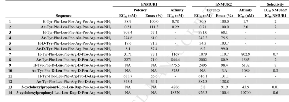
Table 1. Sequences and in vitro activity and affinity of the literature based NMU-8 analogs (first generation) on hNMUR1 and hNMUR2. [double column]
316
hNMUR1 hNMUR2 Selectivity
Potency Affinity Potency Affinity IC50 NMUR2/
Sequence EC50 (nM) Emax (%) IC50 (nM) EC50 (nM) Emax (%) IC50 (nM) IC50 NMUR1
1 H-Tyr-Phe-Leu-Phe-Arg-Pro-Arg-Asn-NH2 38.9 100.0 0.78 30.8 100.0 1.7 2
2 Ac-Tyr-Phe-Leu-Phe-Arg-Pro-Arg-Asn-NH2 0.51 111.3 0.29 0.71 100.0 2.0 7
3 H-Tyr-Phe-Leu-Phe-Ala-Pro-Arg-Asn-NH2 709.4 57.1 - 391.0 68.1 - -
4 Ac-Tyr-Phe-Leu-Phe-Ala-Pro-Arg-Asn-NH2 274.6 61.0 - 242.2 75.5 - -
5 H-D-Tyr-Phe-Leu-Phe-Arg-Pro-Arg-Asn-NH2 18.6 71.3 - 34.3 103.7 - -
6 Ac-D-Tyr-Phe-Leu-Phe-Arg-Pro-Arg-Asn-NH2 8.1 57.4 - 6.2 99.0 - -
7 H-Tyr-Phe-Leu-Phe-Arg-D-Pro-Arg-Asn-NH2 3171 73.1 1167 1079 110.7 802.9 0.7
8 Ac-Tyr-Phe-Leu-Phe-Arg-D-Pro-Arg-Asn-NH2 2271 71.0 844.4 2002 80.9 1365 2
9 H-Tyr-Phe-D-Leu-Phe-Arg-D-Pro-Arg-Asn-NH2 NA NA 775.5 2495 98.4 6132 8
10 Ac-Tyr-Phe-D-Leu-Phe-Arg-D-Pro-Arg-Asn-NH2 NA NA 3755 NA NA 1089 0.3
11 H-Tyr-Phe-Leu-Phe-Arg-Pro-D-Arg-Asn-NH2 683.7 56.6 - 616.1 131.1 - -
12 Ac-Tyr-Phe-Leu-Phe-Arg-Pro-D-Arg-Asn-NH2 343.4 64.1 - 382.3 138.8 - -
13 3-cyclohexylpropionyl-Leu-Leu-Dap-Pro-Arg-Asn-NH2 NA NA 4286 3.8 91.9 43.9 0.01
14 3-cyclohexylpropionyl-Leu-Leu-Dap-D-Pro-Arg-Asn-NH2 NA NA 18320 926.3 100.4 10700 0.6
317
Modifications compared to the native NMU-8 sequence (1) are marked in bold. EC50 values are calculated based on the IP3 SPA data. Emax is the percentage of the maximum 318
response at 10-5 M compared with the NMU-8 response at the same concentration. IC50 values are calculated based on the competitive binding assay. Receptor selectivity is 319
expressed as the ratio of the IC50 value for NMUR2 over the IC50 value for NMUR1 of each NMU-analog. NA: not applicable, no agonist activity up to 10-6 M. -: not 320
determined.
321
M AN US CR IP T
AC CE PT ED
Biological evaluation of the first set of NMU-analogs 322
Evaluation of the agonistic activities at the NMURs of the first set of NMU-analogs was performed in 323
the present study on HEK293 cells transiently expressing hNMUR1 or hNMUR2 using an inositol 324
triphosphate (IP3) SPA assay. The affinity of a selection of analogs for the NMURs was determined in 325
these cells using competitive displacement with [3H]-NMU-8 as radioligand. Table 1 shows the results 326
for the modified peptides (Supporting information, Table S3, for results presented as mean ± SEM).
327
N-terminal acetylation 328
It is reported that enzymatic degradation of the NMU-8 peptide mainly occurs via the N-terminus [14].
329
With the aim to develop NMU-analogs possessing an increased stability, N-terminal modifications 330
were introduced in the peptide sequence. Sakura et al. described that acetylation of the N-terminal Tyr1 331
leads to an increased resistance against proteolysis. Moreover, it has previously been shown that this 332
modification led to an increase in contractile activity on chicken crop smooth muscle preparations 333
[14]. In the present study, we evaluated Ac-NMU-8 (2) in an IP3 accumulation assay on HEK293 cells, 334
an immediate read-out for receptor activation. The introduction of an acetyl group at the N-terminus 335
(2) led to a more than 40-fold increase in potency on both NMURs (with EC50 values of 0.51 nM and 336
0.71 nM for NMUR1 and NMUR2, respectively) when compared to the native NMU-8 (1) (EC50 = 337
38.9 nM for NMUR1 and 30.8 nM for NMUR2). N-terminal acetylation of 1 did not alter the affinity 338
for the NMURs. Acetylation of the N-terminus was systematically carried out in the current study, to 339
present both the acetylated and non-acetylated form of all analogs, and led in general to an increase of 340
the relative activity on both NMURs, when compared to the non-acetylated peptide.
341
[Ala5]-NMU-8 342
A previous study by Funes et al. reported a tool for shifting selectivity towards NMUR2 when 343
replacing Arg5 by an Ala residue [30]. However, in our hands this modification (3) led only to a 344
slightly increased selectivity for NMUR2 (EC50 for NMUR1= 709.4 nM and EC50 for NMUR2 = 345
391.0 nM) instead of the 15-fold shift reported by Funes et al. The loss in potency, compared to 346
NMU-8, came with partial agonism on both NMURs. N-terminal acetylation of this analog (4) 347
increased the potency for both receptors, although activity remained low, as compared to NMU-8.
348
However, partial agonists can be of therapeutic importance since they could be regarded as molecules 349
exerting both agonistic, when low levels of the endogenous ligand is present, and antagonist effects, 350
when present together with a full agonist competing for the same binding site [31].
351
Chirality switches 352
A D-amino acid scan of NMU-8 was performed by Hashimoto et al. and tested on isolated chicken 353
crop smooth muscle preparations [13]. Inspired by the SAR studies which were performed in ‘the 354
early days of NMU research’, we decided to synthesize some of these analogs and evaluate them on 355
M AN US CR IP T
AC CE PT ED
cell cultures expressing the NMURs. [D-Tyr1]-NMU-8 was reported to enhance the contractile activity 356
in the smooth muscle preparation, presumably due to an increased resistance to aminopeptidase-like 357
degradation [13]. In our hands, replacing Tyr1 by D-Tyr led to peptide 5, and its acetylated form 6, 358
with comparable potencies to NMU-8 for NMUR1 and NMUR2, although a decrease of its maximal 359
effect (Emax, effect at 10-5 M compared with the response of the same concentration of NMU-8) on 360
NMUR1 was observed (e.g., 57.4 % for 6). However, rather than an increase in potency compared to 361
the parent peptide, a decreased potency was observed especially when comparing activities of the 362
acetylated forms 6 versus 2. In the literature, [D-Pro6]-NMU-8 and [D-Leu3,D-Pro6]-NMU-8 were 363
found to be weak non-competitive antagonists in chicken crop preparations [13]. We included both 364
molecules and their N-terminal acetylated forms (compounds 7-10) in our study. These ligands indeed 365
did not show potent agonistic activity in the in vitro cell-based assays. To further investigate if these 366
molecules could act as antagonists on the NMURs, their ability to compete with radiolabeled agonist, 367
[3H]-NMU-8, was evaluated. These compounds (7 – 10) all showed a 1000-fold, or even higher, 368
decrease in affinity for NMUR1. On NMUR2, binding studies revealed a major loss in affinity, 369
ranging from a 400 to more than 3000-fold decrease. These results demonstrate that these 370
modifications lead to a loss in affinity and potency of the molecules. To our knowledge, no peptidergic 371
antagonists for NMUR1 or NMUR2 have been proven to exert activity in vivo until now. This 372
correlates with the observation that [D-Pro6]-NMU-8 failed to exert antagonistic activity in vivo [32].
373
However, a small molecule antagonist for NMUR2, R-PSOP, was reported by Liu et al [33].
374
Replacing Arg7 by D-Arg (11, 12) resulted in full agonists with an even higher intrinsic activity than 375
NMU-8 on NMUR2 (Emax values of 131.1 % and 138.8 % for 11 and 12, respectively), but these 376
analogs gave way to a decrease in potency on both receptors. However, these molecules still possessed 377
similar potencies on NMUR1, as compared to the activity on NMUR2, they were only able to partially 378
activate this receptor. Finally, Takayama et al described a selective NMUR2 receptor agonist, namely 379
3-cyclohexylpropionyl-Leu-Leu-Dap-Pro-Arg-Asn-NH2 [18], which was resynthesized as compound 380
13 together with the analog where Pro was replaced by D-Pro (14), in order to verify whether this 381
could lead to selective NMUR2 receptor antagonism. Also in our hands, analog 13 acted as a selective 382
NMUR2 agonist with a lower affinity but conserved potency on NMUR2 as compared to NMU-8. The 383
additional D-amino acid substitution did not give rise to a NMUR2 selective antagonists as compound 384
14 possessed a weak affinity and in consequence a low activity on both NMURs.
385
M AN US CR IP T
AC CE PT ED
Table 2. Sequences and in vitro activity and affinity of the novel NMU-8 analogs (second set) for hNMUR1 and hNMUR2. [double column]
386
387
388
389
390
391
392
393
394
395
396
397
Modifications compared to the native NMU-8 sequence (1) are marked in bold.
EC50 values are calculated based on the IP3 SPA data. Emax is the percentage of the
maximum response at 10-5 M
413
compared with the NMU-8 response at the same concentration. IC50 values are calculated based on the competitive binding assay. Receptor selectivity is expressed as the ratio of the IC50 value for NMUR2 over the IC50 value for NMUR1 of each
414
NMU- analog.
415
hNMUR1 hNMUR2 Selectivity
Potency Affinity Potency Affinity IC50 NMUR2/
Sequence EC50 (nM) Emax (%) IC50 (nM) EC50 (nM) Emax (%) IC50 (nM) IC50 NMUR1
1 H-Tyr-Phe-Leu-Phe-Arg-Pro-Arg-Asn-NH2 38.9 100.0 0.78 30.8 100.0 1.7 2
15 H-7-OH-Tic-Phe-Leu-Phe-Arg-Pro-Arg-Asn-NH2 5.5 103.5 0.086 3.5 115.3 21.4 249
16 Ac-7-OH-Tic-Phe-Leu-Phe-Arg-Pro-Arg-Asn-NH2 5.1 99.3 0.037 13.4 115.8 12.3 332
17 H-2'Nal-Phe-Leu-Phe-Arg-Pro-Arg-Asn-NH2 65.8 95.1 0.97 4.8 106.1 9.9 10
18 Ac-2'Nal-Phe-Leu-Phe-Arg-Pro-Arg-Asn-NH2 1.6 90.9 0.88 1.6 83.9 5.6 6
19 H-Dmt-Phe-Leu-Phe-Arg-Pro-Arg-Asn-NH2 10.4 95.0 0.34 0.83 100.7 4.3 13
20 Ac-Dmt-Phe-Leu-Phe-Arg-Pro-Arg-Asn-NH2 4.0 109.1 0.15 0.70 91.5 2.4 16
21 H-Tyr-Phe-Leu-7-OH-Tic-Arg-Pro-Arg-Asn-NH2 94.5 94.6 8.6 624.3 109.1 1010 117
22 Ac-Tyr-Phe-Leu-7-OH-Tic-Arg-Pro-Arg-Asn-NH2 12.3 88.1 1.2 192.6 86.1 330.8 276
23 H-Tyr-Phe-Leu-1'Nal-Arg-Pro-Arg-Asn-NH2 41.4 101.1 0.31 225.7 109.8 370.5 1195
24 Ac-Tyr-Phe-Leu-1'Nal-Arg-Pro-Arg-Asn-NH2 4.3 105.3 0.41 246.5 86.4 39.1 95
25 H-Tyr-Phe-Leu-2'Nal-Arg-Pro-Arg-Asn-NH2 51.0 103.0 0.51 378.0 104.7 424.4 832
26 Ac-Tyr-Phe-Leu-2'Nal-Arg-Pro-Arg-Asn-NH2 4.2 109.0 0.32 304.2 96.2 195.3 610
27 H-Tyr-Phe-Leu-Dmt-Arg-Pro-Arg-Asn-NH2 77.9 96.1 1.6 1286 103.4 1649 1031
28 Ac-Tyr-Phe-Leu-Dmt-Arg-Pro-Arg-Asn-NH2 7.7 104.4 0.36 3675 96.7 1857 5158
29 H-Tyr-Phe-Leu-Oic-Arg-Pro-Arg-Asn-NH2 184.9 72.2 66.2 472.1 92.0 1546 23
30 Ac-Tyr-Phe-Leu-Oic-Arg-Pro-Arg-Asn-NH2 93.0 83.1 68.6 1830 92.5 253.4 4
31 H-Tyr-Phe-Leu-N(Benzyl)Gly-Arg-Pro-Arg-Asn-NH2 1053 73.7 46.3 1103 103.1 1403 30
32 Ac-Tyr-Phe-Leu-N(Benzyl)Gly-Arg-Pro-Arg-Asn-NH2 172.2 73.6 83.1 1278 85.4 1019 12
33 H-Tyr-Phe-Leu-N(4-OH-Benzyl)Gly-Arg-Pro-Arg-Asn-NH2 593.7 54.6 173.8 1407 69.4 1961 11
34 Ac-Tyr-Phe-Leu-N(4-OH-Benzyl)Gly-Arg-Pro-Arg-Asn-NH2 191.7 60.1 27.9 2686 75.4 1813 65
35 H-Tyr-Phe-Leu-N(Phenethyl)Gly-Arg-Pro-Arg-Asn-NH2 822.7 75.5 30.2 355.7 121.8 261.6 9
36 Ac-Tyr-Phe-Leu-N(Phenethyl)Gly-Arg-Pro-Arg-Asn-NH2 138.9 83.1 62.9 250.0 99.9 316.1 5
37 H-Tyr-Phe-Leu-N(4-OH-Phenethyl)Gly-Arg-Pro-Arg-Asn-NH2 574.4 57.6 100.1 382.2 94.3 428.9 4
38 Ac-Tyr-Phe-Leu-N(4-OH-Phenethyl)Gly-Arg-Pro-Arg-Asn-NH2 216.6 60.6 3.9 339.2 80.7 206.9 53
39 H-Tyr-Phe-Leu-Phe-Arg-Tic-Arg-Asn-NH2 5496 79.0 162.3 45.5 99.3 13.0 0.08
40 Ac-Tyr-Phe-Leu-Phe-Arg-Tic-Arg-Asn-NH2 230.0 72.9 54.4 5.4 91.8 11.2 0.2
41 H-Tyr-Phe-Leu-Phe-Arg-Oic-Arg-Asn-NH2 273.2 106.3 24.3 10.2 90.4 12.7 0.5
42 Ac-Tyr-Phe-Leu-Phe-Arg-Oic-Arg-Asn-NH2 28.8 95.0 11.2 1.8 84.3 13.9 1
M AN US CR IP T
AC CE PT ED
416
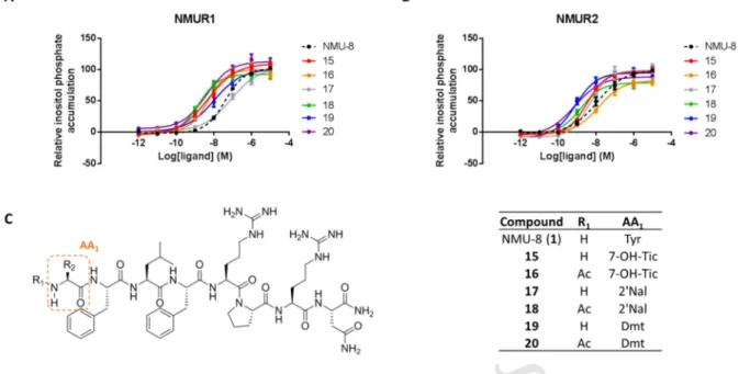
Figure 3. In vitro evaluation of the effect of Tyr1 substitution on receptor activation. IP3 accumulation 417
assay performed on HEK293 cells, transiently expressing A) NMUR1 or B) NMUR2. C) Structures of 418
the novel NMU-analogs 15, 16, 17, 18, 19 and 20. Data are shown as mean ± SEM (n=3). [double 419
column]
420
Biological evaluation of the second set of NMU-analogs 421
Substitution of the N-terminal tyrosine residue 422
To protect the peptides from cleavage by aminopeptidases, the N-terminus of NMU-8 was modified.
423
Figure 3 gives an overview of the NMU-analogs with modification at the Tyr1 residue and their 424
activity at NMUR1 and NMUR2. An analog wherein Tyr1 was replaced by 7-OH-Tic was synthesized 425
together with the N-terminally acetylated peptide, to give compounds 15 and 16. Both molecules 426
showed a very high, picomolar affinity for NMUR1, which is about ten times better than those of the 427
parent compounds 1 and 2, whereas their affinity for NMUR2 decreased. In the activity assay, they 428
were however non-selective, full agonists with a 15 % increase of the maximal effect at NMUR2 429
(Table 2). When Tyr1 was replaced by 2’-naphtylalanine (2’Nal), a non-selective agonist for the 430
NMURs (17) with similar affinity and potency as compared to compound 1 was found. The acetylated 431
analog (18) however, is more potent than NMU-8 at NMUR1 and NMUR2, with a similar activity on 432
both receptors and a 6-fold higher affinity for NMUR1 (Table 2 and Figure 3). Finally, a substitution 433
by Dmt resulted in potent, but non-selective, NMUR agonists 19 and 20. They possessed a 434
subnanomolar affinity for NMUR1, yet an increased potency on NMUR2 was observed.
435
These modifications of the Tyr residue, namely replacement by 7-OH-Tic, 2’Nal or Dmt, led to the 436
discovery of potent NMU-analogs. It indicates that the side chain hydroxyl function in this position is 437
not important for receptor activation. However, an aromatic moiety should be present. Consequently, 438
M AN US CR IP T
AC CE PT ED
when the N-terminus was acetylated, these analogs became even more potent on both receptors, 439
presenting all an increased potency compared to NMU-8, with compound 16 as the only exception. It 440
has an approximately 4-fold lower potency on NMUR2 than the analog with the free amine at the N- 441
terminus (15). However, it was equipotent on this receptor as compared with the native NMU-8 442
peptide. All compounds (15 – 20) possessed higher affinity for NMUR1 although most molecules lead 443
to an increased activity on NMUR2.
444
445
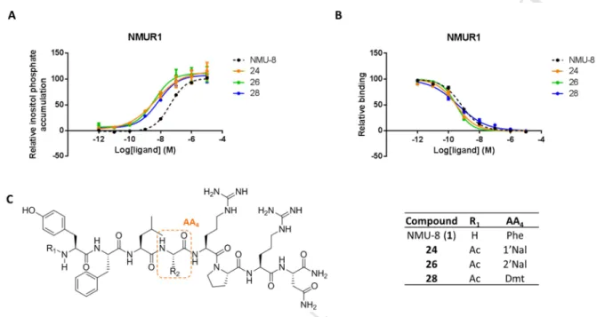
Figure 4. In vitro evaluation of the effect of Phe4 substitution on affinity and receptor activation. A) 446
IP3 accumulation assay performed on HEK293 cells, transiently expressing NMUR1. B) Competitive 447
binding assay with [3H]-NMU-8 carried out on HEK293 cells, transiently expressing NMUR1. C) 448
Structures of the novel NMU-analogs 24, 26 and 28. Data are shown as mean ± SEM (n = 3). [double 449
column]
450
Introduction of unnatural amino acids in position 4 451
Modifications of the Phe residue in position 4 were introduced to prevent the cleavage of the peptide 452
at the biodegradation site Phe-Arg, which was proposed by Takayama et al. after in vitro stability 453
assays in rat serum [19]. By replacing Phe4 by 7-OH-Tic, both the analogs with a free amine N- 454
terminus (21) and the acetylated form (22) were agonists on NMUR1. Compound 21 resulted in an 455
agonist with a minor loss in affinity and potency on this receptor. On the other hand, its acetylated 456
form (22) possessed an increased affinity and potency, as compared to NMU-8, although a loss of 457
more than 10 % of the maximal agonist activity was observed (Table 2). On NMUR2, this 458
modification led to a drop in affinity, and consequently the activity. Moreover, the maximal effect on 459
NMUR2 decreased with more than 10 % for analog 22. When a bulky aromatic residue, 1’Nal (23) or 460
2’Nal (25), was introduced in the sequence, full agonists were obtained for NMUR1 with similar 461
potencies as NMU-8. When these analogs were acetylated at their N-terminus, to give compounds 24 462
M AN US CR IP T
AC CE PT ED
and 26 respectively, potent agonists were found with a 10-fold higher activity at NMUR1 and an 463
increased affinity. For these compounds (23 – 26), a loss in affinity and activity for NMUR2 was 464
found. All potent NMUR1 selective NMU-analogs, obtained by modification of Phe4, are presented in 465
Figure 4. When a bulky Tyr-analog was introduced in the position of Phe4 (i.e. by replacing Phe with 466
Dmt to give 27), a NMUR1-selective agonist with a slightly diminished potency (EC50= 77.9 nM), as 467
compared to NMU-8, was again obtained. The acetylated analog (28) showed a higher potency and 468
affinity for NMUR1 (Figure 4). A last modification which was introduced in this position consisted of 469
the replacement of Phe by octahydroindolecarboxylic acid (Oic), a residue that can be regarded as a 470
saturated and constrained Phe surrogate. Unfortunately, both compounds 29 and 30, were 471
characterized by a lower potency and affinity on both NMURs as compared to NMU-8.
472
Importantly, these results reveal that the modification of Phe in position 4 can serve as a tool to obtain 473
NMU-analogs with an increased selectivity for NMUR1. Moreover, it shows that a hydroxyl function 474
in the side chain of Phe is tolerated since the NMUR1 activation is unaltered when Dmt was 475
introduced. However, when Phe is replaced by 7-OH-Tic, a decrease in affinity for NMUR1 was 476
found. This could be explained by the fact that an unfavorable conformational constraint is imposed, 477
and hence that the side chain is forced in a less favorable position for binding to the NMURs [34].
478
Remarkably, this loss in affinity could be restored by the introduction of an acetyl group at the N- 479
terminus. Moreover, when Phe is replaced by Oic, the potency diminished even more due to a loss in 480
affinity for the NMURs.
481
N-substituted glycines in position 4 482
A series of N-substituted glycines, the so-called ‘peptoid’ residues, was introduced in the NMU-8 483
sequence to mimic the aromatic character of Phe, while aiming to protect the degradation site Phe-Arg 484
by using another strategy than for the analogs of the previous section. Moreover, a shift of the side 485
chain from the α-carbon to the nitrogen in neurotensin(8-13) has been shown to shift the receptor 486
selectivity from NTSR1 to NTSR2 [35]. Hence, benzyl, 4-hydroxy-benzyl, phenethyl and 4-hydroxy- 487
phenethyl moieties were inserted instead of Phe4 in order to verify whether selectivity could be 488
impacted. However, all NMU-analogs with a N-substituted Gly instead of Phe at position 4 489
(compounds 31 – 38) resulted in weak agonists with a loss in potency and a lower Emax compared to 490
our endogenous lead peptide NMU-8. Moreover, a decreased affinity for both NMURs was observed 491
as well. It can be speculated that the backbone amide is necessary for receptor binding and activation.
492
Another explanation for the loss of affinity and activity could be that the side chain is not aligned into 493
the right position to favor binding and, subsequently, to exert an effect on the NMURs. Of note, to 494
compensate for such an unfavorable shift of the side chains, the homologated phenethyl and 4- 495
hydroxy-phenethyl ‘side chains’ were applied, even though unsuccessfully.
496
Substitution of the proline residue
M AN US CR IP T
AC CE PT ED
The naturally constrained Pro6 residue was replaced by the conformationally constrained 1,2,3,4- 498
tetrahydroisoquinoline-3-carboxylic acid (Tic) residue. This bridged residue led to poor partial 499
NMUR1 agonists (39 and 40), which possessed a decreased receptor affinity as compared to NMU-8.
500
Although these analogs had a lower affinity for NMUR2 than compound 1, the non-acetylated peptide 501
was able to fully activate NMUR2 with only a 4.5 fold higher EC50 value. Moreover, compound 40 502
was found to have a 2-fold higher potency at NMUR2, but it was not able to fully activate the receptor 503
(Emax = 91.8 %). Similarly to the insertion of Tic at this position, replacement of Pro by Oic led to a 504
potent agonist (41) on NMUR2, even though it was already stated in literature that the Pro-Arg-Asn- 505
NH2 segment is critical for receptor activation [18]. Moreover, when this peptide was N-terminally 506
acetylated (42), the NMU-8 potency was exceeded. On NMUR1, the substitution of Pro by Oic led to 507
a full agonist with a 7-fold decrease in potency. However, when an acetylation was introduced at the 508
N-terminus, the NMU-8 potency was restored. Both compounds 41 and 42 possessed a decreased 509
affinity for the NMURs. Interestingly, modifications in this, previously described, critical part of the 510
NMU-8 sequence show that other cyclic amino acids are tolerated in this region of the peptide.
511
Substitution of Pro gave rise to molecules with an increased NMUR2 selectivity and high potency at 512
this receptor subtype, which makes it an attractive tool to further develop receptor subtype selective 513
analogs.
514
In vitro plasma stability 515
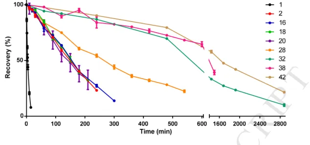
To evaluate the effect of the introduced modifications on the proteolytic stability of the NMU-analogs, 516
in vitro degradation studies in human plasma at 37 °C were performed on a selection of the novel 517
peptides. The percentage of the intact NMU-analog was measured over time by HPLC-UV analysis 518
(Figure 5). Afterwards the samples were analyzed by means of MS and degradation profiles were 519
studied (Figure 6). Depending on the stability of the peptide, samples were taken at different time 520
points (Table S1).
521
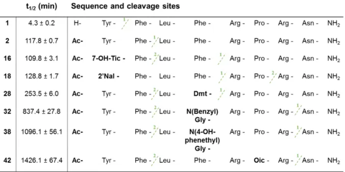
NMU-8 (1) was characterized by a short half-life of only 4.3 ± 0.2 min. It was rapidly degraded at its 522
unprotected N-terminus where Tyr1 was cleaved from the sequence. Since acetylation of the N- 523
terminal amine was systematically performed on all NMU-analogs described in this study, the 524
resistance to biological degradation of compound 2 was evaluated next. When an acetyl group was 525
introduced at the N-terminus of the NMU-8 sequence, a more than 25-fold increase of the half-life 526
(117.8 ± 0.7 min) was observed. The prior cleavage site Tyr1-Phe2 was protected by this N-terminal 527
modification and degradation took place by cleavage of the peptide between the Phe2 and Leu3 528
residues. Substitutions of the Tyr residue did not lead to a further increase of the enzymatic stability 529
with calculated half-lifes of 109.8 ± 3.1 min, 128.8 ± 1.7 min and 154.5 ± 10.2 min for compounds 16, 530
18 and 20, respectively.
531
M AN US CR IP T
AC CE PT ED
532
Figure 5. Percentage of recovery of NMU-analogs in human plasma incubated at 37 °C. The 533
experiments were performed in triplicate and the results are presented as mean ± SEM (n = 3). [single 534
column Figure]
535
Although introduction of a modified amino acid in position 1 gave rise to molecules with a similar 536
half-life compared to Ac-NMU-8 (2), it was observed that the major cleavage site of compound 16 537
was situated in the middle of the sequence, between Phe4 and Arg5. This position was proposed earlier 538
to be an important biodegradation site [19]. Next, the peptide was cleaved further at the same position 539
as described for compound 2, namely after Phe2. The NMU-analog 18 was characterized by the same 540
major cleavage site as for 16, but the introduction of 2’Nal in the sequence led to stabilization of the 541
labile Phe2-Leu3 segment. In a smaller extent the peptide was cleaved between Pro6 and Arg7. When an 542
unnatural amino acid was introduced in position 4, e.g., Dmt instead of Phe for compound 28, a 2-fold 543
increase of the half-life in human plasma was found, as compared to Ac-NMU-8 2 and an almost 60- 544
fold increase when it was compared to the native peptide 1. The major biodegradation site remained in 545
the middle of the sequence, namely at Dmt4-Arg5, but the cleavage was significantly hampered via the 546
replacement of Phe by Dmt at this position. As expected, the introduction of the ‘peptoid’ residues at 547
Phe4 resulted into NMU-analogs with an increased resistance against proteolytic degradation. The 548
stability assay revealed that compound 32 had a half-life of 837.4 ± 27.8 min (or 14.6 ± 0.8 h) in 549
human plasma. When Phe4 was replaced by N(4-OH-phenethyl)Gly (38), it was even higher, namely 550
1096.1 ± 56.1 min (18.3 ± 0.9 h). Moreover, the switch of Phe4 by N-substituted glycines changed the 551
biodegradation profile. The major cleavage site moved towards the C-terminus of the peptide, more 552
precisely between Arg7 and Asn8. A last subset of modifications which was used in this study, is the 553
substitution of the Pro residue. When it was replaced by Oic to give 42, a molecule with a high 554
resistance to enzymatic degradation was found. The half-life of analog 42 in human plasma was 555
1426.1 ± 67.4 min or 23.8 ± 1.1h. The major degradation site for this analog was as well located 556
![Figure 2. Structure and sequence of NMU-8 [single column Figure]](https://thumb-eu.123doks.com/thumbv2/9dokorg/1391633.115722/8.892.117.739.102.394/figure-structure-sequence-nmu-single-column-figure.webp)