Avian Pathology
ISSN: 0307-9457 (Print) 1465-3338 (Online) Journal homepage: https://www.tandfonline.com/loi/cavp20
Development of mismatch amplification mutation assay (MAMA) for the rapid differentiation of
Mycoplasma gallisepticum K vaccine strain from field isolates
Katinka Bekő, Áron Botond Kovács, Zsuzsa Kreizinger, Szilvia Marton, Krisztián Bányai, László Bánáti, Salvatore Catania, Janet Bradbury, Inna Lysnyansky, Olusola Martins Olaogun & Miklós Gyuranecz
To cite this article: Katinka Bekő, Áron Botond Kovács, Zsuzsa Kreizinger, Szilvia Marton, Krisztián Bányai, László Bánáti, Salvatore Catania, Janet Bradbury, Inna Lysnyansky, Olusola Martins Olaogun & Miklós Gyuranecz (2020): Development of mismatch amplification mutation assay (MAMA) for the rapid differentiation of Mycoplasma�gallisepticum K vaccine strain from field isolates, Avian Pathology, DOI: 10.1080/03079457.2020.1744523
To link to this article: https://doi.org/10.1080/03079457.2020.1744523
Accepted author version posted online: 17 Mar 2020.
Submit your article to this journal
View related articles
View Crossmark data
Publisher: Taylor & Francis & Houghton Trust Ltd Journal: Avian Pathology
DOI: 10.1080/03079457.2020.1744523
Development of mismatch amplification mutation assay (MAMA) for the rapid differentiation of Mycoplasma gallisepticum K vaccine strain from field isolates
Katinka Bekő1, Áron Botond Kovács1, Zsuzsa Kreizinger1, Szilvia Marton1, Krisztián Bányai1, László Bánáti1, Salvatore Catania2, Janet Bradbury3, Inna Lysnyansky4, Olusola Martins Olaogun5, Miklós Gyuranecz1,6*
1Institute for Veterinary Medical Research, Centre for Agricultural Research, Hungária körút 21, Budapest 1143, Hungary
2Instituto Zooprofilattico Sperimentale delle Venezie, Via San Giacomo 5, Verona 37000, Italy
3Institute of Veterinary Science, University of Liverpool, Leahurst Campus, Neston CH64 7TE, UK
4Department of Avian Diseases, Kimron Veterinary Institute, POB 12, Beit Dagan 50250, Israel
5Asia-Pacific Centre for Animal Health, Faculty of Veterinary and Agricultural Sciences, The University of Melbourne, Parkville, Victoria 3010, Australia
6 Department of Microbiology and Infectious Diseases, University of Veterinary Medicine, Hungária körút 23-25, Budapest 1143, Hungary
* Corresponding author; e-mail: m.gyuranecz@gmail.com
Abstract
Mycoplasma gallisepticum causes respiratory diseases and reproduction disorders in turkeys and chickens. The infection has considerable economic impact due to reduced meat and egg production. Because elimination programs are not feasible in a large number of poultry farms, vaccination remains the only effective measure of disease control.
Differentiating vaccine strains from field isolates is necessary in the control of vaccination programs and diagnostics. The aim of this study was to develop a polymerase chain reaction (PCR) based mismatch amplification mutation assay (MAMA) for the discrimination of K vaccine strain (K 5831, Vaxxinova Japan K.K.).
After determining the whole genome sequence (WGS) of the K strain, primers were designed to detect seven different vaccine-specific single nucleotide polymorphisms (SNPs). After evaluating preliminary results, the MAMA-K-fruA test detecting a single guanine-adenine substitution within the fruA gene (G88A) was found to be the most applicable assay to distinguish the K vaccine strain from field isolates.
The detected K strain-specific SNP showed genetic stability after serial passage in vitro, but this stability test still needs to be evaluated in vivo as well, investigating a large number of K strain re-isolates. The MAMA-K-fruA assay was tested on a total of 280 culture and field samples. The designed assay had 102 and 103 template copy number/µl sensitivity in melt- curve analysis based and agarose-gel based assays, respectively, and showed no cross reaction with other avian Mycoplasma species. The new MAMA provides a time- and cost-effective molecular tool for the control of vaccination programmes and for diagnostics.
Keywords: avian mycoplasmosis; Mycoplasma gallisepticum; K vaccine strain; molecular biology; mismatch amplification mutation assay (MAMA); single nucleotide polymorphism (SNP)
Introduction
Mycoplasma gallisepticum is a bacterial pathogen with global distribution which causes chronic respiratory disease (CRD) in chickens and infectious sinusitis in turkeys (Levisohn and Kleven, 2000). Infection results in increased mortality, and a reduction in feed intake, weight gain, egg production and hatchability (Raviv and Ley, 2013). Therefore M.
gallisepticum is among the economically most significant mycoplasmas of poultry worldwide (Whithear, 1996). M. gallisepticum can be disseminated horizontally, but the major route of transmission is from infected breeder birds to progeny (Levisohn and Kleven, 2000). Thus, control programs for M. gallisepticum are primarily based on maintaining commercial breeder stocks free of infection (Whithear, 1996). Other feasible options for controlling M.
gallisepticum infection are targeted antibiotic treatment and vaccination (Kleven, 2008).
Bacterins, live vaccines and a recombinant fowl pox vaccine can be used against
M. gallisepticum infection (Ferguson-Noel et al., 2012). Commercially available live M.
gallisepticum vaccine strains are the 6/85 (Nobilis® MG 6/85, MSD Animal Health), the ts-11 (TS-11®, Merial Italia S.p.A.), the F (Cevac® MG-F, Ceva, Inc.) and the most recently developed K strain (K 5831, Vaxxinova Japan K.K.) which is used mainly in Japan. The K strain shows high efficiency in broiler and layer-type chickens in the protection of the respiratory and reproductive systems (Ferguson-Noel and Williams, 2015).
Since live vaccines are used in many parts of the world, differentiation of M. gallisepticum vaccine strains from wild, virulent isolates is essential in control programs (Whithear, 1996).
Previously, DNA fingerprinting methods, such as amplified fragment length polymorphism (AFLP) (Hong et al., 2005) and random amplified polymorphic DNA (RAPD) (Fan et al., 1995) were used to discriminate M. gallisepticum vaccine strains and field isolates. However, these methods have low reproducibility and they require the isolation and a pure culture of the tested organisms (Sulyok et al., 2019). Sequence-based methods such as gene-targeted
sequencing (Ferguson-Noel et al., 2005), TaqMan assay (Raviv et al., 2008) and high- resolution melt (HRM) curve analysis (Ghorashi et al., 2010; 2013; 2015) have higher reproducibility and lower labour intensity. On the other hand, these techniques are expensive or need special equipment (Sulyok et al., 2019). Polymerase chain reaction (PCR) tests also can be used for the differentiation of vaccine strains from field isolates (Evans and Leigh, 2008; Ricketts et al., 2017; Sulyok et al., 2019). For the genotyping of M. gallisepticum isolates a core genome multi-locus sequence typing (cgMLST) system with improved discriminatory power has been established (Ghanem et al., 2017), but this method requires isolation and whole genome sequencing of the bacterium. In addition, a six-gene based MLST assay was described, which proved to be suitable for the discrimination of the K, ts-11, 6/85 and F vaccine strains from each other and from field strains (Bekő et al., 2019).
Recently, mismatch amplification mutation assays (MAMAs) were developed for the simultaneous discrimination of 6/85, ts-11 and F vaccine strains from field isolates (Sulyok et al., 2019). MAMA is a PCR-based technique used for single nucleotide polymorphism (SNP) discrimination in many bacteria (Birdsell et al., 2012). In brief, the technique is based on SNP-specific primers at the 3′ end, one being marked with an additional 15-20 bp long GC- clamp at the 5’ end. A single destabilizing mismatch at the 3′ end of each allele-specific primer enhances the discriminative capacity of the assay (Cha et al., 1992). The GC-clamp increases the melting temperature and the size of the amplicon as well. The temperature shift can be easily detected in the presence of intercalating fluorescent dye on a real-time PCR platform (melt-MAMA) and the difference in sizes of the amplicons can be observed with agarose gel electrophoresis (agarose-MAMA), which enables the differentiation of SNP- specific genotypes (Birdsell et al., 2012).
In the present study, SNPs specific for the M. gallisepticum K vaccine strain have been identified and used for the development of MAMAs to differentiate this M. gallisepticum K vaccine strain from field isolates.
Materials and methods
M. gallisepticum samples. In total, 280 samples were provided for the assay development, including 19 M. gallisepticum whole genome sequences (WGSs) available online in GenBank (strain S6, GenBank accession number: NC_023030.2; strain Rlow, GenBank accession number: AE015450.2; strain Rhigh, GenBank accession number: NC_017502.1; house finch isolates, GenBank accession numbers: NC_018412.1, NC_018409.1, NC_018406.1, NC_018407.1, NC_018408.1, NC_018410.1, NC_018411.1, NC_018413.1; and ts-11 re- isolates,GenBank accession numbers: MAFU00000000, MAFV00000000, MAFW00000000,
MADW00000000, MATM00000000, MATN00000000, MAGQ00000000,
MAGR00000000).
The live M. gallisepticum K vaccine strain (K 5831, Vaxxinova Japan K.K., Tokyo, Japan) was obtained from its commercial distributor. DNA of the M. gallisepticum ATCC 19610 type strain, and the 6/85 (Nobilis® MG 6/85, MSD Animal Health Hungary, Budapest, Hungary), ts-11 (TS-11®, Merial Italia S.p.A., Assago, Italy) and F (Cevac® MG-F, Ceva Inc., Budapest, Hungary) vaccine strains were used as control in the assays.
In addition, DNA of 256 M. gallisepticum samples including pure M. gallisepticum cultures (n=206) and field samples (n=50) were investigated. These samples were collected between 1993 and 2019 and originated from at least 25 countries (Italy, n=80; Spain, n=56; UK, n=23;
Israel, n=23; Hungary, n=9; USA, n=7; Malaysia, n=7; Australia, n=6; India, n=6; the Netherlands, n=4; Thailand, n=4; Germany, n=3; Romania, n=3; Indonesia, n=3; Fiji, n=3;
Portugal, n=2; Ukraine, n=2; Russia, n=2; Iraq, n=2; China, n=2; Philippines, n=2; France,
n=1; Austria, n=1; Slovenia, n=1; Czech Republic, n=1; Albania, n=1; Jordan, n=1), seven avian species (chicken, turkey, partridge, pheasant, goose, guinea fowl, quail) including different production categories of poultry (broiler, layer or breeder chickens, backyard chickens, meat turkeys or turkey breeders).
The 256 samples originated from tracheal swabs or lungs. Ethical approval and specific permissions were not required for the study as all samples were collected during routine diagnostic examinations or necropsies with the consent of the owners. DNA extraction was performed using the Qiamp DNA Mini kit (Qiagen Inc., Hilden, Germany) according to the manufacturers’ instructions for Gram-negative bacteria. The presence of M. gallisepticum DNA in the samples was confirmed by qPCR targeting the mgc2 gene of M. gallisepticum (Raviv and Kleven, 2009). Background information of the 280 tested M. gallisepticum samples is provided in Supplementary Table 1.
Whole genome sequencing, target selection and primer design. Genomic DNA of M.
gallisepticum K strain was extracted from 10 ml of logarithmic-phase broth culture using a QIAamp DNA Mini kit (Qiagen Inc.). Next-generation sequencing was performed on an Illumina NextSeq 500 platform (Illumina Inc., San Diego, USA). Reads were mapped to M.
gallisepticum strain Rlow (GenBank accession number: AE015450.2) as reference genome and annotated by Geneious software version 10.2.3. (Biomatters Ltd., Auckland, New Zealand) (Kearse et al., 2012).
The draft whole genome of M. gallisepticum K strain and 19 published M. gallisepticum genomes were aligned. The SNPs which were present exclusively in the M. gallisepticum K strain were explored by Geneious software (Biomatters Ltd.) (Kearse et al., 2012). Target mutations were selected based on the following criteria: 1) SNP is present in a single-copy gene which can be found in the genomes of all examined M. gallisepticum strains; 2) SNP
results in amino acid change in the protein encoded by the gene 3) SNP is surrounded by conserved regions suitable for primer design. Numbering of nucleotide positions was according to the individual genes of M. gallisepticum strain Rlow (GenBank accession number AE015450.2). The selected SNPs were used as targets for primer design by Geneious software (Biomatters Ltd.) (Kearse et al., 2012). The primers were constructed to limit amplicon lengths of ≤110 bp. The quality of the designed primer sets was assessed by NetPrimer (Premier Biosoft International, Palo Alto, CA).
Preliminary tests of the designed primers. After in silico analysis on the available sequences, preliminary examinations were performed to test the designed primers using the DNA of the K vaccine strain and as controls, DNA of vaccine strains 6/85, ts-11, F, and the M.
gallisepticum ATCC 19610 type strain. Nuclease-free water was used as negative control in all PCR assays.
Melt-MAMA tests were optimized on Applied Biosystems Step-One Plus real-time PCR system with StepOne Software version 2.3 (Thermo Fisher Scientific, Waltham, MA). PCR mixture of Melt-MAMA consisted of 2 μl 5X Colour-less GoTaq Flexi Buffer (Promega Inc., Madison, WI), 1 μl MgCl2 (25 mM), 0.3 μl dNTP (10 mM, Qiagen Inc., Valencia, CA), 0.5 μl EvaGreen (20X, Biotium Inc., Hayward, CA), 0.15 μl of each primer (10 pmol/ μl), 0.08 μl GoTaq G2 Flexi DNA polymerase (5 U/μl; Promega Inc.), nuclease-free water and 1 μl DNA template with a final volume of 10 μl. Thermocycling parameters were 95 °C for 10 min, followed by 30 cycles of 95 °C for 15 s and 60 °C for 1 min. PCR products were subjected to melt analysis using a dissociation protocol comprising 95 °C for 15 s, followed by 0.3 °C incremental temperature ramping from 60 °C to 95 °C. EvaGreen fluorescence intensity was measured at 525 nm at each ramp interval and plotted against temperature.
Agarose-MAMA was performed in C1000™ Touch Thermal Cycler (Bio-Rad Laboratories, Inc., Berkeley, CA) in 25 μl total volume containing 1 μl target DNA diluted in 5 μl 5X Green GoTaq Flexi Buffer (Promega Inc.), 2.5 μl MgCl2 (25 mM, Promeg Inc.), 0.5 μl dNTP (10 mM, Qiagen Inc.), 1 μl of each primer (10 pmol/μl), 0.2 μl GoTaq G2 Flexi DNA polymerase (5 U/μl; Promega Inc.) and nuclease-free water under the following PCR conditions: 95 °C for 5 min followed by 35 cycles at 95 °C for 30 s, 60 °C for 30 s and 72 °C for 30 s. The final elongation step was performed at 72 °C for 5 min. Standard electrophoresis (8 V/cm) was carried out in 3% agarose gel (MetaPhor Agarose, Lonza Group Ltd., Basel, Switzerland) and a 20-bp DNA ladder (O'RangeRuler 20 bp, Thermo Fisher Scientific Inc.) was used as molecular weight marker.
The assays showing the highest discriminating potential were selected for further tests.
Discriminating potential was examined based on the following criteria: 1) peaks of the melting curves and band sizes of the two genotypes (the K vaccine-specific genotype, hereafter referred as “vaccine-type”, and the genotype of other vaccine strains, type strains and field isolates, hereafter referred as “genetic variant”) were distinguishable; 2) negative control was not amplified; 3) the mutation was specific to the vaccine strain when all available samples with known vaccination status were tested.
Stability, specificity and sensitivity tests. In the stability tests of the mutations targeted by the designed assays, the K vaccine strain and the M. gallisepticum ATCC 19610 type strain were subjected to serial passage: 100 µl broth culture was inoculated into 900 µl Frey’s medium 10 times (Frey et al., 1968). Each passage resulted in ~3.322 population doubling (PD=log(Nf/Ni)/log2 where Nf is the final number of cells, Ni is the initial number of cells), thus cumulative population doubling was ~33.22 to the end of the 10-step serial passage (cPD=10PD) (Choi et al., 2017). After the 10th passage, DNAs were extracted, the designed
assays were performed and comparisons were made between genotypes of the parent and derivative strains.
The specificity of the assays was tested using the following avian Mycoplasma species: M.
anatis (ATCC 25524), M. anseris (ATCC 49234), M. sp. 1220 (“M. anserisalpingitis”, ATCC BAA-2147), M. cloacale (ATCC 35276), M. columbinasale (ATCC 33549), M. columbinum (ATCC 29257), M. columborale (ATCC 29258), M. gallinaceum (ATCC 33550), M.
gallinarum (ATCC 19708), M. gallopavonis (ATCC 33551), M. iners (ATCC 19705), M.
imitans (ATCC 51306), M. iowae (ATCC 33552), M. meleagridis (NCTC 10153), and M.
synoviae (ATCC 25204) type strains.
In order to test the sensitivity of the assays, tenfold dilutions of each genotype were used in the range of 106-100 copy number/μl. Copy number was calculated with the help of an online tool (http://cels.uri.edu/gsc/cndna.html; Staroscik, 2004) based on the DNA concentration measured by Nanodrop 2000 Spectrophotometer (Thermo Fisher Scientific Inc.). The lowest template copy numbers yielding melting temperature (Tm) specific to the genotypes were considered as the detection limit of the assays.
In order to assess the capability of the assays to identify mixed population of the two genotypes in a single specimen, different template copy number combinations of the M. gallisepticum ATCC 19610 type strain and the K vaccine strain were tested. The mixtures contained the type strain and the vaccine strain in the following combinations: constant template copy numbers (106 copies/µl) of one strain was paired with a member of a series of 10-fold DNA dilutions (106-103 copies/µl) of the other strain and vice versa.
For the discrimination of K vaccine strain one assay was selected according to the preliminary examinations considering stability, specificity and sensitivity. The DNAs of further 256 M.
gallisepticum samples including pure cultures and field samples (Supplementary Table 1) were submitted to test the reliability of the developed assay.
Sequence data submitted to international databases. The raw nucleotide sequence reads of the K vaccine strain were submitted to the Short Read Archive (SRA) database of National Center for Biotechnology Information (NCBI) (BioProject accession number:
PRJNA592048).
Sequence data of the following genes of the K vaccine strain were submitted to the GenBank of NCBI: aptA (GenBank accession number: MNN627735); dnaA (GenBank accession number: MN627736); dppD (GenBank accession number: MN627737); fruA (GenBank accession number:MN627738); pgi (GenBank accession number:MN627739).
Results
WGS of the K vaccine strain was generated using a total of 463,946 sequence reads with average lengths of 76 bp, the mean coverage was 33.1-fold for the whole genome. The following SNPs met the criteria of target selection: A1469G in aptA; T227C, T350C and C452T in dnaA; G73A in dppD; G88A in fruA; G958A in pgi.
Among these mutations, MAMA targeting the SNP at nucleotide position 88 of the fruA gene (MAMA-K-fruA) was selected based on stability, specificity, sensitivity and discriminating potential. This guanine-adenine substitution is unique for the K strain and induces aspartic acid - asparagine amino acid change in the encoded fructose-specific enzyme (EIIABC component) of the phosphotransferase system (PTS) (Table 1).
The MAMA-K-fruA assay resulted in genetic variant-specific amplicon when the M.
gallisepticum ATCC 19610 type strain or the other three vaccine strains were tested (Figure 1A and 1B). Negative controls did not amplify. After the 10-step serialpassage (resulting in
~33.22 population doubling), K vaccine strain and the M. gallisepticum ATCC 19610 type strain showed identical genotypes as their parent strains in the in vitro stability test. The
MAMA-K-fruA assay was proved to be species-specific, as templates of other avian Mycoplasma species were not amplified.Detection limit for both genotypes was 102 template copy number/µl in melt-MAMA assay and 103 in agarose-MAMA assay. This method was able to identify both genotypes in mixed samples in the following combinations: vaccine- type:genetic variant 106:106, 106:105, 105:106 template copy number/µl. Bimodal melting peaks at the specific melting temperatures or two amplicons with the specific band sizes indicated the presence of both genotypes (Figure 2A and 2B).
The MAMA-K-fruA assay was able to distinguish the two genotypes based on the melting peaks and sizes of the amplicons due to the 14 bp long GC-clamp of the vaccine-specific primer. Peaks of the melting curves were in the range 71.46-72.21 °C for the variant strains, while melting peaks of the K vaccine strain varied from 77.12 to 77.42 °C during the melt- MAMA studies. Accordingly, minimum difference in melting temperatures of the K vaccine strain and variant strains throughout the tests was 4.91 °C. Likewise, 71 (variant strains) and 85 (K vaccine strain) bp long PCR products can be clearly distinguished by agarose-MAMA (Table 2).
The quantity of M. gallisepticum DNA in the 256 samples submitted for further evaluation of the assay varied largely and showed wide range of cycle treshold (Ct) values in the previously performed mgc2 gene based qPCR. All of the tested DNA samples were found to be variant strains (Supplementary Table 1).
Discussion
M. gallisepticum infections have great impact on the poultry industry and vaccination is a cost-effective option to reduce economic losses (Kleven, 2008). The use of M. gallisepticum live vaccines led to the need for a reliable technique, which can differentiate vaccine strains from wild, virulent isolates. This is crucial in epidemiological investigations, vaccination and
eradication programs (Whithear, 1996). This study revealed a mutation in M. gallisepticum K vaccine strain that is absent in all of the other examined M. gallisepticum strains. The targeted mutation is located in the fruA gene, which encodes a PTS enzyme.
The bacterial PTS is a multi-protein system involved in the regulation of a variety of metabolic and transcriptional processes (Västermark and Saier, 2014). This system catalyses the phosphorylation of incoming sugar substrates concomitant with their translocation across the cell membrane (Deutscher et al., 2006). Although direct evidence of its role in virulence is lacking, a connection between the PTS and the virulence of certain pathogens was suggested by the observation that some virulence genes underlie a kind of carbon catabolite repression (CCR) (Trappetti et al., 2017). This is a regulatory mechanism enabling bacteria to increase their fitness by optimizing growth rates in natural environments providing different nutrients (Stülke and Hillen, 1999). Although several virulence genes and factors have been already identified, genetic background of virulence is not completely understood (Szczepanek et al., 2010). Role of the PTS in the virulence of M. gallisepticum has not been described yet, thus relevance of the revealed SNP in the attenuation of the K vaccine strain merits further investigations.
The mutation in the fruA gene showed firm genetic stability through the passages of the K vaccine strain and the M. gallisepticum ATCC 19610 type strain, however, it should be noted, that the in vitro test may not reflect completely the in vivo genetic stability of the strains.
In this study, 280 M. gallisepticum samples were investigated, including 19 whole genome sequences, the M. gallisepticum ATCC 19610 type strain, the K, 6/85, ts-11 and F vaccine strains, and 256 M. gallisepticum DNA samples of clinical specimens. After performing the designed MAMA-K-fruA test, we found that the targeted mutation occurs exclusively in the K vaccine strain. Although vaccination status of the animals was unknown in many cases, none of the tested samples originated from Japan, where K vaccine strain is used. Moreover,
our results are congruent with the earlier published MLST analysis of 130 M. gallisepticum samples, which belonged to the same strain collection, used in this work. In the MLST study genetic diversity and relatedness to vaccine strains were examined based on the analysis of six housekeeping genes (atpG, dnaA, fusA, rpoB, ruvB and uvrA). The M. gallisepticum K vaccine strain had unique sequence type and MLST system could not identify any closely related strains among the tested samples (Bekő et al., 2019). These results confirm the competence of the designed MAMA assay. On the other hand, a limitation of our study is that no field samples obtained from flocks vaccinated with commercial K vaccine was investigated. Evaluation of K strain re-isolates should further increase the reliability of the presented assay.
The new MAMA is suitable for the detection of simultaneous presence of the K vaccine strain and wild, virulent isolates in samples, which is especially useful in monitoring of vaccination programmes. Although it is important to mention, that based on our results, simultaneous detection presumably requires samples containing approximately the same amount of DNA of the two genotypes.
The developed method is highly specific and sensitive enough to be applicable directly on clinical samples avoiding technical problems associated with isolation, which is particularly complicated in case of mycoplasmas. Further advantage of the assay is that it can be performed on basic real-time PCR platforms and on conventional PCR equipment coupled with agarose gel electrophoresis too. The K strain-specific method reported here represents a convenient, rapid and cost-efficient tool for control programs against M. gallisepticum infections.
Acknowledgement
This work was supported by the Lendület program (LP2012-22) of the Hungarian Academy of Sciences, the K_16 (119594), FK_17 (124019) and KKP19 (129751) grants of the National Research, Development and Innovation Fund, Hungary. ZK, SM and MG were supported by the Bolyai János Fellowship of the Hungarian Academy of Sciences. MG was supported by the Bolyai+ Fellowship (ÚNKP-19-4-ÁTE-1) of the New National Excellence Program of the Ministry of Innovation and Technology. The authors wish to thank the British Veterinary Poultry Association for funding part of the UK game bird mycoplasma investigations and David Welchman for his valuable input in organisation of sample collection and protocols.
We also thank Christine Ellis of the University of Liverpool’s Mycoplasma diagnostic facility and Anne Forrester of the Institute of Global Health for their excellent assistance. The funders had no role in study design, data collection and interpretation, or the decision to submit the work for publication.
The authors declare that they have no competing interests.
References
Bekő, K., Kreizinger, Z., Sulyok, K.M., Kovács, Á.B., Grózner, D., Catania, S., Bradbury, J., Lysnyansky, I., Olaogun, O.M., Czanik, B., Ellakany, H. & Gyuranecz, M. (2019).
Genotyping Mycoplasma gallisepticum by multilocus sequence typing. Veterinary Microbiology, 231, 191-196.
Birdsell, D.N., Pearson, T., Price, E.P., Hornstra, H.M., Nera, R.D., Stone, N., Gruendike, J., Kaufman, E.L., Pettus, A.H., Hurbon, A.N., Buchhagen, J.L., Harms, N.J., Chanturia, G., Gyuranecz, M., Wagner, D.M. & Keim, P.S. (2012). Melt analysis of mismatch amplification mutation assays (Melt-MAMA): a functional study of a cost-effective SNP genotyping assay in bacterial models. PLoS One, 7, e32866.
Cha, R.S., Zarbl, H., Keohavong, P. & Thilly, W.G. (1992). Mismatch amplification mutation assay (MAMA): application to the c-H-ras gene. PCR Methods and Applications, 2, 14-20.
Choi, Y.S., Park, Y.B., Ha, C.W., Kim, J.A., Heo, J.C., Han, W.J., Oh, S.Y., Choi, S.J.
(2017).
Different characteristics of mesenchymal stem cells isolated from different layers of full term placenta. PLoS One, 12, e0172642.
Deutscher, J., Francke, C. & Postma, P.W. (2006). How Phosphotransferase System-Related Protein Phosphorylation Regulates Carbohydrate Metabolism in Bacteria. Microbiology and Molecular Biology Reviews, 70, 939-1031.
Evans, J.D. & Leigh, S.A. (2008). Differentiation of Mycoplasma gallisepticum vaccine strains ts-11 and 6/85 from commonly used Mycoplasma gallisepticum challenge strains by PCR. Avian Diseases, 52, 491-497.
Fan, A.H.H., Kleven, S.H. & Jackwood, M.W. (1995). Application of polymerase chain reaction with arbitrary primers to strain identification of Mycoplasma gallisepticum. Avian Diseases, 39, 729-735.
Ferguson-Noel, N.M., Cookson, K., Laibinis, V.A. & Kleven, S.H. (2012). The efficacy of three commercial Mycoplasma gallisepticum vaccines in laying hens. Avian Diseases, 56, 272-275.
Ferguson-Noel, N.M., Hepp, D., Sun, S., Ikuta, N., Levisohn, S., Kleven, S.H. & García, M.
(2005). Use of molecular diversity of Mycoplasma gallisepticum by gene-targeted sequencing (GTS) and random amplified polymorphic DNA (RAPD) analysis for epidemiological studies. Microbiology, 151, 1883-1893.
Ferguson-Noel, N.M. & Williams, S.M. (2015). The efficacy of Mycoplasma gallisepticum K-strain live vaccine in broiler and layer chickens. Avian Pathology, 44, 75-80.
Frey, M.L., Hanson, R.P. & Anderson, D.P. (1968). A medium for the isolation of avian Mycoplasmas. American Journal of Veterinary Research, 29, 2163-2171.
Ghanem, M., Wang, L., Zhang, Y., Edwards, S., Lu, A., Ley, D. & El-Gazzar, M. (2017).
Core genome multilocus sequence typing: a standardized approach for molecular typing of Mycoplasma gallisepticum. Journal of Clinical Microbiology, 56, e01145-17.
Ghorashi, S.A., Bradbury, J.M., Ferguson-Noel, N.M. & Noormohammadi, A.H. (2013).
Comparison of multiple genes and 16S-23S rRNA intergenic space region for their capacity in high resolution melt curve analysis to differentiate Mycoplasma gallisepticum vaccine strain ts-11 from field strains. Veterinary Microbiology, 167, 440-447.
Ghorashi, S.A., Kanci, A. & Noormohammadi, A.H. (2015). Evaluation of the capacity of PCR and high-resolution melt curve analysis for identification of mixed infection with Mycoplasma gallisepticum strains. PLoS One, 10, 1-14.
Ghorashi, S.A., Noormohammadi, A.H. & Markham, P.F. (2010). Differentiation of Mycoplasma gallisepticum strains using PCR and high-resolution melting curve analysis.
Microbiology, 156, 1019-1029.
Hong, Y., García, M., Levisohn, S., Savelkoul, P., Lysnyansky, I., Ley, D.H. & Kleven, S.H.
(2005). Differentiation of Mycoplasma gallisepticum strains using amplified fragment length polymorphism and other DNA-based typing methods published. Avian Diseases, 49, 43-49.
Kearse, M., Moir, R., Wilson, A., Stones-Havas, S., Cheung, M., Sturrock, S., Buxton, S., Cooper, A., Markowitz, S., Duran, C., Thierer, T., Ashton, B., Meintjes, P. & Drummond, A. (2012). Geneious Basic: An integrated and extendable desktop software platform for the organization and analysis of sequence data. Bioinformatics, 28, 1647-1649.
Kleven, S.H. (2008). Control of avian mycoplasma infections in commercial poultry. Avian Diseases, 52, 367-374.
Levisohn, S. & Kleven, S.H. (2000). Avian mycoplasmosis (Mycoplasma gallisepticum).
Revue Scientifique et Technique, 19, 425-442.
Raviv, Z., Callison, S.A., Ferguson-Noel, N.M. & Kleven, S.H. (2008). Strain differentiating real-time PCR for Mycoplasma gallisepticum live vaccine evaluation studies. Veterinary Microbiology, 129, 179-187.
Raviv, Z. & Kleven, S.H. (2009). The development of diagnostic real-time Taqman PCRs for the four pathogenic avian mycoplasmas. Avian Diseases, 53, 103-107.
Raviv, Z. & Ley, D.H. (2013). Mycoplasma gallisepticum infection. In: Swayne, D.E., Glisson, J.R., McDougald, L.R., Nolan, L.K., Suarez, D.L., Nair, V.L. (eds.). Diseases of poultry. Wiley‐ Blackwell, Ames, Iowa, USA. pp. 877-928.
Ricketts, C., Pickler, L., Maurer, J., Ayyampalayam, S., García, M. & Ferguson-Noel, N.M.
(2017). Identification of strain-specific sequences that distinguish a Mycoplasma gallisepticum vaccine strain from field isolates. Journal of Clinical Microbiology, 55, 244- 252.
Stülke, J. & Hillen, W. (1999). Carbon catabolite repression in bacteria. Current Opinion in Microbiology, 2, 195-201.
Sulyok, K.M., Kreizinger, Z., Bekő, K., Forró, B., Marton, S., Bányai, K., Catania, S., Ellis, C., Bradbury, J., Olaogun, O.M., Kovács, Á.B., Cserép, T. & Gyuranecz, M. (2019).
Development of molecular methods for the rapid differentiation of Mycoplasma gallisepticum vaccine strains from field isolates. Journal of Clinical Microbiology, 57, 01084-18.
Szczepanek, S.M., Tulman, E.R., Gorton, T.S.,Liao, X., Lu, Z., Zinski, J., Aziz, F., Frasca, S.
Jr., Kutish, G.F. & Geary, S.J. (2010). Comparative genomic analyses of attenuated strains of Mycoplasma gallisepticum.Infection and Immunity, 78, 1760-1771.
Trappetti, C., McAllister, L.J., Chen, A., Wang, H., Paton, A.W., Oggioni, M.R., McDevitt, C.A. & Paton, J.C. (2017). Autoinducer 2 signaling via the phosphotransferase fruA drives galactose utilization by Steptococcus pneumoniae, resulting in hypervirulence. mBio, 8, 02269-16.
Västermark, A. & Saier, M.H. Jr. (2014). The involvement of transport proteins in transcriptional and metabolic regulation. Current Opinion in Microbiology, 18, 8-15.
Whithear, K.G. (1996). Control of avian mycoplasmoses by vaccination. Revue Scientifique et Technique, 15, 1527-1553.
Tables
Table 1. Sequences of the primers designed for the identification of K vaccine strain-specific SNP in the fruA gene. Single destabilizing mismatches are indicated with bold letters, SNP- specific nucleotides are underlined.
Assay name Primer names Primer sequences
MAMA-K- fruA
fruA-88-gv CGTGTAGCTAGTTCTTTTAAGATATCATC
fruA-88-K ggggcggggcggggCGTGTAGCTAGTTCTTTTAAGATATCCTT
fruA-88-con AAGCCCGAGTTTGTCTTTATTAATC
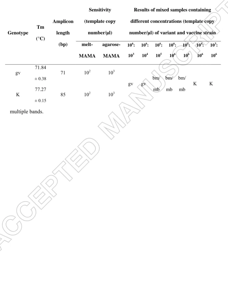
Table 2. Summary table for the results of the MAMA-K-fruA tests. Abbreviations: Tm = melting temperature; gv = genetic variant; K = K vaccine-type; bm = bimodal peak; mb =
multiple bands.
Genotype
Tm (°C)
Amplicon length
(bp)
Sensitivity (template copy
number/µl)
Results of mixed samples containing different concentrations (template copy number/µl) of variant and vaccine strain melt-
MAMA
agarose- MAMA
106: 103
106: 104
106: 105
106: 106
105: 106
104: 106
103: 106
gv 71.84
± 0.38 71 102 103
gv gv bm/
mb bm/
mb bm/
mb K K
K 77.27
± 0.15 85 102 103
Figures
Figure 1. Detection of K vaccine strain-specific SNP in the fruA gene.
Figure 1A. Discrimination of K vaccine strain with agarose-MAMA. Electrophoresis was performed in 3% agarose gel (MetaPhor Agarose, Lonza Group Ltd.). Line 1 and 6: 20-bp DNA ladder (O'RangeRuler 20 bp, Thermo Fisher Scientific Inc.) used as molecular weight marker, Line 2: K vaccine strain yielded 85 bp fragments, Line 3: 71 bp fragments of the M.
gallisepticum ATCC 19610 type strain, Line 4: 71 bp fragments of the M. gallisepticum field sample 19135-M1c, Line 5: negative control (nuclease-free water).
Figure 1B. Discrimination of K vaccine strain with melt-MAMA. Melting curves of the M.
gallisepticum ATCC 19610 type strain (purple line; Tm 72.05 °C), the M. gallisepticum field sample 19135-M1c (blue line; Tm 72.06 °C) and the K vaccine strain (green line; Tm 77.25
°C). Negative control (yellow line) did not amplify. y-axis: derivative reporter, the negative first-derivative of the normalized fluorescence generated by the reporter during PCR amplification; x-axis: temperature melt curve.
Figure 2. Agarose- and melt-MAMA tests of mixed DNA of M. gallisepticum ATCC 19610 type strain and K vaccine strain.
Figure 2A. Samples containing variant:vaccine strain DNA mix in 106:106 (Line 2), 106:105 (Line 3) and 105:106 (Line 4) template copy number/µl resulted two amplicons at specific band sizes by agarose-MAMA test. Electrophoresis was performed in 3% agarose gel (MetaPhor Agarose, Lonza Group Ltd.). Negative control (Line 5) did not amplify. 20-bp DNA ladder (O'RangeRuler 20 bp, Thermo Fisher Scientific Inc.) used as molecular weight marker (Line 1 and 6).
Figure 2B. Samples containing the variant:vaccine strain DNA mix in 106:105 (purple line) and 105:106 (green line) template copy number/µl resulted bimodal peaks in the genotype- specific melting temperatures by melt-MAMA test. Negative control (yellow line) did not amplify. y-axis: derivative reporter, the negative first-derivative of the normalized fluorescence generated by the reporter during PCR amplification; x-axis: temperature melt curve.