R E S E A R C H A R T I C L E Open Access
Prevalence and antimicrobial susceptibility of enterotoxigenic extra-intestinal
Bacteroides fragilis among 13-year collection of isolates in Kuwait
Wafaa Jamal1* , Fatima Bibi Khodakhast1, Ameerah AlAzmi1, Jόzsef Sόki2, Ghayda AlHashem1and Vincent O. Rotimi1
Abstract
Background:Some strains ofBacteroides fragilisspecies are associated with diarrhea as a result of enterotoxin production (bft or fragilysin). Fragilysin is activated by C11 protease (fpn) and together with C10 protease (bfp) play a significant role in its invasiveness. The objectives of this study were to investigate the proportion of clinical isolates from extra-intestinal sources that are toxin producers and characterize the genes mediating toxin production. Clinical isolates submitted to our reference laboratory over the last 13 years were screened for toxin production using PCR technique. All stool isolates were excluded. The isolates were tested for their susceptibility to 8 antimicrobial agents by E test. Carbapenem resistance genecfiAwas detected by PCR.
Results:A total of 421B. fragilisisolates were viable. Out of these,bftwas detected in 210 (49.9%) isolates. Of the 210 bft-positive isolates, 171 (81.4%), 33 (15.7%) and 6 (2.8%) harboredbft-1,bft-2, andbft-3 genes, respectively. Twenty (9.5%) of thebft-positive strains originated from bloodstream infections. Twenty-five, 20 and 9 strains harboredbfp-1, bfp-2 and bfp-3 gene, respectively. Two, 3, 4 bfp isotypes were detected simultaneously in some of strains.
The resistance rates against amoxicillin-clavulanic acid was 32%, clindamycin 62%, cefoxitin 26%, imipenem 11%, meropenem 17%, metronidazole 4%, piperacillin 61% and tigecycline 14%. A chromosomally located cfiA gene that encode metallo-β-lactamase was identified in only 34 isolates (16.2%).
Conclusions: The prevalence of enterotoxin-producingB. fragilis was high among the extra-intestinal isolates.
Metronidazole was the most active agent against all isolates. There was no statistically significance difference between resistance rates among bft-positive and bft-negative isolates except for clindamycin.
Keywords: Bacteroides fragilis, Enterotoxin, Carbapenem-resistance, Kuwait
Backgrounds
Bacteroides species are obligate anaerobic members of the normal microbiota of the human gut. They are the commonest anaerobic bacteria associated with clinical infections particularly those associated with infections of the mucous membranes and adjacent tissues [1]. B.
fragilis is a type species of theBacteroides genus whose numerical population is far less than the other
Bacteroides spp., (e.g. B. thetaiotaomicron, B. distasonis andB. vulgatus), in the normal gut microbiota. However, paradoxically, it is the most common non-spore forming anaerobic bacteria found in routine clinical specimens, e.g. those obtained from deep intra-abdominal abscesses, suppurative skin and soft tissue infections, infections fol- lowing intra-abdominal and vaginal post-hysterectomy surgeries [1]. Recent studies on gut microbiota have suggested that B. fragilis can be the dominant species associated/adherent to the colonic mucosa in healthy individuals [2] contrary to the old believe that its
© The Author(s). 2020Open AccessThis article is distributed under the terms of the Creative Commons Attribution 4.0 International License (http://creativecommons.org/licenses/by/4.0/), which permits unrestricted use, distribution, and reproduction in any medium, provided you give appropriate credit to the original author(s) and the source, provide a link to the Creative Commons license, and indicate if changes were made. The Creative Commons Public Domain Dedication waiver (http://creativecommons.org/publicdomain/zero/1.0/) applies to the data made available in this article, unless otherwise stated.
* Correspondence:wjamal@hsc.edu.kw
1Department of Microbiology, Faculty of Medicine, Kuwait University, P. O.
Box 24923, 13110 Safat, Kuwait
Full list of author information is available at the end of the article
contribution to the anaerobic microbiota of the gut is minimal based on studies on fecal samples.
An important virulence factor of B. fragilis is an en- terotoxin. Enterotoxin-producing B. fragilis (ETBF) strain was first isolated from the stool of neonatal lambs with diarrhea in 1984 [3]. ETBF was later found to cause diarrhea in humans and has been defined as one of the 6 possible causes of diarrhea in children aged 1–5 years [4]. It has also been isolated infrequently from stool specimens of symptomatic adult patients [5]. Although some reports have put the prevalence of extra-intestinal ETBF at 6.2–38% [6, 7], the prevalence of ETBF strains amongB. fragilis isolates from diarrheagenic and extra- intestinal clinical samples in Kuwait is unknown. The enterotoxin produced by B. fragilis is also known as fragilysin. It is a zinc-dependent, non-lethal, heat labile metalloprotease of about 20 kDa [8] which acts by cleav- ing the E-cadherin protein of zonula adherens and tight junctions in the intestinal epithelial cells leading to re- arrangement of the actin of the cytoskeleton of the epi- thelial cells [9, 10]. So far the gene mediating fragilysin (bft) has been well characterized into 4 isotypes, namely bft-1, bft-2, bft-3 and bft-4 [11]; the latter is mainly found in the Far East [7].
In addition,B. fragilis has another virulence factor, an endotoxin/lipopolysaccharide (LPS) with a demonstrable toxicity [12]. OnceB. fragilisis exposed to antibiotics, it liberates endotoxin more than other Bacteroides species which may explain why this species is associated with clinical infections and higher mortality rate [13].
Some studies have demonstrated an association be- tween increased prevalence of ETBF strains and inflam- matory bowel diseases, such as Crohn’s disease and ulcerative colitis [14], while a possible pathogenic role in the etiology of colorectal cancer [15] and bacteremia [16,17] has been suggested. Two cystein peptidase types with pathogenic role have recently been described:bfp1–
4 genes encoding C10 peptidase and fpn gene encoding C11 peptidase (fragipain). Potential link between C10 peptidase and the pathogenesis of inflammatory bowel disease and sepsis has been documented and fragipain has been shown to activateB. fragilisenterotoxin [17,18].
The susceptibility of B. fragilis to metronidazole and carbapenem has been excellent with few anecdotal re- ports of resistance emerging in the literature over the last 3 decades [19–21]. They have remained the drugs extensively used for the treatment of infections caused by this opportunistic pathogen. The emerging reports of B. fragilisisolates resistant to these drugs are causing in- creasing concern to the infectious diseases and clinical microbiology experts worldwide. The trend of resistance to these drugs by B. fragilis in our country, and elsewhere in the Gulf countries, is not clearly defined at molecular level.
This study was designed to investigate the prevalence of ETBF and thebftgenes among B. fragilis isolates col- lected over 13 years in the Anaerobic Reference Labora- tory and to determine the presence of genes mediating carbapenemase (cfiA) production among carbapenem- resistantB. fragilisisolates. It was also planned to investi- gate the prevalence ofbfp1–4 andfpngenes inbft-positive andbft-negative strains.
Results
Bacterial isolates
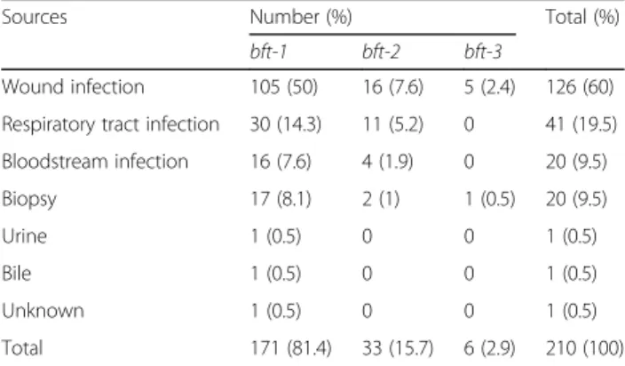
A total of 421B. fragilisisolates were collected from the following different sources: wound infections (WIs), lower respiratory tract infections (LRTIs), bloodstream infections (BSIs), biopsy specimens, urine, and bile. As shown in Table 1, out of the 421 isolates, 210 (49.9%) harbored the bft gene. The majority of the bft-positive isolates were from WIs, followed by LRTIs, BSIs and bi- opsy specimens (BS).
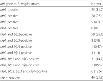
The ages ranged from 1 month to 94 years (mean, 52 years). Of the 210 patients, 137 (65.2%) were males and 73 (34.8%) were females. Out of 39 isolates de- rived from patients with BSIs, 20 (51.2%) were posi- tive for bft gene. The presence of bfp1–4 (C 10 protease gene) and fpn (C11 protease gene) were in- vestigated in the 210 bft-positive B. fragilis strains using PCR. As shown in Table 2, the distribution of C10 protease (bfp) genes was the following: 25 (11.9%) isolates harbored bfp-1 gene, 20 (9.5%) bfp-2 and 9 (4.2%) bfp-3 isotypes alone.
Of the 210, 59 (28.1%) strains carried both bfp-1 and bfp-2 simultaneously, and 31 (14.7%) strains were posi- tive forbfp-1,bfp-2 and bfp-3. In all, 46 (21.9%) isolates did not carry any of the testedbfpgenes.
As shown in Table 3, 170 (80%) B. fragilis were posi- tive forfpn(C11 protease) gene among the 210bft-posi- tive strains, 34 (20%) of which werecfiA-positive.
Table 1Distribution ofB. fragilis bftsubtypes among different sources of infections
Sources Number (%) Total (%)
bft-1 bft-2 bft-3
Wound infection 105 (50) 16 (7.6) 5 (2.4) 126 (60) Respiratory tract infection 30 (14.3) 11 (5.2) 0 41 (19.5) Bloodstream infection 16 (7.6) 4 (1.9) 0 20 (9.5)
Biopsy 17 (8.1) 2 (1) 1 (0.5) 20 (9.5)
Urine 1 (0.5) 0 0 1 (0.5)
Bile 1 (0.5) 0 0 1 (0.5)
Unknown 1 (0.5) 0 0 1 (0.5)
Total 171 (81.4) 33 (15.7) 6 (2.9) 210 (100)
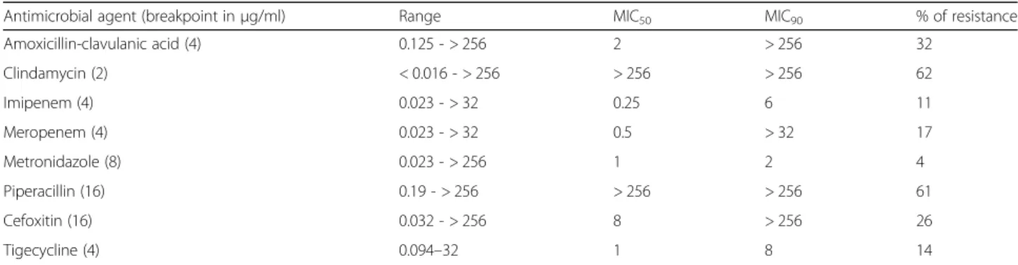
Antimicrobial susceptibility testing and distribution of resistance cfiA gene
MIC range, MIC50 MIC90 and percentage of resistance of the tested antibiotics are shown in Table4.
A total of 261 (62%) and 257 (61.1%) were resistant to clindamycin and piperacillin, respectively. Amoxicillin- clavulanic acid and cefoxitin had unacceptable high MIC values: 135 (32.1%) and 110 (26.1%) were resistant, re- spectively. Resistance rates to metronidazole, tigecycline, imipenem and meropenem were 4, 14, 11, and 17%, re- spectively. When resistance rates amongbft-positive and bft-negative strains were compared, only resistance to clindamycin and tigecycline were higher amongbft-posi- tive than the bft-negative strains but only resistance to clindamycin attained statistically significant level (P= 0.048: CI 18.1–23.21) (Table5).
Further analysis showed that 72 (34.2%) ofbft-positive isolates were multidrug resistant (MDR), that is non- susceptibility to at least one agent in 3 or more different antimicrobial categories [22]. Out of the 72 MDR iso- lates, 64 (88.9%) and 8 (11.1%) werebft-1 andbft-2 sub- types, respectively. None of thebft-3 positive strains was multidrug-resistant.
Thirty-four (8.1%) and 19 (4.5%) of 421 isolates were resistant to meropenem and imipenem, respectively.
Mechanism of carbapenem resistance revealed that all the carbapenem resistant strains were positive for the cfiA gene. This equates with 16.2% of the 210 bft-posi- tive isolates. The sources of these resistant isolates were WIs [23], biopsy samples [4], BSI [3] and LRTIs [3]. A cfiA gene was detected in one B. fragilis strain that showed susceptibility to both imipenem and meropenem with MIC of 2 and 4μg/ml, respectively. This isolate was cultured from a patient with wound infection. In addition, oneB. fragilisisolate was resistant to both imi- penem and meropenem with MIC of 8 and > 32μg/ml, respectively, butcfiA-negative.
Discussion
B. fragiliscan cause serious clinical infections thought to be related to the production of enterotoxin (bft), among other virulence factors. This toxin is activated by C11 (fpn) and C10 proteases (bfp) which help in the invasive- ness of the organism. It has been shown thatbft-positive B. fragilis are more invasive than bft-negative isolates in different types of infections and that blood culture iso- lates are more likely to carry bft enterotoxin gene than other isolates [24]. The prevalence of 49.9% for the en- terotoxin producing-extra intestinal B. fragilis in our study is relatively high when compared with the figures of 14.4% reported in Poland [25], 18.6%, in Japan [16], 6.2–38% in USA [6,7], and 13–25% in Hungary [11,26].
Our data showed that the majority (81.4%) of the isolates contained bft-1 isotype compared with 15.7% of bft-2 and 2.9% bft-3 isotypes. It is pertinent to note that no bft-4 isotype strain was detected in this series. This order of prevalence of the isotypes is partially concordant with previous reports by Scotto d’Abusco et al., [27], Sarvari et al., [11] and Kierzkowska et al., [25]. In their study, Sarvari and colleagues [11] from Hungary reported the prevalence of 10% for the bft-1 isotype compared with 3%bft-2 but, unlike our study, they did not detectbft-3 isotype. This difference in the distribution of bft gene may be related to the severity of illness, prior antimicro- bial therapy, type of the diet and thus gut flora and the method used to detect the enterotoxin. In addition, more than half (51.2%) of the isolates from bloodstream infections in Kuwait were enterotoxin producers which was much higher than previous reports from the USA and Japan (19–28.1% by Claros et al., 2006 and Kato Table 2Distribution ofbfpgenes among 210bft-positiveB.
fragilisstrains
bfpgene inB. fragilisstrains No (%)
bfp1- positive 25 (11.9)
bfp2-positive 20 (9.5)
bfp3-positive 9 (4.2)
bfp4 positive 0 (0)
bfp1 andbfp2-positive 59 (28.1)
bfp1 andbfp3-positive 8 (3.8)
bfp1 andbfp4-positive 1 (0.47)
bfp2 andbfp3-positive 3 (1.4)
bfp1,bfp2 andbfp3-positive 31 (14.7)
bfp1,bfp2 andbfp4-positive 2 (0.95)
bfp1, bfp2, bfp3 andbfp4-positive 6 (12.6)
bfp–negative 46 (21.9)
Table 3Distribution offpnandbfp1–4 genes in thecfiA- positive andcfiA-negativeB. fragilis
B. fragilisstrains (total no) cfiA-positive (no = 34)
cfiA-negative (no = 176)
bfp1- positive (25) 11 14
bfp2- positive (20) 1 19
bfp3- positive (9) 0 9
bfp4- positive (0) 0 0
bfp1 andbfp2-positive (60) 12 48
bfp1 andbfp3-positive (8) 0 8
bfp1 andbfp4-positive (1) 0 1
bfp2 andbfp3-positive (3) 0 3
bfp1,bfp2 andbfp3-positive (31) 6 25
bfp1,bfp2,bfp4-positive (2) 2 0
bfp1,bfp2,bfp3 andbfp3-positive (6) 1 5
bft-negative isolates (46) 1 45
fpn-positive strains (170) 34 136
et al, 1996, respectively) [16,28]. In our study, simultan- eous harboring of 2, 3 and 4bfpisotypes occurred in 71, 33 and 6 isolates, respectively, which is higher than those reported by Sarvari et al., and among the mostbft-posi- tive strains, bfp-1 was the most prevalent isotype. This was discordant with the results of the study reported by Sarvari et al., in which the most common isotype was bfp-2 [11]. In addition, they did not find 4 isotypes in the same isolates [11]. Almost half of our isolates were bft-negative although they were pathogenic in a number of clinical scenarios. The explanation for this may be due to production of other virulence factors e.g. lipo- polysaccharide (LPS) endotoxin especially after exposure to antibiotics [13] or both LPS and capsule that act as adhesion allowing the organism to become established at the site of infection and providing a nidus for abscess formation [1,24].
The majority (60%) of bft-positive B. fragilis isolates were from wound infections which was higher than that reported from Germany and USA (10%; 24) and from Hungary (51%; 11), but lower than that reported in Warsaw, Poland (67.5%; 28). This probably implies that bft-positive strains are more pathogenic in wound infec- tions than thebft-negative strains.
Antimicrobial resistance is a growing problem all over the world including resistance phenomena byB. fragilis.
In this study, resistance to clindamycin (62%) was at an unacceptable level. This shows that B. fragilis in Kuwait are much more highly resistant to this agent than discordant reports elsewhere, such as 28.5% in Europe [29], 48.9% in Taiwan [30], 29.9% in USA [31]
and 36.6% in China [32]. It is conceivable that the over use and abuse of this agent in almost all govern- ment and private hospitals as well as dental clinics in Kuwait is responsible for the alarming high resistance rate. Other relatively high unacceptable resistance level of 26% was recorded against cefoxitin. This is very disturbing finding as this agent is massively used for surgical prophylaxis by most of our surgeons in the country. Its empirical use must therefore be called to question. Metronidazole was the most active non- β-lactam drug with 4% resistance rate. Despite being an active agent in our country, this resistance rate is much higher than those reported from other coun- tries around the world [29–32]. Although the resist- ance rate to amoxicillin-clavulanic acid in our study is marginally higher than those reported around the globe, it is nonetheless at a very uncomfortable high level (32%). Another very interesting but disturbing finding, in our study, is the relatively high tigecycline resistance level of 14%. This is too high when com- pared with findings reported in the European study Table 4Antimicrobial susceptibility for 421B. fragilisisolates
Antimicrobial agent (breakpoint inμg/ml) Range MIC50 MIC90 % of resistance
Amoxicillin-clavulanic acid (4) 0.125 - > 256 2 > 256 32
Clindamycin (2) < 0.016 - > 256 > 256 > 256 62
Imipenem (4) 0.023 - > 32 0.25 6 11
Meropenem (4) 0.023 - > 32 0.5 > 32 17
Metronidazole (8) 0.023 - > 256 1 2 4
Piperacillin (16) 0.19 - > 256 > 256 > 256 61
Cefoxitin (16) 0.032 - > 256 8 > 256 26
Tigecycline (4) 0.094–32 1 8 14
Table 5Antimicrobial resistance amongbft-positive (210) andbft-negative (211)B. fragilis Antibiotic (breakpoint inμg/ml) No (%) of resistantbft-positiveB.fragilis Total no (%)
of resistant bft-positive B. fragilis
No (%) of resistant bft-negative B. fragilis
Pvalue Confidence interval (CI)
bft-1 bft-2 bft-3
Amoxicillin-clavulanic acid (4) 56 (26.7) 2 (1) 2 (1) 60 (28.8) 37 (34.6) 0.354230 [−15.52, 27.12]
Clindamycin (2) 119 (56.7) 20 (9.5) 3 (1.4) 142 (67.6) 120 (56.9) 0.048847 [−1.81, 23.21]
Imipenem (4) 16 (7.6) 1 (0.5) 1 (0.5) 18 (8.6) 27 (12.8) 0.482089 [−18.50, 26.90]
Meropenem (4) 31 (14.8) 2 (1) 1 (0.5) 34 (16.2) 39 (18.5) 0.479721 [−17.83, 22.43]
Metronidazole (8) 10 (4.8) 2 (1) 0 12 (5.7) 6 (2.8) 0.182954 [−28.21, 34.01]
Piperacillin (16) 102 (48.6) 15 (7.1) 4 (1.9) 121 (57.6) 136 (64.5) 0.157430 [−5.81, 19.61]
Cefoxitin (16) 36 (17.1) 2 (1) 1 (0.5) 39 (18.6) 70 (33.2) 0.079984 [−3.85, 33.05]
Tigecycline (4) 29 (13.8) 5 (2.4) 3 (1.4) 37 (17.6) 21 (10) 0.345271 [−13.89, 29.09]
(1.8%) by Nagy et al., [29], 0% in Taiwan study [30]
and 5.4% in USA study [31].
B. fragilisresistance to carbapenem is often associated with production of a class B metallo-β- lactamase encoded by the chromosomal cfiA gene, in addition to outer membrane permeability barrier mechanism [33].
In our study, 11 and 17% of B. fragilis were resistant to imipenem and meropenem, respectively which are much higher than those reported in Europe (1.2% for imipenem) [29], USA (1.1% for imipenem and 2.5% meropenem) [31], Taiwan (8.5% imipenem and 9.9% meropenem) [30].
However, theB. fragilisresistance to imipenem and mero- penem in our study were lower than reported in China (22.7 and 18.2%, respectively) [32]. One of ourB. fragilis isolate was resistant to both imipenem and meropenem in the absence ofcfiA gene which was similar to the report in studies by Soki et al., [34]. Our speculation is that this may be due to other mechanism of resistance, perhaps out membrane permeability problem. Detection of 16.2%cfiA gene is higher than 1.8% which was reported in Poland [25] but lower than that of 36.4% reported in China [32].
A cfiA gene was detected in one B. fragilis isolate that showed susceptibility to both imipenem and meropenem.
This has been reported previously [35,36] and can be ex- plained by the absence of insertion sequence upstream the gene leading to poor expression ofcfiAgene.
Limitation of the study include retrospective collection of isolates and clinical data. Response to therapy could not be determined and insertion sequence elements were not done in the cfiA-positive isolates. Although to our knowledge the most common mechanism of carba- penem resistance inB. fragilis is the production of cfiA metallo-β-lactamase via activation of the cfiA gene by IS elements (high level resistance) or by activation of its putative own promoter other possible mechanisms, such as other carbapenemase genes and AmpC gene, were not investigated.
Conclusions
The prevalence of enterotoxin-producing B. fragilis strains among the clinical isolates of extra-intestinal ori- gin was very high in our study. There was no statistically significance difference in the antibiotic resistance rates among bft-positive and bft-negative isolates except for clindamycin. In this study, metronidazole was the most active antimicrobial agent against enterotoxigenic B. fra- gilisisolates.
Methods Study design
This was a multicenter prospective investigational study of stored B. fragilis isolates from 6 hospital microbio- logical laboratories (Mubarak, Amiri, Al Babtain, Ibn Sina, Adan and Maternity hospitals) in Kuwait. All
clinical isolates obtained from proven cases of infections stored at−80 °C were resuscitated and viable strains in- vestigated for enterotoxin production.
Bacterial strains
The bacterial strains were isolates collected during a 13- years period, from 2006 through 2018, and stored in the Anaerobe Reference Laboratory, Faculty of Medicine, Kuwait University. The viable B. fragilis isolates were from proven cases of intra-abdominal infections, lower respiratory tract infections, bloodstream infections, wound infections and abscesses, managed in the 6 hospi- tals. All isolates were stored, in Brain Heart Infusion (BHI, Oxoid limited, Basingstoke, Hampshire, UK) broth containing 20% glycerol, at -80 °C. During our investiga- tion, isolates were subcultured on Brucella blood agar (Becton Dickinson, Heidelberg, Germany) incubated for 48 h at 37 °C, in an Anoxomat Anaerobic WS800 system™ (MART Microbiology BV, Lichtenvoorde, Netherlands), in an atmospheric condition of 85%
N2, 10% CO2, 5% H2. The identification was confirmed by a Matrix Assisted Laser Desorption/Ionization-Time of Flight Mass Spectrometry (MALDI-TOF MS; bioMérieux, L’Etoile, Marcy, France) analysis.
Antimicrobial susceptibility testing (AST)
The susceptibility of the isolates to 8 anti-anaerobic anti- biotics was investigated by determining the minimum inhibitory concentrations (MICs) of the antibiotics using the E test method (bioMérieux) according to manufac- turer’s instructions. The antibiotics tested were the follow- ing: amoxicillin-clavulanic acid, clindamycin, cefoxitin, imipenem, meropenem, metronidazole, piperacillin and tigecycline. Susceptibility profiles of the isolates were de- termined according to the interpretative criteria recom- mended by the CLSI, 2018 [37]. B. fragilisATCC 25285, and B. thetaiotaomicron ATCC 29741 were included as control in each run. Results for the isolates were accepted if the quality control strains results were within the estab- lished CLSI ranges (CLSI, 2018). MIC50, MIC90and per- centage of resistance were calculated.
Molecular detection of fragilysin (bft) gene
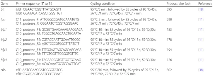
Using previously published procedure, a PCR was per- formed for the detection ofbft gene in all theB. fragilis isolates using bftF and bftR primers [18]. The genes, pri- mer sequences, cycling conditions are shown in Table6.
The following positive controls were used: B. fragilis R19811 (bft-1), B. fragilis1 ATCC 43858(bft-2), andB.
fragilis GAI 96462 (bft-3). Sequencing of the amplicons of the internal fragments of bft-1, bft-2 and bft-3 were performed using a GenAmp PCR system 9700 by cycling sequencing with BigDye® Terminator (AB Applied Bio- systems, Carlsbad, California, USA).
Molecular detection of cfiA carbapenemase–producing gene Production of carbapenemase by the isolates was de- tected in selected number of strains with very high MIC values for meropenem/imipenem using modified Hodge test. All bft-positive strains as well as imipenem and/or meropenem resistantB. fragilis(i.e. MIC≥4μg/ml) were screened for the presence ofcfiA gene and confirmed by PCR, using published primers [38]. The genes, primer sequences, cycling conditions are given in Table6. PCR was carried out in a volume of 25μl. The PCR mix was obtained from Qiagen (Hilden, Germany) and the supernatant of boiled bacterial cells was used as a source of DNA template and the concentration of each primer was 25 pmol. PCR products was sepa- rated by agarose gel electrophoresis and stained with 1% ethidium bromide (Bio-Rad, Hercules, CA, USA) and visualized by UV light.
Molecular analysis of C10 protease (bfp1–4) and C11 protease (fragipain, fpn) genes
For allbft-positive isolates, bfp1–4 and fpn genes in the C10 and C11 proteases, respectively, were investigated.
They were investigated by PCR using the genes, primer sequences, cycling conditions given in Table 6 and the following control strains:B. fragilis638R (bfp1–4) andB.
fragilisATCC 43859 (fpn) were included [17,23,28].
Statistical evaluation
The EpiCalc 2000, version 1.02 (Brixton Heath, Llanidloes, Powys, Wales, UK) was used to compare two proportions- percentages with 95% confidence interval and one sided P-value.
Abbreviations
AST:Antimicrobial susceptibility testing;B. distasonis:Bacteroides distasonis;B.
fragilis:Bacteroides fragilis;B. thetaiotaomicron:Bacteroides thetaiotaomicron;B.
vulgatus:Bacteroides vulgatus;bfp: C10 protease;bft: fragilysin enterotoxin;
BHI: Brain Heart Infusion; BS: Biopsy specimens; BSIs: Bloodstream infections;
cfiA: carbapenem resistance gene; CI: Confidence interval; CLSI: Clinical and Laboratory Standards Institute; CO2: Carbon dioxide; DNA: Deoxyribonucleotide;
ETBF: Enterotoxin-producingBacteroides fragilis;fpn: C11 protease;
H2: Hydrogen; kDa: KiloDalton; LPS: Lipopoltsaccharide; LRTI: Lower respiratory tract infections; MALDI-TOF MS: Matrix Assisted Laser Desorption/Ionization- Time of Flight Mass Spectrometry; MDR: Multidrug resistant; MIC: Minimum inhibitory concentrations; MIC50: Minimum inhibitory concentrations that inhibited 90% of the isolates; MIC90: Minimum inhibitory concentrations that inhibited 90% of the isolates; N2: Nitrogen; PCR: Polymerase chain reaction;
WIs: Wound infections
Acknowledgements
The authors thank all microbiology departments in the 6 hospitals for sending us the strains.
Authors’contributions
WJ: contributes to conception, acquisition and analysis, interpretation of data, and drafted the work. FBK: acquisition, analysis. AA: acquisition, analysis.
JS: substantively revised the work. GA: substantively revised the work. VOR:
contributes to conception, acquisition and analysis, interpretation of data, and drafted the work. All authors have approved the submitted version and the modified version that involves the author’s contribution to the study;
AND have agreed both to be personally accountable for the author’s own contributions and ensure that questions related to the accuracy or integrity of any part of the work, even ones in which the author was not personally involved, are appropriately investigated, resolved and the resolution documented in the literature.
Funding non-funded.
Availability of data and materials
All data generated or analyzed during this study are publicly available and included in this published article.
Ethics approval and consent to participate non-applicable.
Consent for publication non-applicable.
Competing interests
The authors declare that they have no competing interests.
Table 6The genes, primer sequence, cycling condition and reference for fragilysinbftgene,fpn, bfp1–4 genes andcfiA carbapenemase gene
Gene Primer sequence (5′to 3′) Cycling condition Product size (bp) Reference
bft bftF: CGAACTCGGTTTATGCAGTT bftR: GGATACATCAGCTGGGTTGT
95 °C/5 min; followed by 35 cycles of 95 °C/45 s, 56 °C /1 min, 72 °C/45 s, 72 °C/ 7 min
295 [18]
fpn C11_protease_F: ATTCGGCCGATGCAAATGTG C11_protease_R: CGGAATCTCGGTAGGGAAC
95 °C 5 min; followed by 35 cycles of 95 °C/45 s;
56 °C /1 min; 72 °C/45 s, 72 °C/7 min
290 [11]
bfp1 C10_protease_F1: GCGGTGAACAAAGAACGACA C10_protease_R1: TCGCCTGAGCAACTGCAATA
95 °C 10 min; 35 cycles of 95 °C/15 s; 59 °C/30s;
72 °C/47 s; 72 °C/7 min
153 [11]
bfp2 C10_protease_F2: CGTACCAATTGCAATTGCGC C10_protease_R2: AGCTCCCGTGGCTTTATCTT
95 °C 10 min; 35 cycles of 95 °C/15 s; 59 °C/30 s;
72 °C/47 s; 72 °C/7 min
178 [11]
bfp3 C10_protease_F3: TTTGGAGTAGCAGCAGCAGA C10_protease_R3: TTTCTGGTTTCGGGTGTTTC
95 °C 10 min; 35 cycles of 95 °C/15 s; 59 °C/30 s;
72 °C/47 s; 72 °C/7 min
194 [11]
bfp4 C10_protease_F4: TACAACGGTGTTGGTGCAAG C10_protease_R4: ACACAAATGCGCCACTTCAT
95 °C 10 min; 35 cycles of 95 °C/15 s; 59 °C/30s;
72 °C/47 s; 72 °C/7 min
126 [11]
cfiA cfiF: AATCGAAGGATGGGGTATGG cfiR: CGGTCAGTGAATCGGTGAAT
95 °C/10 min, followed by 35 cycles of 95 °C/15 s, 59 °C/30s, 72 °C/ 7 s; 72 °C/7 min
302 [38]
Author details
1Department of Microbiology, Faculty of Medicine, Kuwait University, P. O.
Box 24923, 13110 Safat, Kuwait.2Institute of Clinical Microbiology, University of Szeged, Szeged, Hungary.
Received: 21 September 2019 Accepted: 9 January 2020
References
1. Garrett WS, Onderdonk AB. B.Bacteroides,Prevotella,Porphyromonasand Fusobacteriumspecies (and other medically important anaerobic gram- negative bacilli). In: Mandell GL, Benett JE, Dolin R, editors. Mandell, Douglas and Bennett’s principles and practice of infectious diseases. 8th ed. New York: Churchill Livingston; 2015. p. 2773–80.
2. Jandhyala SM, Talukdar R, Subramanyam C, Vuyyuru H, Sasikala M, Nageshwar RD. Role of the normal gut microbiota. World J Gastroenterol.
2015;21(29):8787–803.https://doi.org/10.3748/wjg. v21.i29.8787.
3. Myers LL, Firehammer BD, Shoop DS, Border MM.Bacteroides fragilis: a possible cause of acute diarrheal disease in newborn lambs. Infect Immunol.
1984;44(2):241–4.
4. Sack RB, Myers LL, Almeido-Hill J, Shoop DS, Bradbury WC, Reid R, et al.
EnterotoxigenicBacteroides fragilis: epidemiologic studies of its role as a human diarrhoeal pathogen. J Diarrhoeal Dis Res. 1992;10(1):4–9.
5. Akpinar M, AktaşE, Cömert F, Külah C, Sümbüloğlu V. Evaluation of the prevalence of enterotoxigenicBacteroides fragilisand the distributionbft gene subtypes in patients with diarrhea. Anaerobe. 2010;16(5):505–9.
https://doi.org/10.1016/j.anaerobe.2010.08.002.
6. Mundy LM, Sears CL. Detection of toxin production byBacteroides fragilis:
assay development and screening of extraintestinal clinical isolates. Clin Infect Dis. 1996;23(2):269–76.
7. Chung GT, Franco AA, Wu S, Rhie GE, Cheng R, Oh HB, et al. Identification of a third metalloprotease toxin gene in extraintestinal isolates of Bacteroides fragilis. Infect Immun. 1999;67(9):4945–9.
8. Sears CL. The toxins ofBacteroides fragilis. Toxicon.2001;39(11):1737–46.
9. Obiso RJ Jr, Azghani AO, Wilkins TD. TheBacteroides fragilistoxin fragilysin disrupts the paracellular barrier of epithelial cells. Infect Immun. 1997;65(4):1431–9.
10. Wu S, Lim KC, Huang J, Saidi RF, Sears CL.Bacteroides fragilisenterotoxin cleaves the zonula adherens protein, E-cadherin. Proc Natl Acad Sci USA.
1998;95(25):14979–84.
11. Sarvari KP, Soki J, Ivan M, Miszti C, Latkoczy K, Melegh SZ, et al. Detection of enterotoxin and protease genes among Hungarian clinicalBacteroides fragilisisolates. Anaerobe.2017;48:98–102.https://doi.org/10.1016/j.
anaerobe.2017.07.005.
12. Delahooke DM, Barclay GR, Poxton IR. A re-appraisal of the biological activity of bacteroides LPS. J Med Microbiol. 1995;42(2):102–12.
13. Rotimi VO, Verghese TL, Al-Sweih N, Khodakhast FB, Ahmed K. Influence of five anti-anaerobic antibiotics on endotoxin liberation by gram-negative anaerobes. J Chemother. 2000;12(1):40–7.
14. Prindiville TP, Sheikh RA, Cohen SH, Tang YJ, Cantrell MC, Silva J Jr.
Bacteroides fragilisenterotoxin gene sequences in patients with inflammatory bowel disease. Emerg Infect Dis. 2000;6(2):171–4.
15. Toprak NU, Yagci A, Gulluoglu BM, Akin ML, Demirkalem P, Celenk T, et al. A possible role ofBacteroides fragilisenterotoxin in the aetiology of colorectal cancer. Clin Microbiol Infect. 2006;12(8):782–6.
16. Kato N, Kato H, Watanabe K, Ueno K. Association of enterotoxigenic Bacteroides fragiliswith bacteremia. Clin Infect Dis. 1996;23(Suppl. 1):S83–6.
17. Choi VM, Herrou J, Hecht AL, Teoh WP, Turner JR, Crosson S, Bubeck, et al. Activation ofBacteroides fragilistoxin by a novel bacterial protease contributes to anaerobic sepsis in mice. Nat Med. 2016;22(5):
563–7.https://doi.org/10.1038/nm.4077.
18. Sóki J, Edwards R, Hedberg M, Fang H, Nagy E, Nord CE, et al. Examination ofcfiA-mediated carbapenem resistance inBacteroides fragilisstrains from a European antibiotic susceptibility survey. Int J Antimicrob Agents. 2006;
28(6):497–502.
19. Jamal W, Al Hashem G, Rotimi VO. Antimicrobial resistance among anaerobes isolated from clinical specimens in Kuwait hospitals: comparative analysis of 11-year data. Anaerobe. 2015;31:25–30.https://doi.org/10.1016/j.
anaerobe.2014.08.012.
20. Jamal WY, Rotimi VO, Brazier JS, Johny M, Wetieh WM, Duerden BI.
Molecular characterization of nitroimidazole resistance in metronidazole-
resistantBacteroidesspecies isolated from hospital patients in Kuwait. Med Princ Pract. 2004;13(3):147–52.
21. Jamal W, Shahin M, Rotimi VO. Surveillance and trends of antimicrobial resistance among clinical isolates of anaerobes in Kuwait hospitals from 2002 to 2007. Anaerobe. 2010;16(1):1–5.https://doi.org/10.1016/j.anaerobe.
2009.04.004.
22. Magiorakos AP, Srinivasan A, Carey RB, Carmeli Y, Falagas ME, Giske CG, et al. Multidrug–resistant, extensively drug-resistant and pandrug-resistant bacteria: an international expert proposal for interim standard definitions for acquired resistance. Clin Microbiol Infect. 2012;18(3):268–81.https://doi.org/
10.1111/j.1469-0691.2011. 03570.x.
23. Thornton RF, Kagawa TF, O’Toole PW, Cooney JC. The dissemination of C10 cysteine protease genes inBacteroides fragilisby mobile genetic elements.
BMC Microbiol. 2010;10:122.https://doi.org/10.1186/1471-2180-10-122.
24. Wexler HM.Bacteroides: the good, the bad and the nitty–gritty. Clin Microbiol Rev. 2007;20(4):593–621.
25. Kierzkowska M, Majewska A, Szymanek-Majchrzak K, Sawicka-Grzelak A, Mlynarczyk A, Mlynarczyk G. The presence of antibiotic resistance genes and bftgenes as well as antibiotic susceptibility testing ofBacteroides fragilis strains isolated from inpatients of infant Jesus teaching hospital, Warsaw during 2007-2012. Anaerobe. 2019;56:109–15.https://doi.org/10.1016/j.
anaerobe.2019.03.003.
26. Szoke I, Dósa E, Nagy E. EnterotoxigenicBacteroides fragilisin Hungary.
Anaerobe. 1997;3(2–3):87–9.
27. Scotto d’Abusco AS, Del Grosso M, Censini S, Covacci A, Pantosti A. The alleles of thebftgene are distributed differently among enterotoxigenic Bacteroides fragilisstrains from human sources and can be present in double copies. J Clin Microbiol. 2000;38(2):607–12.
28. Claros MC, Claros ZC, Tang YJ, Cohen SH, Silva J Jr, Goldstein EJ, et al.
Occurrence ofBacteroides fragilisenterotoxin gene-carrying strains in Germany and the United States. J Clin Microbiol. 2000;38(5):1996–7.
29. Nagy E, Urban E, Nord EC, on behalf of the ESCMID Study Group on Antimicrobial Resistance in Anaerobic Bacteria. Antimicrobial susceptibility of Bacteroides fragilisgroup isolates in Europe: 20 years of experience. Clin Microbiol Infect. 2011;17(3):371–9.https://doi.org/10.1111/j.1469-0691.2010.03256. x.
30. Wang FD, Liao CH, Lin YT, Sheng WH, Hsueh PR. Trends in the susceptibility of commonly encountered clinically significant anaerobes and susceptibilities of blood isolates of anaerobes to 16 antimicrobial agents, including fidaxomicin and rifaximin, 2008-2012, northern Taiwan. Eur J Clin Microbiol Infect Dis. 2014;
33(11):2041–52.https://doi.org/10.1007/s10096-014-2175-y.
31. Snydman DR, Jacobus NV, McDermott LA, Golan Y, Goldstein EJ, Harrell L, et al. Update on resistance ofBacteroides fragilisgroup and related species with special attention to carbapenems 2006-2009. Anaerobe. 2011;17(4):
147–51.https://doi.org/10.1016/j.anaerobe.2011.05.014.
32. Gao Q, Wu S, Xu T, Zhao X, Huang H, Hu F. Emergence of carbapenem resistance inBacteroides fragilisin China. Int J Antimicrob Agents. 2019;53(6):
859–63.https://doi.org/10.1016/j.ijantimicag.2019.02.017.
33. Hurlbut S, Cuchural GJ, Tally FP. Imipenem resistance inBacteroides distasonismediated by a novelβ-lactamase. Antimicrob Agents Chemother.
1990;34(1):117–20.
34. Sóki J, Fodor E, Hecht DW, Edwards R, Rotimi VO, Kerekes I, et al. Molecular characterization of imipenem-resistant,cfiA-positiveBacteroides fragilisisolates from the USA, Hungary and Kuwait. J Med Microbiol. 2004;53(Pt 5):413–9.
35. Yamazoe K, Kato N, Kato H, Tanaka K, Katagiri Y, Watanabe K. Distribution of thecfiAgene amongBacteroides fragilisstrains in Japan and relatedness of cfiAto imipenem resistance. Antimicrob Agents Chemother. 1999;43(11):
2808–10.
36. Edwards R, Read PN. Expression of the carbapenemase gene (cfiA) in Bacteroides fragilis. J Antimicrob Chemother. 2000;46(6):1009–12.
37. Clinical and Laboratory Standards Institute: CLSI Publication Document M100-S28. Performance standards for antimicrobial susceptibility testing:
Twenty eighth Informational Supplement, Vol. 34. Wayne: Clinical and Laboratory Standards Institute; 2018. p. No.1.
38. Eitel Z, Sóki J, Urbán E, Nagy E, the ESCMID Study Group on Anaerobic Infection. The prevalence of antibiotic resistance genes inBacteroides fragilis group strains isolated in different European countries. Anaerobe. 2013;21:
43–9.https://doi.org/10.1016/j.anaerobe.2013.03.001.
Publisher’s Note
Springer Nature remains neutral with regard to jurisdictional claims in published maps and institutional affiliations.