TECHNICAL ADVANCE
Gene modification by fast-track recombineering for cellular localization and isolation of components of plant protein complexes
Zhoubo Hu1, Ajit Ghosh1,2, Sara C. Stolze1, Mihaly Horvath1, Bing Bai1, Sabine Schaefer1, Simone Z€undorf1, Shanda Liu1, Anne Harzen1, Mohsen Hajheidari1,3, Tomasz J. Sarnowski1,4, Hirofumi Nakagami1, Zsuzsa Koncz1and Csaba Koncz1,5,*
1Max-Planck Institute for Plant Breeding Research, Carl-von-Linne-Weg 10, D-50829, Cologne, Germany,
2Department of Biochemistry and Molecular Biology, Shahjalal University of Science and Technology, Sylhet 3114, Bangladesh,
3Botanical Institute, Cologne Biocenter, Cluster of Excellence on Plant Sciences, University of Cologne, D-50674, Cologne, Germany,
4Institute of Biochemistry and Biophysics, Polish Academy of Sciences, Pawinskiego 5A, 02-106, Warsaw, Poland, and
5Institute of Plant Biology, Biological Research Center of Hungarian Academy of Sciences, Temesvari krt. 62, H-6726, Szeged, Hungary
Received 29 April 2019; revised 14 June 2019; accepted 26 June 2019; published online 5 July 2019.
*For correspondence (e-mail koncz@mpipz.mpg.de).
[Correction added on 14 October 2019, after first online publication: ‘Bai Bing’ has been changed to ‘Bing Bai’ in author list.]
SUMMARY
To accelerate the isolation of plant protein complexes and study cellular localization and interaction of their com- ponents, an improved recombineering protocol is described for simple and fast site-directed modification of plant genes in bacterial artificial chromosomes (BACs). Coding sequences of fluorescent and affinity tags were inserted into genes and transferred together with flanking genomic sequences of desired size by recombination intoAgrobacteriumplant transformation vectors using three steps ofE. colitransformation with PCR-amplified DNA fragments. Application of fast-track recombineering is illustrated by the simultaneous labelling of CYCLIN- DEPENDENT KINASE D (CDKD) and CYCLIN H (CYCH) subunits of kinase module of TFIIH general transcription factor and the CDKD-activating CDKF;1 kinase with green fluorescent protein (GFP) and mCherry (green and red fluorescent protein) tags, and a PIPL (His18-StrepII-HA) epitope. Functionality of modifiedCDKF;1gene constructs is verified by complementation of corresponding T-DNA insertion mutation. Interaction of CYCH with all three known CDKD homologues is confirmed by their co-localization and co-immunoprecipitation. Affinity purification and mass spectrometry analyses of CDKD;2, CYCH, and DNA-replication-coupled HISTONE H3.1 validate their association with conserved TFIIH subunits and components of CHROMATIN ASSEMBLY FACTOR 1, respectively.
The results document that simple modification of plant gene products with suitable tags by fast-track recombi- neering is well suited to promote a wide range of protein interaction and proteomics studies.
Keywords: recombineering, site-directed gene modification, fluorescent reporters, affinity purification, TFIIH protein kinases, DNA replication-dependent HISTONE H3.1, technical advance.
INTRODUCTION
The term recombineering refers to cloning technologies that employ phage-encoded recombination enzymes, such as Exo, Beta, and Gam of lambda phage Red system, to achieve in vivo site-specific integration of foreign DNA sequences into genes carried by bacterial chromosomes or plasmids (Thomasonet al., 2014). ThekRed system medi- ates recombination between 50 nucleotide arms of a PCR- amplified DNA fragment and corresponding homologous
sequences flanking the target site, which may be repre- sented by a single nucleotide, a codon triplet, or a longer DNA sequence for generating point mutations, codon exchanges, deletions, and in-frame insertions of suitable tags, respectively. First, a positive-negative selectable mar- ker cassette is inserted into the target gene, and then this cassette is replaced by a desired tag, or with a DNA frag- ment carrying a nucleotide exchange or deletion. The phage genes coding for recombination enzymes are either
©2019 The Authors 411
harboured by a plasmid or stably integrated into the chro- mosome of bacterial host for recombineering. In thekRed E. colihost SW102 (Warminget al., 2005), theexo, beta, andgamgenes of a defective prophage are expressed by thepLpromoter, which is induced by temporal inactivation of thermosensitive cI857tsrepressor. One of the most pop- ular positivenegative selectable markers is the galactoki- nase (galK) gene, the integration of which into the target site is selected for on minimal medium in agalK host.
Subsequently, the exchange of galKmarker with desired sequences is achieved by counter-selection on deoxygalac- tose-containing medium.
Bacterial artificial chromosome (BAC) clones generated during the genome-sequencing projects carry 100 kb or larger genomic DNA segments with multiple genes and can be readily recombined into chromosomes of trans- formed mammalian embryonic stem cells. The modifica- tion of mammalian genes by BACs recombineering became a routine high-throughput approach for the gener- ation of knockout and knock-in lines, especially in trans- genic mice (Sharan et al., 2009; Ciotta et al., 2011;
Narayanan and Chen, 2011). As homologous recombina- tion by BAC DNA transformation did not prove to be feasi- ble in plants, the application of recombineering was coupled to Agrobacterium-mediated gene transfer. This was achieved either by recombineering of plant genes cloned in TACs (transformation competent BACAgrobac- teriumvectors, Shibata and Liu, 2000; Zhou et al., 2011;
Alonso and Stepanova, 2015) or by moving the modified plant genes from BACs intoAgrobacteriumbinary vectors by gap-repair recombination (Bitrian et al., 2011). Both plant BAC-recombineering approaches are, however, rela- tively slow because direct and counter-selection of thegalK exchange marker on minimal medium requires weeks, and the first approach is also affected by low frequency of plant transformation with large chromosomal segments of TACs.
To improve the efficacy of plant BAC recombineering, we replaced the galK marker with antibiotic resistance genes that are either flanked by cleavage sites of the I-SceI homing endonuclease and therefore excisable, or linked to an arabinose-inducibleccdBgyrase-inhibitor killer gene as counterselectable marker. Inhibition of gyrase by ccdB results in the accumulation of DNA double-stranded breaks, causing ultimate cell death. To avoid unnecessary cloning steps, the target plant genes modified by recombi- neering were moved from the BACs into PCR-amplified binary vectors by recombination, and then transferred by Agrobacterium into transgenic plants. The improved recombineering tools were used for exploringin vivointer- actions of Arabidopsis CDKD (CYCLIN-DEPENDENT KINASE D) homologues of human CDK7 with the CDKD-ac- tivating kinase CDKF;1, CYCLIN H (CYCH) and core compo- nents of the RNA polymerase II (RNAPII) general
transcription factor TFIIH. The kinase module (TFIIK) of human TFIIH is composed of CDK7, CYCH and MAT1 (Menage a trois 1) assembly factor subunits. TFIIK plays a key role in the activation of cell cycle kinases and, when bound to TFIIH it phosphorylates serine 5 residues of hep- tapeptide repeats of RNAPII C-terminal domain promoting transcription initiation. Furthermore, TFIIK is targeted to DNA damage sites by TFIIH and modulates both transcrip- tion-coupled and general genome repair (Fisher, 2012, 2019). Whereas the Arabidopsis CDK7 homologues CDKD;1, CDKD;2 and CDKD;3 are activated by CDKF;1-me- diated T-loop phosphorylation and phosphorylate serine 5 residues of RNAPII CTD in vitro (Hajheidari et al., 2012, 2013), their interactions with CYCH and CDKF;1 are not confirmedin vivo. Compared with yeast and animal MAT1 homologues, which mediate interaction of TFIIK with the XPB and XPD helicase subunits of TFIIH, the N-terminal RING domain is missing in the putative Arabidopsis MAT1 (At4 g30820) TFIIK subunit (Umedaet al., 2005). As TFIIH was not yet purified in association with TFIIK from plants, it is unknown whether Arabidopsis TFIIK-TFIIH carries con- served homologues of all TFIIH subunits that were recently characterized by structural studies of human TFIIH (Greber et al., 2019). Therefore, we isolated TFIIH complexes from Arabidopsis using the CDKD;2 and CYCH TFIIK subunits modified by recombineering and analyzed their subunit composition by LC-MS/MS mass spectrometry. In these experiments, we used as nuclear control the DNA replica- tion-dependent HISTONE H3.1 protein, which is located in silent regions of the genome and incorporated into chro- matin during heterochromatin replication (Jacob et al., 2014; Otero et al., 2016). CDKD and CYCH subunits of the TFIIH kinase module were labelled by recombineering with a PIPL (His18-StrepII-HA) epitope, and GFP and mCherry (green and red fluorescent protein) tags. The CDKD-activat- ing kinase CDKF;1 was similarly labelled with GFP and PIPL tags and expressed in thecdkf;1T-DNA insertion mutant to validate functionality of modified gene constructs by genetic complementation. Co-immunoprecipitation and cellular co-localization data indicated that CYCLIN H (CYCH) is associated with the Arabidopsis CDKD;1, CDKD;2 and CDKD;3 kinases, but not with their upstream activating kinase CDKF;1. In contrast, co-immunoprecipitation data confirmed interaction of CDKF;1 with CDKD kinases. Affin- ity purification and mass spectrometry analysis of CYCH:
mCherry and CDKD;2:GFP verified their association with Arabidopsis homologues of conserved core subunits of TFIIH except XPB. Histone H3.1–mCherry purified as nuclear control in the same experiments was identified in complex with three subunits of CAF1 (CHROMATIN ASSEMBLY FACTOR 1, Tagamiet al., 2004; Jiang and Ber- ger, 2017) and ASF1A/B (ANTI-SILENCING FUNCTION 1, Lario et al., 2013). In summary, the results documented that fast-track recombineering is well suited to assist the
isolation and characterization of components of plant pro- tein complexes.
RESULTS
Recombineering usingccdBgene cassettes
In recombineering experiments, selectable markers are inserted into desired positions of genes and then replaced with DNA fragments that either code for suitable tags or carry nucleotide exchanges or deletions. Linear DNA frag- ments of selectable markers and tags used for their replacement are PCR amplified with primers that carry 50 nucleotides (nt) flanks of targeted gene positions. These fragments are transformed intoE. colihosts and following pulse-induction of phage-encoded enzymes are integrated by recombination aided by the 50-nt flanks into the desig- nated target sites. The time requirement of recombineering experiments, which replace traditional cloning with simple E. colitransformation is primarily determined by the selec- tion conditions applied. E. coli hosts with heat-inducible kRed genes need to be cultured at 32°C to avoid constitu- tive expression of recombination enzymes (Warminget al., 2005). Transformation of such strains with BACs carrying the target plant genes is achieved in a day by selecting for the BAC-encoded antibiotic resistance markers in complete medium. However, subsequent selection for insertion and replacement of populargalKmarker on minimal medium requires considerably longer time, hence represents a major bottleneck of recombineering experiments. To accel- erate the recombineering procedure, we replacedgalKwith new exchange markers by linking chloramphenicol (CmR), kanamycin (KmR) and spectinomycin (SpR) resistance genes to accdBDNA-gyrase (gyrA) inhibitor suicide gene, which is transcribed by an arabinose-inducible pBAD pro- moter and controlled by an adjacent araCrepressor gene (Le Roux et al., 2007; see Experimental Procedures, Sup- porting Information Figure S1).
For recombineering, BAC DNAs carrying the genes of TFIIH-associated kinases CDKD;1 (At1g73690 BAC F25P22 KmR), CDKD;2 (At1g66750 BACF4N21 KmR) and CDKD;3 (At1g18040 BAC T10F20, CmR) and their upstream activat- ing kinase CDKF;1 (At4g28980, BAC F25O24 KmR) were verified for the presence of target genes by PCR using pri- mers flanking their stop codons (Figure S2 and Table S1).
The BACs were transformed intoE. coliSW102 by select- ing for their KmR or CmR markers. In the first step of recombineering (Figure 1), the stop codons of CDKF;1, CDKD;1andCDKD;2genes were replaced by theCmR-ccdB marker, and that of the CDKD;3gene with theKmR-ccdB cassette. DNA fragments of ccdB cassettes were PCR amplified with primers including 50nt flanks of target gene stop codons (Figure S2 and Table S1) and transformed into the BAC-containing host strains. Transformants selected for the CmR or KmR markers of ccdBcassettes
were subsequently grown without selecting for the BAC- encoded resistance markers to enhance the loss of empty BAC copies lackingccdBinsertions, which was monitored by colony PCR with flanking gene-specific primers.
In the second step, the ccdB cassettes were replaced with coding sequences of PIPL tag, which is composed of 18 His residues from the Co2+/Ni2+-binding domain of Ara- bidopsis CobW-like protein (At1g15730) linked to StrepII and HA (hemagglutinin) epitopes (Figure S3), or with those of the GFP, or a combination of both. These tags were also adapted to generation of N-terminal fusions for replacing translational start codons (Figure S3), and designed to assist purification of modified plant proteins on Ni2+-agar- ose, Strep-Tactin, anti-HA affinity, and GFP-Trap resins.
Cells carrying only BACs with ccdB cassette insertions were transformed with PCR-amplified fragments of the tags, and then transformants were selected for the BAC-en- coded resistance marker in the presence of 0.2% arabinose to induce the suicide ccdB gene. The exchange events were confirmed by screening for the loss of CmR or KmR markers of ccdB cassettes and colony PCR with primers flanking the target sites.
In the third step, the tagged genes and neighbouring genomic sequences securing their native transcriptional regulation (i.e. including usually two flanking genes) were transferred by gap-repair into pGAP binary vectors (Bitrian et al., 2011; Table S1, Figures 1 and S2). Two flanks, defin- ing the boundaries of modified genes were cloned into pGAP vectors, which were then linearized between the flanks, dephosphorylated and transformed into SW102 cells carrying the BACs. Gap-repair recombination between homologous sequences of flanks of linear vectors and BACs resulted in the integration of modified genes into the binary vectors. Following selection for ampicillin resistant (AmpR) transformants, the resulting pGAP clones were fin- gerprinted by restriction enzyme digestions, and the junc- tions of inserted tags were confirmed by sequencing using the flanking gene-specific primers.
Whereas theccdBcassettes could be similarly inserted into any position of a target gene and replaced also by DNA fragments carrying codon exchanges or deletions, the need for finding transformants with homogeneous BAC populations carrying only theccdBinsertions delimited the speed of the first step in the procedure. The requirement for cloning of homology arms into the binary vectors for gap-repair recombination in the third step represented another bottleneck.
Recombineering with I-SceI insertion cassettes
When designing a fast-track version of recombineering, it was considered that the majority of plant gene modifica- tions aims at labelling the gene products with N- and C-ter- minal fusions to fluorescent or affinity tags. Therefore, we constructed a set of N- and C-terminal insertion cassettes,
in which the KmR and SpR genes flanked by recognition sites of the homing endonuclease I-SceI were fused to cod- ing sequences of GFP, mCherry and GFP–PIPL tags (Fig- ures 2a and S4). To avoid unnecessary cloning steps, PCR- amplifiable binary vectors (6.5–6.7 kb, Figure S5) with a
cosmid replicon, bacterial ampicillin (AmpR)/carbenicillin (CbR) resistance marker, and conditional RK2 conjugational transfer and replication origins (oriTand oriV) were con- structed (pGAPBRKm and pGAPBRHyg; Experimental Pro- cedures). Studies of the T-DNA integration mechanism and
Figure 1.Recombineering withccdBgene cassettes. The work flow of recombineering with theccdBexchange cassettes is illustrated in the example of replace- ment of the stop codon ofCDKD;3(At1g18040) gene by the GFP coding sequences (Figure S2e). TheCDKD;3BAC clone (T10F20) carrying a CmR marker is intro- duced into the recombineering hostE. coliSW102 and the presence of the target gene is verified by PCR amplification with gene-specific primers (green arrows) flanking its stop codon. In the first step of recombineering (1), the SW102 (BAC T10F20) strain is transformed with the DNA fragment of KmR-araC-ccdB cassette (2.7 kb), which is PCR amplified with primers carrying 50 nt flanks of the target stop codon (blue and red bars). KmR transformants are selected and regrown in LB-Km0.5% glucose medium without selecting for the BAC CmR marker, to enhance the loss of BACs lacking theccdBinsertion. Colonies carrying only BACs with theccdBinsertion are identified by PCR (2.7 kb+space between the gene-specific primers). In the second step (2), the obtained SW102 (BAC:
ccdB) strain is transformed with a DNA fragment of GFP coding region, which is PCR amplified with primers carrying the 50 nt flanks of the stop codon (0.82 kb). Transformants are selected and enriched for the BAC CmR marker in LB medium containing 0.2% arabinose to induce the suicideccdBgene expres- sion. Exchange of theccdBmarker with the GFP cassette is monitored by colony PCR (0.72 kb+space between the gene-specific primers). In the third step (3), the modified plant gene is moved by gap-repair into anAgrobacteriumbinary vector. When using pGAPKm or pGAPHyg (Bitrianet al., 2011; Figure S2a), two BAC segments flanking the modified gene (usually located upstream and downstream of neighbouring genes) are PCR amplified asEcoRI-SalI andSalI-BamHI fragments and inserted intoEcoRIBamHI sites of pGAPs. Subsequently, the vectors are linearized bySalI, phosphatase treated and transformed into SW102 (BAC:GFP). Following selection of AmpR transformants, plasmid DNA is prepared and transformed intoE. coliDH5aor DH10B. The presence of modified plant gene is verified by restriction enzyme fingerprinting and sequencing with the gene-specific primers. The verified clone is transformed to theE. colidonor stain MFDpirDTIV lacIq and the conjugated intoAgrobacteriumGV3101 (pMP90RK) for plant transformation. To save time, the gap-repair step (3) is performed with PCR-amplifiable pGAPBRKm and pGAPBRHyg vectors as shown in Figure 2b. BACs carrying a KmR marker are similarly modified using either the SpR-ccdB or CmR-ccdB cassette. The latter was used for modification ofCDKF;1,CDKD;1andCDKD;2genes (Figure S2b–d). TheccdBexchange cassettes can be similarly inserted into any position of a target gene and replaced with DNA fragments carrying point mutations, codon exchanges or deletions.
sequencing plant DNA junctions of T-DNA insertions indi- cated that, compared with the left T-DNA border, the right border is less prone to deletions as it is protected by a covalently linked VirD2 protein during T-DNA transfer from Agrobacteriuminto plants (Gelvin, 2017). To select for the integration of full-length T-DNA inserts into plant chromo- somes, therefore, the plant selectable markers were placed into the vicinity of T-DNA left border, whereas the site used for linearization and PCR amplification of vectors, and inte- gration of tagged plant genes by homologous recombina- tion was positioned close to the right T-DNA border.
We used the I-SceI insertion cassettes for in-frame replacement of stop codons of CYCLIN H (CYCH
At5g27620; BACF15A18 KmR) and HISTONE H3;1 (AT5G65360; BAC MNA5 KmR) genes with coding sequences of GFP and mCherry (Figure S2). In the first step (Figure 2b), PCR-amplified C–mCherrystop-SpR and C- GPFstop-SpR cassette fragments (Figure S4 and Table S1) with corresponding flanks were transformed into E. coli SW102 carrying the verified BACs, and then transformants were selected for the SpR marker of I-SceI cassette inser- tions.
To move the insertion cassette-containing genes into Agrobacterium vectors in the second step, the pGAPBR vectors were linearized byBamHI digestion and PCR ampli- fied with primers that carried 50 nt flanks marking the
Figure 2. Fast-track recombineering using I-SceI insertion cassettes. (a) Schematic presentation of N- and C-terminal KmR and SpR gene-linked I-SceI cassettes (Figure S4) designed for replacement of start and stop codons of target genes with coding regions of GFP, mCherry and PIPL (His18StrepII-HA) epitope. (b) The work flow of fast-track recombineering is illustrated schematically by the replacement of stop codons ofCYCHandH3.1genes (Figure S2f,g), which are carried by BACs with KmR markers. The BAC harbouring the target gene is transformed into the recombineering host SW102 and verified by PCR amplification of a seg- ment of target gene with primers flanking its stop codon (green arrowheads). In the first step of recombineering (1), the C–GFPstop-SpR I-SceI cassette (Fig- ure S4) is PCR amplified with primers carrying 50-nt flanks of the stop codon (red and blue bars) and the cassette DNA fragment (2.07 kb) is transformed into SW102 harbouring the target BAC. Transformants are selected for the SpR marker of the I-SceI cassette and verified by colony PCR with the gene-specific pri- mers. The PCR will detect BACs both with and without cassette insertions (2.07 kb+space between the primers versus distance between the gene-specific pri- mers). In the second step (2), the target gene carrying the I-SceI cassette insertion replacing its stop codon is moved by gap-repair into the pGAPBRHyg (or pGAPBRKm, Figure S5) binary vector. pGAPBRHyg is linearized withBamHI, phosphatase treated (see Experimental Procedures for necessary control step), and PCR amplified with primers that carry 50 nt flanks of BAC sequences designed for transfer into plants linked to the modified target gene (Figure S2f,g). The puri- fied linear pGAPBRHyg is transformed into SW102 (BAC:GFPstop-SpR). Following selection of AmpR transformants, plasmid DNA is prepared and transformed intoE. coliDH10B to purify the pGAPBRHyg clones from the resident BACs. In the third step (3), the pGAPBRHyg clone is fingerprinted with restriction enzymes, cleaved by I-SceI, self-ligated and transformed intoE. coliDH10B. AmpR transformants are screened for the loss of SpR marker and subjected to verification by sequencing the junction of modified plant gene in pGAPBRHyg using the gene-specific primers. Finally, the construct is transferred by conjugation fromE. coli intoAgrobacteriumfor plant transformation as described in Figure 1.
boundaries of plant genomic regions of BACs destined for transfer into plants (Figures S2 and S5). From AmpR SpR colonies obtained by transformation, plasmid DNA was isolated and transformed into a regularE. colihost, such as DH5aor DH10B. The selection for gap-repair of target plant genes that carried the insertion cassettes with the SpR marker made it unnecessary to remove the untagged BACs before performing the gap-repair.
Finally, the SpR selectable marker was removed in the third step from the modified plant genes by I-SceI diges- tion and transformation of self-ligated recombinant binary vectors intoE. coli followed by screening for AmpR and Sp-sensitive colonies. Analogously to the Gateway and Cre/Lox site-specific recombination techniques, the diges- tion left an I-SceI footprint of 27 bp after the integrated GFP and mCherry tags in the modified plant genes. The resulting recombinant vectors were verified by endonucle- ase fingerprinting and sequencing. The RK2 oriTfunction aided easy conjugation of binary vectors fromE. coliinto Agrobacterium GV3101 (pMP90RK, Koncz and Schell, 1986), whereas the RK2 oriVreplication origin secured their maintenance in the latter host, which provided thetrans- actingtrfA replication helper function on the disarmed Ti- plasmid pMP90RK. RK2 conjugation helper functions of pMP90RK also assisted back-conjugation of the vectors fromAgrobacteriumtoE. coli, in order to test their integ- rity before plant transformation. Detailed protocols of recombineering with theccdBexchange marker and I-SceI insertion cassettes are provided in the Experimental Proce- dures.
Expression and cellular localization of proteins labelled by recombineering in Arabidopsis
All genes modified by recombineering were transformed inAgrobacteriumbinary vectors into wild type Arabidopsis plants. Although the insert size in the pGAPBR vectors var- ied from 4.7 kb (HISTONE H3.1 clones) to 14.2 kb (CDKD;2 constructs; Table S1), the transformation efficiencies were similar to those obtained with the empty pGAP and related pPCV binary vectors (the transformation frequencies ran- ged between 0.5% and 1.2% of T1 seed obtained by infiltra- tion of inflorescences; Konczet al., 1994; Rıoset al., 2002).
Transformants showing 3:1 segregation of single T-DNA insertions were propagated to isolate homozygous T3 lines.CDKF;1gene constructs carrying the GFP–PIPL, GFP and PIPL tags were also introduced into the cdkf;1-2/+
(GABI_315A10, Hajheidari et al., 2012) T-DNA insertion mutant. T2 lines carrying single T-DNA inserts of pGAP vectors were screened for homozygous status of sulfadi- azine resistance marker ofcdkf;1-2mutation, and then at least three independent T3 offspring harbouring the kana- mycin or hygromycin resistance markers of complement- ing CDKF;1:GFP–PIPL, CDKF;1:GFP and CDKF;1:PIPL constructs in homozygous form were identified. Compared
with an extreme dwarf phenotype of cdkf;1-2 mutant, all selected T3 lines were wild type indicating genetic comple- mentation of the mutation and verifying full functionality of tagged CDKF;1 gene constructs (Figure 3a). Western blotting of equal aliquots of total protein extracts from ran- domly chosen T3 lines confirmed comparable expression levels of tagged CDKF;1 kinase proteins in the comple- mentedcdkf;1-2mutant (Figure 3b). Following microscopic inspection of GFP and mCherry expression in roots and hypocotyls of wild type T3 seedlings, the expression of CDKF;1:GFP–PIPL; CDKD;2:GFP–PIPL, CDKD;3:GFP, CYCH:
GFP, CYCH:mCherrry and Histone H3;1:mCherry proteins of expected molecular mass was analogously confirmed by western blotting of total protein extracts with anti-GFP and anti-RFP antibodies. As expression levels of CDKD;1:
GFP and CDKD;3:GFP proteins were low, their detection required previous enrichment by affinity purification on GFP-Trap resin (Figure 3c).
Examination of expression patterns of these fusion pro- teins in 15-day-old seedlings by confocal microscopy revealed that CDKF;1, CYCH, CDKDs and the control DNA replication-dependent histone H3.1 showed the highest levels in root tip, division and elongation zones, but lower expression in differentiated cells of lateral roots and hypo- cotyls. Except for CDKF;1 and CDKD;2, the amounts of other examined TFIIH components and histone H3.1 were particularly low in leaves. Compared with CDKD;2, CDKD;1 and CDKD;3 showed low expression in all organs except primary roots (Figure S6). All examined proteins were also detected in the regions of sperm and vegetative cells in pollen grains, various cell types of pistils, and epidermal cells of immature seeds, in which CDKD;1 levels were the lowest (Figure S7). Compared with CDKF;1 and CYCH, CDKDs and histone H3.1 displayed higher level of nuclear localization in the examined cell types.
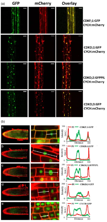
To compare subcellular co-localization of CYCH to those of CDKF;1 and CDKDs, the CYCH:mCherry construct linked to a plant hygromycin resistance marker was transformed into lines carrying the CDKF;1:GFP, CDKD;1:GFP and CDKD;3:GFPgenes linked to KmR gene, and crossed with the CDKD;2–GFP–PIPL line, which harboured a HygR mar- ker. In hypocotyl cells of subsequently isolated double homozygous seedlings, subcellular localization of CDKF;1:
GFP and CYCH:mCherry overlapped yielding orange fluo- rescence in the cytoplasm around the plasma membrane and nuclei. In contrast, signals of CDKD:GFP fusion pro- teins showed an overlap with CYCH:mCherry only in cell nuclei (Figure 4a). These results were corroborated by line intensity profile analysis of fluorescence signals of GFP-la- belled CDKF;1, CDKD and CYCH proteins through the medi- ans of propidium iodine (PI, red) counter-stained single root cells. High cytoplasmic GFP signals were detected close to the peaks of PI-stained red cell wall positions in CDKF;1:GFP expressing cells, and at lower levels in cells
expressing the CYCH:GFP and CDKD;2:GFP–PIPL proteins, respectively. In contrast, the GFP signals of CDKD;1 and CDKD;3 kinases were confined to the area of root cell nuclei (Figure 4b).
Differential interaction of CDKD kinases with CYCH and CDKF;1
It is still an open question whether all three Arabidopsis CDKD kinases are found in similar complexes with CYCH and function as TFIIH-associated kinases phosphorylating the RNAPII CTD. According to Shimotohno et al. (2006), CDKD;1 cannot phosphorylate the RNAP II CTD, and CDKD;3 shows only very weak interaction with CYCH, sug- gesting that the main CYCH-associated TFIIH kinase is CDKD;2. In contrast, Hajheidariet al. (2012) showed that all
three Arabidopsis CDKD kinases were active, phosphory- late serine 5 residues of RNAP II CTD and their activities were increased by CYCH binding and CDKF;1-mediated phosphorylation of their conserved T-loop threonine resi- dues. To examinein vivointeraction of CYCH with CDKDs and CDKF;1 in Arabidopsis, protein extracts from seedlings co-expressing CYCH:mCherry with CDKF;1:GFP–PIPL, CDKD;1:GFP, CDKD;2:GFP–PIPL and CDKD;3:GFP were affinity purified on GFP-Trap and subjected to western blotting with anti-RFP and anti-GFP antibodies. Although CDKD;1:GFP and CDKD;3:GFP were not detectable in the input fractions because their levels were much lower in seedlings compared with CDKD;2 and CDKF;1, the associa- tion of CYCH:mCherry with all three CDKD kinases was clearly demonstrated after GFP-Trap purification by
Figure 3.Genetic complementation ofcdkf;1mutation with modified nativeCDKF;1gene constructs and confirmation of expression of CDKF;1, CDKD, CYCH and HISTONE H3.1 proteins labelled by recombineering in transgenic plants. (a) Comparison of phenotypes ofcdkf;1mutant and genetically complemented mutant lines carrying theCDKF;1:GFP–PIPL, CDKF;1–GFPandCDKF;1:PIPLconstructs. Bar, 7 cm. (b) Comparison of expression levels of CDKF;1:GFP–PIPL, CDKF;1:GFP and CDKF;1:PIPL proteins in the genetically complementedcdkf;1mutant by western blotting with anti-GFP and anti-HA (PIPL cross-reacting) anti- bodies. (c) Confirmation of expression of CDKF;1, CDKD, CYCH and H3.1 proteins labelled by GFP/PIPL and mCherry tags using recombineering in wild type transgenic plants by western blotting with anti-GFP and anti-RFP antibodies. Except for CDKD;1:GFP and CDKD3:GFP, equal aliquots (25lg) of total protein extracts from 15-day-old seedlings were used for western blotting. CDKD;1:GFP and CDKD;3:GFP were isolated by affinity purification on GFP-Trap from 20 mg protein prepared in parallel from seedlings grown under identical conditions.
Figure 4. Co-localization of CYCH:
mCherry with CDKF;1:GFP, CDKD;1:GFP, CDKD:2:GFP–PIPL and CDKD;3:GFP in hypocotyl cells and line intensity profile analysis of subcellular distribution GFP/
PIPL-labelled CDKF;1, CDKD and CYCH proteins in propidium iodine-stained roots cells. (a) Confocal images and overlay of GFP-labelled CDKF;1 and CDKDs and mCherry-labelled CYCH in hypocotyl cells indicate overlapping localization of CDKF;1 with CYCH in the cytoplasm and nuclei. In contrast, CDKDs show nuclear co-localization with CYCH. Bars: 20lm.
(b) Scanning of green GFP and PI-stained red cell wall fluorescence through individ- ual root cells detects CDKF;1:GFP in both cytoplasm and nuclei. In comparison, CDKD;2:GFP and CYCH:GFP show lower accumulation in the cytoplasm, whereas the CDKD;1:GFP and CDKD;2:GFP–PIPL signals are confined to nuclei. White arrows: 10lm.
western blotting with anti-RFP antibody (Figure 5a). In con- trast, CDKF;1: GFP–PIPL failed to pull-down CYCH:mCherry.
To examine the interaction of CDKF;1 with CDKDs, the CDKF;1:PIPL construct was introduced into CDKD;1:GFP and CDKD;3:GFP expressing plants to isolate subsequently double homozygous lines. Protein extracts from these lines were purified by GFP-Trap and subjected to western blot- ting with anti-HA antibody detecting the HA epitope of CDKF;1-fused PIPL tag, as well as with anti-GFP antibody to monitor the CDKD baits. The results confirmed that both CDKD;1 and CDKD;3 were associated with their activating CDKF;1 kinase (Figure 5b).
Identification of CDKD;2:GFP and CYCH:mCherry associated proteins and interacting partners of HISTONE H3.1 by mass spectrometry
Expression of modified TFIIH components in transgenic wild type (wt) and mutant Arabidopsis lines provided suit- able starting materials for affinity purification of corre- sponding proteins and their associated factors. CDKD;2–
GFP–PIPL was purified using standardized amounts of pro- tein extracts from three biological replicates of 4-week-old rosette plants in parallel with similar samples from control wild type and YFPHA (yellow fluorescent
Figure 5.Differential interaction of CDKF;1 with CDKDs and CYCH. (a) CDKD;1:GFP, CDKD;2:GFP–PIPL, and CDKD;3:GFP were affinity purified on GFP-Trap using 20 mg protein extracts prepared from 15-day-old seedlings co-expressing CYCH:mCherry. Western blotting of proteins eluted from the GFP-Trap by anti-RFP and anti-GFP antibodies indicates that CYCH:mCherry is immunoprecipitated by all three CDKDs, but not by CDKF;1. Compared with CDKD;2:GFP–PIPL, CDKD;1:
GFP and CDKD;3:GFP are expressed at lower levels and therefore were not detected in the input protein (50lg) fractions. (b) CDKD;1:GFP and CDKD;3:GFP were affinity purified on GFP-Trap as in (a) from 15-day-old seedlings co-expressing CDKF;1–PIPL. Western blotting of proteins eluted from the GFP-Trap with anti-HA antibody detecting the HA epitope of PIPL tag indicates association of both CDKD;1:GFP and CDKD:3:GFP with CDKF;1–PIPL. Co-immunoprecipitation of CDKF;1 with CDKD;2 was previously shown by Shimotohnoet al. (2004), and is confirmed by identification of CDKF;1 in complex with GFP-Trap purified CDKD;2–GFP– PIPL by LC-MS/MS mass spectrometry (Table S2).
proteinhemagglutinin epitope) expressing plants by affin- ity binding to GFP-Trap. CYCH–mCherry was analogously isolated on RFP-Trap using control extracts from wild type and HISTONE H3.1:mCherry expressing plants. Compar- ison of common components of CDKD;2–GFP–PIPL and CYCH–mCherry complexes identified by mass spectrome- try (Tables S2 and S3) indicated that CDKD;2 and CYCH were associated with each other, as well as with Arabidop- sis homologues of MAT1 (cyclin-dependent kinase-activat- ing kinase assembly factor-related AT4G30820), XPD (50 to 30 helicase XERODERMA PIGMENTOSUM GROUP D, AT1G03190), and p62 (GTF2H1-2, AT1G55750) TFIIH sub- units. In addition, different amounts of p52 (GTF2H4/TFB2, AT4G17020) were pulled down by CDKD;2 and CYCH, which were also associated with comparably lower amounts of p44 (GTF2H2/TF2H5/TTDA, AT1G05055) and p34 (GTF2H3/TFB4, AT1G18340) TFIIH subunits. The small- est TFIIH subunit p8 was detected only in the CYCH:
mCherry pull-down, while corroborating the co-immuno- precipitation data the CDKD-activating kinase CDKF;1 was only found in a complex with CDKD;2–GFP. Intriguingly, both CDKD;2 and CYCH complexes lacked however the 30 to 50 helicase XPB subunit of TFIIH (Figure 6). These data confirmed the results of previous mass spectrometry stud- ies, which identified CDKD;2 in complex with MAT1, CYCH.
XPD, p62 and p52 in Arabidopsis cell suspension (Van Leene et al., 2010) and extended them by showing that CYCH can be isolated in association with all known con- served TFIIH subunits except for XPB (Fan and DuPrez, 2015; Rimel and Taatjes, 2018; Greberet al., 2019).
Simultaneous identification of unique proteins showing co-purification only with H3.1:mCherry, used as nuclear control in the analysis of CYCH-associated factors, con- firmed interaction of HISTONE 3.1 with HISTONE 4 (AT5G59970), as well as with conserved FAS1 (FASCIATA 1, AT1G65470), FAS2 (FASCIATA 2, AT5G64630) and MSI1 (MULTICOPY SUPRESSOR OF IRA1, AT5G58230) subunits of CAF-1 (CHROMATIN ASSEMBLY FACTOR 1; Serra-Car- dona and Zhang, 2018) and their interacting ASF1 partners (ANTI- SILENCING FUNCTION 1A, AT1G66740 and 1B, AT5G38110; Larioet al., 2013). CAF1 and ASF1 play a piv- otal role in the deposition of histone H3/4 core during DNA replication and are involved in the regulation of DNA repair, recombination, endoreduplication, epigenetic imprinting, heterochromatin silencing and cell fate deter- mination (Cheloufi and Hochedlinger, 2017; Jiang and Ber- ger, 2017). Among other candidates of H3.1-interactors (Figure 6, Table S3), the PHD finger protein (AT4G23860) was identified as a homologue of human histone H3-bind- ing UBR7 E3 ubiquitin ligase, which mediates K120 ubiqui- tination of histone H2B and acts as breast cancer tumour suppressor (Kleineret al., 2018; Adhikaryet al., 2019). The tetratricopeptide repeat protein NASP (AT4G37210) was identified as a member of conserved H3-binding SHNi-TRP
domain factors that mediate deposition of H3-variants in yeast and Arabidopsis (Dunleavy et al., 2007; Maksimov et al., 2016), while the DNA-J chaperones ATJ6 and AT3G12170 were found to be closely related to the human nuclear H3-binding factor DNAJC9 (Camposet al., 2015; Lambert et al., 2015). Finally, Ku70 (AT1G16970) and Ku80 (AT1G48050) in the list of putative H3.1-interac- tors represented key factors that bind to DNA ends at double-stranded breaks and interact with components of the nonhomologous end-joining (NHEJ) DNA repair path- way, including the human CAF1 complex (Hoek et al., 2011). Although these protein interactions remained to be confirmed by subsequent studies, the results of mass spectrometry analyses illustrated that fast-track recombi- neering can be applied as a useful tool to assist in the isolation and identification of components of plant pro- tein complexes.
DISCUSSION
Unique advantages of recombineering
Compared with Gateway and Gibson assembly of gene constructs, recombineering offers the advantage that it cir- cumvents PCR amplification of target genes and provides a means for their seamless site-specific modification by homologous recombination in BACs. As it is difficult to predict a priori the localization of full-length functional promoter sequences, larger genomic regions including at least two genes flanking the target are transferred into an Agrobacterium binary vector and then to the plant for maintaining native transcriptional regulation of the stud- ied plant gene. It is particularly important when, for exam- ple, a bidirectional promoter region controls expression of the target and its upstream neighbouring gene (see for example miR159a and CDKD;1 genes in Figure S2), or when 30-UTRs of target and downstream neighbouring gene overlap, suggesting potential generation of natural antisense natsi-RNAs (as is the case for the CDKD;3and AT1G18030PP2Cgenes in Figure S2). Replacement of the galKexchange marker by antibiotic resistance gene-linked cassettes and the use of PCR-amplifiable Agrobacterium binary vectors for gap-repair cloning of modified target genes removed two major bottlenecks from the recombi- neering procedure reducing its total time requirement to 12–14 days, including three E. coli transformation steps with PCR-amplified DNA fragments. From the two proto- cols described, the application ofccdBcassettes (Figures 1 and S1) in combination with PCR-amplifiable pGAPBR gap-repair vectors (Figure S5) is necessary when internal nucleotide or codon exchanges are generated in the target gene. The use of I-SceI insertion cassettes (Figures 2 and S4) simplifies the replacement of translational start or stop codons of target genes with coding sequences of desired tags. N-terminal and C-terminal I-SceI insertion cassettes
were constructed such that their GFP/PIPL and mCherry sequences can be replaced with any other desired tag.
As the most laborious step in recombineering is the preparation of heat-induced electrocompetent cells, its application is particularly effective when all genes in a single BAC are simultaneously modified. This approach could facilitate systematic genome-wide labelling of plant gene products with desired tags. In the examples described above, we illustrated that labelling of interact- ing proteins with GFP and mCherry fluorescent protein tags provides a simple means for their cellular co-local- ization during different developmental stages and in vari- ous cell types. The same fluorescent tags are also amenable to support more detailed fluorescence reso- nance energy transfer (FRET) interaction studies (Alber- tazzi et al., 2009; Liao et al., 2019). In addition, the availability of immobilized alpaca and llama nanobodies against GFP and RFP facilitate simple affinity purification and subsequent mass spectrometry analysis of plant gene products labelled by these tags using recombineer- ing. To assist multistep affinity purification of protein complexes, we also created a combined affinity tag, PIPL, which is applicable for pre-enrichment of tagged proteins e.g., on Ni2+-agarose or on anti-HA matrix following elu- tion by HA-peptide (Farraset al., 2001). Fast-track recom- bineering could particularly accelerate ‘proteomics walking’ by confirmation of interactions between newly identified components of protein complexes and identifi- cation of their novel interacting partners. This application is illustrated in the model study of CDKF;1 interaction with CDKDs and CYCH followed by purification of CDKD;2 and CYCH complexes and identification of their common components.
Interaction of CDKD TFIIH kinases with CDKF;1 and CYCH Arabidopsis CDKD;1–3 are closely related homologues of budding yeast Kin28, fission yeast Msc6 and metazoan CDK7 protein kinases, which together with cyclin H and the MAT1 (Menage a trois 1) assembly factor form the trimeric kinase module (TFIIK) of general transcription factor TFIIH (Rimel and Taatjes, 2018; Kolesnikova et al., 2019). When unbound of TFIIH, TFIIK plays a pivotal role in cell cycle control through activation of cell cycle kinases by T-loop phosphorylation. Cell cycle kinase-activating kinase (CAK) activity of human CDK7 is mediated by autophosphoryla- tion and CYCH binding (Martinezet al., 1997; Fisher, 2012, 2019), whereas for binding of Msc2/cyclin H Msc6 requires its T-loop phosphorylation by the Csk1 CAK-kinase (CAKAK), which has no CAK activity (Hermandet al., 2001;
Devoset al., 2015). In contrast, budding yeast Kin28 has no CAK activity (Cismowskiet al., 1995) and its activating T- loop phosphorylation is mediated by the CDC28 cell cycle kinase-activating kinase Cak1/Cvi1 (Kaldiset al., 1996). Ara- bidopsis CDKF;1 identified as a suppressor of yeast cak1 andcsk1mutations (Umedaet al., 1998; Shimotohnoet al., 2004) phosphorylates the T-loops of CYCH-dependent CDKD TFIIH kinases (Hajheidariet al., 2012), but does not affect the activities of CDKA;1 and CDKB;1/2 cell cycle kinases (Takatsukaet al., 2009). Based on genetic analysis of double and triple mutants, Hajheidariet al. (2012) found that the three Arabidopsis CDKD homologues perform overlapping functions. In contrast, Shimotohno et al.
(2004) reported that CDKD;1 is an inactive enzyme, which is not phosphorylated by CDKF;1, and found that CDKD;3 has higher CDKA;1 phosphorylating activity and lower CYCH-binding capability compared with CDKD;2
CDKD CYCH MAT1 XPB
p52 p62
XPD CDKF;1
p34 p44 p8
TFIIH subunit homologs Gene CDKD2:GFP CYCH:mCherry H3.1:mCherry
CDKF;1 At4g28980 + – –
CDKD;1 At1g73690 – – –
CDKD;2 At1g66750 +++ +++
CDKD;3 At1g18040 – – –
CYCLIN H At5g27620 +++ +++ –
MAT1 AT4G30820 +++ +++ –
XPB1/ERCC3/RAD25/p89 AT5G41370 – – –
XPB2/ERCC2/RAD3/p80 AT5G41360 – – –
XPD/UVH6 AT1G03190 ++ ++ –
p62/GTF2H1-1 AT3G61420 – – –
p62/GTF2H1-2 AT1G55750 ++ ++ –
p52/GTF2H4/TFB2 AT4G17020 + ++ –
p44/TF2H5/TTDA AT1G05055 + ++ –
p34/GTF2H3/TFB4 AT1G18340 +/– +/– –
p8/GTF2H5/TTD; TFB5-1 AT1G12400 – + –
p8/GTF2H5/TTD; TFB5-2 AT1G62886 – – –
HISTONE H3.1 associated
HISTONE H3.1 AT5G65360 – – +++
UBR7 PHD finger protein AT4G23860 – – +++
HISTONE H4 AT5G59970 – – +++
FAS1 AT1G65470 – – +++
FAS2 AT5G64630 – – ++
MSI1 AT5G58230 – – ++
ASF1A/SGA2/SP7 AT1G66740 – – ++
ASF1B/SGA1 AT5G38110 – – ++
Tetratricopepde repeat AT4G37210 – – ++
ATJ6 J-domain protein 6 AT5G06910 – – ++
DnaJ-domain chaperone AT3G12170 – – ++
Ku80 AT1G48050 – – +
Ku70 AT1G16970. – – +
(a) (b)
Figure 6.Summary of results of LC-MS/
MS analyses. (a) Schematic presentation of arrangement of TFIIH subunits based on cryo-electron microscope study of TFIIH structure (Greberet al., 2019). Com- ponents of the kinase module (blue) inter- act through MAT1 with the XPB and XPD helicases of human TFIIH core. However, in Arabidopsis, TFIIH subcomplexes puri- fied by CDKD;2 and CYCH the XPB subunit is absent and the representation of p34 is largely reduced. Compared with XPD and p62, all remaining TFIIH subunits show lower abundance in purified CDKD;2 and CYCH complexes. (b) List of proteins iden- tified in the purified CDKD;2:GFP, CYCH:
mCherry and HISTONE H3.1:mCherry complexes. Representation of peptide peaks of different subunits measured by LC-MS/MS (Tables S2 and S3) is indicated schematically (+signs).
(Shimotohno et al., 2003, 2006). In addition, Takatsuka et al. (2009) observed that the lack of T-loop phosphoryla- tion in thecdkf;1mutant leads to selective transcriptional downregulation and degradation of CDKD;2 but does not affect CDKD;3 levels (Takatsuka et al., 2009). To support the conclusion that CDKDs perform distinct functions, Takatsukaet al. (2015) also reported that acdkd;1/+cdkd;3/
cdkd;3 double mutant segregated progeny with both female and male gametophytic lethality, and even man- aged to complement these segregating defects with the CDKD;3gene.
Our recurrent analysis of expression patterns of native CDKD genes labelled by recombineering indicated that bothCDKD;1andCDKD;3are expressed at notably lower levels compared withCDKD;2in most organs, except roots.
In total protein extracts of seedlings, CDKD;1 and CDKD;3 can only be detected after enrichment on GFP-Trap. All three CDKDs showed nuclear localization, although the most abundant CDKD;2 protein is also detectable at a low level in the cytoplasm. All three CDKDs showed co-im- munoprecipitation and nuclear co-localization with CYCH.
In comparison, the CDKD-activating CDKF;1 kinase, as well as CYCH are detectable in both the cytoplasm and the nucleus, and their cellular localization largely overlaps.
Nonetheless, CDKF;1 did not co-immunoprecipitate with CYCH, but showed similar association with all three CDKD kinases. These results are consistent with the observation that CDKF;1, like yeast Cak1, preferentially binds to and phosphorylates the T-loops of its cyclin-free substrates, which then enhances further interaction of activated kinases with their cyclin partners (Tsakraklides and Solo- mon, 2002). Although binding to the RING domain of MAT1 can also mediate complex formation of unphospho- rylated KIN28 and CDK7 with Ccl1 and CYCH, respectively, T-loop phosphorylation of Kin28 and CDK7 is necessary for stabilization of trimeric CAK complexes in budding yeast and Drosophila (Larochelleet al., 2001; Keoghet al., 2002).
This is probably the case for Arabidopsis CDKD;2 (Takat- suka et al., 2009), but it remains to be clarified how the degradation of CDKDs is regulated during the cell cycle and in different cell types.
TFIIH components isolated by the CDKD;2 and CYCH baits According to mass spectrometry data, CDKD;2, CYCH and MAT1 components of TFIIH kinase module have the high- est abundance in the purified CDKD;2–GFP and CYCH– mCherry complexes. The fourth most abundant compo- nent in the CDKD;2–GFP complex is the Arabidopsis homo- logue of XPD DNA helicase (Table S2), which carries a redox-sensitive 4Fe4S cluster.DrosophilaXPD was identi- fied to interact with the RING and a-helical domains of MAT1 in a cytoplasmic complex (Abdulrahmanet al., 2013;
Greberet al., 2019), and to inhibit cell division promoting activity of the CDK7-CYCH-MAT1 TFIIH kinase module
(Chen et al., 2003; Li et al., 2010). This interaction is dis- rupted by components of the cytosolic MMXD ironsulfur assembly complex that recruit XPD from CAK to mitotic spindles regulating chromosome segregation (Ito et al., 2010; Houten et al., 2016; Nag et al., 2018). It is still an open question whether homologues of ironsulfur assem- bly factors (Yuanet al., 2010; Hanet al., 2015) would anal- ogously regulate CAK activity of CDKDCYCHMAT1 complexes in Arabidopsis. Although synthesized and partly assembled in the cytoplasm, the kinase module and remaining core components of Drosophila TFIIH form a common complex only in the nucleus (Aguilar-Fuentes et al., 2006). When targeted to the RNAPII pre-initiation complex (PIC) by TFIIH in the nucleus, the kinase module mediates phosphorylation of Ser5 and Ser7 residues of RNAPII CTD stimulating transcription initiation. The kinase module is also directed by TFIIH to DNA damage sites.
During transcription-coupled and general genome repair, the XPD-inhibitor kinase module is evicted from the TFIIH complex by the repair factor XPA (Coinet al., 2008; Compe and Egly, 2016).
Although we did not detect CDKF;1 in the complex with CYCH–mCherry, Hajheidariet al. (2012) demonstrated that CDKF;1 is required for in vivophosphorylation of RNAPII CTD Ser7 residues, as well as for maintenance of CDKD-de- pendent Ser5 CTD phosphorylation. CDKF;1 is co-immuno- precipitated with CDKDs but not with CYCH. It is therefore possible that either targeting the C-terminus of CYCH for pull-down or CYCH binding to CDKDs disrupts or weakens, respectively, the interaction of CDKF;1 with the CDKD kinase module of TFIIH. In budding yeast, Cak1 is not part of the TFIIH (Kaldis et al., 1996) but nonetheless remains associated with RNAPII and mediates T-loop phosphoryla- tion and activation of Kin28, Bur1 and Ctk1 RNAPII CTD kinases (Espinozaet al., 1998; Kimmelmanet al., 1999; Yao and Prelich, 2002; Ostapenko and Solomon, 2005). During PIC formation, TFIIH mediates ATP hydrolysis-dependent opening of promoter region around the transcription start site (TSS) (Rimel and Taatjes, 2018; Kolesnikova et al., 2019). In the horseshoe-like structure of TFIIH (Figure 6), which is recruited by the Mediator to PIC, the terminal XPB and XPD DNA helicases interact with each other and the MAT1 subunit of kinase module (Greberet al., 2019). Inter- action of XPD with p62 and p44 forms one arm of the horseshoe structure whereas, in the other arm, XPB is bound to the p52, p8 and p34 TFIIH subunits. The central p44 and p34 subunits play a major role in stabilization of the complex by multiple interactions with the p62, p54 and p8 subunits (Greberet al., 2019). In the TFIIH holocomplex, MAT1 interacts with XPD and its activator p44, and inhibits the helicase activity of XPD (Sandrock and Egly, 2001).
Upon entry into the PIC, the interaction of XPB with XPD is interrupted by TFIIE-binding to XPB, which abolishes its contact with MAT1 and positions of the XPB-p52-p8
helicase module towards opening the promoter DNA. At the same time, the kinase module is displaced between the hook and shoulder positions of the Mediator close to the RNAPII CTD domain (Schilbachet al., 2017). This prevents inhibition of CDK7 kinase through phosphorylation of its CYCH subunit by the Mediator-associated cyclin C-depen- dent CDK8 kinase (Akoulitchevet al., 2000).
Comparison of representation of TFIIH subunits isolated by the help of CDKD;2–GFP and CYCH–mCherry baits (Tables S2 and S3) indicates a tight association of kinase module with XPD and its immediate binding partner p62, which mediates TFIIH interactions with numerous tran- scription factors and the XPC sensor of DNA lesions (Okudaet al., 2016). The representation of TFIIH stabilizing p44, and especially p34 subunit is surprisingly lower, simi- larly to their p52 and p8 interacting partners. The low abundance of latter TFIIH subunits might reflect their prox- imity order compared with the CDKD;2 and CYCH baits used for purification. However, the complete absence of XPB from both CDKD;2 and CYCH complexes contradicts this argumentation, as XPB is an immediate binding part- ner of MAT1 and XPD. From the two Arabidopsis XPB homologues, XPB2 is known to complement the yeast rad25DNA repair deficiency (Morganteet al., 2005). How- ever, participation of XPBs in transcription-committed TFIIH complexes is not yet confirmed experimentally.
Although ATP-dependent helicase activity of XPB is essen- tial for transcription, recently Alekseevet al. (2017) demon- strated that chemical inhibition and induced degradation of XPB does not prevent RNAPII transcription, whereas XPB functions as transcription inhibitor in the absence of ATP. Further labelling of other TFIIH subunits, including XPBs by recombineering should be helpful to determine whether displacement of the XPB-p52-p8 arm from the kinase module can be prevented by modification of TFIIH isolation conditions. Combination of this approach with induction of DNA lesions might also be helpful to identify components of plant TFIIH-associated DNA repair factors.
EXPERIMENTAL PROCEDURES
Plant materials, transformation and growth conditions Wild type (Col-0) and cdkf;1/+ (GABI_315A10, Hajheidari et al., 2012) Arabidopsis plants were grown in MSAR medium (Koncz et al., 1994) in a controlled culture room at 22°C with 120 mol m2sec1 light intensity and a photoperiod of 8 h light and 16 h darkness. Seedlings fromin vitro cultures were trans- ferred into soil and grown under standard greenhouse conditions (12 h light/12 h of dark period; 22–24°C day temperature and 18°C night temperature, 200lEinstein m2sec1 irradiance). For crosses andAgrobacterium-mediated transformation by vacuum infiltration (Bechtoldet al., 1993), Arabidopsis seedlings planted into soil were grown under short-day conditions (8 h light/16 h dark) for 14–16 days and then transferred to long-day conditions to induce flowering. T1cdkf;1/+ plants transformed withCDKF;1 gene constructs carrying the GFP–PIPL, GFP and PIPL tags were
self-pollinated and the resulting T2 lines were screened for homozygous status of the SuR (sulfadiazine resistance) marker of cdkf;1mutation. Subsequently, the progeny of derived T3 lines were screened for homozygous status of the KmR or HygR mark- ers of complementing CDKF;1 gene constructs. The CYCH– mCherry(HygR) construct was transformed to homozygous lines carrying the CDKF;1:GFP, CDKD;1:GFP and CDKD;3:GFP genes linked to KmR selectable marker to isolate homozygous HygR and KmR T2 progeny. Homozygous CYCH–mCherry (HygR) and CDKD;2–GFP–PIPL(HygR) plants were crossed to identify homozy- gous T2CYCH–mCherrylines showing 100% mCherry and segre- gating GFP expression in their roots, and then screened for T3 progeny carrying both markers in homozygous form.
Construction ofccdBexchange and I-SceI insertion cassettes and PCR-amplifiable binary vectors for recombineering
To constructccdBcassettes, first a CmR gene linked to an I-SceI site was PCR amplified from plasmid pEL04 (Leeet al., 2001; pri- mers CmF and CmR-I-SceI, Table S1) and inserted asNaeI frag- ment into the NcoI site of pGEM-T Easy (Promega). Next, the araC-pBAD-ccdBcassette was amplified from pSW8197 (Le Roux et al., 2007; primersSpeI-araC andXbaI-ccdB) and inserted as an SpeISacI fragment into the adjacentXbaISacI sites resulting in pGEM-CmR-araC-ccdB (Figure S1). The CmR gene of the latter vector was removed byNcoI cleavage and replaced by an ampli- fiedBspHI fragment pACYC177KmRgene linked to an I-SceI site (primers KmF and KmR-I-SceI) to yield pGEM-KmR-araC-ccdB. A SphIEcoRI fragment of the latter plasmid carrying theKmRgene was replaced by theSpRgene of pER8 linked to an I-SceI site (Zuo et al., 2000; primers SpF and SpR-I-SceI) to construct pGEM-SpR- araC-ccdB (Figure S1).
To assemble the coding region of PIPL tag, the Co2+/Ni2+-bind- ing domain of Arabidopsis CobW-like protein (At1g15730) carrying 18 His residues was linked to two copies of the StrepII epitope separated by a Gly-rich linker by annealing partially complemen- tary primers P1 and P2 (Table S1). After filling in the ends with T4 DNA polymerase, the resulting fragment was extended and PCR amplified using P1 and a third partially overlapping primer P3, and cloned into theEcoRV site of pUC57 (Genscript). A GFP–PIPL cassette for generation of C-terminal fusions was constructed by simultaneous insertion of the PCR-amplifiedEcoRISalI fragment of the GFP coding region without the stop codon (primers GFPc1F/R) and an amplifiedSalISacI fragment of PIPL coding region (primers PIPLc1F/R) with start and stop codons intoEcoR- ISacI sites of pBSKII. A PIPL–GFP cassette for generation of N- terminal fusions was created by insertion an amplified (primers PIPLc2F/R)EcoRISalI fragment of the PIPL coding region without a stop codon and aSalISacI fragment of GFP coding region with start but with no stop codon intoEcoRISacI sites of pBSKII (Fig- ure S3).
DNA fragments amplified from pGAPHyg and pGAPKm (Bitrian et al., 2011) with linker primers for joining the adjacent segments (Table S1) were assembled into the binary vectors pGAPHyg2 and pGAPKm2 (Figure S5) using a Gibson assembly master mix (NEB). UponSalI cleavage, T4 DNA polymerase fill in andEcoRI digestion, the multicopy pUC9 replicons of the latter vectors were exchanged for a pHC79 cosmid replicon, which was isolated by BglII cleavage, fill in and EcoRI digestion from pPCV6NFHyg (Konczet al., 1989) to construct the more stable and lower copy number binary vectors pGAPBRHyg and pGAPBRKm (Figure S5).
To construct the N-terminal KmR–GFP–PIPL I-SceI insertion cas- sette, the KmR gene amplified (primers BamSceKm5 and Km3Spe;