Genomic analysis of a novel picornavirus from a migratory waterfowl, greater white ‑ fronted goose (Anser albifrons)
Ákos Boros1,2, Péter Pankovics1,2, Peter Simmonds3, Tamás Kiss4, Tung Gia Phan5,6, Eric Delwart5,6, Gábor Reuter1,2
1 Regional Laboratory of Virology, National Reference Laboratory of Gastroenteric Viruses, ÁNTSZ Regional Institute of State Public Health Service, Pécs, Hungary
2 Department of Medical Microbiology and Immunology, Medical School, University of Pécs, Szigeti út 12., H-7624 Pécs, Hungary
3 Nuffield Department of Medicine, Peter Medawar Building for Pathogen Research, University of Oxford, South Parks Road, Oxford, UK
4 Hungarian Ornithological and Nature Conservation Society, Budapest, Hungary 5 Blood Systems Research Institute, San Francisco, CA, USA
6 University of California, San Francisco, CA, USA Gábor Reuter reuter.gabor@gmail.com
The GenBank[/EMBL/DDBJ] accession number for the study sequence: MF358731.
Abstract
The complete genome of goose picornavirus 1 (GPV-1) strain goose/NLSZK2/HUN/2013 (MF358731) was determined by RT-PCR and next-generation sequencing from a cloacal sample of a migratory waterfowl, greater white-fronted goose (Anser albifrons) in Hungary. The genome of GPV-1 shows an L-3-3-4 organization pattern with a 5’-terminal origin of replication (ORI) region, a type-IV IRES, and an Hbox/NC-type 2A protein. This virus showed the highest overall sequence identity to the members of the genus Kobuvirus, although the phylogenetic position of GPV-1 is different in the analyzed P1, 2C and 3CD phylogenetic trees, which further increases the diversity of known avian picornaviruses.
Long-distance migratory bird species such as greater white- fronted goose (Anser albifrons) of the family Anatidae live in large flocks that include birds of different species [1]. These mixed flocks could carry the risk of spreading mostly unknown zoonotic pathogens as well as pathogenic avian viruses [2, 3]. The family Picornaviridae consists of genetically diverse viruses of vertebrates, including birds, with a small single-stranded RNA genome of positive polarity [4, 5].
One of the most diverse avian picornavirus phylogenetic clusters is the passerivirus cluster, which includes members of the genera Passerivirus, Gallivirus, Oscivirus and Sicinivirus [3].
Picornaviruses of the passerivirus cluster share a same L(leader)-3-3-4 genome organization pattern with a type-II or type-V internal ribosomal entry site (IRES) and predominantly a single, Hbox/NC- type 2A [3]. Here, we report the complete genome sequence of the novel goose picornavirus 1 (GPV-1, MF358731) from a greater white- fronted goose, determined by viral metagenomics and conventional and 5’/3’ RACE RT-PCR techniques as described previously [6, 7].
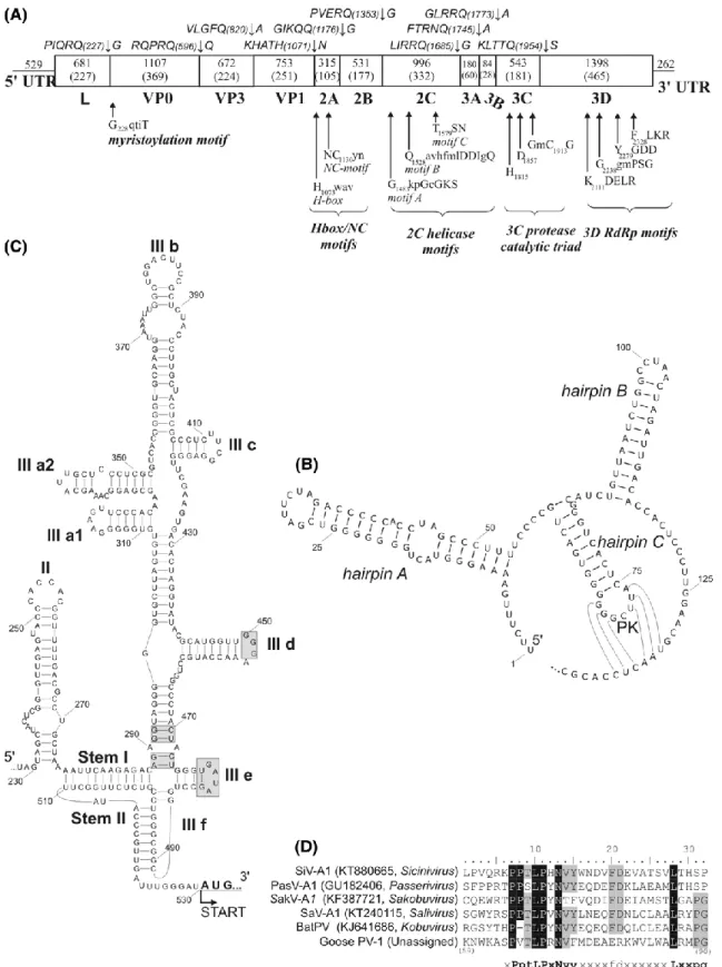
The analyzed cloacal sample was collected from an apparently healthy, one-year-old greater white-fronted goose (Anser albifrons) at Lake Zabszék, Szabadszállás, Hungary, in November 2013. The 8051-nt-long complete genome of GPV-1 strain goose/NLSZK2/HUN/2013 was predicted to have a L-3-3-4 genome layout: 5′UTR-L-P1(VP0-VP3- VP1)-P2(2A-C)-P3(3A- D)-3′UTR (Fig. 1a). At the 5’-terminal end, the presence of three hairpins and a possible pseudoknot were predicted using Mfold (Fig. 1b). This structure strongly resembles the origin of replication (ORI) structures of kobu-, sali-, and osciviruses [8]. Based on the results of Mfold analysis and 5’UTR sequence alignments of different IRES elements, the 5’UTR-IRES of GPV-1 is most likely to belong to the hepacivirus/pestivirus-like type-IV (Fig. 1c) [8]. The 227-aa-long leader (L) contains the conserved motifs P65VTLPRNV and L86RMPG. Similar motifs are encoded in the genomes of other phylogenetically related picornaviruses as well (Fig. 1d). The P1 region was predicted to encode only three capsid proteins: a myristoylated VP0, VP3 and VP1 (Fig. 1a).
The P2 is predicted to contain a 105-aa long H-box/NC-type 2A peptide (Fig. 1a), and the 2C helicase, the 3C protease and the 3D RNA polymerase are predicted to contain all known functional motifs (Fig. 1a).
Fig. 1 (A) The main genomic features, the conserved picornaviral motifs, and the predicted cleavage sites of GPV-1 (MF358731). (B and C) The predicted secondary RNA structures of 5’-terminal origin of replication (ORI) region (B) and the 5’UTR-IRES (C) of the 5’UTR. “PK” indicates a pseudoknot. Grey boxes indicate conserved motifs of type-IV IRES elements [8]. (D) Sequence alignment of the leader proteins of GPV-1 and related viruses. Conserved amino acids are indicated with black and grey backgrounds
The P1 of GPV-1 shows highest pairwise aa sequence identity to aichivirus A1 of the genus Kobuvirus (43.4%) (Table S1). In the case of the 2C and 3CD peptides, the high- est identities are found with sicinivirus A1 (2C: 46.9%) and passerivirus A1 (3CD: 47.3%), although only slightly lower values were found with bat picornavirus 1 of the genus Kob- uvirus (2C, 46.7%; 3CD, 46.2%; Table S1). GPV-1 shows a distant phylogenetic relationship to kobuviruses in the P1 phylogenetic trees and to siciniviruses in the 2C phylogenetic trees (Fig. 2a, and b), while in the 3CD region, GPV-1 is clustered together with the osciviruses (Fig. 2c).
Fig. 2 Phylogenetic analysis of GPV-1 (indicated in bold and with an arrow), representative members of the family Picornaviridae (P1), and the closest relatives of the strain from this study (2C and 3CD trees) based on amino acid sequences of the picornavirus proteins P1 (A), 2C (B) and 3CD (C). The trees were generated using the maximum- likelihood method based on the Le_Gascuel_2008 model of MEGA Ver.7.0 [9]. Bars indicate amino acid substitutions per site. A-F designations for the genus Kobuvirus represent the members of the species Aichivirus A-F
In this study, the complete genome sequence of a novel goose picornavirus (MF358731) detected from a cloacal sample of a greater white-fronted goose (Anser albifrons) was determined and characterized. GPV-1 shows an L-3-3-4 genome layout with a characteristic 5’-terminal ORI structure, a type-IV IRES and an Hbox/NC-type 2A protein. The conserved aa motifs found in the L proteins of GPV-1 and various mammalian and avian picornaviruses suggest a similar but currently unknown function and a common origin of these proteins. Based on the results of sequence comparisons, GPV-1 could be assigned to the genus Kobuvirus, although the phylogenetic position of the virus in the 2C and 3CD phylogenetic trees do not support this classification.
Acknowledgements This work was supported by a grant from the Hungarian Scientific Research Fund (NKFIH/OTKA K111615). Á.B. and P.P. are supported by the János Bolyai Research Scholarship of the Hungarian Academy of Sciences.
Compliance with ethical standards
Funding This study was funded by Hungarian Scientific Research Fund (OTKA/NKFIH K111615).
Conflict of interest The authors declare that they have no conflict of interest.
Ethical approval All applicable international, national, and/or institutional guidelines for the care and use of animals were followed.
References
1. Carboneras C (1992) Family Anatidae. In: Hoyo J, Elliott A, Sar- gatal J (eds) Handbook of birds of the world. volume 1: ostrich to ducks. Lynx Editions, Barcelona, pp 536–629
2. Woo PC, Lau SK, Yuen KY (2006) Infectious diseases emerging from Chinese wet-markets:
zoonotic origins of severe respiratory viral infections. Curr Opin Infect Dis 19:401–407
3. Boros Á, Pankovics P, Reuter G (2014) Avian picornaviruses: molecular evolution, genome diversity and unusual genome features of a rapidly expanding group of viruses in birds. Infect Genet Evol 28:151–166
4. Zell R (2017) Picornaviridae Arch Virol, pp 1–19. https://doi.org/10.1007/s00705-017-3614-8 5. Palmenberg A, Neubauer D, Skern T (2010) Chapter 1: Genome organization and encoded
proteins. In: Ehrenfeld E, Domingo E, Roos RP (eds) The picornaviruses. ASM Press, Washington, DC, pp 3–17
6. Phan TG, Vo NP, Boros A, Pankovics P, Reuter G, Li OTW, Wang C, Deng X, Poon LLM, Delwart E (2013) The viruses of wild pigeon droppings. PLoS One 8(9):e72787
7. Boros Á, Pankovics P, Knowles NJ, Reuter G (2012) Natural inter- species recombinant bovine/porcine enterovirus in sheep. J Gen Virol 93:1941–1951
8. Asnani M, Kumar P, Hellen CUT (2015) Widespread distribution and structural diversity of Type IV IRESs in members of Picor- naviridae. Virology 478:61–74
9. Kumar S, Stecher G, Tamura K (2016) MEGA7: Molecular Evolutionary Genetics Analysis version 7.0 for bigger datasets. Mol Biol Evol 33:1870–1874
Table S1: Pairwise amino acid sequence identities (%) between the P1, 2C and 3CD proteins of GPV-1 strain goose/NLSZK2/HUN/2013 (MF358731) compared to the representative members of the 35 officially recognized and 7 candidate picornavirus genera. Boldface numbers indicate the highest levels of amino acid identities.
Genus Type/virus name [Species] Accession Number
P1 2C 3CD
"Aalivirus" Duck picornavirus GL12 KJ000696 13.2 23.7 22.0
Ampivirus Ampivirus A1 KP770140 14.4 17.8 20.7
Aphthovirus Foot-and-mouth disease virus 1 AF308157 20.4 26.5 26.6
Aquamavirus Aquamavirus A1 EU142040 13.6 22.2 21.2
Avihepatovirus Duck hepatitis A virus 1 DQ226541 15.0 18.4 21.2
Avisivirus Avisivirus A1 KC465954 15.4 22.5 25.9
Cardiovirus Encephalomyocarditis virus 1 M81861 20.1 23.7 27.7
Cosavirus Cosavirus A1 FJ438902 18.2 27.7 28.3
"Crohivirus" Crohivirus 1 AB937989 13.6 19.5 21.0
Dicipivirus Cadicivirus A1 JN819202 19.0 27.4 32.2
Enterovirus Poliovirus 1 V01149 19.6 21.5 28.2
Erbovirus Equine rhinitis B virus 1 X96871 21.6 25.7 29.0
Gallivirus Gallivirus A1 JQ691613 21.9 41.0 44.3
Harkavirus Falcovirus A1 KP230449 14.5 20.8 19.4
Hepatovirus Hepatitis A virus 1 M14707 13.5 25.2 22.6
Hunnivirus Hunnivirus A1 JQ941880 21.5 26.2 27.9
Kobuvirus Aichivirus A1 [Aichivirus A] AB010145 43.4 44.0 44.9 Kobuvirus Bovine kobuvirus 1 [Aichivirus B] AB084788 40.1 44.1 45.5 Kobuvirus Porcine kobuvirus 1 [Aichivirus C] EU787450 40.3 44.4 43.3 Kobuvirus Kagovirus 1 [Aichivirus D] LC055961 39.9 42.5 42.8 Kobuvirus Rabbit picornavirus 1 [Aichivirus E] KT325852 39.6 46.2 45.3 Kobuvirus Bat picornavirus BtMr-PicoV
[Aichivirus F]
KJ641686 41.4 46.7 46.2 Kunsagivirus Kunsagivirus A1 KC935379 16.4 23.1 21.6
"Lesavirus" Lesavirus 1 KM396707 22.1 25.0 26.9 Limnipivirus Limnipivirus B1 KF306267 12.4 18.4 20.3
"Livupivirus" Livupivirus 1 KX463670 34.5 37.4 41.3 Megrivirus Melegrivirus A1 (THV-1) HM751199 16.7 30.4 35.0
Mischivirus Mischivirus A1 JQ814851 21.3 29.2 27.2
Mosavirus Mosavirus A1 JF973687 22.3 25.1 29.3
"Orivirus" Orivirus A1 KM203656 14.6 21.3 18.8
Oscivirus Oscivirus A1 GU182408 21.7 41.2 45.2
Parechovirus Human parechovirus 1 AJ005695 14.0 23.4 21.2
Pasivirus Pasivirus A1 JQ316470 13.5 24.2 20.0
Passerivirus Passerivirus A1 GU182406 25.8 43.0 47.3
"Poecivirus" Poecivirus 1 KU977108 15.5 30.5 31.1 Potamipivirus Eel picornavirus 1 KC843627 11.2 20.9 22.5
Rabovirus Rabovirus A1 KP233897 18.3 21.1 28.3
"Rafivirus" Tortoise rafivirus A1 KJ415177 26.9 32.6 37.5
Rosavirus Rosavirus A1 JF973686 17.2 31.1 31.4
Sakobuvirus Sakobuvirus A1 KF387721 37.4 42.6 45.3
Salivirus Salivirus A1 GQ179640 36.8 43.8 37.1
Sapelovirus Porcine sapelovirus 1 AF406813 20.0 19.9 28.1 Senecavirus Seneca Valley virus 1 DQ641257 19.7 25.4 26.9
Sicinivirus Sicinivirus A1 KF741227 23.9 46.9 43.7
Teschovirus Porcine teschovirus 1 AJ011380 19.9 24.8 27.5 Torchivirus Tortoise picornavirus 1 KM873611 21.4 27.6 29.9 Tremovirus Avian encephalomyelitis virus 1 AJ225173 14,6 22,9 23.3