R E S E A R C H A R T I C L E Open Access
Isolation of Mycoplasma anserisalpingitidis from swan goose (Anser cygnoides) in China
Miklós Gyuranecz1,2* , Alexa Mitter1, Áron B. Kovács1, Dénes Grózner1, Zsuzsa Kreizinger1, Krisztina Bali1, Krisztián Bányai1and Christopher J. Morrow3
Abstract
Background:Mycoplasma anserisalpingitidiscauses significant economic losses in the domestic goose (Anser anser) industry in Europe. As 95% of the global goose production is in China where the primary species is the swan goose (Anser cygnoides), it is crucial to know whether the agent is present in this region of the world.
Results:Purulent cloaca and purulent or necrotic phallus inflammation were observed in affected animals which represented 1–2% of a swan goose breeding flock (75,000 animals) near Guanghzou, China, in September 2019.
From twelve sampled animals the cloaca swabs of five birds (three male, two female) were demonstrated to beM.
anserisalpingitidispositive by PCR and the agent was successfully isolated from the samples of three female geese.
Based on whole genome sequence analysis, the examined isolate showed high genetic similarity (84.67%) with the European isolates. The antibiotic susceptibility profiles of two swan goose isolates, determined by microbroth dilution method against 12 antibiotics and an antibiotic combination were also similar to the European domestic goose ones with tylvalosin and tiamulin being the most effective drugs.
Conclusions:To the best of our knowledge this is the first description ofM. anserisalpingitidisinfection in swan goose, thus the study highlights the importance of mycoplasmosis in the goose industry on a global scale.
Keywords:Antibiotic, China, Mycoplasma, Swan goose, Phallus inflammation, Venereal disease, Whole genome
Background
Production of geese is very important in many parts of the world. Meat and eggs of waterfowl provide foods with high nutritional quality and unique flavour which is believed to be delicious [1]. Waterfowl are also widely used as a source for down and feathers. In some coun- tries, like France and Hungary, geese also produce foie gras comprising engorged fatty goose liver. In Europe and North America geese products are considered as premium quality food sold at high prices while in the
Far-East waterfowl are marketed at relatively low prices being a bulk meat source.
Mycoplasma diseases cause enormous economic losses to the goose industry in Europe [2]. The estimated yearly loss of the Hungarian goose industry (25.8 thousand tonnes per year production) [3] inflicted by mycoplas- mosis ranged between 2 and 2.5 million euro in the last decades. Numerous Mycoplasma species have been iso- lated from adult geese in association with reproductive disorders. Mycoplasma infection of geese suffering from salpingitis was first reported by Kosovac and Djurisic [4]. Since then beside Acholeplasma species,M. anseris, M. anatis,M. cloacale,unidentifiedMycoplasmaspecies and most frequently M. anserisalpingitidis were identi- fied and associated with reproductive diseases in water- fowl [1, 5–8]. After the first laying period about 15 to 20% of the ganders harbour M. anserisalpingitidisin the
© The Author(s). 2020Open AccessThis article is licensed under a Creative Commons Attribution 4.0 International License, which permits use, sharing, adaptation, distribution and reproduction in any medium or format, as long as you give appropriate credit to the original author(s) and the source, provide a link to the Creative Commons licence, and indicate if changes were made. The images or other third party material in this article are included in the article's Creative Commons licence, unless indicated otherwise in a credit line to the material. If material is not included in the article's Creative Commons licence and your intended use is not permitted by statutory regulation or exceeds the permitted use, you will need to obtain permission directly from the copyright holder. To view a copy of this licence, visithttp://creativecommons.org/licenses/by/4.0/.
The Creative Commons Public Domain Dedication waiver (http://creativecommons.org/publicdomain/zero/1.0/) applies to the data made available in this article, unless otherwise stated in a credit line to the data.
* Correspondence:m.gyuranecz@gmail.com
1Institute for Veterinary Medical Research, Centre for Agricultural Research, Hungária körút 21, Budapest 1143, Hungary
2Department of Microbiology and Infectious Diseases, University of Veterinary Medicine, Hungária körút 23-25, Budapest 1143, Hungary Full list of author information is available at the end of the article
phallus lymph, cloaca and/or trachea. During the laying periods when the ganders are sexually active and under stress, up to 50 to 100% become clinically diseased showing cloaca and phallus inflammation and testicular atrophy [9]. Less frequently salpingitis and vaginitis are observed in the infected breeders [10]. Fertile egg pro- duction also decreases.M. anserisalpingitidis can induce lethal pathological changes in the embryos and vertical transmission may also occur. Sometimes airsacculitis and peritonitis are seen, even in young birds. General signs such as changes in thirst, decreased food consump- tion, body weight loss, weakness, nasal discharge, im- paired breathing, conjunctivitis, diarrhoea and nervous signs were also described in the affected waterfowl flocks [1,11,12].
The annual goose meat production of the world is over 2.5 million tonnes and it is dominated by China (2.4 million tonnes) [3]. In contrast to Europe where the domestic goose (Anser anser) is farmed, in China the swan goose (Anser cygnoides) is the primarily breeding species. The aim of our study was to investigate myco- plasmosis in swan goose in China with a focus on the presence ofM. anserisalpingitidis.
Results
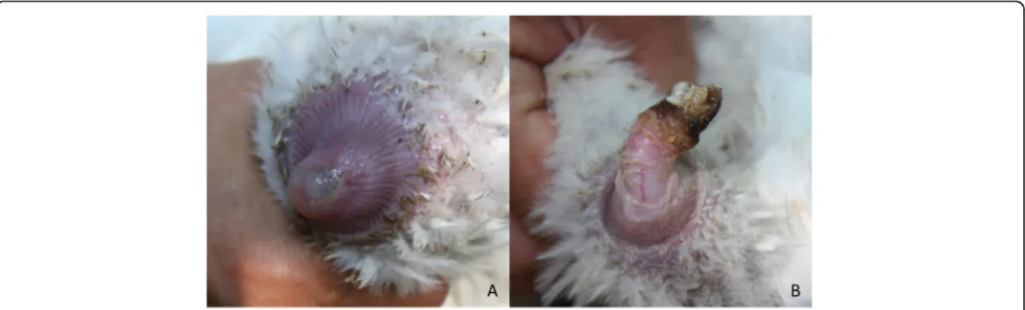
The detailed results of the investigation are summarized in Table 1. Similarly to the European domestic goose, purulent cloaca and purulent or necrotic phallus inflam- mation were observed in the diseased animals (Fig. 1).
According to the owner 1 to 2% of the birds are affected with the disease in each year.M. anserisalpingitidis was detected by PCR in the cloaca swab samples of two fe- male and three male birds. The agent was isolated from three geese (three females). From four ganders M. cloa- cale was detected by PCR and was isolated as well. An undetermined Mycoplasma species, with 92% 16S–23S
sequence similarity to a Mycoplasmasp. isolated from a Humboldt penguin in Austria in 2003 (KX863539) was also cultured from one of the ganders.
The sequencing of strain MYCAV785 resulted in more than 5.9 million single reads with the average Phred score over 30 (base call accuracy over 99.9%). The reads have been uploaded to the sequence read archive (SRA) under Bioproject number PRJNA602206. The MAUVE alignment found more than 86,000 single nucleotide polymorphism (SNPs) and over 9000 of these were only present in MYCAV785, approximately 1% of the whole genome of the strain. Based on the BLAST search done with BRIG and the alignment done by MAUVE, MYCAV785 showed highest similarity (84.67% based on MAUVE) with strain MYCAV93, isolated from the inflammated phallus of a domestic goose in Hungary in 2011 [13] (Fig. 2). Although it is important to note that the MYCAV785 genome contains ambiguous nucleo- tides which may be the reason behind some of the gaps in the BRIG analysis.
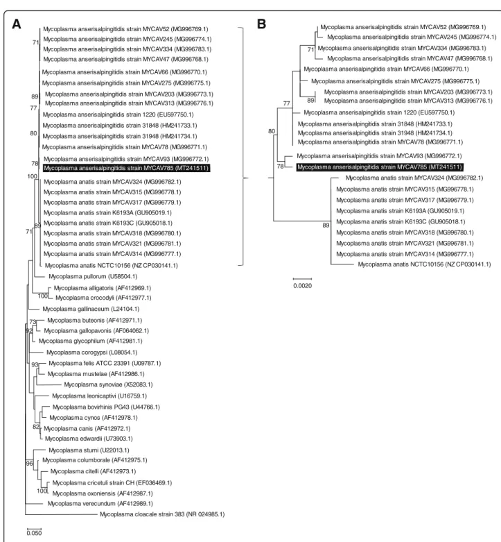
The phylogenetic analysis of MYCAV785 based on the 16S rRNA region [15] supported the close relationship between the swan goose isolate and MYCAV93 within the M. anserisalpingitidis clade (Fig. 3). Sequence simi- larity analysis of the rpoB gene [8, 17] showed 96.95–
97.85% identity between MYCAV785 and M. anserisal- pingitidis strains available in GenBank, while 90.14–
90.24% identity was detected between MYCAV785 and the publicly available M. anatis strains. GenBank se- quence accession numbers of the 16S rRNA and rpoB gene fragments of MYCAV785 are MT241511 and MT241512, respectively (Supplement material).
The M. anserisalpingitidis strain MYCAV785, isolated from a non-medicated female goose (12A), showed low MIC values for all tested antibiotics (enrofloxacin, difloxacin, spectinomycin, lincomycin, doxycycline,
Table 1Information of sampled swan geese and results of diagnostic examination
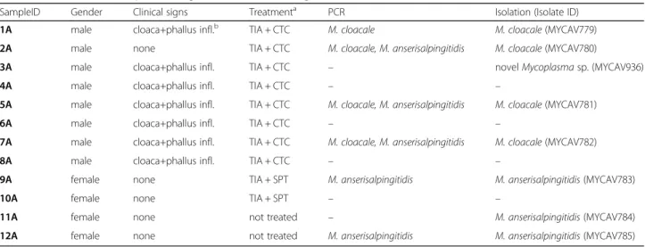
SampleID Gender Clinical signs Treatmenta PCR Isolation (Isolate ID)
1A male cloaca+phallus infl.b TIA + CTC M. cloacale M. cloacale(MYCAV779)
2A male none TIA + CTC M. cloacale, M. anserisalpingitidis M. cloacale(MYCAV780)
3A male cloaca+phallus infl. TIA + CTC – novelMycoplasmasp. (MYCAV936)
4A male cloaca+phallus infl. TIA + CTC – –
5A male cloaca+phallus infl. TIA + CTC M. cloacale, M. anserisalpingitidis M. cloacale(MYCAV781)
6A male cloaca+phallus infl. TIA + CTC – –
7A male cloaca+phallus infl. TIA + CTC M. cloacale, M. anserisalpingitidis M. cloacale(MYCAV782)
8A male cloaca+phallus infl. TIA + CTC – –
9A female none TIA + SPT M. anserisalpingitidis M. anserisalpingitidis(MYCAV783)
10A female none TIA + SPT – –
11A female none not treated – M. anserisalpingitidis(MYCAV784)
12A female none not treated M. anserisalpingitidis M. anserisalpingitidis(MYCAV785)
aTIATiamulin,CTCChlortetracycline,SPTspectinomycinbinfl.Inflammation
oxytetracycline, chlortetracycline, tilmicosin, tylosin, tylvalosin, tiamulin, florfenicol and the combination of lincomycin and spectinomycin, at a ratio of 1:2), while isolate MYCAV783 isolated from a treated bird (9A) showed elevated MIC values against all drugs except tylvalosin and tiamulin (Table 2). The growth
of the M. cloacale strain (MYCAV781) originating from a treated swan goose (5A) was inhibited by low concentrations of the majority of the examined anti- biotics, but it showed increased resistance to fluoro- quinolones and a slight elevation to tetracyclines and florfenicol.
Fig. 1Purulent cloaca (a) and necrotic phallus (b) inflammation in swan goose
Fig. 2Whole-genome based comparison ofM. anserisalpingitidisstrains from China and Europe. Comparison of the whole genome sequences of strain MYCAV785 isolated from swan goose in China with clinical isolates from domestic goose from Hungary (MYCAV93&177) and the type strain (ATCC BAA-2147). BLAST Ring Image Generator (BRIG) [14]
Discussion
M. anserisalpingitidis is one of the most important disease agent of domestic goose causing significant economic losses of the industry in Europe. It was un- known if this disease agent was present in China where 95% of the global goose production is made
using a different species, the swan goose. In previous Chinese publications studying venereal disease in geese the lesions were either correlated with Escheri- chia coli infection, or when Mycoplasma isolation was performed as well only unidentified Mycoplasma-like cultures were determined [18, 19].
Mycoplasma anserisalpingitidis strain MYCAV785 (MT241511) Mycoplasma anserisalpingitidis strain MYCAV52 (MG996769.1) Mycoplasma anserisalpingitidis strain MYCAV245 (MG996774.1) Mycoplasma anserisalpingitidis strain MYCAV334 (MG996783.1) Mycoplasma anserisalpingitidis strain MYCAV47 (MG996768.1) Mycoplasma anserisalpingitidis strain MYCAV66 (MG996770.1) Mycoplasma anserisalpingitidis strain MYCAV275 (MG996775.1)
Mycoplasma anserisalpingitidis strain MYCAV203 (MG996773.1) Mycoplasma anserisalpingitidis strain MYCAV313 (MG996776.1) Mycoplasma anserisalpingitidis strain 1220 (EU597750.1) Mycoplasma anserisalpingitidis strain 31848 (HM241733.1) Mycoplasma anserisalpingitidis strain 31948 (HM241734.1) Mycoplasma anserisalpingitidis strain MYCAV78 (MG996771.1)
Mycoplasma anserisalpingitidis strain MYCAV93 (MG996772.1)
Mycoplasma anatis strain MYCAV324 (MG996782.1) Mycoplasma anatis strain MYCAV315 (MG996778.1)
Mycoplasma anatis NCTC10156 (NZ CP030141.1) Mycoplasma anatis strain MYCAV317 (MG996779.1) Mycoplasma anatis strain K6193A (GU905019.1) Mycoplasma anatis strain K6193C (GU905018.1) Mycoplasma anatis strain MYCAV318 (MG996780.1) Mycoplasma anatis strain MYCAV321 (MG996781.1) Mycoplasma anatis strain MYCAV314 (MG996777.1)
Mycoplasma pullorum (U58504.1) Mycoplasma alligatoris (AF412969.1)
Mycoplasma crocodyli (AF412977.1) Mycoplasma gallinaceum (L24104.1)
Mycoplasma buteonis (AF412971.1) Mycoplasma gallopavonis (AF064062.1) Mycoplasma glycophilum (AF412981.1)
Mycoplasma corogypsi (L08054.1) Mycoplasma felis ATCC 23391 (U09787.1) Mycoplasma mustelae (AF412986.1)
Mycoplasma synoviae (X52083.1) Mycoplasma leonicaptivi (U16759.1) Mycoplasma bovirhinis PG43 (U44766.1) Mycoplasma cynos (AF412978.1) Mycoplasma canis (AF412972.1)
Mycoplasma edwardii (U73903.1) Mycoplasma sturni (U22013.1) Mycoplasma columborale (AF412975.1)
Mycoplasma citelli (AF412973.1)
Mycoplasma cricetuli strain CH (EF036469.1) Mycoplasma oxoniensis (AF412987.1) Mycoplasma verecundum (AF412989.1)
Mycoplasma cloacale strain 383 (NR 024985.1) 71
89 77
78 80
89 100
71
100
73 92
93
82
100 96
0.050
A
Mycoplasma anserisalpingitidis strain MYCAV785 (MT241511) Mycoplasma anserisalpingitidis strain MYCAV334 (MG996783.1)
Mycoplasma anserisalpingitidis strain MYCAV47 (MG996768.1) Mycoplasma anserisalpingitidis strain MYCAV66 (MG996770.1)
Mycoplasma anserisalpingitidis strain MYCAV275 (MG996775.1) Mycoplasma anserisalpingitidis strain MYCAV203 (MG996773.1) Mycoplasma anserisalpingitidis strain MYCAV313 (MG996776.1) Mycoplasma anserisalpingitidis strain 1220 (EU597750.1)
Mycoplasma anserisalpingitidis strain 31848 (HM241733.1) Mycoplasma anserisalpingitidis strain 31948 (HM241734.1) Mycoplasma anserisalpingitidis strain MYCAV78 (MG996771.1) Mycoplasma anserisalpingitidis strain MYCAV93 (MG996772.1)
Mycoplasma anatis strain MYCAV324 (MG996782.1) Mycoplasma anatis strain MYCAV315 (MG996778.1)
Mycoplasma anatis NCTC10156 (NZ CP030141.1) Mycoplasma anatis strain MYCAV317 (MG996779.1) Mycoplasma anatis strain K6193A (GU905019.1) Mycoplasma anatis strain K6193C (GU905018.1) Mycoplasma anatis strain MYCAV318 (MG996780.1) Mycoplasma anatis strain MYCAV321 (MG996781.1) Mycoplasma anatis strain MYCAV314 (MG996777.1) 71
77 89
78 80
89
0.0020
Mycoplasma anserisalpingitidis strain MYCAV245 (MG996774.1) Mycoplasma anserisalpingitidis strain MYCAV52 (MG996769.1)
B
Fig. 3Phylogenetic analysis of theM. anserisalpingitidisstrain from China within the Synoviae cluster. The dendrograms were assessed using Generalised Time Reversible model with Gamma distribution with invariant sites (GTR + G + I) and 1000 bootstrap in Mega X software [16].
Bootstrap values of≥70 are shown. Strain MYCAV785, isolated from a swan goose in China is highlighted in black.a.Dendrogram based on the nucleotide sequences of 16S rRNA gene ofMycoplasmaspecies within the Synoviae cluster. Sequence of theM. cloacalewas used as outgroup.
b.Dendrogram based on the nucleotide sequences of 16S rRNA region ofM. anserisalpingitidisandM. anatisstrains
Thus to the best of our knowledge this is the first report describingM. anserisalpingitidisinfection in swan goose.
Similar to the domestic goose, cloaca and phallus inflam- mation were observed in the diseased birds in China.M.
anserisalpingitidis was detected and/or isolated from the cloaca swabs of the sampled clinically ill and healthy swan geese like in domestic goose in Europe [20,21].
The draft whole genome based genetic characterization of the isolated strain showed high similarity with Hungar- ian M. anserisalpingitidis isolates (GenBank accession numbers: CP042295.1, CP041663.1 and CP041664.1) which is interesting considering the geographic distance and different host species. The rpoBgene sequence simi- larity was suggested to be used for interspecies discrimin- ation before, using the cut off value ≥96% [8, 17]. The rpoB gene sequence comparison confirmed that MYCAV785 isolate belonged to the speciesM. anserisal- pingitidis. The whole genome comparison, and the 16S rRNA and rpoB genes based analyses revealed that MYCAV785 showed closest relationship with the domes- tic goose isolate from Hungary, MYCAV93, forming a unique subclade among theM. anserisalpingitidisstrains.
The antibiotic susceptibility profile of the isolated swan gooseM. anserisalpingitidisstrains also showed high simi- larity with the European isolates [20]. The quick develop- ment of multi-resistance ofM. anserisalpingitidis against different antibiotics is regularly experienced in Europe, and it was confirmed by the comparison of the antibiotic susceptibility profile ofM. anserisalpingitidisisolates from treated and non-treated birds from China also. Tylvalosin and tiamulin are the most potent antibiotics against myco- plasmosis in goose in both China and Europe. The ele- vated MIC values against a commensal bacterium, M.
cloacaleand the detected rapid development of resistance in M. anserisalpingitidis highlight that susceptibility test based targeted antibiotic therapy is strongly recommended in the geese farms.
Conclusions
Our study highlights the importance ofM. anserisalpin- gitidis infection in the goose industry on a global scale.
The extensive/semi-intensive production system of goose hamper the eradication of the agent from a farm thus antibiotic therapy is the primary option of disease control. Unfortunately it not only means disease
treatment but often prophylactic antibiotic application as well which quickly ends up in diverse antimicrobial resistance inM. anserisalpingitidisand probably in other bacteria colonizing the goose including zoonotic agents.
Thus the development of a vaccine, as a long-term dis- ease control option has to be considered.
Methods
Samples were collected from a swan goose breeding farm with 75,000 animals 300 km southwest from Guanghzou in China in September 2019. A total of 12 cloaca swabs were collected from eight male (seven birds showing cloaca and phallus inflammation and one clinic- ally healthy gander) and four female (all clinically healthy) swan geese, from live animals (Table 1). Ac- cording to the written declaration (reference number:
IVMR/2019/0023) of the Ethics Committee of the Insti- tute for Veterinary Medical Research, Centre for Agri- cultural Research ethical approval was not required for the study as the samples were taken during routine diag- nostic examinations with the written consent of the owner. The swabs were put on FTA cards (Whatman, Sigma-Aldrich Inc., Germany), first for future DNA ex- traction and then were also put in 2 ml of Mycoplasma broth medium (pH 7.8) (ThermoFisher Scientific Inc./
Oxoid Inc./, Waltham, MA) supplemented with 0.5% (w/v) sodium pyruvate, 0.5% (w/v) glucose and 0.005% (w/v) phe- nol red, 0.15% L-arginine hydrochloride (w/v), filtered through a 0.65μm pore size syringe filter (Sartorius GmbH, Goettingen, Germany), transported to the laboratory (1 week) and incubated at 37 °C. The cultures were inoculated onto solid Mycoplasma media (Thermo Fisher Scientific Inc./Oxoid Inc./) supplemented with 0.15% L-arginine hydrochloride (w/v) after color change of the broth and were incubated at 37 °C and 5% CO2until visible colonies appeared (1–2 days). Purification of mixed cultures was performed by one-time filter cloning, minimizing the in vitro adaptation and mutations of the isolates. The QIAamp DNA Mini Kit (Qiagen Inc., Hilden, Germany) was used for DNA extraction according to the manufac- turers’ instructions from the cultures and from the FTA cards. Species-specific PCR systems targetingM. anserisal- pingitidis, M. anatis, M. anseris, M. cloacale and Achole- plasma species were used for screening and identification [21,22]. The purity of the cultures was confirmed and the Table 2Minimum inhibitory concentration values of aM. cloacaleand twoM. anserisalpingitidisstrains isolated from swan geese
MIC (μg/ml)
Isolate ID Species Treatment ENRa DIF DOX OTC CTC SPT TYL TIL TLV LIN LIN-SPT (1:2) TIA FLO
MYCAV781 M. cloacale TIA-CTC 2.5 5 0.625 2 4 2 ≤0.25 ≤0.25 ≤0.25 ≤0.25 ≤0.25 ≤0.039 4
MYCAV783 M. anserisalpingitidis TIA-SPT 10 10 10 64 64 8 8 64 ≤0.25 > 64 16 0.625 4
MYCAV785 M. anserisalpingitidis not treated 1.25 0.625 0.078 4 2 2 ≤0.25 4 ≤0.25 ≤0.25 0.5 ≤0.039 0.5
aENREnrofloxacin,DIFDifloxacin,DOXDoxycycline,OTCOxytetracycline,CTCChlortetracycline,SPTSpectinomycin,TYLTylosin,TILTilmicosin,TLVTylvalosin,LIN Lincomycin,TIATiamulinFLOFlorfenicol
unknown species were identified by a universal Myco- plasmaPCR system targeting the 16S/23S rRNA intergenic spacer region in Mycoplasmatalesfollowed by sequencing on an ABI Prism 3100 automated DNA sequencer (Applied Biosystems, Foster City, CA), sequence analysis and BLAST search [23,24].
Whole genome sequencing of an isolated strain (MYCAV785 from bird 12A) was performed on NextSeq 500 Illumina equipment (Illumina, Inc. San Diego, CA USA) using NextSeq 500/550 High Output Kit v2.5 re- agent kit, resulting in 150 bp long single reads. The reads were quality checked with FastQC software version 0.11.8 [25]. A draft genome was assembled with the SPAdes soft- ware version 3.13.0 [26] and aligned with the three currently available M. anserisalpingitidisstrains from the NCBI database (Accession number: CP042295.1 for ATCC:BAA-2147, CP041663.1 for MYCAV93 and CP041664.1 for MYCAV177). The alignment was per- formed with MAUVE version 20150226 [27] and the sin- gle nucleotide polymorphisms (SNP) found in the alignments were exported.
The Medusa web server [28] was used to create contigs from the previously assembled scaffolds. The longest con- tig was 0.941 Mbps similar in size to the otherM. anseri- salpingitidis whole genomes, as such this contig was chosen for use in further study. The genomes were also analyzed with BLAST Ring Image Generator (BRIG) [14].
The 16S rRNA sequence of M. anserisalpingitidis strain MYCAV785 was compared with other Myco- plasma species in the Synoviae cluster [8, 29] with M.
cloacale strain 383 (GenBank accession number: NR_
024985.1) chosen as outgroup. The genetic sequences were aligned with MAFFT algorithm [30] with the Uni- pro Ugene software version 33.0 [31]. A Maximum like- lihood phylogenetic tree was constructed with the MEGA X software version 10.1.5 [16] using Generalised Time Reversible model with Gamma distribution with invariant sites (GTR + G + I) and 1000 bootstrap.
The sequence similarity of a 1997 bp fragment of the rpoB gene of strain MYCAV785 was examined with NCBI BLASTn search tool [24, 29]. Sequence identity was evaluated when query cover was 100%.
The following antimicrobial agents were examined during the microbroth dilution tests: the fluoroquino- lones: enrofloxacin (batch SZBA336XV) and difloxacin (SZBD178XV); the aminocyclitol: spectinomycin (batch SZBB166XV); the lincosamide: lincomycin (batch SZBC340XV); the tetracyclines: doxycycline (batch SZBD007XV), oxytetracycline (batch SZBC320XV) and chlortetracycline (batch BCBR8687V); the macrolides:
tilmicosin (batch SZBC345XV) and tylosin (batch SZBB160XV); the pleuromutilin: tiamulin (batch SZBC026XV); and the phenicol: florfenicol (batch SZBC223XV); all products originated from VETRANAL,
Sigma-Aldrich Inc. The macrolide tylvalosin (Aivlosin, ECO Animal Health Ltd., UK; LOT M102A) was also in- cluded in the examinations. Lincomycin and spectino- mycin were applied also in combination at a ratio of 1:2.
Twofold dilutions were prepared in the range of 0.039–
10μg/ml for fluoroquinolones, doxycycline and tiamulin, 0.25–64μg/ml for spectinomycin, lincomycin, lincomycin- spectinomycin (1:2) combination, oxytetracycline, chlor- tetracycline and macrolides and 0.125–32μg/ml for florfenicol.
The microbroth dilution examinations on 104–105 (color changing unit) CCU/ml of the strains were per- formed according to Hannan [32]. The strains were tested in duplicates and the plate contained a duplicate of the M. anserisalpinigitidis type strain (ATCC BAA- 2147) as a quality control. MIC was established as the lowest antibiotic concentration where no color change of the broth was observed as a consequence of the ab- sence of bacterial metabolism. MIC values were re- corded when color change of the broth media of the growth control was visible (initial MIC).
Supplementary information
Supplementary informationaccompanies this paper athttps://doi.org/10.
1186/s12917-020-02393-5.
Additional file 1.
Abbreviations
BRIG:BLAST ring image generator; CCU: Color changing unit; SNP: Single nucleotide polymorphism; SRA: Sequence read archive
Acknowledgements
The authors thanks the help of the Chinese farmers and veterinarians in sample collection.
Authors’contributions
MG and CJM designed the study, collected the samples, analysed the data and wrote the manuscript. AM and DG processed the samples, isolated the strains and edited the manuscript. ÁBK, KB1 and KB2 performed the whole genome sequencing, analysed the sequences and edited the manuscript. ZK processed the samples, analysed the data and edited the manuscript. All authors read and approved the final version of the manuscript.
Funding
This work was supported by the Lendület program (LP2012–22) of the Hungarian Academy of Sciences, the K_16 (119594), FK_17 (124019) and KKP19 (129751) grants of the National Research, Development and Innovation Office, Hungary. MG and ZK were supported by the Bolyai János Fellowship of the Hungarian Academy of Sciences, and MG was supported by the Bolyai+ Fellowship (ÚNKP-19-4-ÁTE-1) of the New National Excellence Program of the Ministry of Innovation and Technology. The funders had no role in study design, data collection and interpretation, or the decision to submit the work for publication.
Availability of data and materials
All data generated or analysed during this study are included in this published article. The sequence reads of strain MYCAV785 gained by sequencing on NextSeq 500 Illumina equipment have been uploaded to the SRA under Bioproject number PRJNA602206. GenBank accession numbers of the 16S rRNA andrpoBgene fragments of MYCAV785 are MT241511 and MT241512, respectively (Supplement material).
Ethics approval and consent to participate
According to the written declaration (reference number: IVMR/2019/0023) of the Ethics Committee of the Institute for Veterinary Medical Research, Centre for Agricultural Research ethical approval was not required for the study as the samples were taken during routine diagnostic examinations with the written consent of the owner. The final study and the manuscript was submitted and approved in a written declaration by the Ethics Committee of the Institute for Veterinary Medical Research, Centre for Agricultural Research.
Consent for publication Not applicable.
Competing interests
The authors declare that they have no competing interests.
Author details
1Institute for Veterinary Medical Research, Centre for Agricultural Research, Hungária körút 21, Budapest 1143, Hungary.2Department of Microbiology and Infectious Diseases, University of Veterinary Medicine, Hungária körút 23-25, Budapest 1143, Hungary.3Faculty of Veterinary and Agricultural Sciences, The University of Melbourne, Melbourne, Victoria 3010, Australia.
Received: 12 February 2020 Accepted: 28 May 2020
References
1. Stipkovits L, Szathmary S.Mycoplasmainfection of ducks and geese. Poult Sci. 2012;91(11):2812–9.
2. Grózner D, Gyuranecz M. Kacsák és ludakMycoplasma-fertőzései. Magy Állatorvosok Lapja. 2019;141(8):495–504 Article in Hungarian.
3. Food and Agriculture Organization of the United Nations, FAOSTAT.http://
www.fao.org/faostat/en/#data/QLAccessed 24 March 2020..
4. Kosovac A, Djurisic S. Disease of female genital tract of geese and mycoplasmosis. Belgrade: IVth Congress WVPA; 1970. p. 429–40.
5. Benčina D, Dorrer D, Tadina T.Mycoplasmaspecies isolated from six avian species. Avian Pathol. 1987;16(4):653–64.
6. Bradbury JM, Vuillaume A, Dupiellet JP, Forrest M, Bind JL, Gaillard-Perrin G.
Isolation ofMycoplasma cloacalefrom a number of different avian hosts in Great Britain and France. Avian Pathol. 1987;16(1):183–6.
7. Carnaccini S, Ferguson-Noel NM, Chin RP, Santoro T, Black P, Bland M, et al.
A novelMycoplasmasp. associated with phallus disease in goose breeders:
pathological and bacteriological findings. Avian Dis. 2016;60(2):437–43.
8. Volokhov D, Grózner D, Gyuranecz M, Ferguson-Noel N, Gao Y, Bradbury J, et al.Mycoplasma anserisalpingitidissp. nov., isolated from European domestic geese (Anser anser domesticus) with reproductive pathology. Int. J.
Syst. Evol. Microbiol. 2020;70:2369–81.https://doi.org/10.1099/ijsem.0.
004052.
9. Stipkovits L, Varga Z, Czifra G, Dobos-Kovács M. Occurrence of mycoplasmas in geese affected with inflammation of the cloaca and phallus. Avian Pathol.
1986;15(2):289–99.
10. Dobos-Kovács M, Varga Z, Czifra G, Stipkovits L. Salpingitis in geese associated withMycoplasmasp. strain 1220. Avian Pathol. 2009;38(3):239–43.
11. Stipkovits L, Glavits R, Ivanics E, Szabo E. Additional data onMycoplasma disease of goslings. Avian Pathol. 1993;22(1):171–6.
12. Hinz K, Pfützner H, Behr K. Isolation of mycoplasmas from clinically healthy adult breeding geese in Germany. J Vet Med Ser B. 1994;41(1–10):145–7.
13. Grózner D, Forró B, Kovács ÁB, Marton S, Bányai K, Kreizinger Z, et al.
Complete genome sequences of threeMycoplasma anserisalpingitis (Mycoplasmasp. 1220) strains. Microbiol Resour Announc. 2019;8(37):
e00985–19.
14. Alikhan N-F, Petty NK, Ben ZNL, Beatson SA. BLAST Ring Image Generator (BRIG): simple prokaryote genome comparisons. BMC Genomics. 2011;12(1):
402.
15. Volokhov DV, George J, Liu SX, Ikonomi P, Anderson C, Chizhikov V.
Sequencing of the intergenic 16S-23S rRNA spacer (ITS) region of Mollicutes species and their identification using microarray-based assay and DNA sequencing. Appl Microbiol Biotechnol. 2006;71(5):680–98.
16. Kumar S, Stecher G, Li M, Knyaz C, Tamura K, Mega X. Molecular evolutionary genetics analysis across computing platforms. Mol Biol Evol.
2018;35(6):1547–9.
17. Adékambi T, Drancourt M, Raoult D. TherpoBgene as a tool for clinical microbiologists. Trends Microbiol. 2009;17(1):37–45.
18. Shao G, Mao H, Jin H, Qian J, Pan S, Xia Q. Isolation and primary identification ofMycoplasmafrom dead goose embryos. China Vet Sci Technol. 1994;24(11):20 Article in Chinese.
19. Wang Y, Tian H. Prevention and treatment of common goose epidemic diseases. Aquat Poult World. 2006;(March):1673–1085(2006)03–0012–09.
[Article in Chinese].
20. Grózner D, Kreizinger Z, Sulyok KM, Rónai Z, Hrivnák V, Turcsányi I, et al.
Antibiotic susceptibility profiles ofMycoplasmasp. 1220 strains isolated from geese in Hungary. BMC Vet Res. 2016;12(1):170.
21. Grózner D, Sulyok KM, Kreizinger Z, Rónai Z, Jánosi S, Turcsányi I, et al.
Detection ofMycoplasma anatis,M. anseris,M. cloacaleandMycoplasmasp.
1220 in waterfowl using species-specific PCR assays. PLoS One. 2019;14(7):
e0219071.https://doi.org/10.1371/journal.pone.0219071.
22. Gioia G, Werner B, Nydam DV, Moroni P. Validation of a mycoplasma molecular diagnostic test and distribution ofMycoplasmaspecies in bovine milk among New York state dairy farms. J Dairy Sci. 2016;99(6):4668–77.
23. Lauerman LH, Chilina AR, Closser JA, Johansen D. AvianMycoplasma identification using polymerase chain reaction amplicon and restriction fragment length polymorphism analysis. Avian Dis. 1995;39(4):804–11.
24. NCBI BLAST web services.https://blast.ncbi.nlm.nih.gov/Accessed 24 March 2020.
25. Babraham Bioinformatics.https://www.bioinformatics.babraham.ac.uk/
projects/fastqc/Accessed 24 March 2020.
26. Bankevich A, Nurk S, Antipov D, Gurevich AA, Dvorkin M, Kulikov AS, et al.
SPAdes: a new genome assembly algorithm and its applications to single- cell sequencing. J Comput Biol. 2012;19(5):455–77.
27. Darling ACE, Mau B, Blattner FR, Perna NT. Mauve: multiple alignment of conserved genomic sequence with rearrangements. Genome Res. 2004;
14(7):1394–403.
28. Medusa web server.http://combo.dbe.unifi.it/medusa/Accessed 24 March 2020.
29. Volokhov DV, Simonyan V, Davidson MK, Chizhikov VE. RNA polymerase beta subunit (rpoB) gene and the 16S-23S rRNA intergenic transcribed spacer region (ITS) as complementary molecular markers in addition to the 16S rRNA gene for phylogenetic analysis and identification of the species of the family Mycoplasmataceae. Mol Phylogenet Evol. 2012;62(1):515–28.
30. Katoh K, Misawa K, Kuma K, Miyata T. MAFFT: a novel method for rapid multiple sequence alignment based on fast Fourier transform. Nucleic Acids Res. 2002;30(14):3059–66.
31. Okonechnikov K, Golosova O, Fursov M, Team U. Unipro UGENE: a unified bioinformatics toolkit. Bioinformatics. 2012;28(8):1166–7.
32. Hannan PC. Guidelines and recommendations for antimicrobial minimum inhibitory concentration (MIC) testing against veterinary mycoplasma species. International research Programme on comparative Mycoplasmology. Vet Res. 2000;31(4):373–95.
Publisher’s Note
Springer Nature remains neutral with regard to jurisdictional claims in published maps and institutional affiliations.