Contents lists available atScienceDirect
Veterinary Microbiology
journal homepage:www.elsevier.com/locate/vetmic
Development of molecular biological tools for the rapid determination of antibiotic susceptibility of Mycoplasma hyopneumoniae isolates
Orsolya Felde
a, Zsuzsa Kreizinger
a, Kinga Maria Sulyok
a, Eniko Wehmann
a, Miklos Gyuranecz
a,b,*
aInstitute for Veterinary Medical Research, Centre for Agricultural Research, Budapest, Hungary
bDepartment of Microbiology and Infectious Diseases, University of Veterinary Medicine, Budapest, Hungary
A R T I C L E I N F O
Keywords:
HRM MAMA
Mycoplasma hyopneumoniae SNP
Swine
A B S T R A C T
Mycoplasma hyopneumoniaeis the etiologic agent of porcine enzootic pneumonia, a contagious respiratory dis- ease, causing significant economic losses worldwide. Antibiotic treatment is commonly utilised in the pig in- dustry to controlM. hyopneumoniaeinfection. Since the conventional antibiotic susceptibility test is time-con- suming, taking up to weeks’period, antibiotics are usually empirically chosen.
Certain single nucleotide polymorphisms in theparC(C239A/T, G250A) andgyrA(G242C, C247 T, A260 G) genes show correlation with decreased fluoroquinolone susceptibility by the change of the target site.
Furthermore, the nucleotide alteration A2059 G in the 23S rRNA sequence correlates with significantly de- creased macrolide and lincosamide susceptibility ofM. hyopneumoniae. Mismatch amplification mutation assays (MAMA) and high resolution melt (HRM) analysis, capable to detect the mentioned resistance markers, were developed in the present study, in order to provide susceptibility data in a considerably shorter time than the conventional methods. The results of the MAMA and HRM assays were congruent with the results of the con- ventional antibiotic susceptibility method of the testedM. hyopneumoniaefield isolates. The sensitivity of the MAMAs was 103-104copy numbers, while that of the HRM assay was 105-106copy numbers.
To the best of our knowledge this was thefirst time that MAMA and HRM assays were developed for the rapid detection of decreasedfluoroquinolone, macrolide or lincosamide susceptibility inM. hyopneumoniaestrains.
1. Introduction
Mycoplasma hyopneumoniae is the causative agent of enzootic pneumonia, a contagious respiratory disease causing significant eco- nomic losses worldwide (Silva et al., 2019). Beside vaccination, which is not capable to completely prevent colonisation of the respiratory tract (Thacker et al., 1998;Maes et al., 2017), antibiotic treatment is frequently used in the disease control. Several antibiotics (e.g. pleur- omutilines or tetracyclines) are effective against M. hyopneumoniae (Maes et al., 2008,2017), however mycoplasmas are naturally resistant against certain agents (e.g. ß-lactams, glycopeptides) (Gautier- Bouchardon, 2018). Since isolation andin vitroantibiotic susceptibility testing ofM. hyopneumoniaeis time-consuming and fastidious, the an- tibiotics are rather empirically chosen. However, the inappropriate use of the agents may contribute to the emergence of resistance nor is it cost-effective. Acquired resistance ofM. hyopneumoniaewas observed against fluoroquinolones, macrolides and lincosamides in the past decades (Stakenborg et al., 2005;Le Carrou et al., 2006;Vicca et al., 2007;Gautier-Bouchardon, 2018).
Single nucleotide polymorphisms (SNPs) can cause conformation changes of the target regions of certain antibiotics, leading to resistance against the agent (Pirmohamed and Park, 2001). SNPs resulting in amino acid alterations in theparCandgyrAgenes show correlation with decreased fluoroquinolone susceptibility of M. hyopneumoniae (Felde et al., 2018). Furthermore, substitutions in the 23S rRNA sequence play an important role in decreased susceptibility to macrolides and linco- samides in mycoplasmas (Stakenborg et al., 2005;Felde et al., 2018).
Molecular biological methods like mismatch amplification mutation assay (MAMA) or high resolution melt (HRM) analysis are able to detect the SNPs correlating with the decreased antibiotic susceptibility (Sulyok et al., 2018). The aim of the study was to design molecular biological assays for the rapid detection of nucleotide substitutions showing relation with decreased antibiotic susceptibility in M. hyop- neumoniae.
https://doi.org/10.1016/j.vetmic.2020.108697
Received 4 March 2020; Received in revised form 15 April 2020; Accepted 16 April 2020
⁎Corresponding author at: Institute for Veterinary Medical Research, Centre for Agricultural Research, Budapest, Hungary.
E-mail address:m.gyuranecz@gmail.com(M. Gyuranecz).
0378-1135/ © 2020 The Authors. Published by Elsevier B.V. This is an open access article under the CC BY license (http://creativecommons.org/licenses/BY/4.0/).
T
2. Materials and methods 2.1. Samples
Porcine lung samples with typical mycoplasmal lesions were col- lected from Hungarian slaughterhouses with the permission of the owners. Friis broth medium was used for the isolation ofM. hyopneu- moniae(Friis, 1975) as previously described (Felde et al., 2018). DNA extraction was performed from stationary phase broth media with a maximum of three passages, using the QIAamp DNA mini kit (Qiagen Inc., Hilden, Germany) according to the manufacturer’s instructions.
The DNA samples were used as templates for all assays described in the study.
Antibiotic susceptibility profiles of 44 HungarianM. hyopneumoniae isolates and the type strain (NCTC 10,110) were defined previously by microbroth dilution method according to Hannan (2000), and de- creased susceptibility tofluoroquinolones, macrolides and lincosamides was detected in certain strains (Felde et al., 2018). Since there is no official breakpoint for the antibiotic susceptibility testing ofM. hyop- neumoniae,MIC values suggested in previous publications were used to categorize the susceptibility of the strains (Felde et al., 2018). There- fore, strains, which were inhibited by≥2μg/ml MIC values offluor- oquinolones and/or ≥4 μg/ml MIC values of macrolides, were con- sidered resistant, while strains inhibited by lower MIC values were considered susceptible according toHannan et al. (1997). Both initial MIC values (defined when the growth control changed colour) andfinal MIC values (defined in the absence of further colour change) were re- corded. The type strain with low minimum inhibitory concentration (MIC) values andfield isolates with significantly increased MIC values offluoroquinolones (MycSu1; 15; 18; 20; 39; 50), macrolides and lin- cosamides (MycSu18) were involved in the development of the mole- cular assays. The initial and final MIC values of fluoroquinolones, macrolides and lincosamides are summarised in Supplementary Table 1.
2.2. MAMA and HRM design
Previously determined genetic markers, correlating with decreased fluoroquinolone (C239 T/A and G250A in theparCgene; G242C, C247
T and A260 G in thegyrAgene, according to numbering ofEscherichia coli strain K-12 substrain MG1655, GenBank accession number CP014225), macrolide and lincosamide (A2059 G in the 23S rRNA;
Supplementary Table 1) susceptibility in M. hyopneumoniae (Felde et al., 2018) were targeted by mismatch amplification mutation assay (MAMA) and high resolution melt (HRM) assay.
In the present study, MAMAs were designed and tested for the de- tection of SNPs related tofluoroquinolone, macrolide and lincosamide resistance using competing primers (Birdsell et al., 2012;Sulyok et al., 2018). The assays were designed to show more than 2 °C difference of the melting temperatures of the different genotypes, containing either the original nucleotide sequence or the SNP. PCR mixture of the assays composed of 2 μl 5X Colour-less GoTaq Flexi Buffer (Promega Inc., Madison, WI), 1.5μl MgCl2(25 mM), 0.3μl dNTP (10 mM, Qiagen Inc., Valencia, CA), 0.5 μl EvaGreen (20X, Biotium Inc., Hayward, CA), primers (10 pmol/μl,Table 1), 0.08μl GoTaq G2 Flexi DNA polymerase (5 U/μl; Promega Inc.) and 1μl DNA template with afinal volume of 10 μl. Thermocycling parameters were the following for the MAMAs, 95 °C for 10 min, followed by 30 cycles (gyrA242; 260, 23S rRNA) or 34 cycles (parC239;250,gyrA247) of 95 °C for 15 s and 45 s of 60 °C (gyrA247), or 60 s of 55 °C (parC239;250)/ 56 °C (gyrA242;260)/ 60 °C (23S rRNA). The PCR products were subjected to melt analysis using a dissociation protocol comprising the steps 95 °C for 15 s, followed by incremental temperature ramping of 0.3 °C from 60 °C to 95 °C. Eva- Green fluorescence intensity was measured at 525 nm at each ramp interval and plotted against temperature. The nucleotide sequences of the primers and the utilised primer ratios are summarised inTable 1.
Melt-MAMAs were carried out on an Applied Biosystems Step-One Plus real-time PCR system with StepOne Software™v2.2.2.
Beside individual SNPs, a“hot-spot”region with two possible mu- tations in theparCgene was identified in strains with declinedfluor- oquinolone susceptibility (Le Carrou et al., 2006). This region of the parCgene (containing the nucleotide positions 239 and 250) was tar- geted by HRM assay (Palais et al., 2005;Sulyok et al., 2018). The pri- mers used during an HRM assay are summarised inTable 1. PCR mix- ture of the assays composed of 2μl 5X Colour-less GoTaq Flexi Buffer (Promega Inc., Madison, WI), 1.5μl MgCl2(25 mM), 0.3μl dNTP (10 mM, Qiagen Inc., Valencia, CA), 0.5μl EvaGreen (20X, Biotium Inc., Hayward, CA), primers (10 pmol/μl,Table 1), 0.08μl GoTaq G2 Flexi
Table 1
Characteristics of the primers and products used in the developed MAMA and HRM assays.
Assay type Antibiotic group
Primer name with target gene and SNPa
Primer sequence (5’-3’)
Primerb(μl) Ta
(°C)
Type Tm
(°C)
Amplicon (bp)
MAMA Fluoroquinolones gyrA_242C TTGAGCCATTCGCACCA 0.15 56 S
R
77.5-79.2 82.9-83.1
58 73
gyrA_242S GAAAATACCATCCTCACGG 0.15
gyrA_242R ggggcggggcggggcGAAAATACCATCCTCAAGC 0.6
gyrA_247C TCCGCTAGAATTGTTGGTGA 0.15 60 S
R
77.1-78.1 81.0-81.7
73 88
gyrA_247S ACCATCGATTCATAGACAGCAG 0.3
gyrA_247R ggggcggggcggggcACCATCGATTCATAGACAGCAA 0.15
gyrA_260C TTGTTGGTGATGTTCTTGG 0.15 56 S
R
77.9-79.2 82.6-82.8
70 85
gyrA_260S CATTCGCACCATCGCTT 0.6
gyrA_260R ggggcggggcggggcCATTCGCACCATCGTTC 0.15
parC_239C AATCTGCTAGAGTTGTCGGTG 0.15 55 S
R
75.0-75.5 79.8-80.1
74 86
parC_239S CAAGAGCATC/TATAGATTGgAG 0.15
parC_239R ggggcggggcggggcCAAGAGCATC/TATAGATTGcAA/T 0.15
parC_250C AATCTGCTAGAGTTGTCGGTG 0.15 55 S
R
77.0-77.9 80.7-81.1
84 96
parC_250S GCAAGTCTGACAAGAGCgTC 0.15
parC_250R ggggcggggcggggcGCAAGTCTGACAAGAGCcTt 0.15
Macrolides, Lincosamides
23S_2059C CCACCTATCCTACACATAATAAACC 0.15 60 S
R
76.9-77.2 79.0
85 97
23S_2059S GTTA/TCCCGCATCAAGACaAA 0.15
23S_2059R ggggcggggcggggcGTTA/TCCCGCATCAAGACtAg 0.6
HRM Fluoroquinolones parC_239-250_R CATTCCTGGGCAAGTCTG 0.25 55 S
R
78.6-78.7 78.1-78.5
93
parC_239-250_F AATCTGCTAGAGTTGTCGGTG 0.25
Nucleotide sequences, annealing temperatures and ratios of the primers used for the assays and results including melting temperatures, sizes of the amplicons.
Artificial GC-tail of the primers, enabling the differentiation of the resistant genotypes are presented with lowercased letters.
Abbreviations: S-sensitive; R-resistant; C-consensus; Ta-annealing temperature; Tm-melting temperature.
a-nucleotide position according toE. colinumbering; b-primer amounts per sample in melt-MAMAs and HRM.
DNA polymerase (5 U/μl; Promega Inc.) and 1μl DNA template with a final volume of 10μl. Thermocycling parameters were the following for the HRM assay, 95 °C for 10 min followed 34 cycles of 95 °C for 15 s and 55 °C for 60 s. The PCR products were subjected to melt analysis, with an incremental temperature ramping of 0.1 °C from 60 °C to 95 °C. HRM profiles were analysed using High Resolution Melt software, version 3.0.1 (Thermo Fisher Scientific Inc.). Fluorescent values were normal- ised according to user-adjustable pre- and post-melting temperature intervals.
2.3. Validation of the assays
In order to test the sensitivity of the MAMA and HRM assays, tenfold dilutions of the type strain and strains showing increased MIC values (MycSu1, 15, 18, 20, 39, and MycSu50) were used in the range of 106- 101copy number/μl. The template copy number was calculated with the help of an online tool (Staroscik, 2004) based on the length of the whole genome sequence and concentration of DNA of pureM. hyop- neumoniae cultures measured by Nanodrop 2000 Spectrophotometer (Thermo Fisher Scientific Inc.). The lowest DNA concentrations (tem- plate copy number) yielding melting temperature (Tm) specific to the genotype were considered the detection limits of the assays. The spe- cificity of the assays was tested by including pathogen and non-pa- thogen porcine Mycoplasmaspecies in the analysis: M. hyorhinis, M.
hyosynoviaeandM.flocculare. The developed assays were tested onM.
hyopneumoniaeisolates with previously accomplished microbroth dilu- tion method also. Genotypes identified by the MAMA and HRM assays were compared with MIC values of the 38M. hyopneumoniaeisolates.
3. Results
All of the developed MAMA and HRM assays successfully differ- entiated the antibiotic sensitive and resistant genotypes (Fig. 1), and correlated with the results of the previously accomplished microbroth dilution method. Melting temperatures and cycle threshold (ct) values of the assays are presented in SupplementaryTable 1.
Five melt-MAMAs (targeting SNPs in thegyrAandparCgenes) and an HRM assay (targeting“hot-spot” region in theparCgene (Fig. 2) were developed for the rapid detection of decreasedfluoroquinolone susceptibility ofM. hyopneumoniaeisolates and one melt-MAMA (tar- geting SNP in the 23S rRNA region) was established for the detection of macrolide and lincosamide susceptibility. Since thegyrAgene contained several silent mutations beside the mentioned sense mutations, this region was not suitable for the development of an HRM assay.
The sensitivity of the assays was 103 copy numbers/reaction for both the sensitive and resistant genotypes in case of MAMAs targeting the nucleotide substitution C247 T in thegyrAand G250A in theparC gene. However, the sensitivity of the MAMAs targeting the alterations A260 G in thegyrAgene and C239 T/A in theparCgene was 103copies/
reaction for the sensitive genotype and 104for the resistant genotype.
Furthermore, the sensitivity was 103copies/reaction for the resistant genotype and 104for the sensitive genotype in case of MAMA targeting the nucleotide change G242C in thegyrAgene. The sensitivity of the MAMA differentiating sensitive and resistant genotypes according to the nucleotide alteration A2059 G in the 23S rRNA sequence was 103 copies/reaction for both genotypes. Normalization interval of 74.0–74.5 °C and 81.0–81.5 °C were used in the HRM analysis of the amplicons. The sensitivity of the HRM assay was 105copies/reaction for the sensitive genotype and for most types of the resistant genotype (C239A or G250A), while the sensitivity of the system was 106copies per reaction in case of C239 T alteration of the parCgene (resistant genotype). Melting temperature ranges and amplicon sizes are sum- marised in Table 1. Cross-reactions were only observed using the MAMAs targeting the nucleotide substitutions G250A in theparCgene (M.flocculare) and A2059 G in the 23S rRNA sequence (M.flocculare,M.
hyorhinis), none of the other assays showed false positivity.
4. Discussion
Since vaccination does not provide complete protection againstM.
hyopneumoniaeinfection (Meyns et al., 2004,2006;Stakenborg et al., 2006;Villarreal et al., 2009), targeted antimicrobial therapy plays an important role in the control of enzootic pneumonia in the pig industry.
Antimicrobials are able to reduce clinical symptoms, moderate the mortality rate and increase weight-gain of the animals (Maes et al., 1996; Pallarés et al., 2015). However, conventional antibiotic sus- ceptibility testing is usually not accomplished before the therapy, be- cause it is extremely time-consuming, could take even up to months (Hannan, 2000). Therefore, the choice of the antimicrobial is usually based on earlier experiences.
In the present study, six MAMAs and one HRM assay were suc- cessfully designed for the fast detection of SNPs correlating with fluoroquinolone, macrolide and lincosamide resistance inM. hyopneu- moniae. Although the mentioned agents are discouraged to be used in veterinary praxis, these agents showed decreased effectiveness against M. hyopneumoniae before (Gautier-Bouchardon, 2018; Felde et al., 2018) and SNPs showing correlation with decreased antibiotic sus- ceptibility ofM. hyopneumoniaewere described previously only in case of the mentioned antibiotics (Gautier-Bouchardon, 2018). The results of the MAMA and HRM assays were congruent with that of the conven- tional microbroth dilution test. MAMA systems are cost-effective and widely available methods for diagnostic purposes. The developed HRM assay serves as confirmation option in well-equipped laboratories. Both MAMA and HRM are standard methods providing results in con- siderably shorter time (hours after isolation) compared to the conven- tional antibiotic susceptibility tests (requiring months to perform and special conditions which still need to be standardized).Furthermore, these MAMA and HRM systems can serve as examples for further assay developments. Since HRM assays are designed to examine“hot spot” regions, containing closely localised SNPs within a sequence; and only one SNP showed correlation with decreased macrolide and lincosamide susceptibility of the Hungarian isolates, therefore no HRM test was developed for the investigation of the mentioned antibiotics.
According to the number of the observed amino acid alterations, two subgroups of thefluoroquinolone resistant genotype were defined.
The presence of SNP in theparCgene correlated with moderately in- creased MIC values offluoroquinolones, while the increase of MIC va- lues was more pronounced in case of double substitutions in theparC andgyrAgenes. Since both the single and double substitutions show correlation with increased MIC values, the use of allfive MAMAs is suggested for the exclusion of decreasedfluoroquinolone susceptibility.
The target positions in theparCgene can be investigated either by HRM assay (detecting both C239A/T and G250A substitutions in a single investigation; although it requires special laboratory equipment), or by the two melt-MAMAs using the same temperature profile. Furthermore, two of the three nucleotide substitutions in thegyrAgene can be de- tected simultaneously, thus application of the developed assays can provide results within 10–12 hours. According to their sensitivity, the assays are suggested to be utilised forM. hyopneumoniaeisolates instead of clinical material, therefore the observed cross-reactions can be ig- nored.
Although certain mutations in the target regions of the antibiotics seem to correlate with increasedin vitroMIC values (Stakenborg et al., 2005;Le Carrou et al., 2006;Vicca et al., 2007;Gautier-Bouchardon, 2018), PCR-based susceptibility testing ofM. hyopneumoniae has not been published before. Due to the limited number of publications and sequence data of M. hyopneumoniae strains showing decreased anti- biotic susceptibility, the development of the MAMA and HRM assays is also limited and challenging. The MAMA and HRM assays described in the present study offer reliable guidance for antibiotic therapy against M. hyopneumoniae. The application of the developed assays for the determination of antibiotic susceptibility of circulatingM. hyopneumo- niae strains would improve the targeted treatment of enzootic
pneumonia.
Funding
This work was supported by the Lendület program (LP2012-22) of the Hungarian Academy of Sciences, the K_16 (119594),FK_17 (124019) andKKP19 (129751) grants of the National Research, Development and Innovation Office, Hungary. ZK and MG were sup- ported by the Bolyai János ResearchFellowship of the Hungarian Academy of Sciences. MG was supported by the Bolyai+ Fellowship (ÚNKP-19-4-ÁTE-1) of the New National Excellence Program of the
Ministry of Innovation and Technology. The funders had no role in study design, data collection and interpretation, or the decision to submit the work for publication.
Declaration of Competing Interest
The authors declare that they have no competing interests.
Appendix A. Supplementary data
Supplementary material related to this article can be found, in the Fig. 1. Mismatch amplification mutation assay targeting the nucleotide substitution G242C in thegyrAgene offluoroquinolone sensitive and resistantM.
hyopneumoniaestrains.
Fluoroquinolone sensitive type (blue lines) (Tm = 78.1–78.4 °C),fluoroquinolone resistant type (green lines) (Tm = 82.9–83.1 °C) and negative control (grey line)
online version, at doi:https://doi.org/10.1016/j.vetmic.2020.108697.
References
Birdsell, D.N., Pearson, T., Price, E.P., Hornstra, H.M., Nera, R.D., Stone, N., Gruendike, J., Kaufman, E.L., Pettus, A.H., Hurbon, A.N., Buchhagen, J.L., Harms, N.J., Chanturia, G., Gyuranecz, M., Wagner, D.M., Keim, P.S., 2012. Melt analysis of mismatch amplification mutation assays (melt-MAMA): a functional study of a cost- effective SNP genotyping assay in bacterial models. PLoS One 7.https://doi.org/10.
1371/journal.pone.0032866.
Felde, O., Kreizinger, Z., Sulyok, K.M., Hrivnák, V., Kiss, K., Jerzsele, Á., Biksi, I.,
Gyuranecz, M., 2018. Antibiotic susceptibility testing ofMycoplasma hyopneumoniae field isolates from Central Europe forfifteen antibiotics by microbroth dilution method. PLoS One 1–13.https://doi.org/10.1371/journal.pone.0209030.
Friis, N.F., 1975. Some recommendations concerning primary isolation ofMycoplasma suipneumoniaeandMycoplasmafloccularea survey. Nord. Vet. 27, 337–339.
Gautier-Bouchardon, A.V., 2018. Antimicrobial resistance inMycoplasmaspp. Microbiol.
Spectrum 6, 1–21.https://doi.org/10.1128/microbiolspec.ARBA-0030-2018.
Hannan, P.C.T., Windsor, G.D., De Jong, A., Schmeer, N., Stegemann, M., 1997.
Comparative susceptibilities of various animal-pathogenic mycoplasmas tofluor- oquinolones. Antimicrob. Agents 2037–2040 Ch. 41.
Hannan, P.C.T., 2000. Guidelines and recommendations for antimicrobial minimum in- hibitory concentration (MIC) testing against veterinary mycoplasma species. Vet. Res.
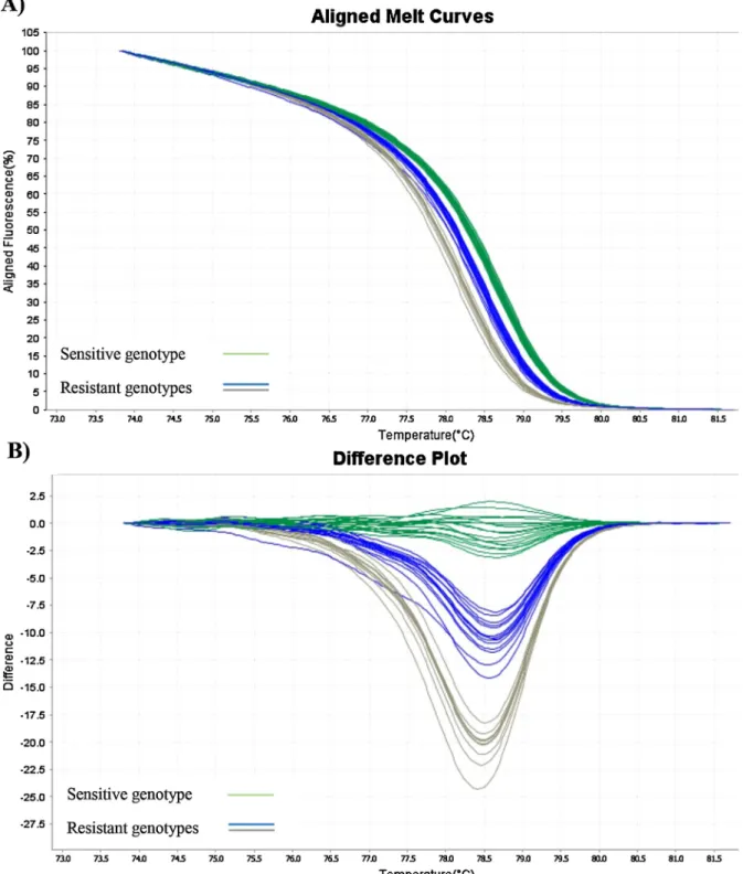
Fig. 2. High resolution melt analyses ofM. hyopneumoniaestrains for the detection offluoroquinolone resistance.
A - Aligned melt curves and B - difference plots of the assayparC(hot spot region 239–250 ofparC). Fluoroquinolone sensitive type (green lines) (Tm = 78.6–78.7 °C) andfluoroquinolone resistant type C239 T (blue lines) (Tm = 78.4–78.5 °C), C239A or G250A (grey lines) (Tm = 78.1–78.2 °C)
31, 373–395.https://doi.org/10.1051/vetres:2000100.
Le Carrou, J., Laurentie, M., Kobisch, M., Gautier-Bouchardon, A.V., 2006. Persistence of Mycoplasma hyopneumoniaein experimentally infected pigs after marbofloxacin treatment and detection of mutations in theparCgene. Antimicrob. Agents 1959–1966.https://doi.org/10.1128/AAC.01527-05.Ch. 50.
Maes, D., Segales, J., Meyns, T., Sibila, M., Pieters, M., Haesebrouck, F., 2008. Control of Mycoplasma hyopneumoniaeinfections in pigs. Vet. Microbiol. 126, 297–309.https://
doi.org/10.1016/j.vetmic.2007.09.008.
Maes, D., Sibila, M., Kuhnert, P., Segalés, J., Haesebrouck, F., Pieters, M., 2017. Update onMycoplasma hyopneumoniaeinfections in pigs: knowledge gaps for improved dis- ease control. Transbound. Emerg. Dis. 1–15.https://doi.org/10.1111/tbed.12677.
Maes, D., Verdonck, M., Deluyker, H., de Kruif, A., 1996. Enzootic pneumonia in pigs.
Vet. Res. Forum 18, 104–109.https://doi.org/10.1080/01652176.1996.9694628.
Meyns, T., Dewulf, J., De Kruif, A., Calus, D., Haesebrouck, F., Maes, D., 2006.
Comparison of transmission ofMycoplasma hyopneumoniaein vaccinated and non- vaccinated populations. Vaccine 24, 7081–7086.https://doi.org/10.1016/j.vaccine.
2006.07.004.
Meyns, T., Maes, D., Dewulf, J., Vicca, J., Haesebrouck, F., De Kruif, A., 2004.
Quantification of the spread ofMycoplasma hyopneumoniaein nursery pigs using transmission experiments. Prev. Vet. Med. 66, 265–275.https://doi.org/10.1016/j.
prevetmed.2004.10.001.
Palais, R.A., Liew, M.A., Wittwer, C.T., 2005. Quantitative heteroduplex analysis for single nucleotide polymorphism genotyping. Anal. Biochem. 346, 167–175.https://
doi.org/10.1016/j.ab.2005.08.010.
Pallarés, F.J., Lasa, C., Roozen, M., Ramis, G., 2015. Use of tylvalosin in the control of porcine enzootic pneumonia. Vet. Rec. Open 2, 1–6.https://doi.org/10.1136/
vetreco-2014-000079.
Pirmohamed, M., Park, B.K., 2001. Genetic susceptibility to adverse drug reactions.
Trends Pharmacol. Sci. 22, 298–305.https://doi.org/10.1016/S0165-6147(00) 01717-X.
Silva, G.S., Yeske, P., Morrison, R.B., Linhares, D.C.L., 2019. Benefit-cost analysis to es- timate the payback time and the economic value of twoMycoplasma hyopneumoniae elimination methods in breeding herds. Prev. Vet. Med. 168, 95–102.https://doi.
org/10.1016/j.prevetmed.2019.04.008.
Stakenborg, T., Vicca, J., Butaye, P., Maes, D., Minion, F.C., Peeters, J., De Kruif, A., Haesebrouck, F., 2005. Characterization ofin vivoacquired resistance ofMycoplasma hyopneumoniaeto macrolides and lincosamides. Microb. Drug Resist. 11, 290–294.
https://doi.org/10.1089/mdr.2005.11.290.
Stakenborg, T., Vicca, J., Maes, D., Peeters, J., De Kruif, A., Haesebrouck, F., Butaye, P., 2006. Comparison of molecular techniques for the typing ofMycoplasma hyopneu- moniaeisolates. J. Microbiol. Meth. 66, 263–275.https://doi.org/10.1016/j.mimet.
2005.12.002.
Staroscik, A., 2004. Calculator for Determining the Number of Copies of a Template.
http://cels.uri.edu/gsc/cndna.html.
Sulyok, K.M., Bekő, K., Kreizinger, Z., Wehmann, E., Jerzsele, Á., Rónai, Z., Turcsányi, I., Makrai, L., Szeredi, L., Jánosi, S., Nagy, S.Á., Gyuranecz, M., 2018. Development of molecular methods for the rapid detection of antibiotic susceptibility ofMycoplasma bovis. Vet. Microbiol. 213, 47–57.https://doi.org/10.1016/j.vetmic.2017.11.026.
Thacker, E.L., Thacker, B.J., Boettcher, T.B., Jayappa, H., 1998. Comparison of antibody production, lymphocyte stimulation, and protection induced by four commercial Mycoplasma hyopneumoniaebacterins. Swine Health Prod. 6, 107–112.
Vicca, J., Maes, D., Stakenborg, T., Butaye, P., Minion, F., Peeters, J., de Kruif, A., Decostere, A., Haesebrouck, F., 2007. Resistance mechanism againstfluor- oquinolones inMycoplasma hyopneumoniaefield isolates. Microb. Drug Resist. 13, 166–170.https://doi.org/10.1089/mdr.2007.716.
Villarreal, I., Maes, D., Meyns, T., Gebruers, F., Calus, D., Pasmans, F., Haesebrouck, F., 2009. Infection with a low virulentMycoplasma hyopneumoniaeisolate does not protect piglets against subsequent infection with a highly virulentM. Hyopneumoniae isolate. Vaccine 27, 1875–1879.https://doi.org/10.1016/j.vaccine.2008.12.005.