CARBON SOURCE UTILISATION AND EVALUATION OF THE BIOLOG SYSTEM IN THE IDENTIFICATION OF
ACTINOBACILLUS PLEUROPNEUMONIAE
László MAKRAI*, Rita SÁRKÖZI and László FODOR
Department of Microbiology and Infectious Diseases, University of Veterinary Medicine, H-1581 Budapest, P.O. Box 22, Hungary
(Received 4 March 2019; accepted 31 July 2019)
Sixty-eight Actinobacillus pleuropneumoniae strains were isolated from porcine acute pleuropneumonia cases from different parts of Hungary between 2000 and 2014. A total of 41 isolates were identified as A. pleuropneumoniae bio- type I and 27 strains as biotype II based on cultural, morphological and biochemi- cal characteristics. The aim of this study was to evaluate metabolic fingerprinting in the species-level identification of A. pleuropneumoniae isolates. Utilisation of carbon sources by these field isolates and six reference strains was characterised by the Biolog system (GN2 Microplate, MicroLog3 Version 4.20.05 software).
Twenty-nine field strains were correctly identified by the Biolog system as A.
pleuropneumoniae, 36 strains as A. lignieresii, two strains as H. paraphrohaemo- lyticus and one strain as A. equuli after 24 h of incubation. Among the six A. pleu- ropneumoniae reference strains the Biolog system identified one strain as A. pleu- ropneumoniae, four as A. lignieresii and one as H. paraphrohaemolyticus. There was no correlation between biotypes and serotypes of A. pleuropneumoniae and the carbon source utilisation pattern and species identification by the Biolog sys- tem. Our data indicate that the efficacy of the Biolog system used here could be improved by including phenotypes of more A. pleuropneumoniae strains repre- senting a wider geographical occurrence into the database.
Key words: Actinobacillus pleuropneumoniae, carbon source utilisation, Biolog system, identification
Actinobacillus pleuropneumoniae is one of the most important bacterium species causing respiratory disease in swine all over the world. Acute haemor- rhagic-necrotic pneumonia with fibrinous pleuritis can usually be seen among 12- to 16-week-old pigs, but actually the acute or peracute form can be recog- nised in all ages, while the chronic form generally develops after the disappear- ance of acute signs. Both forms can cause huge economic losses (Marsteller and Fenwick, 1999; Christensen and Bisgaard, 2004; Gottschalk, 2012).
*Corresponding author; E-mail: makrai.laszlo@univet.hu; Phone: 0036 (1) 251-9900;
Fax: 0036 (1) 251-9260
Actinobacillus pleuropneumoniae strains are facultative anaerobic, medi- um-sized (< 2 µm), non-motile rods covered by a polysaccharide capsule; they can infect only swine (Markey et al., 2013). The bacterium has two biotypes;
biotype I strains require nicotinamide adenine dinucleotide (NAD, V-factor) for growth, while biotype II strains are NAD independent. On the basis of surface soluble capsular polysaccharide antigens 18 serovars have been described (Niel- sen, 1986a,b; Fodor et al., 1989; Nielsen et al., 1997; Blackall et al., 2002; Sárközi et al., 2015; Bossé et al., 2018).
All 18 serovar reference strains are able to express some of the four differ- ent Apx toxins belonging to the pore-forming repeats-in-toxin (RTX) group (Shin et al., 2011; Sárközi et al., 2015; Bossé et al., 2018). Three toxins, ApxI, ApxII and ApxIII are responsible for haemolysis and cytotoxic damage of the lung cells (Sthitmatee et al., 2003), but these toxins are produced by other bacterium spe- cies, too, not only by A. pleuropneumoniae (Schaller et al., 2001). The ApxIV toxin gene is species specific, it can be found only in A. pleuropneumonae strains, and ApxIV toxin is produced only in live animals (Schaller et al., 2001).
Although A. lignieresii is the species most closely related to A. pleuropneumoni- ae as determined by DNA-DNA hybridisation and comparison of the 16S rRNA sequences (Borr et al., 1991), there is no apxIVA gene in A. lignieresii (Schaller et al., 2001).
There are six members of the Actinobacillus genus which are nowadays recognised as significant causes of diseases in animals: A. pleuropneumoniae, A.
suis, A. lignieresii, A. equuli, A. seminis, and A. capsulatus (Rycroft and Garside, 2000). Many species of the Actinobacillus genus other than A. pleuropneumoniae can be found in tonsils of swine, such as A. minor, A. porcinus, Bisgaard’s Taxon 10, A. rossii, and A. porcitonsillarum (Lowe et al., 2011; Gottschalk, 2012). Ac- tinobacillus muris, A. hominis and A. ureae are species of little veterinary impact (Christensen and Bisgaard, 2004).
Actinobacillus lignieresii can be found on the mucous membranes of cattle and is able to cause wooden tongue or lesions in the oral cavity and the regional lymph nodes (Gottschalk, 2012). Differentiation of A. pleuropneumoniae biotype II strains and A. lignieresii is difficult because of their close phylogenetic rela- tionship and common characteristics (Rycroft and Garside, 2000).
There are different methods for the identification of bacteria. In addition to the detection of genus-, species- and serotype-specific genes, identification based on phenotypic characteristics is also widely used. Besides the traditional identifi- cation using cultural, morphological, biochemical and serological features (Bar- row and Feltham, 2003), several identification systems based on the examination of phenotypic characteristics are available on the market. Different manual bio- chemical identification systems like API (Bio-Mérieux, Lyon, France), RapID System (Thermo Fisher Scientific, Lenexa, KS, USA), BD BBL Identification System (Becton Dickinson, Franklin Lakes, NJ, USA) and automated identifica-
tion systems, like VITEK (Bio-Merieux), BD Phoenix System (Becton Dickin- son), Sherlock Microbial ID System (MIDI Inc., Newark, DE, USA) and matrix- assisted laser desorption ionisation-time of flight mass spectrometry (MALDI- TOF MS) methods are widely used.
The Biolog Microbial Identification Systems (Biolog Inc., Hayward, CA, USA) are available in manual, semi-automated and fully automated forms; they identify the bacteria on the basis of utilisation of carbon sources (Wong et al., 1992). Their databases include A. pleuropneumoniae, A. lignieresii, A. hominis, A. equuli and A. suis from the Actinobacillus genus.
The aim of this study was to examine metabolic fingerprinting of field iso- lates and reference A. pleuropneumoniae strains based on the utilisation of 95 carbon sources and to evaluate this method in the identification of A. pleuro- pneumoniae strains.
Materials and methods
Bacterium strains
Sixty-eight A. pleuropneumoniae field isolates were included in the exam- ination; all were isolated from lung samples collected in slaughterhouses and from postmortem cases of acute porcine pleuropneumonia, submitted to our la- boratory from different Hungarian swine farms between 2000 and 2010. One of them was suggested as reference strain of serotype 16 isolated in 2014. Six sero- type reference strains of A. pleuropneumoniae K17 (serotype 5a), L20 (serotype 5b), CVI13261 (serotype 9), D13039 (serotype 10), 56153 (serotype 11) and N273 (serotype 13) provided by Dr. Ø. Angen (Danish Veterinary Laboratory, Copen- hagen) were included in the examinations.
The A. pleuropneumoniae strains were isolated on Tryptone Soya Agar (TSA, Biolab Ltd., Hungary) cross-inoculated with Staphylococcus aureus, and subcultured on chocolate agar with added 50 μg/ml NAD (Biolab Ltd., Hungary), both containing 10% defibrinated sheep blood. Cultures were incubated at 37 °C for 24 h in aerobic environment with the addition of 5% carbon dioxide. They were identified using standard methods (Barrow and Feltham, 2003), and most strains were serotyped (Sárközi et al., 2018). After identification, the isolated A.
pleuropneumoniae strains were stored at –80 °C until further examination.
Carbon source utilisation
A 96-well automated MicroLog MicroStation System with GN2 Micro- plates (Biolog Inc., Hayward, CA, USA) was used for the characterisation of car- bon source utilisation. Microplates were set up and analysed following the manu- facturer’s instructions with minor modification. Single colonies of biotype I of A.
pleuropneumoniae were subcultured three times on Biolog Universal Growth
(BUG) agar plates with NAD, and biotype II strains were cultured on BUG agar containing 10% defibrinated blood. Two pure colonies from the third subculture of each strain were inoculated on two chocolate plates with NAD or blood agar plates evenly covering the whole surface of the plate. Plates were cultured at 37 °C and 5% CO2. After 24-h incubation, the thin and confluent layer of A.
pleuropneumoniae was collected with a cotton swab and suspended in 18 ml in- oculation fluid (GN/GP IF) to obtain a homogeneous suspension. The turbidity of the bacterium suspensions was set to 20 ± 2% using the Biolog Turbidimeter.
GN2 MicroPlates were inoculated with 150 µl bacterium suspension per well and incubated at 37 °C in 5% CO2 atmosphere. Metabolic activity was determined by visual reading of the plates after 24 h. The results were evaluated and a dendro- gram showing the metabolic relationships between the strains was produced by Biolog MicroLog3 software (Biolog Inc., USA, Version 4.20.05).
Results
Identification of bacterium isolates
All 68 field isolates were Gram-negative, < 2 µm, coccoid rods. They pro- duced small grey colonies surrounded by a narrow β-haemolytic zone. They all produced urease, they were oxidase positive but catalase negative. All strains proved to be A. pleuropneumoniae, 41 strains needed NAD, and 27 strains were able to grow without NAD.
Carbon source utilisation
The carbon source utilisation of the A. pleuropneumoniae strains is pre- sented in Table 1. There were no major differences between biotype I and II strains of A. pleuropneumoniae in carbon source utilisation pattern. All strains were able to metabolise 20 carbon sources and 1–99% of the strains could utilise 27 further carbon sources after 24-hour-long incubation. Twenty-nine out of the 68 field isolates were identified by the Biolog system as A. pleuropneumoniae, 36 strains as A. lignieresii, two strains as H. paraphrohaemolyticus, and one strain as A. equuli.
The A. pleuropneumoniae reference strain 9 was identified as A. pleuro- pneumoniae, type strains 5a, 10, 11 and 13 as A. lignieresii, and 5b as H. para- phrohaemolyticus.
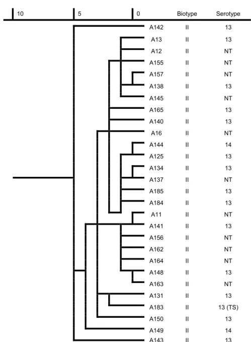
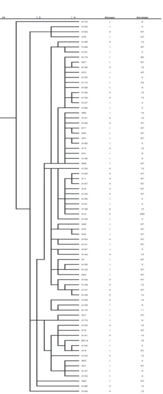
The dendrograms show the similarity of all the A. pleuropneumoniae strains and that of biotype I and II ones (Figs 1, 2 and 3).
There was no connection between biotype and serotype and identification with the Biolog system.
Table 1
Carbon source utilisation of 68 field isolates (biotypes I and II of A. pleuropneumoniae) and 6 reference strains
Carbon sources Total* (74)
(68) Biotype I* (41)
(41) Biotype II* (27)
(27) Reference strains* (6) (6)
Dextrin 100 100 100 100 N-Acetyl-D-Glucosamine 99 98 100 100
L-Arabinose 99 98 100 100
D-Arabitol 4 5 4 0
D-Cellobiose 100 100 100 100 D-Fructose 100 100 100 100 L-Fructose 97 98 96 100 D-Galactose 100 100 100 100 Gentiobiose 10 12 7 0
α-D-Glucose 100 100 100 100
m-Inositol 7 7 7 0
α-D-Lactose 100 100 100 100
Lactulose 100 100 100 100 Maltose 100 100 100 100 D-Mannitol 100 100 100 100 D-Mannose 100 100 100 100 D-Melibiose 3 2 4 0 D-Psicose 100 100 100 100 D-Raffinose 100 100 100 100 L-Rhamnose 94 98 89 100 D-Sorbitol 100 100 100 100 Sucrose 100 100 100 100 D-Trechalose 38 46 26 50 Turanose 97 100 93 100
Xylitol 1 0 4 17
Pyruvic Acid Methyl Ester 94 90 100 100 Succinic Acid Mono-Methyl Ester 56 71 33 83 Acetic Acid 71 71 70 17 Formic Acid 100 100 100 100 D-Galacturonic Acid 54 54 56 33 D-Gluconic Acid 93 90 96 83 D-Glucosaminic Acid 19 17 22 17 D-Glucuronic Acid 50 59 37 33
α-Hydroxybutyric Acid 97 98 96 100
p-Hydroxy-phenylacetic Acid 1 0 4 17
α-Ketobutyric Acid 99 100 96 83
α-Ketoglutaric Acid 40 41 37 50
α-Ketovaleric Acid 1 0 4 0
D,L-Lactic Acid 100 100 100 100 Propionic Acid 1 0 4 0 Quinic Acid 1 0 4 0 Succinic Acid 99 98 100 83 Bromosuccinic Acid 49 51 44 17
Glucuronamide 74 76 70 33 L-Asparagine 68 68 67 67 L-Aspartic Acid 79 78 81 83
Glycyl-L-Aspartic Acid 1 2 0 0
L-Proline 75 71 81 67 L-Threonine 84 83 85 83 Urocanic Acid 68 61 78 50
Inosine 100 100 100 100 Uridine 100 100 100 100 Thymidine 100 100 100 100 Phenylethyl-amine 4 7 0 0
Putrescine 1 0 4 0
D,L,α-Glycerol Phosphate 1 0 4 0
α-D-Glucose-1-Phosphate 57 49 70 83
D-Glucose-6-Phosphate 100 100 100 83
*proportion of the strains utilising carbon sources (%)
Average similarity value
10 5 0 Biotype Serotype 85/14 I 16 A126 I 2 A19 I NT A133 I 2 A67 I NT A130 I 2 A42 I NT A40 I NT A41 I NT A129 I 9 A167 I 2 A173 I 5b (TS) A70 I NT A136 I 2 A186 I 2 A146 I 9 A72 I NT A122 I 2 A7 I NT A68 I NT A166 I 2 A169 I 9 A27 I NT A127 I 2 A37 I NT A63 I 2 A48 I NT A181 I 2 A71 I NT A74 I NT A174 I 9 (TS) A168 I NT A170 I 9 A147 I 9 A132 I 2 A45 I NT A182 I 2 A31 I 9 A123 I NT A151 I NT A89 I 10 (TS) A187 I 2 A44 I NT A172 I 5a (TS) A78 I NT A175 I 11 (TS) Fig. 1. Dendrogram based on the metabolic fingerprint of biotype I strains (n = 46).
NT: non-typeable, TS: type strain
Average similarity value
Fig. 2. Dendrogram based on the metabolic fingerprint of biotype II strains (n = 28).
NT: non-typeable, TS: type strain
10 5 0 Biotype Serotype A142 II 13
A13 II 13 A12 II NT A155 II NT A157 II NT A138 II 13 A145 II NT A165 II 13 A140 II 13 A16 II NT A144 II 14 A125 II 13 A134 II 13 A137 II NT A185 II 13 A184 II 13 A11 II NT A141 II 13 A156 II NT A162 II NT A164 II NT A148 II 13 A163 II NT A131 II 13 A183 II 13 (TS) A150 II 13 A149 II 14 A143 II 13
Fig. 3. Dendrogram based on the metabolic fingerprint of 74 Actinobacillus pleuropneumoniae strains. NT: non-typeable, TS: type strain (A89, 172–175, 183)
Discussion
In addition to widely used nucleic acid typing methods, identification sys- tems based on the detection of various phenotypic characteristics are also availa- ble on the market and used both in human and veterinary medicine. Biolog Mi- crobial Identification Systems are used successfully for the identification of Gram-positive and Gram-negative bacteria (Gyuranecz et al., 2010; Zasada and Mosiej, 2018).
Our results show that identification of the primary pig pathogen A. pleuro- pneumoniae based on carbon source utilisation using the Biolog system has only limited value due to the high similarity of A. pleuropneumoniae and A. ligniere- sii. If the metabolic fingerprint shows questionable results, these two bacterium species can be appropriately differentiated by taking into consideration the pathological origin of the bacterial isolate (A. pleuropneumoniae: haemorrhagic, necrotic fibrinous pleuropneumonia of swine, A. lignieresii: granulomatous mas- titis of pigs, or tongue, lymph node, ruminal wall or skin lesions of ruminants) and some cultural (growth on MacConkey agar of A. lignieresii) and haemolytic features [haemolysis on blood agar and positive CAMP test with Staphylococcus aureus (A. pp.)] of the isolates.
There was no correlation between the biotypes and serotypes of A. pleuro- pneumoniae and carbon source utilisation pattern and species identification by the Biolog system.
Comparing our results with the Biolog standard of A. pleuropneumoniae and A. lignieresii, it is evident that some patterns of A. pleuropneumoniae strains included in the Biolog database have not been represented in our study, as certain carbon sources were not utilised at all by our isolates. No major difference could be seen between the carbon source utilisation of biotype I and II strains of the field isolates.
The dendrograms based on carbon source utilisation show a high level of similarity, especially in the case of biotype II strains of A. pleuropneumoniae, where the difference was below 5%. A higher variability was seen in the case of biotype I strains, but the difference was below 7.5% in this case as well. Our data confirm the results of other authors on the low variability of A. pleuropneumoni- ae strains (Fussing et al., 1998; Kokotovic and Angen, 2007; Sassu et al., 2018;
Ito et al., 2018).
Actinobacillus pleuropneumoniae strains show a high level of antigenic variability in different geographical locations (Gottschalk, 2012; Perry et al., 2012), and a similar metabolic variability could be expected in the utilisation of carbon sources, too. The efficacy of the Biolog system could be improved by in- cluding phenotypes of more A. pleuropneumoniae strains representing a wider geographical occurrence into the database.
Acknowledgement
This work was supported by the Hungarian Scientific Research Fund (OTKA 84220 and OTKA 112826).
References
Barrow, G. I. and Feltham, R. K. A. (eds) (2003): Cowan and Steel’s Manual for the Identification of Medical Bacteria. 3rd edition. Cambridge University Press, Cambridge. 331 pp.
Blackall, P. J., Klaasen, H. L. B. M., Bosch, H. V. D., Kuhnert, P. and Frey, J. (2002): Proposal of a new serovar of Actinobacillus pleuropneumoniae: serovar 15. Vet. Microbiol. 84, 47–52.
Borr, J. D., Ryan, D. A. and MacInnes, J. I. (1991): Analysis of Actinobacillus pleuropneumoniae and related organisms by DNA-DNA hybridization and restriction endonuclease finger- printing. Int. J. Syst. Bacteriol. 41, 121–129.
Bossé, J. T., Li, Y., Sárközi, R., Fodor, L., Lacouture, S., Gottschalk, M., Casas Amoribieta, M., Angen, Ø., Nedbalcova, K., Holden, M. T. G., Maskell, D. J., Tucker, A. W., Wren, B. W., Rycroft, A. N. and Langford, P. R. (2018): Proposal of serovars 17 and 18 of Actinobacil- lus pleuropneumoniae based on serological and genotypic analysis. Vet. Microbiol. 217, 1–6.
Christensen, H. and Bisgaard, M. (2004): Revised definition of Actinobacillus sensu stricto isolated from animals. A review with special emphasis on diagnosis. Vet. Microbiol. 99, 13–30.
Fodor, L., Varga, J., Molnár, É. and Hajtós, I. (1989): Biochemical and serological properties of Actinobacillus pleuropneumoniae biotype 2 strains isolated from swine. Vet. Microbiol.
20, 173–180.
Fussing, V., Barfod, K., Nielsen, R., Møller, K., Nielsen, J. P., Wegener, H. and Bisgaard, M. (1998):
Evaluation and application of ribotyping for epidemiological studies of Actinobacillus pleuropneumoniae in Denmark. Vet. Microbiol. 62, 145–162.
Gottschalk, M. (2012): Actinobacillosis. In: Zimmerman, J. J., Karriker, L. A., Ramirez, A., Schwartz, K. J. and Stevenson, G. W. (eds): Diseases of Swine. Wiley-Blackwell, Chiches- ter. pp. 653–669.
Gyuranecz, M., Erdélyi, K., Fodor, L., Jánosi, K., Szépe, B., Füleki, M., Szőke, I., Dénes, B. and Makrai, L. (2010): Characterisation of Francisella tularensis strains, comparing their car- bon source utilization. Zoonoses Public Hlth 57, 417–422.
Ito, H., Takahashi, S., Asai, T., Tamura, Y. and Yamamoto, K. (2018): Isolation and molecular characterization of a urease-negative Actinobacillus pleuropneumoniae mutant. J. Vet. Di- agn. Invest. 30, 172–174.
Kokotovic, B. and Angen, Ø. (2007): Genetic diversity of Actinobacillus pleuropneumoniae assessed by amplified fragment length polymorphism analysis. J. Clin. Microbiol. 45, 3921–3929.
Lowe, B. A., Marsch, T. L., Isaacs-Cosgrove, N., Kirkwood, R. N., Kiupel, M. and Mulks, M. H.
(2011): Microbial communities in the tonsils of healthy pigs. Vet. Microbiol. 147, 346–357.
Markey, B. K., Leonard, F. C., Archambault, M., Cullinane, A. and Maguire, D. (2013): Actinobacil- lus species. In: Clinical Veterinary Microbiology. Mosby Elsevier, Edinburgh. pp. 297–305.
Marsteller, T. A. and Fenwick, B. (1999): Actinobacillus pleuropneumoniae disease and serology.
Swine Health Prod. 7, 161–165.
Nielsen, R. (1986a): Serology of Haemophilus (Actinobacillus) pleuropneumoniae serotype 5 strains: establishment of subtypes A and B. Acta Vet. Scand. 27, 49–58.
Nielsen, R. (1986b): Serological characterization of Actinobacillus pleuropneumoniae strains and proposal of a new serotype: serotype 12. Acta Vet. Scand. 27, 453–455.
Nielsen, R., Andresen, L. O., Plambeck, T., Nielsen, J. P., Krarup, L. T. and Jorsal, S. E. (1997):
Serological characterization of Actinobacillus pleuropneumoniae biotype 2 strains isolated from pigs in two Danish herds. Vet. Microbiol. 54, 35–46.
Perry, M. B., Angen, Ø., MacLean, L. L., Lacouture, S., Kokotovic, B. and Gottschalk, M. (2012):
An atypical biotype I Actinobacillus pleuropneumoniae serotype 13 is present in North America. Vet. Microbiol. 156, 403–410.
Rycroft, A. N. and Garside, L. H. (2000): Actinobacillus species and their role in animal disease.
Vet. J. 159, 18–36.
Sárközi, R., Makrai, L. and Fodor, L. (2015): Identification of a proposed new serovar of Actino- bacillus pleuropneumoniae: serovar 16. Acta Vet. Hung. 63, 444–450.
Sárközi, R., Makrai, L. and Fodor, L. (2018): Actinobacillus pleuropneumoniae serotypes in Hun- gary. Acta Vet. Hung. 66, 343–349.
Sassu, E. L., Bossé, J. T., Tobias, T. J., Gottschalk, M., Langford, P. R. and Hennig-Pauka, I.
(2018): Update on Actinobacillus pleuropneumoniae – knowledge, gaps and challenges.
Transbound. Emerg. Dis. 65 (Suppl. 1), 72–90.
Schaller, A., Djordjevic, S. P., Eamens, G. J., Forbes, W. A., Kuhn, R., Kuhnert, P., Gottschalk, M., Nicolet, J. and Frey, J. (2001): Identification and detection of Actinobacillus pleuro- pneumoniae by PCR based on the gene apxIVA. Vet. Microbiol. 79, 47–62.
Shin, M. K., Cha, S. B., Lee, W. J. and Yoo, H. S. (2011): Predicting genetic traits and epitope analysis of apxIVA in Actinobacillus pleuropneumoniae. J. Microbiol. 49, 462–468.
Sthitmatee, N., Sirinarumitr, T., Makonkewkeyoon, L., Sakpuaram, T. and Tesaprateep, T. (2003):
Identification of the Actinobacillus pleuropneumoniae serotype using PCR based-Apx genes. Mol. Cell. Probes 17, 301–305.
Wong, J. D., Janda, J. M. and Duffey, P. S. (1992): Preliminary studies on the use of carbon sub- strate utilization patterns for identification of Brucella species. Diagn. Microbiol. Infect.
Dis. 15, 109–113.
Zasada, A. A. and Mosiej, E. (2018): Contemporary microbiology and identification of Corynebac- teria spp. causing infections in human. Lett. Appl. Microbiol. 66, 472–483.
This is an open-access article distributed under the terms of the Creative Commons Attribution 4.0 International License (https://creativecommons.org/licenses/by/4.0/), which permits unrestricted use, distribution, and repro- duction in any medium, provided the original author and source are credited, a link to the CC License is provid- ed, and changes – if any – are indicated. (SID_1)