Investigation of liver function during portal vein occlusion-induced liver regeneration
Doctoral thesis
Tibor Kovács MD
Semmelweis University
Doctoral School of Clinical Sciences
Consultant: Attila Szijártó M.D., Ph.D., associate professor
Opponents: Gábor Jancsó M.D., Ph.D., associate professor Violetta Kékesi M.D., Ph.D., associate professor
Head of the Examination Board: László Kóbori M.D., Ph.D., professor Members of the Examination Board: Andrea Ferencz M.D., Ph.D., associate
professor
Kristóf Dede M.D., Ph.D., chief surgeon, assistant professor
Budapest
2019
2 1. Introduction
Being the sixth most frequent, and second in cancer-related deaths, primary liver tumours, as well as metastatic hepatic neoplasia with like prospects and even higher incidence, embody an immense medical problem on all levels of healthcare systems and societies worldwide. Despite the consensual, modern concept of multidisciplinarity, treatment of liver tumours is overwhelmingly tied to the hands of (hepato-pancreatico-biliary) surgeons, for, bearing survival and oncological aspects in mind, the curative treatment approach is nearly exclusively related to the surgical removal of tumorous tissue. Along with the encouraging results, however, incredibly demanding professional and economical requirements of liver transplantation, liver resection still remains the mainstay of surgical therapy in all regions of the world. While over the years, the astounding advancements of intensive care and other concomitant disciplines allowed the technical execution of heroic liver resections, the limit of resection has dominantly become the amount of residing liver parenchyma of the patient, or
’future liver remnant (FLR)’, because of posthepatectomy liver failure, which remains the chief cause of postoperative morbidity and mortality. In line with this, the paradigm of hepatic surgery abruptly shifted, with the crucial question ‘How much to resect?’ becoming ‘How much to preserve?’. Regarding this, the actual, consensus criteria for safe resection is an FLR volume ratio surpassing 20-25% or 40-50% of tumor-free total liver volume in ‘healthy’ or
‘compromised’ liver parenchyma, respectively, as measure by computed tomography volumetry. Besides neoadjuvant chemotherapy, patients, who are primarily unfit for resection based on this recommendation, may be treated (on a curative intent) with portal vein occlusive procedures (PVO). Regarding these, the surgical ligation (portal vein ligation, PVL) or percutaneous interventional embolization (portal vein embolization, PVE) of portal vein tributaries of the tumorous liver lobes induce ipsilateral atrophy and contralateral (FLR) hypertrophy, which ultimately enable the second-step surgical resection of the tumorous tissue.
Unfortunately, because of the functional inhomogeneity of the liver, hepatic volume does not necessarily reflect liver function. Therefore, during PVO-induced liver regeneration, monitoring of liver function, the main determining factor of postoperative morbidity and mortality, is of crucial importance for the optimal timing of surgical resection as well as for the improvement of clinical outcome. With reference to hepatic biosynthesis, metabolism and elimination, liver function is a complex entity, which may subsequently be measured by a number of different functional tests. Beyond the unequivocally ineffective, (single- or multiple) serum parameter-based ‘static tests’, several ‘clinical score systems’ (Child-Turcotte-Pugh score, MELD-score), combining serum parameters with clinical symptoms, are widely applied, which however still fall short of reliable and sophisticated functional analysis. Instead, the
3
prevailing role in liver functional analysis is fulfilled by ‘dynamic tests’, which are mostly based on either the conversion, elimination or nuclear imaging of a previously injected exogenous agent. The latter two are fundamentally based on the organic anion transport of the liver, which, as the core, energy-dependent pathway for the hepatic elimination of endogenous (bilirubin, bile acids, etc.) and exogenous (alkaloids, toxins, drugs, etc.) substances, allows the specific and sensitive analysis of liver function. The indocyanine-green (ICG) clearance test, based on the hepatic elimination of ICG from the bloodstream, assumes a distinguished role, as the most popular functional test worldwide, and by some also proposed as the basis of decision- making in liver surgery, however, the test is only capable of measuring liver function as a whole. By enabling the anatomical localisation of functional signals through the utilisation of bloodstream-injected, radionuclide-bound liver-specific organic substances, nuclear imaging- based tests opened new horizons, by allowing the specific measurement of any individual, circumscript liver regions. 99mTc-mebrofenin hepatobiliary scintigraphy (HBS) maintains a distinguished role, which, by the specific and sensitive analysis of uptake- and excretory capacities of the FLR, as well as the whole liver, shows promising initial results in the monitoring of hepatic function throughout liver regeneration induced by PVO. Furthermore, liver functional assessment may also be realised by novel procedures, such as MRI- and
13metacetin-based methods, as well as ‘confocal laser endomicroscopy’, an optical/fluorescent imaging method originally developed for the endoscopic diagnostics of the gastrointestinal and bronchoalveolar diseases, which, to the best of our knowledge, has not been applied to date.
Moreover, it is also of chief significance to identify the changes in hepatic drug metabolism during the regenerative process, whose key superfamily and subfamilies of cytochrome P450 (CYP) enzymes also possess central role in conversion tests, whereas exhibit considerable interspecies differences. CYP enzyme activities, which show dynamic and wide-ranging shifts (either increasing or decreasing) in response to diverse pathological changes, are critical factors in the development, hence the potential prevention of drug-related complications, therefore an improved awareness is of utmost clinical relevance.
4 2. Aims
In our studies, the goal was the complex evaluation of the changes of hepatic morphology and function during liver regeneration induced by PVL in a rat model. During Experiment I, the focus of investigation was centered on the organic anion transport system of the liver by the utilisation of in vitro cell cultures, ex vivo tissue samples and modern in vivo imaging, whereas Experiment II was aimed at the analysis of the CYP-mediated drug metabolism of the liver.
We sought answers the following questions:
1. Could viable hepatocytes, capable of the generation of primary cell cultures, be isolated from ligated (LL) as well as non-ligated (NLL) lobes at each time point of regeneration?
2. If yes, how do the morphology, transporter expression and transport function of hepatocytes in LL and NLL cell cultures change following PVL?
3. How does PVL affect the ‘global’ function of the whole liver, with a specific scope on the relevant parameters of pentobarbital sleeping test, indocyanine-green clearance test and 99mTc-mebrofenin hepatobiliary scintigraphy?
4. What are the effects of PVL on the ‘regional’ function of the LL, and more importantly, on the NLL; with a specific interest in their relevant parameters of functional tests quantifying organic anion transport (99mTc-mebrofenin HBS, CLE), as well as drug metabolism (CYP enzyme expression and activity, CYP mRNA expression)?
5.
Could confocal laser endomicroscopy (Cellvizio, MaunaKea Technologies, Paris, France), a method originally developed for the diagnostics of other gastroenterological and bronchoalveolar conditions, be applied for the targeted measurement of regional liver function?5 3. Methods
3.1. Animals and ethics
The experiments were conducted in accordance with international animal welfare guidelines and the mutual approval of local, as well as institutional ethical boards (permit numbers: PEI/001/313-4/2014; PE/EA/2895-6/2016; PE/EA/2893-6/2016) was also granted.
Male Wistar rats (Σn = 106) weighing 200–240 g (Central Animal Facility, Semmelweis University, Budapest, Hungary) were kept in a specifically appointed, conventional-level, isolated, temperature- (22-24°C) and humidity- controlled room, in cages conforming the animal welfare standards of the European Union, with a 12-hour light-dark cycle and surroundings-enrichment, regular housing and caretaking, as well as free access to standard rat chow and water.
3.2. Surgical procedure – portal vein ligation (PVL)
Surgical procedures and analytical tests were carried out under general inhalative isoflurane anesthesia (Experiment I) [(Fortec, Cyprane Ltd., Keighley, England; 1–1.5 l/min oxygen containing 2–2.5% isoflurane (Vetflurane 1000 mg/g, Virbac, Carros, France)], as well as general intraperitoneal anesthesia (Experiment II) [intraperitoneal (i.p.) injection of 75 mg/body weight (bw) kg ketamine and 7.5 mg/bw kg xylazine in 1.5 mL saline). Rats were subjected to PVL corresponding to approximately 80% of total liver parenchyma (n = 91) according to the pre-established model of our workgroup. In a supine position, following abdominal shaving, disinfection and medianlaparotomy, the abdominal cavity is exposed.
Secondary to bowel isolation, the liver, its hilar structures and their portal debranching patterns are identified. With an operation microscope (Leica M6, Wild Heerbrugg, Heerbrugg, Switzerland), microsurgical instruments and atraumatic preparation, the trunk of the portal vein supplying the median-, left lateral- and caudate liver lobes is dissected, as well as atraumatically isolated from the concomitant artery and bile duct and finally selectively ligated with a 6-0 polypropylene surgical suture (Atramat, Ciudad du México, Mexico). After abdominal saline lavage, antibiotic [10 mg/body weight kilograms (bwkg) metronidazole intraperitoneally] and postoperative analgesic (1 mg/bwkg nalbuphine subcutaneously, repeated once 24 h later) administration, a double-layered abdominal closure (abdominal muscles, skin) was performed with running 4-0 silk sutures, and animals were rested.
6
3.3. Experiment I – Liver function as reflected by hepatic organic anion transport 3.3.1. Experiment setup
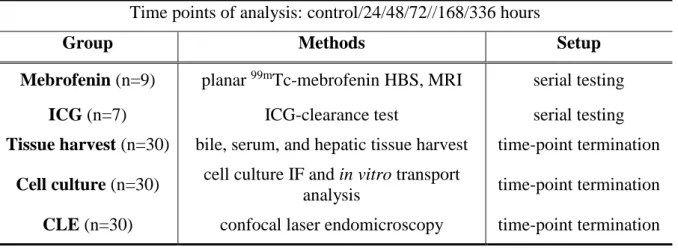
Analyses were performed preoperatively (‘control’), as well as 24/48/72/168/336 hours after PVL. Animals (n=106 db) were assigned to five groups (Table 1). In the first two groups, for more precise and standard analysis, ICG-clearance test („ICG” group, n=7) and planar dynamic 99mTc-mebrofenin HBS and subsequent magnetic resonance imaging (MRI)-based liver volumetry („MEB” group, n=9) were performed repeatedly as part of a serial testing pattern. Three further groups, utilising time-point termination setups based on the characteristics of the applied methods, allowed the acquiring of in vitro cell cultures (n=30), bile, blood and tissue samples (n=30), as well as CLE data (n=30).
Table 1. Group order of Experiment I.
Time points of analysis: control/24/48/72//168/336 hours
Group Methods Setup
Mebrofenin (n=9) planar 99mTc-mebrofenin HBS, MRI serial testing ICG (n=7) ICG-clearance test serial testing Tissue harvest (n=30) bile, serum, and hepatic tissue harvest time-point termination
Cell culture (n=30) cell culture IF and in vitro transport
analysis time-point termination CLE (n=30) confocal laser endomicroscopy time-point termination 3.3.2. In vitro experiments
3.3.2.1. Isolation of hepatocytes and generation of cell cultures
For in vitro experiments, hepatocytes were isolated by collagenase digestion as previously described with a modification of using ‘retrograde’ liver perfusion through the suprahepatic inferior vena cava allowing perfusion of both ligated and non-ligated lobes.
The liver was flushed with Earle’s balanced salt solution (EBSS) (self-produced), firstly with, and subsequently, without the Ca2+-chelating agent EGTA, and finally, with both Ca2+ and type IV collagenase (prepared from Clostridium histolyticum, Sigma-Aldrich, St. Louis, MO) for approximately 5 min. The preoxygenised, temperature-controlled (37°C) and 30 ml/min flow rate perfusion protocol, leading to the blanching of the whole organ, reached its endpoint when the tissue was visibly digested and the (Glissonian) capsule started to separate from the liver surface after approximately 5 mins. Following the precise separation and physical manipulation
7
of the LL and NLL, and trypan blue exclusion-determined cell viability, only suspensions of hepatocytes from preparations surpassing 90 % viability were plated at a density of 2.0 x 10⁶ cells/well on 6-well plates, and 0.36 x 10⁶ cells/well on 24-well plates, in William’s Medium E containing 5% of fetal calf serum, 0.1μM insulin, 0.05 μM glucagon, 0.05 mg/ml gentamicin, 30 nM Na2SeO3, and 0.1μM dexamethasone. Calf serum was omitted after the first 24h. Cells were maintained at 37°C in a humidified atmosphere of 95% air and 5% CO2. Twenty-four hours after plating, cells were overlaid with matrigel basement membrane matrix (Matrigel Matrix; SoftFlow Hungary, Pecs, Hungary) at a concentration of 0.25 mg/ml in 2 ml of ice-cold William’s Medium E supplemented with insulin, glucagon, gentamicin, dexamethasone, and Na2SeO3 to achieve sandwich configuration. The culture medium was replaced every 24h.
3.3.2.2. Immunofluorescence
Cryosections were generated from liver tissue samples of the LL and NLL excised from identical anatomical loci following termination. Representative cryosections of distinctive time points (control, 72 h as mid-time, and 336 h as end-stage) of LR, as well as isolated hepatocytes 72 h in culture from all time points were fixed, permeabilised, blocked, and subjected to primary antibodies against Bsep and Ntcp (both antibodies from Bruno Stieger, University Hospital Zürich, Switzerland), Mrp2 (George Scheffer, Free University Medical Center, Amsterdam, Netherlands), as well as the tight junction protein ZO-1 (Merck Millipore, Billerica, MA). After washes, the samples were stained with fluorophore-conjugated goat anti-rabbit, anti-mouse, and anti-rat secondary antibodies (Thermo Fisher, Waltham, MA), as well as with 4',6-diamidino- 2-phenylindole (DAPI) for nuclear staining. The blue, green, and red fluorescence images of stained hepatocytes and liver sections were acquired by wide-field and confocal microscopy (Leica, Wetzlar, Germany), respectively. As negative controls, samples subjected to no primary antibodies were used. Images were analysed by ImageJ 1.51 h (National Institutes of Health, Bethesda, MD).
3.3.2.3. In vitro taurocholate and bilirubin transport analysis
Bilirubin and taurocholate transport experiments were performed 72 h after culturing, when functional bile canaliculi were formed between adjacent cells, as described previously.
For bilirubin transport analysis, cells were provided 10 μM bilirubin in Hank’s balanced salt solution (HBSS) for 5 min, followed by washing and an efflux period in either standard or Ca2+/ Mg2+-free HBSS for 10 min. Then the cells were lysed with an acetonitrile/water solution. The bilirubin and its mono- and diglucuronide conjugate content of both the efflux media and the cell-lysate were determined by high-performance liquid chromatography (HPLC). From these
8
results, basolateral uptake, canalicular- and sinusoidal efflux transport rates, as well as intracellular accumulation of bilirubin were calculated. The experimental procedure for taurocholate transport analysis was similar to that of the bilirubin transport, while applying 3H- taurocholate as substrate at 1 μM for 1 min. The efflux lasted for 10 min, and the cells were lysed with 0.5% Triton X-100 in phosphate-buffered saline. Taurochoalte content of the efflux media and the lysates were determined by liquid scintillation counting.
3.3.3. In vivo experiments
3.3.3.1. Liver lobe weight and volume
The wet weights of the LL and NLL were determined gravimetrically (AG 245, Mettler- Toledo LLC, Columbus, OH; confidence: 0.01mg/0.1mg). In vivo liver lobe volumes were determined with MRI volumetry [coronal T1-weighed gradient echo sequencing, 128 axial slices of 0.4 mm thickness] (nanoScan PET/MRI; Mediso Ltd., Budapest, Hungary) by the manual delineation of LL and NLL on each axial slice and three-dimensional reconstruction in a 160x160 matrix. Both parameters (weight and volume) were ultimately expressed as a fraction of body weight.
3.3.3.2. Bile excretion
After median laparotomy, selective biliary drainage of the LL and NLL was realised with the insertion and glue-fixation of 1.8 F polyethylene cannulas (PE10, Harvard Apparatus, Holliston, MA) into anatomically corresponding bile ducts under an operation microscope. The amount of bile produced by the LL and NLL over 40 min was measured gravimetrically (AG 245, Mettler-Toledo LLC) and expressed as a fraction of bw. Furthermore, citrate- anticoagulated serum samples were prepared with routine laboratory techniques from the blood acquired during exsanguination. The amounts of UCB and BG were determined by HPLC from both bile and serum samples.
3.3.3.3. Indocyanine-green clearance test
The indocyanine-green (ICG)-clearance test was performed similarly to the previous study of our workgroup. The medial side of the left upper thigh was shaved, and a neonatal laser probe (PV50200; PULSION Medical Systems, Feldkirchen, Germany) of a commercially available analysing device (PC5000 LiMON; PULSION Medical Systems) for ICG densitometry was fixed with an elastic bandage. Following calibration, 1 ml/bwkg of 1.5 mg ICG/ml distilled water was injected into the lateral tail vein. Results were displayed 5-6 minutes later as plasma disappearance rate (PDR) and 15-minute retention (RT15) values.
9
3.3.3.4. Planar dynamic 99mTc-hepatobiliary scintigraphy
99mTc-mebrofenin [99mTc-2,4,6 trimethyl-3-bromo iminodiacetic acid, produced by combining Bromo-Biliaron radiopharmaceutical kit (Medi-Radiopharma Ltd., Budapest, Hungary) and 99mTc isotopes, generated with an Ultra-Technekow Technetium Generator (Mallinckrodt Medical, Petten, The Netherlands), was injected in 150 MBq dosage in 0.3 ml saline into the tail vein. Thereafter, planar HBS (NanoSPECT/CT Silver Upgrade, Mediso Ltd., Budapest, Hungary) was acquired as projections from four separate angles in a resolution of 256 x 256 using Ultrahigh Resolution (UHR) parallel septal collimator (NanoSPECT-UHR, Mediso Ltd, Budapest, Hungary). A dynamic protocol of three different phases was used including 20/6/2 frames per minutes for 2/4/35 minutes, respectively, to monitor the rapid uptake and the canalicular elimination of the tracer. Recordings were evaluated with manual allocation of elliptic regions of interest (ROI) to the projection corresponding to the blood pool, the duodenum, as well as the LL and NLL. The characteristic parameters derived from the kinetics curves included the blood halflife (B1/2), first duodenal appearance (DSTART), as well as regional-specific information of LL and NLL – peak count (PC), time of maximum (TMAX), tracer half-life (T1/2), and relative ratio of LL or NLL peak counts (PC) to corresponding blood counts (RC).
3.3.3.5. Confocal laser endomicroscopy
Following laparotomy, the inferior right lateral lobe of the liver was mobilised, and carefully placed on a fastened plastic foil, elevated to the level of about the median axillary plane, to attenuate respiration-associated movement. The endoscopic wire probe of the laser unit (Cellvizio, MaunaKea Technologies, Paris, France) was harmlessly pressed against and stabilised on the medial surface of the inferior right lateral lobe. After an injection of ICG (1 ml/bwkg of 1.5 mg ICG/ml distilled water) into the tail vein, a 40-minute time lapse video was acquired with intermittent light excitation and registration (3 frames/10 seconds). Video evaluation was performed by the registration of signal intensities of 10 ROI, manually allocated to liver acini. Exponential growth and decay were used to determine the values reflecting the uptake (time of signal maximum, TMAX), as well as excretion (ICG half-life, T1/2) transport activities of the NLL.
10
3.4. Experiment II – Liver function as reflected by hepatic drug metabolism 3.4.1. Experiment setup
Male Wistar rats (n=24) were divided into six groups according to their intended survival times (Table 2). In a ‘control’ group (n = 4), spared PVL and any sort of operative procedure, immediately, whereas in the remaining five groups (n=4-4 each), 24/48/72/168/336 hours after PVL, an in vivo functional pentobarbital sleeping test was performed, followed by exsanguination under repeated general ip. anesthesia and the harvest of liver samples.
Table 2. Group order of Experiment II.
Group Animals PVL Sleeping test Pattern
Control n=4 - immediately time-point termination 24h n=4 + 24th postoperative hour time-point termination 48h n=4 + 48th postoperative hour time-point termination 72h n=4 + 72nd postoperative hour time-point termination 168h n=4 + 168th postoperative hour time-point termination 336h n=4 + 336th postoperative hour time-point termination
3.4.2. Pentobarbital sleeping test
The “pentobarbital sleeping test” was induced by a single i.p. injection of pentobarbital (30 mg/bw kg in 1 mL saline), and performed to determine the “sleeping time”; the start, as well as the end of which was indicated by the loss, as well as the spontaneous reappearance, respectively, of the instinctive reaction of the supine-positioned rat of turning to one side, referred to as „righting reflex”.
3.4.3. Liver lobe weight
The gravimetrical, labouratory scale-measurement of liver lobe weights of the LL, NLL and the whole liver (TMT), as well as their displaying were performed matchingly to that of described throughout Experiment I.
3.4.4. Preparation of microsomes and cytochrome P450 enzyme activity assays
Liver tissues from both the LL and NLL were homogenized in 0.1 M Tris-HCl buffer (pH 7.4) containing 1 mM EDTA and 154 mM KCl (Reanal Finechemicals Co., Budapest, Hungary). Hepatic microsomal fractions were prepared by the method of differential
11
centrifugation. Liver homogenates were centrifuged at 10.000g for 30 min. The supernatant (S9 fraction) was centrifuged az 105.000g for 1 hour, after which the pellet was re-suspended in 0,1 M Tris-HCl and re-centrifuged with identical parameters as the latter. Microsomes were stored at –80 ° C until further analysis. The protein content of the microsomes was determined by the method of Lowry et al., with bovine serum albumin as the standard (Sigma-Aldrich GmbH, Deisenhofen, Germany). Subsequently, CYP selective enzyme activities were determined by previously published methods, involving ethoxyresorufin O-deethylation for CYP1As, pentoxyresorufin O-dealkylation for CYP2Bs, tolbutamide 4-hydroxylation for CYP2Cs, and midazolam 1′- and 4-hydroxylation for CYP3As, respectively. The incubation mixture contained an NADPH-generating system (1 mM NADPH, 10 mM glucose 6-phosphate, 5 mM MgCl2, and 2 IU/mL glucose 6-phosphate dehydrogenase), rat liver microsomes, and the respective CYP isoformspecific substrate (ethoxyresorufin, pentoxyresorufin, tolbutamide, or midazolam for CYP1As, CYP2Bs, CYP2Cs, and CYP3As, respectively). After sixty minutes, enzyme reactions were terminated by either ice-cold methanol or acetonitrile. HPLC analyses were performed while adhering to published methods. The fluorescence of resorufin was quantified at 550 nm excitation and at 589 nm emission wavelengths. Finally, beyond the determination of “intrinsic” microsomal CYP isoform activities, expressed as [pmol metabolite/(mg microsomal protein × min)], the total CYP isoform activities of the LL and the NLL were also extrapolated after scaling with the corresponding weights of the LL or NLL, and displayed in relative units (% of control).
3.4.5. RNA Isolation and quantitative real-time polymerase chain reaction
Total RNA was isolated from the liver samples (TRI reagent, Molecular Research Center Inc., Cincinnati, OH, USA) according to the manufacturer’s instructions. The purity and the concentration of the RNA samples were determined spectrophotometrically (NanoDrop 1000, Thermo Fisher Scientific Inc., Waltham, MA, USA). RNA (3 μg) was reverse-transcribed into single-stranded cDNA (Maxima First Strand cDNA Synthesis Kit, Thermo Fisher Scientific Inc.), and RT-PCR was performed (Maxima SYBR Green qPCR Master Mix, Thermo Fisher Scientific Inc.) using the respective primers for CYP isoforms: CYP1A2, CYP2B1, CYP2B2, CYP3A1, CYP2C6, CYP2C11, CYP2C13. The quantity of the target mRNA relative to that of the housekeeping gene hypoxanthine phosphoribosyltransferase (HPRT) was determined.
12 3.4.6. Immunohistochemistry
Liver samples were excised from identical anatomical parts of the LL and the NLL, fixed in 4% neutral buffered formaldehyde, and embedded in paraffin. Sections of 3–5 μm were cut, deparaffinized (EZ Prep 10×, F. Hoffmann-La Roche, Basel, Switzerland) and rehydrated.
Antigen retrieval was performed for 30 min with a prediluted solution (Cell Conditioning 1, Ventana Medical Systems, F. Hoffmann-La Roche) designated for automated immunohistochemical slide stainers. A standard immunohistochemical reaction was performed in an automated immunohistochemical reaction slide staining system (BenchMark ULTRA, Ventana Medical Systems, F. Hoffmann-La Roche) with a respective detection kit (ultraView Universal DAB Detection Kit, F. Hoffmann-La Roche) involving a standard horseradish peroxidase-diaminobenzidine reaction. Rabbit polyclonal primary antibodies against CYP3A1 (BML-CR3310–0025; Enzo Life Sciences Inc., Farmingdale, NY, USA) were applied at 42 ° C for 32 min in a dilution of 1: 1,000. The manufacturer’s instructions were always followed.
Sections were counterstained with hematoxylin and scanned to produce digital material.
3.5. Statistical analysis
Data distribution was tested with the Shapiro-Wilk normality test. All data was found to display normal distribution. Parametric data was expressed as mean ± standard deviation.
For statistical analysis, analysis of variance was performed with Bonferroni’s post hoc test to correct for multiple comparisons, whereas Pearson’s correlation test was used to correlate parametric data. P-values of less than 0.05 were considered statistically significant. Data management, calculations and visualisation were performed with GraphPad Prism 6 (GraphPad Software Inc., La Jolla, CA) and Origin (OriginLab Corporation, Northampton, MA).
13 4. Results
4.1. Experiment I – Liver function as reflected by hepatic organic anion transport 4.1.1. In vitro experiments
4.1.1.1. Cell culture immunofluorescence
A substantial number of viable hepatocytes (at least 50 million) could be isolated at all time points from both the NLL and LL. Immunofluorescence staining for ZO-1, Ntcp, and Bsep demonstrated that isolated cells from all time points and both liver regions are able to generate polarised cultures with normal morphology, forming bile canaliculi, expressing and properly localising the key Ntcp and Bsep liver transporters. Negative antibody control tests testified for a specific antibody binding.
4.1.1.2. In vitro taurocholate and bilirubin transport analysis
Taurocholate uptake and canalicular efflux were significantly deteriorated at 48–72 h in both lobes. Likewise, the uptake and canalicular excretion of bilirubin were also significantly reduced at 48 h in LL. Considering the bilirubin transport of NLL, a very similar, though non- significant tendency was detected. However, intracellular load of both TC and bilirubin remained constant throughout the observation period.
4.1.2. In vivo experiments
4.1.2.1. Liver lobe weight and volume
The LL progressively atrophysized with significantly decreasing liver lobe weights, whereas parallelly, the NLL continually hypertrophysized along with significantly increasing lobe weights, resulting in essentially unchanged total liver lobe weights throughout the regenerative period. In vivo liver lobe volumes as seen by serial postoperative MRI scans showed close correlation with the weight of both the LL (r = 0.976; p = 0.001) and NLL (r = 0.96; p = 0.002).
4.1.2.2. Bile excretion
With matching characteristics, bile output and total biliary BG content displayed progressive deterioration in the LL and in the meantime, progressive gain in the NLL, leading to undisturbed overall hepatic bile- and BG production. Based on the analysis of serum and bile samples, serum levels of BG, unconjugated and total bilirubin; as well as the fractional share of unconjugated bilirubin (unconjugated/total) in both serum and bile remained unchanged.
14 4.1.2.3. In vivo immunofluorescence
Immunofluorescence of key hepatic organic anion transporters Ntcp and Mrp2 carried out on frozen sections of the LL and NLL of control rats demonstrated normal acinar outlines with proper canalicular and basolateral localisation of Mrp2 and Ntcp, respectively. 72 h after PVL, the LL displayed large patches with abrogated staining for Mrp2 or Ntcp, likely corresponding to necroapoptotic lesions. Meanwhile NLL sections at 72 h lacked any regional differences; however, expression level of both proteins was generally reduced. After 336 h, transporter expression was restored in both lobes. However, acinar structures became divergent;
LL acini were markedly shrunk, while NLL acini became expanded. The described changes were well-reflected on the accessory, representative hematoxylin-eosin histological slides.
4.1.2.4. Global liver function
The ICG-clearance test exhibited a marked drop between 24–72 h (diminished plasma disappearance rate [PDR], 5.8-fold increased retention at 15 minutes [RT15]), which rapidly normalised by 168 h. In parallel, with significantly prolonged blood half-life (B1/2) and first duodenal appearance (DSTART) at 48-72 h, the planar dynamic HBS test closely echoed these results, indicating a transient drop in global hepatic uptake and -excretion capacity. Whereas the normalisation of B1/2 was complete by 168 h, the re-convergence of DSTART lagged, and was still tendentiously elevated at 336 h.
4.1.2.5. Regional liver function
As illustrated by representative HBS registries, the peaks in both LL and NLL decreased and widened after PVL. However, by 336 h, the kinetics of drug elimination normalised in the NLL, whereas further flattened in LL resulting in a solid gap between their curves. Accordingly, these changes were well-reflected in the relative peak counts, reporting on regional hepatic function, of the LL and NLL. A transitional regional functional depression was observed in both lobes at 24–72 h, which was gradually restored in the NLL at 336 h, but remained diminished in LL, resulting in a significant difference between LL and NLL regional functions after 48 h. Considering the CLE-assisted measurement of NLL regional function, TMAX
(~uptake) and half-life T1/2 (~excretion) values, derived from intensity curves, confirmed a transitional drop of NLL regional uptake and excretory function at 24–72 h, after which both parameters gradually reached preoperative levels.
15
4.2. Experiment II – Liver function as reflected by hepatic drug metabolism 4.2.1. Pentobarbital sleeping test
Sleeping time gradually increased after PVL with a significantly elevated peak at 72 h (p = 0.0451), and thereafter normalized to control levels.
4.2.2. Liver lobe weight
Following PVL, the weight of the LL, constituting 76 ± 1.83% of total liver weight and 3.77 ± 0.27% of bw in controls, progressively decreased to only 15.25 ± 7.18% of total liver weight and 0.63 ± 0.33% of bw by 336 h (p < 0.0001). Meanwhile, NLL weights significantly increased from the control values of 23.25 ± 1.71% of total liver weight and 1.17 ± 0.05% of bw to 84.25 ± 6.4% of total liver weight and 3.37 ± 0.29% of bw by the end of the experiment (p < 0.0001). After a slight but significant decrease at 24 h, total liver weight remained essentially unchanged thereafter.
4.2.3. Cytochrome P450 enzyme activity
“Intrinsic” microsomal enzyme activities [pmol/mg protein × min] of CYP1A and CYP3A displayed very similar characteristics. After a general significant increase at 24 h and a subsequent bilobar downward tendency up to 72 h, enzyme activities of the LL and the NLL became solidly divergent. Accordingly, in the NLL, a significant increase in CYP1A as well as in CYP3A was observed at 168 h compared to control values, followed by convergence to baseline by 336 h; meanwhile enzyme activities in LL were permanently deteriorated, resulting in a significant discrepancy of LL and NLL enzyme activities as early as 24–48 h, but becoming the most prominent at the late time points of 168–336 h. CYP2B enzyme activities showed some similarities to that of CYP1A and CYP3A; however, both the late rise of CYP2B activity in NLL, and concomitantly the discrepancy of LL and NLL activities, were moderate and only tendentious. CYP2C activities displayed markedly different features. CYP2C activity of both the LL and the NLL underwent a sharp and significant decrease immediately post-PVL.
However, after 168–336 h, enzyme activity in the NLL showed a late convergence to baseline, while the LL remained permanently depressed, culminating in a gaping discrepancy and a significant difference of LL and NLL CYP2C enzyme activities at 168–336 h.
Changes of the extrapolated, weight-scaled total CYP activities were generally very similar to the corresponding ‘intrinsic’ microsomal CYP activities; however, the “up-scaling”
with weights further amplified the discrepancies of LL and NLL activities primarily in the later stages. Thereby, due to the shift in lobe weights, while the lowered activity of the LL further
16
sank, the progressively increased activity of the NLL became more prominent, resulting in significantly elevated total NLL CYP1A, CYP2C, and CYP3A function even at 336 h, as well as greater interlobar discrepancy, also in terms of CYP2B. Subsequent to the surge in total CYP activity of the NLL, the combined (LL+NLL) total CYP1A and CYP3A activities of the liver were also significantly elevated at 168 h compared to control values. The redistribution of total liver CYP function was observable in terms of all isoforms, reflected in a rise from the 28 ± 2%
control functional contribution of the NLL to the total extrapolated CYP function of the liver to 96 ± 2% by 336 h (p < 0.0001).
4.2.4. Cytochrome P450 mRNA expression
The mRNA levels of CYP2B1, CYP2B2, and CYP3A1 reflected generally similar changes. Here, mRNA levels of the NLL showed an early, steep, and significant increase in the 24- to 48-h period, followed by a rapid redistribution to baseline levels. Meanwhile, mRNA levels in the LL showed either only a modest increment (CYP2B1 and CYP2B2) or were unchanged (CYP3A1). Hence, a significant difference was verified between LL and NLL mRNA expression levels, confined to the early postoperative period of 24–48 h. While a gap of LL and NLL mRNA expression levels was also present in the case of CYP1A2, instead of only an early localization, it was observable throughout the whole postoperative period from 24 to 336 h, which was generated by a dramatic, immediate, and definite reduction of CYP1A2 mRNA expression in the LL, in parallel to an upheld (and at 48 h, tendentiously increased) mRNA expression of the NLL. Again, CYP2C6, CYP2C11, CYP2C13 mRNA concentrations displayed different characteristics. While the mRNA levels of the LL were always significantly and permanently depressed, CYP2C mRNA expression in the NLL was either maintained (CYP2C6) or showed only a temporary decrement with later reconvergence either bordering (CYP2C13), or even reaching the baseline levels (CYP2C11). As a result, a significantly higher mRNA expression in the NLL was once again displayed dominantly in the later stages of 168–
336 h, whereas also present at 48 h in terms of CYP2C6 and CYP2C13.
4.2.5. Immunohistocemistry
Consistent with physiological conditions, CYP3A1 was in all cases expressed with a dominantly pericentral localisation. CYP3A1 positivity of the LL decreased after PVL and remained permanently lower. While positivity in the NLL was also lower as compared to controls at 168 h, a maintained expression between 24 and 72 h resulted in a difference of CYP3A1 positivity between LL and NLL in the early postoperative period.
17 5. Conclusions
Summarising the results of our animal research studies (in rat), investigating the effects of PVL-induced liver regeneration on liver morphology and liver function, therein hepatic organic anion transport and drug metabolism, the following statements can be made corresponding to the pre-emptively posed questions:
1. Following PVL, a large number of viable hepatocytes, with a sustained potential to form primary cell cultures, could be isolated at all time points and both lobes.
2. In cell cultures from each and every postoperative time points of both lobes, hepatocytes exhibited adequate morphology, polarisation, potential for proper bile canaliculi development and maintained transporter expression, whose in vitro (taurocholate and bilirubin) transport displayed only a temporary impairment, whereas bilirubin transport of the NLL was upheld.
3. PVL led to the temporary impairment of global liver function, which was well-reflected in the reduced plasma elimination of ICG, and prolonged global uptake- and global elimination (the latter showing lagging normalisation) of 99mTc-mebrofenin, as well as transitionally increased pentobarbital sleeping test values.
4. After PVL, the regional liver function of ligated lobes was permanently reduced, whereas in non-ligated lobes, after a temporary setback, regional liver function was solidly increased, accounting for the re-establishment of global liver function, as supported by both 99mTc-mebrofenin (HBS) and ICG (CLE) transport, as well as CYP enzyme activities of hepatic drug metabolism. Furthermore, the NLL also displayed an adaptive transcriptional CYP mRNA upregulation, absent however in the LL. Liver function became overwhelmingly redistributed, along with a shift towards the NLL.
5. Confocal laser endomicroscopy proved to be a viable method for the targeted, experimental assessment of regional liver-function, posing the possibility of its potential clinical application in the future.
18 5.1. Novel findings
Based on our conclusions, the following new statements could be made:
1. Throughout the full duration of liver regeneration induced by PVL in rat, viable hepatocytes persist in both lobes, therefore also in ligated lobes, which cells maintain their potential to generate primary cell cultures and therein develop adequate structure, polarisation, bile canaliculi and specific transporter expression, whereas their in vitro function suffers only a temporary setback.
2. Considering the established modalities of induced liver regeneration (PVO, ALPPS, etc.), the transitional postoperative deterioration of global-, as well as bilateral regional liver function could also be verified experimentally by 99mTc-mebrofenin hepatobiliary scintigraphy, also in relation of PVL. This functional depression is reversed by an enormous gain in the functional output of non-ligated lobes, ultimately leading to expressive liver functional inhomogeneity and a shift towards non-ligated lobes.
3. Following PVL in rat, an adaptive, positive transcriptional mechanism of non-ligated lobes could be accounted for the re-establishment of temporarily reduced total heptic drug metabolism in an isotype-dependent manner, which is characterised by the transitional suppression of CYP2C enzymes and the simultaneous preference of CYP1A, CYP2B and CYP3A enzymes.
4. Confocal laser endomicroscopy is a suitable method for the minimal invasive, probe- based assessment of regional liver function in experimental settings.
19 6. List of own publications
6.1. Publications related to the present thesis
1. Kovacs T*, Mathe D*, Fulop A, Jemnitz K, Batai-Konczos A, Veres Z, Torok G, Veres DS, Horvath I, Szigeti K, Homolya L§, Szijarto A§
Functional shift with maintained regenerative potential following portal vein ligation SCIENTIFIC REPORTS 7: Paper 18065. 14 p. (2017)
* Kovacs T, Mathe D: shared first authors; § Homolya L, Szijarto A: shared senior authors
IF: 4.122
2. Kovács T, Déri M, Fülöp A, Pálházy T, Háfra E, Sirok D, Kiss Á, Lotz G, Szijártó A, Monostory K:
Isoform-Dependent Changes in Cytochrome P450-Mediated Drug Metabolism after Portal Vein Ligation in the Rat
EUROPEAN SURGICAL RESEARCH 59: (5-6) pp. 301-319. (2018) IF: 1.343
6.2. Other publications
3. Lauber DT, Tihanyi DK, Czigany Z, Kovacs T, Budai A, Drozgyik D, Fulop A, Szijarto A Liver regeneration after different degrees of portal vein ligation.
JOURNAL OF SURGICAL RESEARCH 203:(2) pp. 451-458. (2016) IF: 2.187
4. Budai A, Fulop A, Hahn O, Onody P, Kovacs T, Nemeth T, Dunay M, Szijarto A Animal Models for Associating Liver Partition and Portal Vein Ligation for Staged Hepatectomy (ALPPS): Achievements and Future Perspectives
EUROPEAN SURGICAL RESEARCH 58:(3-4) pp. 140-157. (2017) IF: 1.343
5. Lauber DT, Fulop A, Kovacs T, Szigeti K, Mathe D*, Szijarto A*
State of the art in vivo imaging techniques for laboratory animals
LABORATORY ANIMALS: THE INTERNATIONAL JOURNAL OF LABORATORY ANIMAL SCIENCE AND WELFARE 51:(5) pp. 465-478. (2017)
*Attila Szijártó and Domokos Máthé are joint senior authors
IF: 1.450
20
6. Koos O, Kovacs T, Fulop A, Pekli D, Onody P, Lukovich P, Harsanyi L, Kupcsulik P, Hahn O, Szijarto A
A posztoperatív keringésváltozások jelentősége a májsebészetben [The importance of postoperative circulatory alterations in hepatic surgery]
ORVOSI HETILAP [HUNGARIAN MEDICAL JOURNAL] 156:(48) pp. 1938-1948.
(2015) IF: 0.291
7. Rosero O, Ónody P, Kovács T, Molnár D, Lotz G, Tóth Sz, Turóczi Zs, Fülöp A, Garbaisz D, Harsányi L, Szijártó A
Impaired Intestinal Mucosal Barrier upon Ischemia-Reperfusion: “Patching Holes in the Shield with a Simple Surgical Method”
BIOMED RESEARCH INTERNATIONAL 2014: Paper 210901. 11 p. (2014) IF: 1.579
8. Rosero O, Onody P, Kovacs T, Molnar D, Fulop A, Lotz G, Harsanyi L, Szijarto A Postconditioning: "Toll-erating" mesenteric ischemia-reperfusion injury?
SURGERY 161:(4) pp. 1004-1015. (2017) IF: 3.574
9. Rosero Olivér*, Kovács Tibor*, Ónody Péter, Harsányi László, Szijártó Attila Bakteriális transzlokáció: rés a pajzson [Bacterial translocation: gap in the shield]
ORVOSI HETILAP [HUNGARIAN MEDICAL JOURNAL] 155:(8) pp. 304-312. (2014)
* Olivér Rosero and Tibor Kovács contributed equally to the realisation of the study.
10. Ónody P., Rosero O., Kovács T., Garbaisz D., Hegedüs V., Lotz G., Harsányi L., Szijártó A: Posztkondicionálás – A távoli szervi dysfunctiók ellenszere? [Postconditioning -- effective method against distant organ dysfunction?]
MAGYAR SEBÉSZET [HUNGARIAN JOURNAL OF SURGERY] 65 (4); 222-229 (2012)