Investigation of the Ca -inducible mitochondrial permeability transition pore and Bongkrekate sensitivity
in distantly related animal species
Ph.D Thesis
Csaba Konrád, Pharm.D
Semmelweis University
János Szentágothai Doctoral School of Neurosciences
Supervisor: Dr. Christos Chinopoulos, Associate Professor, Ph.D.
Official reviewers: Dr. Csaba Sőti, Associate Professor, Ph.D.
Dr. Beáta Sperlágh, Deputy Director, Head of Laboratory, Ds.C.
Chairman of the comittee: Dr. József Mandl, Head of Depratment, Ds.C.
Members of the comittee: Dr. Tibor Zelles, Associate Professor, Ph.D.
Dr. Erzsébet Ligeti, Deputy Head of Depratment, DsC.
Dr. László Drahos, Senior Research Fellow, Ph.D.
Budapest
2014
1
1. List of contents
1. List of contents ... 1
2. Abbreviations ... 6
3. Introduction ... 8
3.1. General aspects of mitochondria ... 8
3.2. Mitochondrial Ca2+ management ... 11
3.2.1. Mitochondrial Ca2+ sequestration ... 11
3.2.2. The nature of Ca2+ precipitates in the mitochondrial matrix ... 13
3.2.3. Release mechanisms of sequestered Ca2+ ... 16
3.2.4. Physiological relevance of mitochondrial Ca2+ handling ... 17
3.2.5. Pathological effects of Ca2+ involving mitochondria ... 19
3.3. The mitochondrial permeability transition pore ... 20
3.3.1. Consequences of prolonged permeability transition on mitochondrial biochemistry and signaling... 20
3.3.2. Effectors of permeability transition ... 21
3.3.3. The proteins proposed to be structural elements of the PTP ... 26
3.3.4. Possible physiological role ... 30
3.3.5. Pathological aspects of the PTP ... 31
3.3.6. Species lacking the PTP ... 35
4. Objectives ... 37
2
5. Materials and methods... 39
5.1. Mitochondrial isolation ... 39
5.1.1. Artemia franciscana ... 39
5.1.2. Vertebrates (Xenopus laevis, mouse and rat)... 40
5.1.3. Drosophila melanogaster and Caenorhabditis elegans ... 40
5.1.4. Freshwater crustaceans (Cyclops vicinus vicinus and Daphnia pulex) ... 41
5.1.5. Marine species ... 41
5.2. ΔΨm determination ... 42
5.3. Extramitochondrial [Ca2+] determination by Ca-Gr 5N fluorescence ... 43
5.4. Mitochondrial swelling ... 43
5.5. Determination of free mitochondrial [Mg2+] ([Mg2+]f) by Magnesium Green fluorescence in the extramitochondrial volume of isolated mitochondria and conversion of [Mg2+]f to ADP–ATP exchange rate mediated by ANT... 43
5.6. Transmission electron microscopy (TEM) ... 44
5.7. Energy-filtered transmission electron microscopy (EFTEM)... 44
5.8. Mitochondrial respiration ... 45
5.9. Mitochondrial matrix pH (pHi) determination of mouse liver and Artemia cyst mitochondria ... 45
5.10. Cloning of ANT expressed in Artemia franciscana ... 46
5.11. Partial sequencing of mRNA from Crangon crangon and Palaemon serratus transcribing the ANT ... 47
3
5.12. Multiple sequence alignment of protein sequences isoforms of ANT expressed
in different species ... 48
5.13. Reagents ... 48
6. Results ... 49
6.1. Artemia franciscana ... 49
6.1.1. Artemia mitochondria lack the PTP ... 49
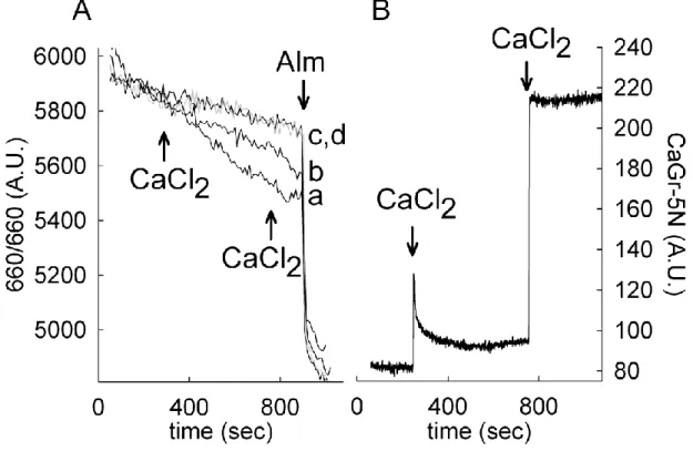
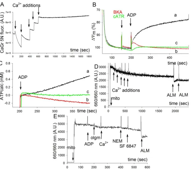
6.1.2. Effect of adenine nucleotides on Ca2+ sequestration of Artemia mitochondria . ... 50
6.1.3. Demonstration of the function of ANT in Artemia franciscana by the ADP– ATP exchange rate–ΔѰm profile ... 53
6.1.4. ANT of Artemia franciscana is refractory to BKA ... 54
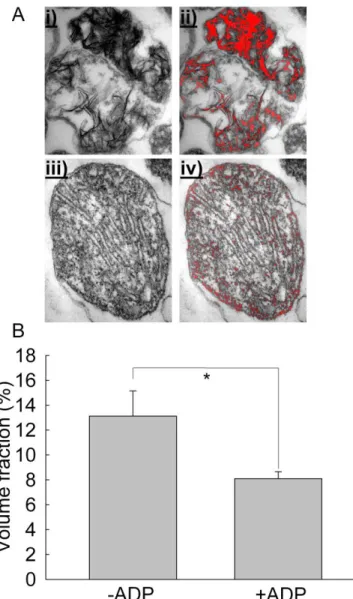
6.1.5. Ca2+-Pi matrix complexation in Artemia mitochondria shows a unique morphology ... 55
6.1.6. Mitochondria isolated from Xenopus liver reveal a classical Ca2+-induced PTP that is sensitive to cyclosporin A and BKA ... 58
6.2. PTP and BKA sensitivity in species related to Artemia: the crustacean subphylum ... 60
6.2.1. Cyclops vicinus vicinus ... 60
6.2.2. Daphnia pulex ... 64
6.2.3. Crangon crangon ... 66
6.2.4. Palaemon serratus ... 69
6.2.5. Carcinus maenas ... 72
4
6.2.6. Pagurus bernhardus ... 74
6.2.7. The lack of Ca2+ induced PTP and insensitivity to BKA are unrelated characteristics of mitochondria isolated from Artemia ... 76
6.3. PTP and BKA sensitivity in non-crustacean species ... 78
6.3.1. Asterias rubens ... 79
6.3.2. Paracentrotus lividus ... 81
6.3.3. Caenorhabditis elegans ... 83
6.3.4. Nephtys hombergii ... 84
6.3.5. Mytilus edule ... 86
6.3.6. Cerastoderma edule ... 87
6.3.7. Patella vulgata ... 89
6.3.8. Drosophila melanogaster ... 91
6.3.9. Branchiostoma lanceolatum ... 92
6.4. The BKA binding site remains elusive ... 92
6.4.1. Sequencing the ANT of Artemia franciscana ... 93
6.4.2. Partial sequences of the ANT of Crangon crangon and Palaemon serratus . 93 6.4.3. Comparison of the primary sequence of Artemia ANT with that of other species 94 6.4.4. Comparison of the predicted three-dimensional structure of Artemia ANT with that of bovine ANT ... 96
6.4.5. The Artemia ANT expressed in yeast ... 97
5
7. Discussion ... 99
8. Conclusions ... 104
9. Summary ... 106
10. Összefoglalás ... 107
11. References... 108
12. Publications ... 142
12.1. Related to the thesis... 142
12.2. Independent from the thesis ... 143
13. Acknowledgements ... 144
6
2. Abbreviations
AD: Alzheimer‟s disease ADP: adenosine diphosphate ALM: alamethicin
ALS: Amyothropic lateral sclerosis ANT: adenine nucleotide translocator AP5A: diadenosine pentaphosphate ATP: adenosine triphosphate
BCECF: 2',7'-Bis-(2-Carboxyethyl)-5-(and-6)-Carboxyfluorescein BKA: Bongkrekik acid
BSA: bovine serum albumin CaGr 5N: Calcium Green 5N cATR: carboxy-atractyloside CSA: cyclosporine A CypD: cyclophillin D DMSO: dimethyl-sulfoxide DNP: 2,4-Dinitrophenol
EDTA: ethylene-diamine-tetraacetic acid
EFTEM: energy-filtered transmission electron microscopy EGTA: ethylene-glycol-tetraacetic acid
ER: endoplasmatic reticulum
FCCP: Trifluorocarbonylcyanide Phenylhydrazone FKBP: FK506 binding protein
HEPES: 4-(2-hydroxyethyl)-1-piperazineethanesulfonic acid IMM: inner mitochondrial membrane
KO: knock-out
MgGr 5N: Magnesium Green 5N
7 mHTT: Mutated huntingtin protein
NAD(P)+: nicotinamide-adenine dinucleotide (phosphate) NMDA: N-methyl-D-aspartate receptor
OMM: outer mitochondrial membrane
PGC-1α: peroxisome proliferator-activated receptor gamma co-activator 1α pHin: mitochondrial matrix pH
pHo: extramitochondrial pH Pi: inorganic phosphate pmf: proton-motive force PT: permeability transition PTP: permeability transition pore ROS: reactive oxygen species
SF 6847: (Tyrphostin 9, RG-50872, Malonaben):
2-[[3,5-bis(1,1-dimethylethyl)-4-hydroxyphenyl]methylene]-propanedinitrile SOD1: superoxide dismutase 1, Cu-Zu superoxide dismutase
TEM: transmission electron microscopy TSPO: 18 kDa translocator protein VDAC: voltage-dependent anion channel
WT: wild type
ΔpH: pH gradient across the inner mitochondrial membrane ΔμH+: proton electrochemical potential gradient
ΔѰm: membrane potential
8
3. Introduction
3.1. General aspects of mitochondria
Mitochondria are the prime source of ATP in the majority of eukaryotes. The energy requirement of the endothermic reaction of converting ADP + Pi to ATP is covered by an electrochemical gradient across the inner mitochondrial membrane (IMM). This electrochemical gradient is maintained by the electron transport chain, which converts the energy from the oxidation of reducing equivalents (NADH + H+, FADH2) into a proton gradient through pumping mechanisms, from the mitochondrial matrix to the intermembrane space. The electrons finally reduce molecular oxygen, thus the name, terminal oxidation. The consumption of oxygen by mitochondria is termed respiration. The respiratory chain consists of four complexes. Complexes I, III, and IV function as proton pumps, acting as a sequential path for the electron flux. The fall in redox potential of the electrons passing through these complexes is used to generate a proton electrochemical potential gradient, ΔμH+, usually expressed in electrical potential units of mV as the proton- motive force (Δp, Eq.1) [1].
Equation 1
Where ΔѰm is the mitochondrial membrane potential and ΔpH is the pH gradient across the inner membrane (conventionally the matrix is referenced to the intermembrane space), and R, T and F refer to the gas constant (8.31 J/(mol*K)), the absolute temperature (310.15 K in human), and the Faraday constant (9.65*104 C/mol), respectively. Under most conditions, ΔѰm is the dominant component of Δp, accounting for 150–180 mV of the total proton-motive force of 200–220 mV, in energized, non-phosphorylating isolated mitochondria [3, 4]. The ΔѰm of mitochondria within cells is lower, as it is being utilized
9
by energy demanding processes. In cultured rat cortical neurons it follows a Gaussian distribution, ranging from -108 to -158 mV, with an average of -139 mV at rest [5].
The respiration generated proton-motive force drives the phosphorylation of ADP into ATP, in functional mitochondria by the FoF1-ATP synthase, residing in the IMM. This protein complex has a proton channel activity, and while allowing a flow of protons into the matrix, converts the pmf into kinetic energy by spinning a central shaft in the molecule which serves as a flexible coupling that transfers the kinetic energy causing conformational changes in the active site, and driving the endothermic reaction of phosphorylation yielding ATP. The less the consumption of the pmf by other processes is, the more tightly respiration and phosphorylation are coupled. Fig. 1 demonstrates the path of electrons and protons in the process of oxidative phosphorylation, through the respiratory complexes and the FoF1-ATP synthase.
A variety of substances, categorized as uncouplers, such as Trifluorocarbonylcyanide Phenylhydrazone (FCCP), 2,4-Dinitrophenol (DNP) or SF6847 (Tyrphostin 9), are capable of dissipating the pmf by increasing the proton permeability of the IMM. The idea of the two reactions being coupled by an electrochemical gradient originates from Peter Mitchell,
Figure 1: The flow of electrons and protons through the respiratory complexes. Reproduced from [1];
no permission required.
10
in 1961 [6]. The coupled reaction is termed oxidative phosphorylation, and is quantified by a dimensionless number, the P/O ratio, indicating how many phosphate molecules are used for ATP synthesis per the reduction of a single oxygen atom. The utilization of FADH2 in well coupled mitochondria yields a P/O ratio of 1.5, while that of NADH+H+ yields 2.5.
The substrates of the FoF1-ATP synthase are inorganic phosphate (Pi) and ADP, which need to be imported across the IMM for ATP production. Pi is transported by the H2PO3-
/OH- antiporter. Cytosolic ADP3- is exchanged for matrix ATP4- in a 1:1 stoichiometry, by the adenine nucleotide translocator (ANT). The FoF1-ATP synthase utilizes 3 H+ to produce a matrix ATP from ADP. The Pi import causes the decrease of the ΔpH, while the exchange of ADP for ATP results in a loss of a charge, leading to the decrease of the (ΔѰm).
Therefore the production of a cytosolic ATP costs a total of 4 H+ flowing back into the matrix.
11
3.2. Mitochondrial Ca
2+management
3.2.1. Mitochondrial Ca2+ sequestration
The mitochondrion is a major player in regulating and shaping cellular Ca2+ levels.
Mitochondrial Ca2+ handling has become a re-emerging topic in recent years (for an extensive review see [2]). Mitochondria from all tissues of mammals possess the ability to sequester divalent cations, like Ca2+, Sr2+ and Ba2+, but Ca2+ is the only one having physiological relevance. Mitochondrial Ca2+ uptake is not only important for buffering transient cytosolic Ca2+ loads, but sequestered Ca2+ is also an activator pyruvate- dehydrogenase, isocitrate-dehydrogenase and α-ketoglutarate-dehydrogenase, regulating the production of reducing equivalents for the energy-demanding uptake of Ca2+ (for review see [7]).
Mitochondria both bind and sequester Ca2+, however the quantity of Ca2+ specifically binding to uptake regulatory sites and substrate transporters, plus the aspecifically binding to membranes is miniscule compared to the uptake capacity. The uptake capacity varies between different tissues, and in the brain, even between mitochondria from different regions [8].
The driving force for Ca2+ uptake is the high electrical potential across the IMM [9, 10].
Indeed, for a ΔΨm of −180 mV, the Nernst equation would predict at equilibrium a mitochondrial [Ca2+]in/[Ca2+]out ratio of about 106 fold, i.e., given a cytosolic Ca2+
concentration of 100 nM at rest, the mitochondrial matrix [Ca2+] should be as high as 0.1 M [11]. In reality such extreme concentrations of free matrix [Ca2+] are never reached, due to the formation of Ca2+ containing precipitates, above 100 nM of [Ca2+], which stabilize matrix [Ca2+] at 1-5 µM. Counter-ions for Ca2+ are primarily Pi, provided by the H2PO3- /OH- antiporter and OH-.
Ca2+ uptake by isolated mitochondria has a relatively low affinity: half maximal uptake speed is achieved at µMs of [Ca2+] [12, 13]. This fact was the main reason for abandoning mitochondrial Ca2+ handling for many years, as cytosolic [Ca2+] are within the mid nM
12
concentrations. The field regained interest when mitochondrial [Ca2+] was measured directly during stimulus. It was done by the transfection of the Ca2+ sensitive photoprotein aequorin, which was fused in frame with a mitochondrial presequence [14].
Mitochondrial Ca2+ uptake can be selectively blocked by Ru 360, the active compound of Ruthenium Red, which is an inorganic dye originally used to stain mucopolysaccharides [15, 16]. The machinery residing in the inner mitochondrial membrane and responsible for Ca2+ uptake was named the calcium uniporter. It has been only until recently, that its proper electrophysiological characterization has been made and its molecular identity has been revealed [17].
The idea that the uniporter is a gated ion channel was first proposed by Bragadin in 1979, on the basis of the dependence characteristics of mitochondrial Ca2+ kinetics on temperature [18]. For decades no major advances were made in identifying the uniporter, until 2004, when direct methods proved his' theory to be correct. Electrophysiological experiments were carried out on mitoplasts, small derivates of mitochondria devoid of the OMM. A highly selective ion channel, with an affinity for Ca2+ of 2 nM, which accounts for the properties of the uniporter was successfully identified. Its relative divalent ion conductance was Ca2+≈Sr2+>>Mn2+≈Ba2+, however it was completely impermeable to Mg2+
and monovalent cations, except Na+ at extremely low [Ca2+]. It was also found that it is forwardly rectifying and sensitive to nanomolar concentrations of Ruthenium Red or Ru360 [17].
In 2010 a regulatory protein of Ca2+ uptake was identified in mitochondria. The protein has two EF-hand domains, the function of which is calcium sensing and one transmembrane domain, which does not support the notion that it could have a channel activity. Its silencing was found to affect Ca2+ uptake activity dramatically. The protein was named MICU1 [19]. The successful molecular identification of the uniporter was achieved by two different groups, using similar methods: both groups highlighted candidate proteins in silico, one by comparing databases of mitochondrial proteins to that of Saccharomyces cerevisiae, in which the uniporter is naturally absent [20], and the other by finding proteins
13
that could be related to the MICU1 [21]. Search results yielded a handful of proteins, of which one, an IMM protein, with two transmembrane regions, plus an intervening loop enriched in acidic residues and with no known function, was further investigated. siRNA and overexpression experiments showed the protein to have a great influence on Ca2+
uptake both in vitro and in vivo. Fluorescent tagging of the protein verified expression in mitochondria. Finally the purified protein was reconstituted in lipid bilayers, for electrophysiological investigation, the results of which are in line with the properties of the uniporter. All these results support, that the 40-kDa protein identified is in fact the mitochondrial calcium uniporter.
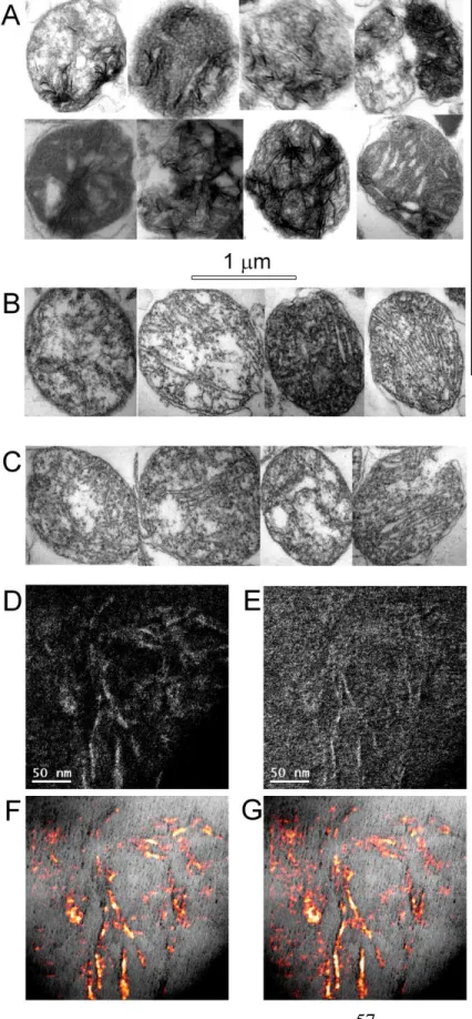
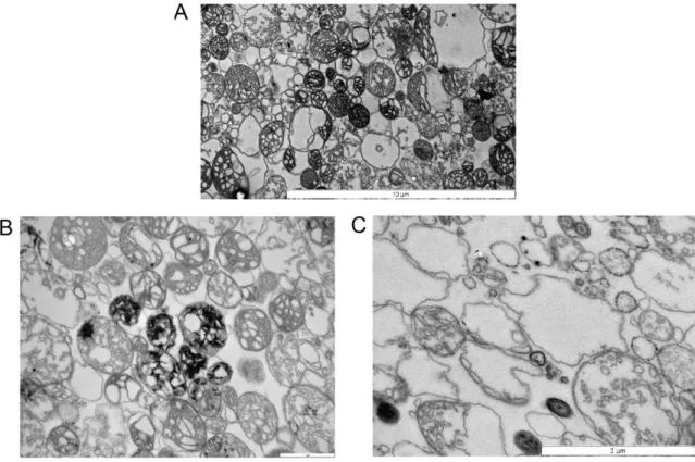
3.2.2. The nature of Ca2+ precipitates in the mitochondrial matrix The phenomenon of precipitation allows an approximately 1 M of [Ca2+]total to be sequestered by mitochondria inside their matrix [22]. Precipitates in mitochondria loaded with Ca2+ can be observed on electron-microscopic images, as amorphous electron-dense granules or ring-like structures. Thread- or needle-like formations were reported in rabbit heart mitochondria, in media devoid of Mg2+ [23], and Artemia franciscana (see below).
Chemical and X-ray diffraction analysis in early studies showed the inorganic content of the precipitates to resemble a mixture hydroxyapatite (Ca10(PO4)6OH2) and whitlockite (Ca3(PO4)2). Mg2+ and CO32- were present in significant quantities, furthermore organic compounds like ADP, ATP and proteins were also found [24]. Recent observations, using more advanced techniques conclude, that stoichiometry between Ca2+ and Pi can vary greatly depending on conditions of Ca2+ sequestration, inhibitors used and the tissue mitochondria originate from [25].
Several anions serve as counter ions for Ca2+, however the most abundant are Pi and OH-. The uptake of ADP or ATP is also necessary [26]. Lack of rapid Ca2+ sequestration by isolated mitochondria incubated in media, in which Pi is absent, demonstrates its relevance in the process [13, 27, 28]. Acidification of the matrix effects precipitate formation, which can be due to the loss of the ΔpH or also, as the ΔpH is constant, due to acidification of the cytosol. The slightly alkaline nature of the matrix favors precipitation, as hydroxide
14
concentration is higher, and also, the dissociation constants of calcium-phosphate salts are pH dependent. During acidification the concentration of hydroxide is decreased and the dissociation constants of calcium-phosphate salts are increased. Furthermore, Pi is imported into mitochondria in exchange for OH-, which leads to a decrease in free Pi when the matrix is acidified [22]. Our laboratory has found however, that even though matrix acidification is an important contributor, it is not a major player in Ca2+ release under the collapse of the ΔpH, induced by uncouplers [29].
Precipitates also contain a significant amount of protein. The role of proteins and their functions remains unknown, and rise numerous questions regarding mitochondrial Ca2+
precipitation. It has been suggested these proteins are regulators or base points of precipitate formation. Interestingly, there have been no Ca2+ binding proteins reported in mitochondria, that would be analogous to proteins, such as calsequestrin, calreticulin or Bip in the endoplasmic reticulum [11].
Another open question is the role of hydroxyapatite and whitlockite forming amorphous precipitates instead of normal crystalline structures. A hypothesis is the presence of lattice
Figure 2: Electron microscopic images of Ca2+ precipitate morphology. A) rat cortical mitochondria fixed after active Ca2+ uptake, 12 000x magnification. B)Rabbit cardiac mitochondria fixed after active Ca2+
uptake in the absence of Mg2+. Reproduced from [2]; no permission required.
15
breaker compounds present in mitochondria. A known lattice breaker, phosphocitrate, might be responsible for this feature [30, 31]. It was shown to be present in cell homogenates [32] and also mitochondria [33].
16
3.2.3. Release mechanisms of sequestered Ca2+
It was described in the late 1970s, that mitochondria loaded with Ca2+ prior to addition of ruthenium red demonstrate a slow Ca2+ release. Furthermore Na+ enhanced release in mitochondria from specific tissues [34-36]. Release of Ca2+ is a slow process, compared to uptake. As they are thermodynamically unfavorable, due to the high electrochemical potential for Ca2+ provided by the ΔѰm and low free matrix Ca2+caused by precipitate formation, these mechanisms are required to be coupled to other electrochemical gradients [37]. Ca2+ release involves two antiporters: the mitochondrial Na+/Ca2+ exchanger and the Ca2+/H+ exchanger, both of which reside in the IMM. The Ca2+/H+ exchanger dissipates pmf directly, while the low matrix Na+ required by the Na+/Ca2+ exchanger is maintained by the Na+/H+ exchanger. The energy required to extrude one atom of Ca2+ can be supplied by the exchange of at least two protons or two Na+ ions. The primarily pathway of Ca2+
release in excitable tissues i.e. heart, brain, skeletal muscle is Na+ dependent Ca2+ efflux, while in liver, kidney, lung and smooth muscle, the Na+/Ca2+ exchanger is not expressed and Ca2+ release is slower and Na+ independent. Characterization of the Ca2+ efflux in excitable tissues showed that the rate of Ca2+ efflux by the Na+/Ca2+ exchanger remains unchanged if Na+ is substituted by Li+, furthermore the antiporter is sensitive to inhibition by CGP-37157.
The molecular identity of the mitochondrial Na+/Ca2+ exchanger is known: the NCLX, which was first described in the plasma membrane, however its unique phylogenetic and functional properties triggered the attention of the group of Sekler. Overexpression, silencing, western blot and immune-electron microscopic experiments confirmed the NCLX to be the Na+/Ca2+ antiporter in mitochondria [38]. The characterization of the NCLX is yet incomplete, with the most pressing question still being the exchange stoichiometry. The molecular identity of the Na+/H+ antiporter remains unknown.
Another player in Ca2+ efflux is a H+/Ca2+ antiport mechanism. A mitochondrial protein, Letm1 was proposed to be the H+/Ca2+ antiporter, however these findings are debated, as Letm1 was previously described as a K+/H+ exchanger [39], which is sensitive to
17
Ruthenium Red and insensitive to CGP-37157 [40]. By immobilized calcium affinity chromatography, the partially purified protein fraction at 66 kDa showed H+/Ca2+ antiport activity when reconstituted in lipid bilayers [41]. Similarly to the Na+/Ca2+ exchanger, there is no consensus in exchange stoichiometry.
Also, the flickering of the mitochondrial permeability transition pore (PTP) could be yet another efflux mechanism, on which phenomena this thesis focuses in later chapters.
Furthermore, a diacylglycerol induced release mechanism was reported, which was unaffected by Ruthenium Red, CGP-37157 or PTP inhibition. The phenomenon could not be demonstrated on mitochondria from sweet potato (Ipomoea batatas), indicating biodiversity, and suggesting it is unlikely to be an artifact [42].
3.2.4. Physiological relevance of mitochondrial Ca2+ handling
Mitochondria couple cellular metabolic state with Ca2+ transport processes. They therefore control not only their own intra-organelle [Ca2+], but also influence the entire cellular network of Ca2+ signaling, including the endoplasmic reticulum, the plasma membrane, and the nucleus [43]. Close interactions with other organelles create so-called microdomains, which can have properties different from that of the cytosol, when rapid changes occur, due to diffusion barriers. The first report of close contacts between mitochondria and the ER was on the basis of overlapping of fluorophores, specific to these organelles, by confocal microscopy [44]. In these regions the Ca2+ concentration can elevate to levels where mitochondrial uptake is active. Lately an elegant method was developed, to directly visualize organelle contacts. The approach used an FKBP-FRB heterodimerization system, targeting the FKBP to the OMM and the FRB to the ER. Introduction of rapamycin was used to cause heterodimerization where FKBP and FRB were close, and close contacts were visualized by FRET [45].
The microdomains between organelles are essential for their coordinated action.
Mitochondrial Ca2+ uptake limits the rapid rise of Ca2+ in these microdomains, otherwise flooded by reticular stores or extracellular Ca2+. Mitochondrial Ca2+ release prolongs the
18
recovery from the Ca2+ stimulus [46]. The microdomains between mitochondria and the endoplasmic reticulum (ER) or the plasma membrane explain the Ca2+ buffering ability of mitochondria in vivo, despite the low affinity of the uptake machinery, as high enough Ca2+
concentrations are reached in these subcellular regions [14].
Early experiments by polarographic Clark electrodes demonstrated mitochondria to take up Ca2+ completely prior to phosphorylating ADP, when challenged with both [13]. However the maintenance of ATP levels is crucial during transients of Ca2+, because the restoration of the ion balance of the cytosol by the ER and the plasmamembrane has a high energy demand. Normal restoration of reticular Ca2+ stores cannot be achieved in experimental conditions where oxidative phosphorylation is blocked and high ATP is made available by permeabilization of cells which supports the importance of high ATP concentration in microdomains between the mitochondria and the ER [47].
The matching of energy production to requirements of the pumping mechanisms is due to the regulatory effect of Ca2+ in the mitochondrial matrix. Ca2+ activates the citric acid cycle in mitochondria by stimulating the pyruvate dehydrogenase complex through the pyruvate dehydrogenase phosphatase, the isocitrate dehydrogenase and the α-ketoglutarate dehydrogenase (for review see [7]). These enzymes all have high flux control coefficients.
A flux control coefficient is a relative measure of how much an infinitely small perturbation of a system parameter affects a system variable: enzymes with a high (close to one) flux control coefficient have a great impact on the rate of an enzymatic pathway. An increase in NADH autofluorescence can be detected for several minutes upon depolarization of excitable cells, like neurons [48]. This increase in reducing equivalents helps maintaining membrane potential during Ca2+ sequestration.
Furthermore organelle interactions is further complicated their motility. The relevance of morphology and dynamics regarding signaling with other organelles has been acknowledged in recent years. The advances mainly in fluorescent microscopic imagery revealed the mitochondria being under constant movement, fusion and fission in the cell.
Elevation in the cytoplasmic [Ca2+] induces translocation of the dynamin-like DRP-1 to the
19
OMM, which is responsible for mitochondrial fission [49]. Also, Rho GTPases (Miro 1 and 2) were found to effect mitochondrial homeostasis, most likely through the mitochondrial trafficking apparatus, as they connect mitochondria to the microtubular network. These proteins exhibit EF-hand domains, the mutation of which influences mitochondrial morphology and distribution in the cell [50].
3.2.5. Pathological effects of Ca2+ involving mitochondria
Ca2+ is implicated in mitochondrial pathologies by two different mechanisms: the mitochondrial permeability transition, on which this thesis focuses and reactive oxygen species (ROS) generation. ROS are the byproducts of mitochondrial respiration and have been implicated in aging and in the pathophysiology of a wide variety of diseases, ranging from diabetes to neurodegeneration. To avoid the malignant effects of ROS, the cell is equipped with scavengers of ROS, such as superoxide-dismutase, catalase, glutathione oxidase, thioredoxin and peroxiredoxin [51].
The effect of Ca2+ on ROS production is controversial: increased [52-55], decreased [56- 58] and unaffected ROS production [59-61] were all reported upon Ca2+ stimulation.
Measured ROS production and elimination of isolated mitochondria in vitro depends on many factors, e.g. substrates, the presence of adenine nucleotides, NADH/NAD+ ratio, inhibitors, and ΔѰm (for extensive review see [51]). As discussed above, Ca2+ has an impact on mitochondrial parameters that have an effect ROS production, which might be an explanation for the inconsistency of the results in the field.
20
3.3. The mitochondrial permeability transition pore
Mammalian mitochondria can only handle a certain amount of Ca2+, before a regulated permeabilization (permeability transition, PT) of the normally impermeable inner membrane occurs, on which most processes of a mitochondrion relies. The entity responsible for the phenomenon was termed the mitochondrial permeability transition pore (PTP). It is commonly associated with various types of disease, and is therefore an intensely researched field [62-71].
3.3.1. Consequences of prolonged permeability transition on mitochondrial biochemistry and signaling
Two types of permeability transition of the IMM were described in mitochondria: the low- and the high-conductance pore. The low-conductance pore allows the pmf to dissipate and Ca2+ to be released, but no swelling or unselective passing of small solutes is observed [70, 72]. The high conductance pore, on the other hand, has a cut-off size of 1500 kDa, allowing ions and small molecules driven by their electrochemical gradients to equilibrate between the matrix and the cytosol. An increase in ROS can be detected during permeability transition, due to the loss of glutathione, impairing ROS scavenging, and NAD+, leaving enzymes, most importantly the α-ketoglutarate dehydrogenase complex without a co-factor to properly reduce [73, 74].
Sequestered Ca2+ is also released in a rapid, unregulated manner. The pmf, similarly to low conductance PTP, is dissipated. PTP opening leaves only a few mV-s of membrane potential, which is caused by the higher abundance of negatively charged proteins in the matrix, too large of size to pass freely. The mitochondrial processes relying on the pmf are therefore severely affected by the opening of the PTP.
As the mitochondrial matrix is hyperosmotic compared to the cytosol, during PT water content of the matrix increases, which is accompanied by swelling. The IMM being folded in mitochondria to form cristae, has a high surface, and can withstand the expansion of the matrix, while the OMM ruptures. This allows proteins residing in the intermembrane space
21
to be released to the cytosol, of which some are pro-apoptotic factors, e.g. cytochrome c, Smac/Diablo, Endonuclease G, or AIF. When a large fraction of mitochondria simultaneously open the PTP in response to stress, the resulting ATP depletion makes it impossible to coordinate the apoptotic machinery, leading to necrosis induction [75, 76]. As a result, transition from apoptosis to necrosis is possible, and the two mechanisms are not always easily distinguishable [77].
3.3.2. Effectors of permeability transition
The effect of a wide variety of chemicals, and changes in different mitochondrial parameters on the probability of PTP opening is known, most of which were documented when the phenomenon was first described. In the 1950s the ability of Ca2+ to cause swelling of isolated mitochondria was first noted, however presumed to be due to non-osmotic mechanisms [78]. It was only in 1976, that a reversible increase in the permeability of the IMM was discovered in beef heart mitochondria, by Hunter and Haworth [79]. Their later work gives a remarkably extensive characterization of the PTP, using swelling experiments almost exclusively [80-82].
Tables 1 and 2 [83] summarize the categories of inducers and inhibitors of permeability transition. Ca2+ alone is sufficient to trigger PT, at least in liver, heart, kidney and adrenal cortex mitochondria, but only if it can be sequestered by mitochondria; inhibition of the Ca2+ uniporter by Ruthenium Red or Ru360 protects mitochondria from the opening of the PTP. Likewise if the counter-ion of Ca2+ does not favor precipitation e.g. if phosphate is substituted by acetate PTP is also inhibited. By this principal, Pi considered as an inducer by many authors.
A number of oxidizing agents have also been reported to be PTP inducers. The mechanism of their action is likely to be identical, but they can be divided into different subgroups.
Furthermore, the distinction between oxidizers and SH reagents may be inappropriate, since there is now reason to think that the effect of the various oxidizers results from the
22
conversion of critical SH groups to disulfide bridges (see, e.g., [84, 85]). SH reducing agents and radical scavengers can be PTP inhibitors.
The effect of uncouplers, like FCCP, suggests a dependence of opening probability on ΔѰm. The PTP inducing effect of ROS was emphasized, and increased generation of ROS was later suggested to be the cause of FCCP induced PTP on mitochondria loaded with Ca2+ [52, 55, 86], however uncouplers trigger PTP even when ROS is eliminated with catalase [87], proving that oxidization by ROS is not responsible for the effect of uncouplers on PT. Both voltage and ROS sensing mechanisms responsible for PTP inducing is unknown.
ADP and ATP are powerful PTP inhibitors [25, 88, 89]. Agents increasing ATP/ADP ratio or decreasing adenine nucleotide content of the matrix are PTP inducers. Pyrophosphate [90] and phosphoenolpyruvate [91] are both known to deplete the matrix adenine nucleotide pool, by exchange on the ANT. Thus, they induce the PT by reducing the level of a protecting agent [83]. Oligomycin also affects nucleotide levels, as the blocking of the FoF1-ATP synthase results in a decrease of the matrix ATP/ADP ratio and hyperpolarization of mitochondria.
PTP opening is also strongly dependent on matrix pH [92]. The optimum for pore opening is pH 7.4. Opening probability decreases sharply both below pH 7.4 (through reversible protonation of critical histidyl residues that can be blocked by diethylpyrocarbonate) and above pH 7.4 (through an unknown mechanism). [93]. Inhibition could be substantially eliminated by pretreatment of the mitochondria with diethyl pyrocarbonate, which effect is most likely due to modification of histidyl residues regulating the pore open-closed probability [94, 95]. The mechanism by which alkaline pH inhibits pore formation is unknown [83].
How Mg2+ and other cations mediate their effect is unknown. Mg2+ was shown to potentiate the effect of both Cyclosporin A and ADP [96]. Cations might act by competitive inhibition on Ca2+ binding sites.
23
Cyclosporin A (CsA) is an immonosuppressant and its effects are caused by the Ca2+- calmodulin-dependent inhibition of calcineurin (a cytosolic phosphatase) by the complex of the drug with cytosolic cyclophilin A [97]. In turn, this prevents dephosphorylation and nuclear translocation of nuclear factor of activated T cells and other transcription factors that are essential for the activation of T cells [93]. CsA also binds to cyclophillin D (CypD), in the matrix, and inhibits PTP formation. The discovery of the effect of cyclosporine A on kidney mitochondria was due to the search for its nephrotoxic adverse effect [98-100].
24
Group Sub-group Agent
Counterions of calcium Phosphate
Oxidizing
agents of pyridyine nucleotides acetoacetate
oxaloacetate
hydroperoxides: t-butylhydroperoxide
cumene hydroperoxide
hydrogen peroxide
radical-generating species menadione
alloxan
allantoin and uric acid
adriamycin and derivatives
nitrofurantoin
Fe2+ Fe3+
xanthine and xanthine oxidase
5-aminolevulinic acid
GSH/GSSG and disulfide hormones
ascorbate
SH reagents heavy and transition metals and their complexes Hg2+ and mercurials
mersalyl
Cd2+
Cu2+
Zn2+
cross-linkers and disulfide bridge formers diamide
arsenite
phenylarsine oxide
other NEM
Adenine nucleotide translocator ligands atractyloside and carboxyatractyloside
pyridoxal-5-phosphate
acyl-CoA's (and acylcarnitines)
Agents causing depletion of matrix adenine nucleotides Pyrophosphate
Phosphoenolpyruvate
Transrnembrane potential-reducing agents uncouplers
lysophospholipids
fatty acids
Other thyroxine
Table 1: inducers of the permeability transition pore
25
Its effect on Ca2+ uptake capacity due to its PTP blocking properties was shortly recognized, by several different groups [101-103]. NIM811 is a CsA derivate causing only PTP inhibition, which makes it unlikely that calcineurin is involved in PTP regulation [104].
Polyamides are documented to protect against PT [105], however the mechanism by which they effect the PTP is not well understood [83].
Inhibitors of the ANT are discussed in the chapter below.
Group Agent
Inhibitors of Ca2+ uptake Ruthenium red, Ru360
Adenine nucleotides ADP
ATP
Ligands of the ANT Bongkrekate
Ligands of the FoF1-ATP synthase Oligomycin Divalent and trivalent cations Mg2+
Sr2+
Mn2+
Ba2+
La3+
Competitors of Ca2+ binding phenothiazines
local anesthetics
Radical scavengers butylhydroxytoluene
Polyamines spermine and spermidine
Carnitine and acylcarnitine
SH-reducing agents dithiothreitol
Ligands of cyclophillin D Cyclosporin A
Fatty acid binder BSA
acidic pH
Table 2: inhibitors of the permeability transition pore
26
3.3.3. The proteins proposed to be structural elements of the PTP Since its discovery, more than 30 years ago, there has been tremendous effort put into identifying the structure of the PTP. The current chapter discusses the most important proteins proposed to be structural elements, and the most relevant experiments unraveling their connection with PTP (for extensive review see [93]).
3.3.3.1. The Adenine nucleotide translocator
The adenine nucleotide translocator (ANT) is the transporter responsible for the exchange of ADP and ATP in a 1:1 stoichiometry between the matrix and the cytosol. Its specificity towards its ligands is far higher, than other ATP utilizing proteins, and also in contrast to them binds the nucleotides in their fully deprotonated form (ATP4-/ ADP3-), without Mg2+
[106]. The transport is therefore electrogenic, and besides numerous other less dynamic mitochondrial parameters, depends on ΔѰm [107]. The transporter can work in both directions, the exchange of cytosolic ADP for matrix ATP is the forward mode. The reversal potential (the membrane potential at which net transport is zero) is approximately - 100 mV [107]. It is the most abundant protein in the IMM, responsible for 10% of its protein content [108].
The ANT was first sequenced in 1982, isolated from beef heart, which consists of 297 amino-acids [109]. It has six trans-membrane domains, and both terminals face towards the cytosol [110]. Whether the ANT functions as homodimers [111, 112], or monomers [113, 114] is debated. The bovine ANT has been solved at a resolution of 2.2 Å [115].
It was also first shown in bovine, that ANT has different organ specific isoforms [116, 117]. There are four human isoforms known. ANT1 is primarily expressed in skeletal and heart muscle [118], ANT2 is found in proliferating cells [119], ANT3 is ubiquitously present in all tissues [120], and ANT4 is expressed the testis [121]. The ANT isoforms show a fair overall homology with the phosphate carrier and the uncoupling proteins, suggesting a common origin of solute carriers in mitochondria [122].
27
There are two known groups of inhibitors of the ANT, atractylosides and bongkrekate derivatives. Atractyloside is found in the toxic thistle Atractylis gummifera. It was originally discovered to cause inhibition of the respiration of mitochondria [106], and it was not long until it was realized, that it mediates its action through blocking the transport of ATP [123]. It binds on the outer surface of the ANT to the translocator site, competitively to ADP [115, 124, 125]. Atratylosides are specific and the ANT has a high affinity towards them. Other than inhibition of the ANT, they also predispose mitochondria to pore opening.
Bongkrekic acid (BKA) is produced by the bacterium Pseudomonas cocovenenans, which infects coconut [126, 127]. It is known to act on the mitochondrial side, but its exact binding site on the ANT is unknown. Only the fully protonated BKAH3 is lipophilic enough to pass through membranes, therefore acidic pH augments the effect of BKA [128, 129]. Low temperature slows diffusion and causes a long offset in the effect of BKA [130, 131]. In contrast with atractyloside, BKA is an inhibitor of PTP opening.
To date the ANT has been the most promising candidate as structural element of the PTP [132]. Numerous effectors of the translocator have been described, and without an exception all having an impact on pore open probability. Three endogenous ligands – ADP, ATP (both inhibiting the PTP) [89, 133], and acyl-CoA and its esters (opening the PTP) [134, 135] – plus four poisons – atractyloside, carboxyatractyloside (cATR) (both favoring pore opening), bongkrekic acid (BKA) and isobongkrekic acid (both promoting pore closure) [136-138] – have been identified. Other, less well-characterized, inhibitors of ANT have also been reported [139]. Therefore the ANT has been under thorough investigation.
The patch-clamping of reconstituted purified bovine heart ANT containing lipid bilayers yielded characteristics similar to that of the PTP [140, 141] described earlier. Ca2+, pH and voltage dependence were all measured [142]. Heterologously expressed ANT of Neurospora crassa in E. coli showed pore opening dependence on cypD [143].
However it was later rejected, as transgenic mice, expressing no ANT1 nor ANT2 isoforms in their liver were shown to exhibit CsA sensitive permeability transition [144]. Based on
28
the mentioned results, the consensus regarding the role of the ANT in PTP is that it is regulatory and nonessential.
3.3.3.2. Cyclophilin D
Cyclophillin D (CypD) gained interest when it was discovered to bind CsA [145, 146]. It is a peptidyl-prolyl cis-trans isomerase located in the matrix of mitochondria, however its substrate is unknown. Cyclophillin D is encoded by the Ppif gene, for which knock-out mice have been successfully generated [62-65]. Surprisingly phenotypic changes in Ppif -/- mice manifest in high levels of anxiety/emotionality in open field and elevated plus maze tests; facilitation of the learning in tasks of active and passive avoidance; increased incidence of adult-onset obesity [147]; prosperity to heart failure, lung edema and reduced survival in response to sustained exercise stimulation [148].
Mitochondria harvested from various tissues of Ppif -/- mice however show a delay in pore opening, and also increased Ca2+ uptake capacity. CsA had no effect on the Ppif knockouts, furthermore wild type mitochondria treated with CsA show similar Ca2+ kinetics to Ppif -/- mice [62]. Ischemia/reperfusion damage is greatly reduced in Ppif -/- mice [63].
A binding site for CypD was recently identified. It was found that CypD associates to the oligomeric forms of the ATP synthase and specifically interacts with OSCP, subunit d, and subunit b of the lateral stalk, decreasing its enzymatic activity, binding being favored by Pi, and that CsA displaces CypD from the ATP synthase complex, resulting in stimulation of the enzyme [149]. This does not translate to changes in mitochondrial ATP output upon treatment of CsA, due to the ∼2.2 fold lower flux control coefficient of the FoF1-ATP synthase compared to that of the ANT [150, 151]. This implies that the inhibition of the FoF1-ATP synthase by CypD is rather serves a function of an in-house control for regulating the matrix ATP/ADP ratio. The mechanism by which CypD regulates the PTP could therefore be indirect, through effecting ATP/ADP ratio, and therefore changing the extent of inhibition.
29
The above findings indicate that CypD is a dispensable regulator, but not a component, of the PTP, the structure of which is unlikely to be altered by the absence of CypD.
3.3.3.3. Voltage-dependant anion channel
The voltage-dependant anion channel (VDAC) is a highly expressed OMM protein. It has 3 highly redundant isoforms in mammals. Several lines of evidence suggested the VDAC could be structural element of the PTP. Purified VDAC incorporated into planar phospholipid bilayers forms channels with properties comparable to those of the PTP [152, 153]; furthermore VDAC channel activity is affected by known modulators of the PTP.
Furthermore chromatography of mitochondrial extracts on a CypD affinity matrix allowed purification of VDAC and the ANT, leading to the conclusion of these proteins being structural elements of the PTP [154].
Though the above findings gave a firm base to conclude, further experiments elaborated below invalidated the concept of the VDAC being an indispensible structural element of the pore. Genetic ablation of VDAC isoforms had been attempted. Mice missing VDAC1, VDAC3 or both were viable showing isoform-specific phenotypes, but were susceptible to permeability transition [155-157], while the unconditional elimination of VDAC2 resulted in embryonic lethality [158]. VDAC was irrevocably rejected to be a structural candidate after mouse embryonic fibroblasts null for Vdac1/Vdac3 were treated with a siRNA against VDAC2, resulting in an in vitro model essentially lacking all three VDAC isoforms. These cells showed no defect in PTP [159].
3.3.3.4. 18 kDa translocator protein
The 18 kDa translocator protein (TSPO), formally called peripheral benzodiazepine receptor is a hydrophobic OMM protein. As the former name implies, it binds benzodiazepine with high affinity and it is thought to be responsible for the adverse effects of benzodiazepines on the immune system, however the structure, localization and function is different [160]. TSPO plays a role in importing cholesterol, protoporphyrins and porphyrins into mitochondria [161-164]. Selective ligands exhibiting PTP inducing activity
30
have been described [165, 166], furthermore porphyrin-induced phototoxicity was shown on isolated mitochondria to be mediated through the TSPO [167], but had no effect on mitoplasts, devoid of the OMM, where TSPO is found [168]. This suggests TSPO is an important regulator, but not structural element of the PTP.
3.3.3.5. F
oF
1-ATP synthase
The finding of a CypD binding site on the FoF1-ATP synthase had raised suspicion that it could be involved in pore formation. An earlier study demonstrated that purified subunit c of the FoF1-ATP synthase has pore activity when reconstituted in lipids [169]. It was recently demonstrated that susceptibility to permeability transition correlates with the expression level (altered by overexpression or silencing) of the ring-forming subunit c of the membrane spanning Fo unit of the FoF1-ATP synthase [170].
Another recent paper also identifies the FoF1-ATP synthase as the PTP. In the study CypD binding to OSCP is reconfirmed. OSCP was knocked down by siRNA, which resulted in premature pore opening. Finally purified FoF1-ATP synthase was reconstituted in lipid bilayers examined by electrophysiology. Pore formation similar to PTP was observed exclusively when dimers of the FoF1-ATP synthase were used [171, 172].
Though these conclusions are based on well-designed experiments and are nevertheless intriguing; many questions regarding the actual mechanism, components and regulation remain open. Furthermore these findings are yet to be proven by direct evidence, such as transgenic in vivo models to support them. The FoF1-ATP synthase being responsible for
∼90% of the ATP produced in the cell will hardly be an easy target for genetic manipulation [173, 174].
3.3.4. Possible physiological role
Despite the PTP being a highly conserved evolutionary trait [175], there is no well- established physiological function, other than cell death. One hypothesis is that the short transients (“flickering”) of permeability transition may serve as rapid Ca2+ release pathway [75, 176]. The phenomenon was demonstrated on isolated mitochondria [177] and intact
31
cells [178]. This notion is supported by the finding that both CsA treatment [179] and genetic ablation of CypD [63, 64] inhibits mitochondrial Ca2+ release.
It is also worth noting, that the PTP was described to be involved in transport of DNA in plants [180].
3.3.5. Pathological aspects of the PTP
A large body of evidence implies the relevance of permeability transition in a variety of untreatable diseases. Here a short overview is given on the subject.
The involvement of PTP in neurodegenerative diseases such as Alzheimer‟s, Parkinson‟s Huntington‟s disease and amyotropic lateral sclerosis is widely accepted. The increase in PTP opening probability is primarily attributed to mitochondrial mutations, bioenergetic impairment, oxidative stress, deficient mitochondrial quality control or errors in mitochondrial dynamics, which as a consequence lead to permeability transition, and cell death [181, 182].
Abnormalities in Huntington`s disease is attributed to the mutated form of the huntingtin protein (mHTT) exhibiting polyglutamine repeats. The exact mechanism by which mHTT expression leads to the clinical phenotype is unknown, but there is evidence of its interaction with mitochondria. mHTT was found to be localized to the outer surface of mitochondria and also to form aggregates inside the matrix [183], leading to decreased membrane potential, Ca2+ handling capacity, mitochondrial trafficking and increased pore opening probability [184-186]. These findings were also confirmed on transgenic mouse models of the disease [187-189]. It was suggested that mHTT affects the peroxisome proliferator-activated receptor gamma co-activator 1α (PGC-1α), a key regulator of mitochondrial biogenesis, energy homeostasis and adaptive thermogenesis [190]. CsA was shown to improve cognitive functions in rodents in the 3-nitropropionic acid induced model of the disease.
32
The proteins shown on Table 3 [191] have been linked to the familiar form of Parkinson`s disease. Many of these proteins play an important role in mitochondrial physiology. PINK1
and Parkin are involved in mitophagy and are therefore essential in the maintenance of mitochondrial quality. Genetic knock down of either of these proteins leads to impaired bioenergetics parameters, which increase PTP opening probability [192-194]. α-synuclein was shown to be localized to the mitochondria causing oxidative stress and increase autophagy [191, 195-197].
Alzheimer's disease (AD) is the most common cause of dementia. The most conspicuous and well known trait in AD is excessive neuronal death and the presence of amyloid beta aggregates in the affected cells. More recent results however indicate that decreased synaptic mitochondrial motility, synaptic dysfunction and the loss of synapses are prior pathological hallmarks to cell death and brain tissue atrophy in AD [198]. Synapses are sites of high energy demand and extensive calcium fluctuations; accordingly, synaptic transmission requires high levels of ATP and causes constant calcium fluctuation [199].
There is a growing number of evidence associating decreased mitochondrial function and increased production of ROS with the disease [200-204]. If axonal mitochondria become less competent in providing energy for Ca2+ handling, the overload will result in permeability transition. The genetic ablation of CypD was shown to rescue axonal
Locus Gene Chromosome Inheritance Probable function
PARK1 and PARK4 α-Synuclein 4q21 AD Presynaptic protein, Lewy body PARK2 Parkin 6q25.2-27 AR Ubiquitin E3 ligase
PARK3 Unknown 2p13 AD Unknown
PARK4 Unknown 4p14 AD Unknown
PARK5 UCH-L1 4p14 AD Ubiquitin C-terminal hydrolase
PARK6 PINK1 1p35-36 AR Mitochondrial kinase
PARK7 DJ-1 1p36 AR Chaperone, Antioxidant
PARK8 LRRK2 12p11.2 AD Mixed lineage kinase
PARK9 ATP13A2 1p36 AR Unknown
PARK10 Unknown 1p32 AD Unknown
PARK11 Unknown 2q36-37 AD Unknown
PARK12 Unknown Xq21-q25 Unknown Unknown
PARK13 HTRA2 2p12 Unknown Mitochondrial serine protease Table 3: Gene loci identified for Parkinson's disease. AD, autosomal dominant; AR, autosomal recessive. Reprinted from [193], no permission required.
33
mitochondrial transport in affected neurons [205], which put an emphasis on the relevance of PTP in the pathology of AD.
Amyothropic lateral sclerosis (ALS) is the most prevalent adult onset motoneuron disease, characterized by selective loss of upper and lower motor neurons [206].
Mitochondria from the spinal cord and muscle of ALS patients revealed decreased activity of the respiratory chain complexes, which fact [207-210]. Studies on familiar types of the disease (accounting for approximately 5% [211] of the cases) provide some insight into the role of mitochondria in the pathology of ALS. There have been 12 genes identified that can cause the disease phenotype, of which the most relevant ones are SOD1, TDP-43 and FUS [212, 213]. Mutated Cu-Zu superoxide dismutase (SOD1) is responsible for 10% of familiar ALS [214]. The disease mechanism by which SOD1 causes ALS is unknown, but genetic ablation of SOD1 does not lead to disease phenotypes in cells [215] or mouse [216], suggesting that mutations cause ALS by a toxic “gain of function” rather than by loss.
Mutant SOD1 proteins were shown to be associated to mitochondria in higher quantities than the wild type form, mostly on the outer membrane and intermembrane space, but also in the matrix [217-226]. The localization of mutant SOD1 is thought to interfere with mitochondrial functions. Mutant SOD1 overexpressing mouse show large, membrane- bound vacuoles derived from degenerating mitochondria in motor neurons [227-229].
These vacuoles show increased immunoreactivity for mutant SOD1, suggesting that accumulation of mutant SOD1 by mitochondria is responsible for their degradation [221].
Another sign of decreased mitochondrial function is the increased cytoplasmatic concentration of Ca2+ in in neuroblastoma cells transfected with mutant SOD1 [230]. In summary the findings above describe conditions in the disease models of ALS in which mutant SOD1 is expressed that favor PTP opening. The crossing of mutant SOD1 expressing mice with CypD knockouts, in which pore opening probability is reduced, results in increased lifespan and later onset of the disease, which findings put an emphasis on the relevance of the PTP in the pathology of ALS .
34
Ischaemia is caused by insufficient or completely lost blood perfusion, which can affect any tissue and leads to necrosis and inflammation. Damage is most devastating and rapid when affecting highly aerobic and energy dependent organs like the brain or the heart.
Furthermore the capacity of the tissues of these organs to regenerate are nearly zero.
The inevitable bioenergetic failure during ischaemia leads to the unavailability of cytosolic ATP, which causes an insufficient activity of plasma membrane pumping mechanisms, resulting in elevated cytosolic Na+ and Ca2+ and consequently, also matrix Ca2+ levels due to mitochondrial Ca2+ uptake. A popular notion was that because both the ANT and the FoF1-ATP synthase can reverse, during ischemia mitochondria could become cytosolic ATP consumers, further contributing to cytosolic ATP depletion, however due to the inhibition of the FoF1-ATP synthase by IF1, this is not likely to occur [231]. The opening of the PTP and the conseqential swelling of mitochondria are well described events that manifest primarily when the ichaemic region is reperfused [232-237]. Reperfusion is accompanied by high oxidative stress, elevation of the pH (therefore the loss of the inhibitory effect of acidic pH on PT [238, 239]) and further mitochondrial overload of Ca2+. Cell death in the brain by necrosis expands to a greater region than primarily affected by ischaemia- reperfusion due to the phenomenon of glutamate excitotoxicity. Glutamatergic neurons depleated of ATP are incapable of polarizing their plasma membrane and therefore release glutamate at synapses from their axons, which by overexciting glutamate receptors like the N-methyl-D-aspartate receptor (NMDA) causes Ca2+ overload and necrosis on postsynaptic neurons [234].
There are two treatments offering some level of protection in ischemia/reperfusion injury, ischemic preconditioning is a process where short episodes of ischemia are applied, while in ischemic postconditioning reperfusion is interrupted by periods of ischemia. PTP is involved in the mechanism these phenomena [240, 241]. Attempts to inhibit PT by CsA or knocking down CypD were shown to reduce infarct size in cerebral ichaemia [63, 242- 245]. Interestingly, CypD KO mice could not be further protected by preconditioning, suggesting CypD is the mediator of protection [246].
![Figure 1: The flow of electrons and protons through the respiratory complexes. Reproduced from [1];](https://thumb-eu.123doks.com/thumbv2/9dokorg/1370366.112255/10.918.139.783.535.833/figure-flow-electrons-protons-respiratory-complexes-reproduced.webp)