O R I G I N A L A R T I C L E
Penicillium expansum strain isolated from indoor building material was able to grow on gypsum board and emitted guttation droplets containing chaetoglobosins and
communesins A, B and D
M.J. Salo1 , T. Marik2, R. Mikkola1 , M.A. Andersson1, L. Kredics2 , H. Salonen1 and J. Kurnitski1,3
1 Department of Civil Engineering, Aalto University, Aalto, Finland
2 Department of Microbiology, Faculty of Science and Informatics, University of Szeged, Szeged, Hungary 3 Department of Civil Engineering and Architecture, Tallinn University of Technology, Tallinn, Estonia
Keywords
Cytotoxicity, fungal contamination, mycotoxins,Penicillium, toxins.
Correspondence
Marja Johanna Salo, Department of Civil Engineering, Aalto University, Box 12100, FI-00076 Aalto, Finland.
E-mail: johanna.72salo@gmail.com
2019/0621: received 7 February 2019, revised 30 May 2019 and accepted 1 June 2019 doi:10.1111/jam.14369
Abstract
Aims: Emission of toxic metabolites in guttation droplets of common indoor fungi is not well documented. The aims of this study were (i) to compare mycotoxins in biomass and guttation droplets from indoor fungi from a building following health complaints among occupants, (ii) to identify the most toxic strain and to test if mycotoxins in guttation liquids migrated trough air and (iii) to test if toxigenic Penicillium expansum strains grew on gypsum board.
Methods and Results: Biomass suspensions and guttation droplets from individual fungal colonies representing Aspergillus, Chaetomium, Penicillium, Stachybotrys and Paecilomyces were screened toxic to mammalian cells. The most toxic strain, RcP61 (CBS 145620), was identified as Pen. expansum Link by sequence analysis of the ITS region and a calmodulin gene fragment, and confirmed by the Westerdijk Institute based on ITS and beta-tubulin sequences. The strain was isolated from a cork liner, was able to grow on gypsum board and to produce toxic substances in biomass extracts and guttation droplets inhibiting proliferation of somatic cells (PK-15, MNA, FL) in up to 20 000-fold dilutions. Toxic compounds in biomass extracts and/or guttation droplets were determined by HPLC and LC-MS. Strain RcP61 produced communesins A, B and D, and chaetoglobosins in guttation droplets (the liquid emitted from them) and biomass extracts. The toxins of the guttation droplets migrated c.1 cm through air and condensed on a cool surface.
Conclusions: The mycotoxin-containing guttation liquids emitted by Pen.
expansumgrown on laboratory medium exhibited airborne migration and were
>100 times more toxic in bioassays than guttation droplets produced by indoor isolates of the genera Aspergillus, Chaetomium, Stachybotrys and Paecilomyces.
Significance and Impact of the Study: Toxic exudates produced by Pen.
expansum containing communesins A, B and D, and chaetoglobosins were transferable by air. This may represent a novel mechanism of mycotoxin dispersal in indoor environment.
Journal of Applied Microbiology127, 1135--1147©2019 The Authors.Journal of Applied Microbiologypublished by John Wiley & Sons Ltd 1135
Introduction
Penicillium expansum is a ubiquitous filamentous fungus causing the serious postharvest disease known as blue mould in harvested apples, peaches and hazels (Julca et al.2015). It is known to produce the potent mycotox- ins communesins, chaetoglobosins and patulin when growing on fruits (Andersen et al. 2004; Nielsen et al.
2004; Tannous et al. 2016). As Penicillium expansum results in fruit losses and is a public health issue (Tan- nous et al. 2016), its ecology as a fruit pathogen and its mycotoxin production on fruits have been extensively studied. On artificially contaminated apples, the optimal conditions for growth have been predicted to occur near 25°C, pH 51 and at a high water activity (aw) value of 099. Growth of Pen. expansum strains occurred between 4–30°C and optimal patulin production was recorded at 16°C (Tannouset al.2016).
It is likely that any cellulolytic, saprophytic or biodete- riogenic fungi can grow in indoor environment if finding suitable substrate and moisture (Li et al. 2015). Penicil- lium expansum has been shown to produce cellulolytic enzymes as b-glucanase, endoglucanases, cellobiohydro- lases and b-glucosidase and has been isolated from sur- face-sterilized timber and deteriorating cedar wood in historical buildings (Duncan et al. 2006; Zayne et al.
2009). This indicates that Pen. expansum may be able to colonize indoor building materials in addition to fruits.
However, growth of Pen. expansum on modern building materials and toxin production by indoor isolates identi- fied by modern molecular methods as Pen. expansum have not been documented.
Moisture and indoor growth of ascomycetous fungi, considered together, are positively associated with respira- tory illness according to studies performed in many geo- graphical regions (Korkalainen et al. 2017; Mendell and Kumagai 2017; Caillaudet al.2018; T€ahtinenet al.2019).
Irrespective of definition, moisture and mould damage are internationally common and are estimated to occur in 18–50% of buildings (Mendell et al. 2011; Norb€ack and Miller 2013). Moisture or dampness and mould damage are found in 25–26% of Finnish buildings, being most prevalent (12–26%) in public educational buildings and healthcare facilities (Annilaet al.2017).
Growth of filamentous fungi indoor depends on mois- ture enabling cell division, mycelial growth, sporulation, formation of membrane-surrounded liquid-containing organelles (vesicles, vacuoles and peroxisomes), synthesis of secondary metabolites and emission of volatile organic compounds (Kenne et al. 2014; Kistler and Broz 2015;
Bennet and Inamdar 2015). Mould odour has been con- nected to unhealthy indoor air and is a possible indicator of active fungal growth (Mendell and Kumagai 2017).
However, no guidelines for unhealthy levels of indoor mould exposure have been defined (Bennet and Inamdar 2015; Hurraßet al.2016).
Biologically active fungal secondary metabolites may be toxic to the producer organism, transported to the cell surface and liberated to the exterior by membrane-sur- rounded organelles like vacuoles and vesicles (Kenne et al. 2014; Kistler and Broz 2015; Bennet and Inamdar 2015). Compared to conidia and hyphal fragments, mem- brane-surrounded organelles of indoor fungi have gained little attention and their impact on indoor air quality is not understood yet. Also, occurrence of Pen. expansum isolates in indoor environments has been of less concern compared to other indoor Penicillium species such as Pen. chrysogenum, and the toxigenic Aspergillus species (Nielsen 2003).
In our preliminary study we found that isolates ofPen.
expansum are able to produce chaetoglobosins and com- munesins, which are secreted in membrane-surrounded vesicles and liberated as liquid exudates (Salo et al.
2015). Recently we showed that indoor Trichoderma strains produce toxic vesicles and guttation droplets con- taining peptaibols (Castagnoliet al.2018b). The reported connection between exposure to toxins and weakened immune tolerance (Genius 2010; Tuuminen and Lohi 2019), as well as our preliminary findings aroused our interest in microbial toxigenesis and vesicle formation and guttation in indoor environments. In this study we investigated toxicities in vesicles and exudates from indoorPenicilliumand Aspergillusstrains properly identi- fied by molecular methods.
Materials and methods
Sampling and microbiological protocols
Rooms in a large, mechanically ventilated office building (200 rooms) associated with severe adverse health effects of several occupants were investigated. The building has a concrete frame, mineral wool and cork board isolation and a tile facßade. Office B23 was on the 1stfloor, C35 on the 2nd and others on the ground floor of a building erected in 1959–1967 and renovated in 1997–2000. For cultivation, the samples (Table 1) were grown on 2%
malt extract agar (35 ml per plate, Ø 9 cm); sealed with gas-permeable adhesive tape to slow moisture loss during the 2–4 weeks culturing at a relative humidity (RH) of 30–40% and a temperature of 22–24°C.
Fungal isolates of the Aspergillus niger complex, Pen.
expansum MH 6, Chaetomium sp. MH52 and Tricho- derma sp. were identified by microscopy (Samson et al.
2002), fluorescence emission of biomass suspensions, tox- icity profiles in three toxicity assays and comparison to
strains identified by DSMZ (Deutche Sammlung f€ur Mikroorganismen und Zellkulturen, Braunschweig, Ger- many), and deposited in the culture collection HAMBI (University of Helsinki, www.helsinki.fi/hambi). The ref- erence strains Aspergillus versicolor GAS226, Aspergillus westerdijkiae PP2, Paecilomyces variotii Paec 2 and Pen.
expansum SE1 were identified to species level by DSMZ and deposited in the HAMBI culture collection.
Strains Acrostalagmus luteoalbus P0b8, Aspergillus cali- doustusMH34,Pen. expansumRcP61 and SE1, as well as Pen. chrysogenum RUK2/3 were identified by sequence analysis of the ITS region (Anderssonet al. 2009) and a calmodulin gene fragment (Hong et al. 2006; Pildain et al. 2008). Sequences were deposited in the GenBank Nucleotide database (http://www.ncbi.nlm.nih.gov) under the following accession numbers: Acr. luteoalbus P0b8:
KM853014; Asp. calidoustus MH34: KM853016; Pen.
expansum RcP61: KP889005, MK201596, Pen. expansum SE1: MK217414, MK201595, Pen. chrysogenum RUK2/3:
KM853015, MK217415. These strains were deposited both in the Szeged Microbiology Collection (http://www.szmc.
hu) in Hungary and the HAMBI (www.helsinki.fi/hambi/
collection) in Finland.
In addition, the identity of the strain Pen. expansum RcP61 was confirmed by the Westerdijk Institute based on ITS and beta-tubulin sequences as Pen. expansum Link, and the strain was deposited in the Westerdijk
Institute strain collection under the accession number CBS 145620.
Analytical procedures
For initial toxicity screening, a loop (10ll) containing 10–20 mg biomass (wet weight) of each colony on the primary culture plates was tested for toxicity. The fungal biomass was dispersed into 02 ml of ethanol, the vial sealed and incubated in a water bath for 10 min at 60
°C. The obtained ethanolic lysates were used to expose porcine spermatozoa (obtained from an artificial insemi- nation station) and kidney tubular epithelial cells (PK- 15). The lysate was considered toxic when 25 vol% (boar sperm) or 5 vol% (PK-15) of the lysate inhibited target cell functions: motility (sperm, within 30 min or 1 day) and proliferation (PK-15, 2 days) (Castagnoli et al.
2018a). The colonies that displayed toxicity were streaked pure and identified to genus or species level.
Exudate droplets harvested from mycelial surfaces with micropipettes and diluted (step=2) in ethanol were incubated at 60 °C for 10 min and then serially diluted (step=2) for toxicity testing. The in vitro toxicity test was performed using porcine cells (sperms, somatic cell lines) as indicators according to Bencsiket al.(2014) and Ajao et al. (2015). Fluorescence emission of the fungal secondary metabolites was photographed and illuminated at 360 nm. Mycotoxins were identified using LC-MS methods (Mikkolaet al.2012; Mikkolaet al.2015).
Toxicity tests of ethanol-soluble substances extracted from plate-grown biomass and the identification of the toxins by LC-MS were described previously (Mikkola et al.2012; Mikkolaet al.2015; Castagnoliet al.2018a).
Other protocols
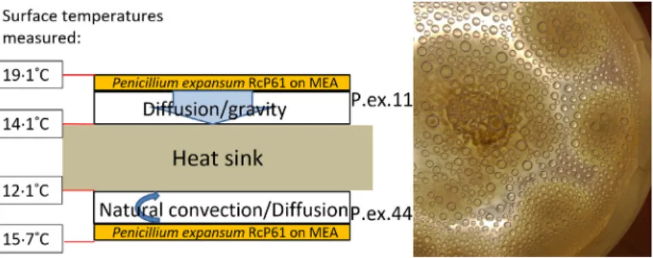
Translocation of fungal metabolites by water vapour from one surface to another was measured using an experi- mental setup as shown and explained in Fig. 1.
To cultivate fungi, moisturized gypsum boards, 100 cm2, thickness of 12 mm, were seeded with spores from a 20-day-old plate culture suspended in 01 vol% Tween 80. The seeded board was incubated in a steel chamber (125 l) and sealed with a glass lid at RH 95% and 201 °C. Biomass (2 mg) was scraped with a micro- scopic slide from the surface of the gypsum liner and dis- persed in 40ll ethanol, incubated at 60 °C for 10 min and then tested for toxicity to PK-15 cells using inhibi- tion of proliferation as endpoint and applying a fluoro- metric readout confirmed with microscopic examination as described in Bencsik et al. (2014). Concentrations of conidia (2lm) and hyphal fragments (larger than 01lm) in biomass lysates and guttation droplets were
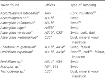
Table 1 Moulds from a university office building where several occu- pants reported severe, building-related adverse health symptoms
Taxon found Offices Type of sampling
Acrostalagmus luteoalbusa A46 Cork insulationbd
Acrostalagmussp.a A31ba Swab
Aspergillus calidoustusa A31ba Swabbc
Aspergillus nigera A45ba Swab
Aspergillus versicolora A31ba,C35a Swab, cork, dust Aspergillus westerdijkiaea C35a Dust, mineral wool
insulation Chaetomium globosuma A31ba,A45ba Swab, fallout Penicillium expansuma A31ba,A45ba Swabbb, corkba, fallout,
impactor Penicilliumsp.a A31aa, A34 Swab
Rhizopussp.b A34, B23 Swab
Trichodermasp.a C35a Dust, mineral wool insulation
Offices associated with building-related health complaints are marked in italics.
aIndicates the sample from which the strains were isolated. aPen.
expansum RcP61, bPen. expansum MH6; cAsp. calidoustus MH34;
dAcr. luteoalbusP0b8.
bIndicates that the majority (>60%) of the isolates/office produced toxic metabolites.
cPlates overgrown withRhizopusmay have contained other species.
calculated as the average from 10 microscopic fields by phase contrast microscope (Olympus CKX41 Tokyo Japan) with 400 9magnification.
HPLC-mass spectrometry analysis
HPLC-electrospray ionization ion trap mass spectrometry analysis (ESI-IT-MS) was performed using an MSD-Trap- XCT plus ion trap mass spectrometer equipped with an Agilent ESI source and Agilent 1100 series LC (Agilent Technologies, Wilmington, Del., USA) in positive mode with the mass range of m/z 50–2000. The column used was a SunFire C18, 21950 mm, 25lm (Waters, Mil- ford, MA, USA). Separation of the compounds from exu- date droplets and condensates of Pen. expansum RcP61 was performed using an isocratic method of solution A:
H2O with 01% (v/v) formic acid and B: methanol in a ratio of 40/60 (v/v) for 15 min and a subsequent gradient of 100% B for 30 min at a flow rate of 02 ml min 1.
Chemicals and suppliers
Boar semen, 279106 sperms per ml in MRA extender, was purchased from Figen Ltd., Tuomikyl€a, Finland. The porcine kidney (PK-15), murine neuroblastoma (MNA) and feline lung (FFL) cells retrieved from EVIRA (Echard 1974; Andersson et al. 2009) were grown in a tissue cul- ture facility as described in detail by Ajao et al. (2015).
Malt extract agar media contained 15 g malt extract (Sharlab, Barcelona, Spain) and 12 g agar (Amresco, Dal- las, USA) in 500 ml of H2O. Tween 80 was from Sigma Aldrich, St Louis, Missouri, USA.
The UV illuminator was from UVA Finland Ltd., Kau- niainen. The toxic fungal droplets were photographed using a Dino-Lite microscopic loupe (Taiwan), magnifi- cation 2009, connected by USB to a laptop computer.
Results
Toxic droplets emitted by the indoor fungusPenicillium expansum
Indoor dust and materials were collected from offices where occupants had complained of severe, building-as- sociated health symptoms. Among the biomass suspen- sions of the 122 colonies from primary culture plates seeded with samples from the affected offices, 63–100%
were toxic ex vivo towards porcine sperm cells and in vitro towards somatic PK-15, FFL and MNA cell lines (Table 1). The fungi corresponding to the toxic biomass suspensions found in the screening procedure were iden- tified as Pen. expansum, Acr. luteoalbus, Asp. calidoustus, Asp. niger, Asp. versicolor, Asp. westerdijkiae, Trichoderma sp. and Chaetomium sp. (Table 1). Guttation droplets produced by the single colonies were screened for toxic- ity: toxic droplets were produced by Acrostalagmus sp., Trichodermasp., Chaetomium sp. and thePen. expansum isolate. For the Pen. expansum RcP61 isolate, concentra- tions of conidia and toxicity endpoints, as EC100 against PK-15 cells, between biomass lysate and guttation liquid were compared. Biomass lysate and guttation liquid con- tained 29107 and 19104conidia per ml, respectively, the toxicity titres were 160 and 640 respectively. The gut- tation droplets containing 1000 times less conidia were more toxic than the biomass lysate. Particles classified as hyphal fragments were detected in the biomass dispersal at an estimated concentration below 104 particles per ml.
Hyphal fragments were not detected in the guttation liq- uid (<1 particle in 10 microscopic fields). This indicated that the liquid of the guttation droplet contained toxic substances.
Toxin-producing Pen. expansum grew from cork insu- lation boards sampled from holes bored inside the walls
Figure 1 Experimental set-up to study transit of toxicity of guttation droplets through air. Two malt agar plates were inoculated withPenicillium expansumRcP61 and sealed with adhesive tape to prevent drying out. A thermostatically controlled cooled steel plate was sandwiched between the lids of the culture plates, with the lid facing down (top plate) and lid facing up (bottom plate). The measured surface temperatures are shown. The right panel shows the droplets condensing in 14 days on the inner surface of the lid facing up. Condensates on both lids were har- vested and analysed using LC-MS (Fig. 3, Table 4). [Colour figure can be viewed at wileyonlinelibrary.com]
of rooms and settled dust from the offices named A31a, A31b, A45b (Table 1) sharing the health problem and the building history. A marked finding was that the colonies of Pen. expansum on plates seeded from samples from the offices associated with serious health concerns extruded amber-coloured guttation droplets of viscous liquid from the mycelium. The droplets emitted blue flu- orescence under UV light.
These droplets emitting blue fluorescence were col- lected from surfaces of the cultured biomass and from the lids of the Petri dishes (Fig. 2). Interestingly the dro- plets proved to be highly toxic to porcine spermatozoa as well as PK-15, FFL and MNA cells. A pooled vesicle har- vest (dry weight content of 8–9 mg ml 1) was cytotoxic towards each of the three somatic cells up to dilutions of 5000–20 000-fold (Table 2). Motility of spermatozoa was lost by exposure to approximately 1ll of the vesicle liq- uid of 50% of 279 106spermatozoa within 1 h, indicat- ing that the vesicles contained compounds exerting immediate toxic action.
Identification of the toxins in guttation droplets produced byPenicillium expansum
Considering the high toxicity of Pen. expansum vesicles towards mammalian cells (Table 2) and the scarcity of information about indoor isolates of this species, gutta- tion droplets were analysed using HPLC-MS. Guttation droplets produced by indoor isolates from other build- ings; Asp. versicolor, Asp. calidoustus, Asp. westerdijkiae, Chaetomiumsp., Pae. variotii, Pen. expansum, Pen. chryso- genumandStachybotryssp.were analysed for reference. It is evident from Table 2, that the vesicles emitted byPen.
expansum RcP61 contained chaetoglobosins and com- munesins A, B and D (Table 3). The main mycotoxin identified in the droplet liquid was chaetoglobosin, repre- senting 56% of the total ion intensity.
Interestingly, ethanol extracts from the biomass and vesicles of Pen. expansum RcP61 contained the same mycotoxins, chaetoglobosins and communesins A, B and D. Biomass extracts of Asp. sp. K20 and Asp. calidoustus MH34 were very toxic to the cells tested and contained mycotoxins (sterigmatocystin, ophiobolins H and K) (Table 3), but the contents of the guttation vesicles were not toxic to the tested cells (Table 2). Also, the guttation droplets from chaetoglobosin-producing Chaetomium sp.
MH1, and melinacidin-producing Acr. luteoalbus POB8 exhibited very low toxicities compared to Pen. expansum RcP61 (Table 2).
Mycotoxin-containing liquids are generated on hyphal surfaces and mobilize into the air
To test whether the toxin contents ofPen. expansumgut- tation droplets would mobilize from a mouldy surface into the air, we set-up a system (Figs 1 and 2) where the aerosolization of toxic exudates from a culture plate of Pen. expansum RcP61 was detected across a column of air. To generate natural air convection and to condense the humidity, the lids of the culture plates were set a few degrees cooler than the culture plates themselves (Fig. 1).
To distinguish between the toxic droplets’ translocation driven by natural air convection and diffusion driven by gravity, one culture plate was placed with its lid facing upwards and the other with its lid facing downwards.
After 8 days, the liquids condensed on the lids were dec- anted and subjected to mass spectrometric analysis. Boar sperm-toxic exudate droplets and condensates of Pen.
expansum RcP61 upwards (P.ex.44) was analysed using HPLC-UV and electrospray ionization mass spectrometry (ESI-MS). Peak 1 (46 min) ofPen. expansumRcP61 exu- date droplet (Fig. 3) contained protonated mass ion [M+H]+ at m/z 4575 of communesin A. Peak 2 (170 min) contained protonated mass ion [M+H]+ at
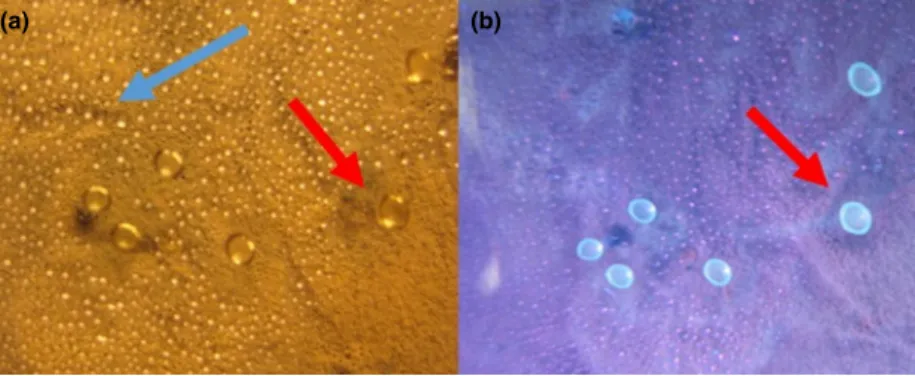
(a) (b)
Figure 2 Photographs of condensates on the lid of a Petri dish containing a 4-week-old culture ofPenicillium expansumRcP61 inspected under visible light (a) and UV-light (b). Droplets emitting blue fluorescence (red arrows) were toxic in>1009dilutions compared with the non-fluores- cent droplets (blue arrow, panel a) to boar sperm and porcine kidney cells PK-15. [Colour figure can be viewed at wileyonlinelibrary.com]
m/z4575, sodiated mass ion [M +Na]+atm/z5294, pro- tonated mass ion at m/z5115 [M +H-H20]+(represent- ing loss of water from protonated mass ion in ESI source) and potassium adduct [M +K]+atm/z5673 of chaetoglo- bosin. Peak 3 (205 min) contained protonated mass ion [M +H]+atm/z5095 and sodiated mass ion [M+Na]+ atm/z5315 of communesin B. Corresponding adduct ions of communesin A atm/z4575 (Fig. 3d), chaetoglobosin at m/z 5115, 5295, 5504 and 5674 (Fig. 3f) and com- munesin B at m/z 5095 and 5315 (Fig. 3g) in peaks 1 (44 min), 2 (170 min) and 3 (205 min), respectively, were found from a condensate ofPen. expansumRcP61.
The total ion chromatograms derived from HPLC-MS analysis of the exudate droplet (Fig. 3a) and condensate of Pen. expansum RcP61, experimental upwards set-up (P.ex.44) (Fig. 3a) were similar. Therefore, it was shown that toxic metabolites (chaetoglobosin and communesin A and B) of exudate droplets of Pen. expansum RcP61
are able to transfer by air. Similarly analysed, condensed water collected from experimental downwards set-up (P.ex.11) also showed that communesin A and chaetoglo- bosin were transferred by air.
It can be concluded, that the toxic contents of Pen.
expansum RcP61 guttation droplets aerosolized and drifted through air (10 mm) both vertically upwards and downwards from the mycelial culture to the respective cooled lids.
Figure 4 shows micrographs of the guttation droplets, visualizing guttation droplet biomass (first left), trapped on the inner surface of the lid (second left), drying of the droplets (third left) and germination of the conidia (last right) on the lid of the Petri plate.
Gypsum board is a common indoor surface material of buildings in Finland. To test ifPen. expansumRcP61 pro- duces droplets while growing on gypsum board, sections of moistened liner-covered gypsum board were seeded with
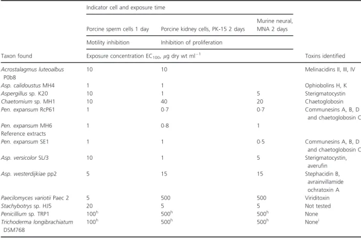
Table 2 Mammalian cell toxicities of vesicles emitted by indoor moulds isolated from offices listed in Table 1, associated with severe building-re- lated ill health of the occupants, and of reference substances
Isolate and culture age Indicator cell and exposure time
Porcine sperm cell, 1 h
Porcine kidney, PK15, 2 days
Feline lung, FFL, 2 days
Murine neural, MNA 2 days Toxicity end-point
Motility inhibition Inhibition of proliferation Penicillium expansumRcP61evesicle Highest dilution causing maximal toxic effect,9
10009 ≥20 4809 ≥10 2409 51209
Exposure concentration
EC50ll vesicle liquid ml 1(EC100,lg dry wt. ml 1)f
Pen. expansumRcP61 7 days 25 008 008 016
Pen. expansumRcP61 22 days 25 (21) 004 (≤04) 008 (<08) 016 (16)
Pen. expansumRcP61 35 dayse (42) (16) (16) (16)
Chaetomiumsp. MH52 22 days 50 25
Acrostalagmus luteoalbusPOB8 14 days 50 50
Aspergillussp. K20 13 days >50 >50 >50 >50
Asp. calidoustusMH34 14 days >50 50 >50
Reference substances
Asp. versicolorGAS/226 14 days >50 >50 >50 >50
Asp. westerdijkiaepp2 14 days >50 >50 >50 >50
Paecilomyces variotiiPaec 2 14 days >50 >50 >50 5
Pen. chrysogenumRUK2/3 exudate droplets 14 days (160) 50 (325) 50 25
Pen. chrysogenumRUK2/3 liquid squeezed from the mycelial biomass 14 daysg
(210) (210)
Pen. expansumSE1 2 01 01 2
Stachybotryssp.HJ5 14 days >50 20 20
Triclosan (mitochondriotoxic reference) (2) (8) (8) (4)
aContaining 86lg ml 1of communesins A, B, D and 480lg ml 1of chaetoglobosins.
bNumbers in brackets indicate dry weight of the liquid in the vesicle.
c22 days grown plate culture containing 4200lg ml 1of meleagrin.
spore suspension, followed by incubation inside moistened climate chambers sealed with glass lids and rubber seals.
After 6 days the gypsum boards appeared visibly mouldy, whereas after 10 days the guttation droplets were visible by naked eye (Fig. 5), having accumulated on the mouldy sur- face of the gypsum board, independently of whether the board was, or was not, autoclaved before inoculation. Bio- mass (2 mg) was collected from the surface of the gypsum liner (Fig. 5a) and the biomass lysates were found toxic to the PK-15 cells at a concentration of 05% (v/v).
Toxins in guttation droplets of reference fungi from a culture collection of toxigenic fungi isolated from buildings
The currently studied office building had no major moisture-damage and was not visibly mouldy, however, the settled dust contained Asp. versicolor-like strains and Asp. calidoustus. These strains contained highly toxic sterigmatocystin and ophiobolins in their biomass extracts and produced visible guttation droplets
(Table 3), but no toxicities were detected in the dro- plet liquids.
We also used the primary isolates of toxigenic fungi (Pen. chrysogenum RUK2/3 and Pen. expansum SE1, Stachybotrys sp. HJ5, Asp. westerdijkiae PP2, Asp. versi- color GAS226 and Pae. variotii Paec 2) isolated from indoor dusts and building materials, deposited in the HAMBI culture collection for testing toxicity and droplet formation (Table 2). Guttation droplets (dry weight con- tent 6–8 mg ml 1) were produced by isolatePen. chryso- genum RUK2/3 and contained up to 4 mg ml 1 of meleagrin. Despite the high concentration and substantial amount of the secondary metabolites (meleagrin) of the Pen. chrysogenum RUK2/3 guttation droplets, the toxic effects were modest: the droplets inhibited growth of PK- 15 100–500-fold less effectively, whereas sperm motility and proliferation of feline pulmonary cells were 5–10-fold less inhibited (Table 2). The reference strainPen. expan- sum SE1 produced chaetoglobosin and communesins A, B and D in the biomass and its guttation droplets were as toxic as those produced by the Pen. expansum strain
Table 3 Toxicity and toxins identified from ethanol extracts from biomass of indoor moulds isolated from offices (Table 1) and of reference extracts
Taxon found
Indicator cell and exposure time
Toxins identified Porcine sperm cells 1 day Porcine kidney cells, PK-15 2 days
Murine neural, MNA 2 days Motility inhibition Inhibition of proliferation
Exposure concentration EC100,lg dry wt ml 1 Acrostalagmus luteoalbus
P0b8
10 10 Melinacidins II, III, IV
Asp. calidoustusMH4 1 1 Ophiobolins H, K
Aspergillussp.K20 10 1 5 Sterigmatocystin
Chaetomiumsp.MH1 10 40 20 Chaetoglobosin
Pen. expansumRcP61 1 07 07 Communesins A, B, D
and chaetoglobosin C
Pen. expansumMH6 1 08 1
Reference extracts
Pen. expansumSE1 1 1 05 Communesins A, B, D
and chaetoglobosin C
Asp. versicolorSL/3 10 1 5 Sterigmatocystin,
averufin
Asp. westerdijkiaepp2 5 15 15 Stephacidin B,
avrainvillamide ochratoxin A
Paecilomyces variotiiPaec 2 5 500 500 Viriditoxin
Stachybotryssp.HJ5 20 5 5 Not tested
Penicilliumsp.TRP1 100h 500h 500h None
Trichoderma longibrachiatum DSM768
100h 500h 500h Nonei
aRepresented the nonspecific upper limit of the assays.
bDetection limit 001 mg ml 1.
0·0 2 Intens.
(a)
(b)
(c)
(d)
(e)
(f)
(g)
(h)
×107
Intens.
Intens. +MS, 4·4min
+MS, 4·6min
+MS, 17·0min
+MS, 20·5min
+MS, 20·5min +MS, 17·0min 0·5
0
0
0 2
4 430
433·3
439·3
443·4 449·4 453·3 467·3
463·3
567·3
567·4 471·5 475·2 479·4
471·4 475·3 479·4
Communesin A
Communesin A
Communesin B
Communesin B Chaetoglobosin
Chaetoglobosin 489·3 457·5 [M+H]+
551·4 [M+Na]+
[M+Na]+
[M+Na]+ 551·4 [M+Na]+
[M+K]+
[M+K]+ 511·4 [M+H-H20]+
511·5 [M+H-H20]+
529·4 [M+H]+
529·5 [M+H]+
509·5 [M+H]+
509·5 [M+H]+ 457·5 [M+H]+ 439·1
440 450 460 470 480
430
510 520 530 540 550 560
510
460 470 480 490 500 510 520
523·5
523·5
531·5
531·5
539·5
539·5
551·5
551·4
530 540 550
460 470 480 490 500 510 520 530 540 550 560m/z
520 530 540 550 560 570
570
440 450 460 470 480
m/z
m/z
m/z
m/z
m/z 1
0 1
0 1
×108
×105
Intens.
Intens.
Intens.
Intens.
×105
×106
×106
×105
0 1 Intens.
×105
HPLC-MS analysis of the boar sperm cell toxic exudate droplet of P.expansum
HPLC-MS analysis of the condensate of P.expansum
Mass spectrum of peak 1 (exudate droplet of P.expansum)
Mass spectrum of peak 1 (condensate of P.expansum)
Mass spectrum of peak 2 (exudate droplet of P.expansum)
Mass spectrum of peak 2 (condensate of P.expansum)
Mass spectrum of peak 3 (exudate droplet of P.expansum)
Mass spectrum of peak 3 (condensate of P.expansum)
2·5 5·0 7·5 10·0
1
1
2
2
3
3
12·5 15·0 17·5 20·0 22·5 Time [min]
0·0 2·5 5·0 7·5 10·0 12·5 15·0 17·5 20·0 22·5 Time [min]
Figure 3 Comparison of HPLC-ESI-MS analyses of the exudates in guttation droplets and vapour condensates ofPenicillium expansumRcP61 from Fig. 2 (P.ex.44). HPLC chromatograms of the exudate droplets (a) and of the vapour condensates (b). The peaks 1, 2, 3 in panels a and b were identified as communesin A (peak 1), chaetoglobosin (peak 2) and communesin B (peak 3). Patulin was not measured. Molecular ion [M + H]+of peak 1 ism/z= 4575 (c, d), of peak 2 ism/z= 5514 (e, f) and of peak 3 ism/z5095 (g, h).
RcP61. The guttation droplets from other reference strains of toxigenic indoor isolates, viriditoxin-producing Pae. variotii Paec 2, and Stachybotrys sp. HJ5 producing yet unidentified toxins were 100 times less toxic than the guttation droplets of the Pen. expansum reference strains SE1 and Pen. expansum strain RcP61. Summarizing the results, we established that an indoor strain of Pen.
expansumemitted substantial amounts of toxins in gutta- tion droplets, furthermore, the toxins migrated aerially.
Discussion
Penicillium expansum is known to produce some of the most potent mycotoxins within the genusPenicillium, the communesins and chaetoglobosins, which are produced when growing on fruits (Andersenet al.2004; Nielsenet al.
2004). We report here for the first time thatPen. expansum strains isolated from building material and dust from an office associated with health complaints also produced communesins and chaetoglobosins. Furthermore, we demonstrated that these toxins migrated through the air (Figs 1 and 2, Table 4). Grown on laboratory medium, the in vitro- and ex vivo-measured specific toxicity of Pen.
expansum emitted in guttation droplets was 100 times
higher than those guttation droplets of toxic indoor iso- lates of Aspergillus, Chaetomium, Acrostalagmus, Pae- cilomyces and Stachybotrys,and is, to our knowledge, the highest among the indoor moulds reported to date. We also show that the strain produced guttation droplets when growing on gypsum liner, and that lysates of biomass grown on gypsum liner, containing guttation droplets and conidia were toxicin vitro.The toxicity value was five times lower than the threshold value for toxicity defined for the screening test for microbial biomass lysates.
The term guttation has long been known as a virulence mechanism of phytopathogenic and entomopathogenic micro-organisms (Hutwimmer et al. 2010; Singh and Singh 2013; Singh 2014).
Culture collection isolates ofPen. verrucosum and Pen.
nordicumwere reported to emit droplets containing 001–
9lg ml 1 of ochratoxins A and B, which is 7–10-fold higher than the concentrations in the mycelial mass of the producer fungus (Gareis and Gareis 2007). For indoor fungi, toxic emission of guttation droplets have been hitherto sparsely reported. Stachybotrys sp. chemo- type S culture collection strains originating from various indoor habitats were reported to produce droplets con- taining 3–5lg ml 1 satratoxin G and H per ml (Gareis 1 mm
Figure 4Life-cycle of toxin-containing vesicles fromPenicillium expansumRcP61. From left to right: a large, amber-coloured vesicle extrudes from the mycelial biomass ofPen. expansumRcP61. The vesicle meets air convection, becomes airborne, hits the polypropylene lid of the culture plate, among tiny droplets of airborne moisture (2ndfrom left). Air moisture is low, RH 30%, the droplet empties and desiccates (3rdfrom left). Nine days later, the vesicle has propagated a new generation ofPen. expansumconidiophores (last right). [Colour figure can be viewed at wileyonlinelibrary.com]
1 cm
(a) (b) (c)
100 cm 70 µm
Figure 5Penicillium expansumRcP61 mycelium grown on gypsum board emits guttation droplets. (a) Visible mould growth, 3 weeks, on the gypsum plate; (b) stereo micrographs of thePen. expansumconidiophores; (c) amber-coloured guttation droplet (arrow) extruding from the mycelial biomass grown on the gypsum board (a). [Colour figure can be viewed at wileyonlinelibrary.com]
and Gottschalk 2014). Gareis and Gottschalk (2014) first observed that guttation droplets of Stachybotrys char- tarum chemotype S contained roridins and satratoxins and produced low (02–04 ng m 3) but measurable air- borne concentrations of satratoxins G and H75.
It is impossible to directly predict in vivo mammalian toxicity based onin vitrotoxicity results. In this study we used in vitroand ex vivotoxicity tests to compare toxici- ties of biomass extracts and guttation droplets produced by selected fungal isolates. Consistently high toxicity was obtained with a continuous lung cell line, FFL, a continu- ous kidney epithelial cell line, PK-15, a malignant cell line, MNA, as well as with an ex vivo assay using boar sperm for Pen. expansum strains RcP61 (CBS 145620) and SE1. Using bioassays in combination with chemical analysis we were able to identify the toxins as chaetoglo- bosins and communesins. The cork liner used as isolation material in the office building may have been the source of Pen. expansum. Penicillium spp. are reported as com- mon contaminants in both moisture-damaged and non- damaged indoor spaces (Pasanen et al. 1992; Gravesen et al. 1995; Kaarakainenet al. 2009; Salonen 2009; Adan et al. 2011; Andersen et al. 2011; Nielsen and Frisvad 2011; Samson 2011), but little attention has been paid to Pen. expansum and emissions of chaetoglobosins and communesins in indoor air and their potential associa- tion with adverse health effects in moisture damaged buildings.
Using chicken tracheas, Pieckova and Wilkins (2004) have demonstrated that indoorChaetomiumsp. produced very potent, ciliostatically active metabolites. Mycotoxins from indoor Chaetomium globosum strains and pure chaetoglobosin A from C. globosum also inhibited sperm motility by a sublethal ciliostatic mechanism, very likely by inhibiting sugar transport affecting glycolytic and mitochondrial energy production (Vicente-Carrillo et al.
2015; Castagnoliet al.2017; Castagnoliet al.2018b). The chaetoglobosin-containing guttation droplets inhibited sperm motility, possibly indicating ciliostatic activity.
Thus, the risk of respiratory toxicity represented by air- borne chaetoglobosin shown toxic to the primary lung cell line FFL and exhibiting ciliostatic activity is highly
speculative, but cannot be excluded. The risk of airborne respiratory toxicity can be directly evidenced by in vivo experiments exposing laboratory animals to known air- borne concentrations of chaetoglobosins and commune- sisns, which was out of scope for this article.
Chaetoglobosins known as cytochalasins (McMullin et al. 2013) exert their cytotoxicity by capping the grow- ing end of actin microfilaments, thereby destroying the cytoskeleton of mammalian cells (Ueno 1985; Scherlach et al. 2010). Communesins are neurotoxic, insecticidal indole alkaloids (Hayashi et al. 2004; Kerzaon et al.
2009). Chaetoglobosin B was reported to be toxic to human erythrocyte membranes by competitively inhibit- ing glucose transport activity (Scherlach et al. 2010).
Inhibition of glucose transport was also reported for chaetoglobosin A (Nielsen and Frisvad 2011). Thus, it is possible that the observed complete blocking of prolifera- tion of the somatic cells PK-15, FFL and MNA (Table 2), as well as the motility inhibition of boar sperm by expo- sure to thePen. expansumRcP61 vesicle fluid was caused by blocked glucose transport depriving the cells of energy.
This study is, to our knowledge, the first where a Pen.
expansumstrain, RcP61 (CBS 145620), growing on build- ing material inside the construction in a building with reported health complaints, was shown to produce toxic guttation droplets on laboratory media and on gypsum liner. Toxin concentrations in guttation droplets emitted by Pen. expansum grown on laboratory medium, in the current work, appear 100–1000-fold higher than previ- ously demonstrated from any fungus.
The weakened immune tolerance reported from urban environments caused by depleted and poor outdoor microbial diversity (Moore 2015; Schuijs et al.2015; von Hertzen et al. 2015; Haahtela et al. 2015; Adams et al.
2016; Mhuireach et al. 2016; Stein et al. 2016; Li et al.
2018) may be attenuated by exposure to indoor microbes producing toxins inducing loss of tolerance (TILT; Miller 1997; Genius 2010). We were tempted to speculate that the absence of a diverse protective microbiome in combi- nation with exposure to microbial TILT may be a poten- tial factor to contribute to the diverse indoor air-related
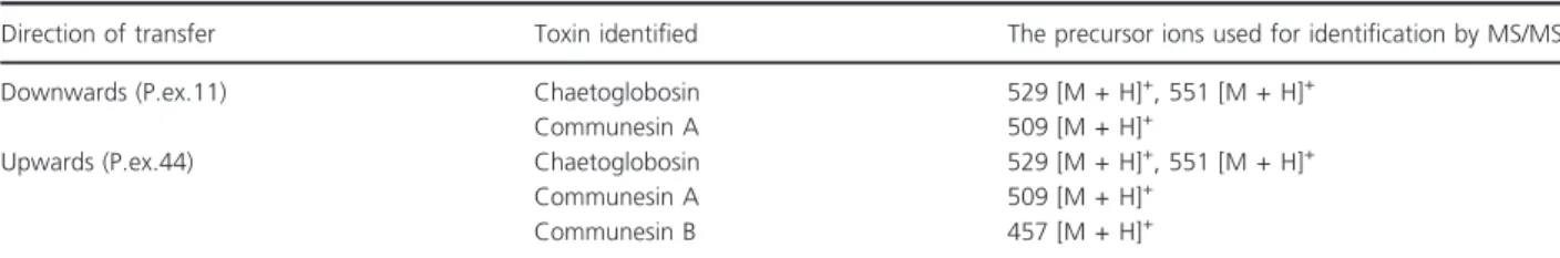
Table 4 The airborne transit of mycotoxins contained in exudate droplets ofPenicillium expansumRcP61 from the mycelial mass towards a cool surface in the experimental set-up shown in Fig. 4
Direction of transfer Toxin identified The precursor ions used for identification by MS/MS
Downwards (P.ex.11) Chaetoglobosin 529 [M + H]+, 551 [M + H]+
Communesin A 509 [M + H]+
Upwards (P.ex.44) Chaetoglobosin 529 [M + H]+, 551 [M + H]+
Communesin A 509 [M + H]+
Communesin B 457 [M + H]+
health symptoms experienced in mould-damaged urban buildings.
Results of this study call for continued research on how mycotoxin-containing guttation droplets can be spread in indoor air. The toxicity and migration of gutta- tion droplets in the air were shown in this study, but their spreading in the rooms needs to be studied in future research.
Acknowledgements
The authors warmly thank Riikka Holopainen at the Fin- nish Food Safety Authority (EVIRA) for providing the somatic cell lines PK-15, FFL and MNA. The authors thank Prof. Tari Haahtela, Prof. Mirja Salkinoja-Salonen and Prof. Martti Viljanen for their valuable advice and support during the work. The authors acknowledge the financial support of the Finnish Work Environment Fund by grant #112134, the Academy of Finland grants
#253727 and #289161 and the Business Finland (the Fin- nish Funding Agency for Innovation, grant number 4098/
31/2015). LK was supported by the GINOP-23.2-15- 2016-00012 grant (Szechenyi 2020 Programme, Hungary) and the Janos Bolyai Research Scholarship (Hungarian Academy of Sciences).
Conflict of Interest
No conflict of interest declared.
References
Adams, R., Bhangar, S., Dannemiller, K., Eisen, J., Fierer, N., Gilbert, L., Green, J., Linsey, C.et al. (2016) Ten questions concerning the microbiomes of buildings.Build Environ109, 224–234.
Adan, O.C.G., Huinink, H.P. and Bekker, M. (2011) Water relations of fungi in indoor environments. In
Fundamentals of Mold Growth in Indoor Environments and Strategies for Healthy Livinged. Adan, O.C.F. and Samson, R.A. pp. 41–65. Wageningen: Wageningen Academic Publishers.
Ajao, C., Andersson, M., Teplova, V.V., Nagy, S., Gahmberg, C.G., Andersson, L.C., Hautaniemi, M., Kakasi, B.et al.
(2015) Mitochondrial toxicity of triclosan.Toxicol Rep2, 624–637.
Andersen, B., Smedsgaard, J. and Frisvad, J.C. (2004) Penicillium expansum: consistent production of patulin, chaetoglobosins and other secondary metabolites in culture and their natural occurrence in fruit products.J Agric Food Chem52, 2451–2428.
Andersen, B., Frisvad, J.C., S€ondergaard, I., Tarmussen, I.S.
and Larsen, L.S. (2011) Associations between fungal
species and water-damaged building materials.Appl Environ Microbiol77, 4180–4188.
Andersson, M.A., Mikkola, R., Raulio, M., Kredics, L., Maijala, P. and Salkinoja-Salonen, M.S. (2009) Acrebol, a novel toxic peptaibol produced by anAcremonium exuviarum indoor isolate.J Appl Microbiol106, 909–923.
Annila, P.J., Hellemaa, M., Pakkala, T., Lahdensivu, J., Suonketo, J. and Pentti, M. (2017) Extent of moisture and mould damage in structures of public buildings.Case Stud Constr Mater6, 103–108.
Bencsik, O., Papp, T., Berta, M., Zana, A., Forgo, P., Dombi, G., Andersson, M.A., Salkinoja-Salonen, M.S.et al. (2014) Ophiobolin A fromBipolaris oryzaeperturbs motility and membrane integrities of porcine sperm and induces cell death on mammalian somatic cell lines.Toxins6, 2857– 2871.
Bennet, J. and Inamdar, A. (2015) Are some volatile organic compound (VOCs) mycotoxins?Toxins7, 3785–3804.
Caillaud, D., Leynaert, B., Keirsbulck, M. and Nadif, R. (2018) Indoor mould exposure, asthma and rhinitis: findings from systematic reviews and recent longitudinal studies - a review.Eur Respir Rev27, 170–199.
Castagnoli, E., Andersson, M.A., Mikkola, R., Kredics, L., Marik, T., Kurnitski, J. and Salonen, H. (2017). Indoor Chaetomium-like isolates: resistance to chemichals, fluorescence and mycotoxin production. Conference paper: Sis€ailmastoseminaari Volume: SYI report 35 March 2017 Helsinki, Finland.
Castagnoli, E., Marik, T., Mikkola, R., Kredics, L., Andersson, M.A., Salonen, H. and Kurnitski, J. (2018a) Indoor Trichodermastrains emitting peptaibols in guttation droplets.J Appl Microbiol125, 1408–1422.
Castagnoli, E., Salo, J., Toivonen, M.S., Marik, T., Mikkola, R., Kredics, L., Vicente-Carrillo, A., Nagy, S.et al. (2018b) An evaluation of boar spermatozoa as a biosensor for the detection of sublethal and lethal toxicity.Toxins10, 463.
Duncan, S., Farrell, R., Thwaites, J., Held, B., Arenz, B., Jurgens, A. and Blanchette, R. (2006) Endoglucanase- producing fungi isolated from Cape Evans historic expedition hut on Ross Island, Antarctica.Environ Microbiol8, 1212–1219.
Echard, G. (1974) Chromosomal banding patterns and karyotype evolution in three pig kidney cell strains (PK- 15, F and RP).Chromosoma45, 133–149.
Gareis, M. and Gareis, E.-M. (2007) Guttation droplets of Penicillium nordicumandPenicillium verrucosumcontain high concentrations of the mycotoxins ochratoxin A and B.Mycopathologia163, 207–214.
Gareis, M. and Gottschalk, C. (2014)Stachybotrysspp. and the guttation phenomenon.Mycotox Res30, 151–159.
Genius, S.J. (2010) Sensitivity-related illness: the escalating pandemic of allergy, food intolerance and chemical sensitivity.Sci Total Environ408, 6047–6061.
Gravesen, S., Frisvad, J.C. and Samson, R.A. (1995) Important moulds in damp buildings. InMicrofungied. Gravesen, S.,
Frisvad, J.C. and Samson, R.A. pp. 15–26. København:
Munksgaard.
Haahtela, T., Laatikainen, T., Alenius, H., Auvinen, P., Fyhrquist, N., Hanski, I., von Hertzen, L., Jousilahti, P.
et al. (2015) Hunt for the origin of allergy - comparing the Finnish and Russian Karelia.Clin Exp Allergy45, 891–
901.
Hayashi, H., Matsumoto, H. and Ariyama, K. (2004) New insecticidal compounds, communesins C, D and E. from Penicillium expansumLink MK-57.Biosci Biotechnol Biochem68, 753–756.
von Hertzen, L., Beutler, B., Bienenstock, J., Blaser, M., Cani, P.D., Eriksson, J., F€arkkil€a, M., Haahtela, T.et al. (2015) Helsinki alert of biodiversity and health.Ann Med47, 218–225.
Hong, S.B., Cho, H.S., Shin, H.D. and Frisvad, J.C. (2006) NovelNeosartoryaspecies isolated from soil in Korea.Int J Syst Evol Microbiol56, 477–486.
Hurraß, J., Heinzow, B., Aurbach, U., Bergmann, K.-C., Bufe, A., Buzina, W., Cornely, O., Engelhart, S.et al.(2016) Medical diagnostics for indoor mold exposure.Int J Hyg Environ Health220, 305–328.
Hutwimmer, S., Wang, H. and Strasser, H. (2010) Formation of exudate droplets byMetarhizium anisopliaeand the presence of destruxins.Mycologia102, 1–10.
Julca, I., Droby, S., Sela, N., Marcet-Houben, M. and
Gabaldon, T. (2015) Contrasting genomic diversity in two closely related postharvest pahogens:Penicillium digitatum andPenicillium expansum.Genome Biol Evol8, 218–227.
Kaarakainen, P., Rintala, H., Veps€al€ainen, A., Hyv€arinen, A., Nevalainen, A. and Meklin, T. (2009) Microbial content of house dust samples determined with qPCR.Sci Total Environ407, 4673–4680.
Kenne, G.J., Chakraborty, P. and Chanda, A. (2014) Modeling toxisome protrusions in filamentous fungi.JSM Environ Sci Ecol2, 1010–1012.
Kerzaon, I., Pouchus, Y.F., Monteau, F., Le Bizec, B., Nourisson, M.-R., Biard, J.-F. and Grovel, O. (2009) Structural investigation and elucidation of new
communesins from marine-derivedPenicillium expansum Link by liquid chromatography/electrospray ionization mass spectrometry.Rap Commun Mass Spectrom23, 3928–3938.
Kistler, H. and Broz, K. (2015) Cellular compartmentalization of secondary metabolism.Front Microbiol6, 68.
Korkalainen, M., Naarala, J., Kirjavainen, P., Koistinen, A., Hyv€arinen, A., Komulainen, H. and Viluksela, M. (2017) Synergistic proinflammatory interactions of microbial toxins and structural components characteristic to moisture-damaged buildings.Indoor Air27, 13–23.
Li, D.-W., Johanning, E. and Yang, C. (2015) In Airborne fungi and mycotoxins. Chapter 3.2.5. InManual of Environmental Microbiology(4th edn) ed. Yates, M., Nakatsu, C., Miller, R. and Pillai, S. Washington DC:
ASM Press.
Li, G., Sunc, G.-X., Rena, Y., Luo, X.-S. and Zhua, Y.-G.
(2018) Urban soil and human health: a review.Eur J Soil Sci69, 196–215.
McMullin, D.R., Sumarah, M.W. and Miller, J.D. (2013) Chaetoglobosins and azaphilones produced by Canadian strains ofChaetomium globosumisolated from the indoor environment.Mycotox Res29, 47–54.
Mendell, M.J. and Kumagai, K. (2017) Observation-based metrics for residential dampness and mold with dose– response relationships to health.Indoor Air27, 506–517.
Mendell, M.J., Mirer, A.G., Cheung, K., Tong, M. and Douwes, J. (2011) Respiratory and allergic health effects of dampness, mold, and dampness-related agents: a review of the epidemiologic evidence.Environ Health Perspect119, 748–756.
Mhuireach, G., Johnson, B.R., Adam, E., Altrichter, A.E., Ladau, J., Meadow, J.F., Pollard, K.S. and Green, J.L.
(2016) Urban greenness influences airborne bacterial community composition.Sci Total Environ571, 680–687.
Mikkola, R., Andersson, M.A., Kredics, L., Grigoriev, P., Sundell, N. and Salkinoja-Salonen, M. (2012) 20-Residue and 11-residue peptaibols from the fungusTrichoderma longibrachiatumare synergistic in forming Na+/K+ permeant channels and in adverse action towards mammalian cells.FEBS J279, 4172–4190.
Mikkola, R., Andersson, M.A., Hautaniemi, M. and Salkinoja- Salonen, M. (2015) Toxic indole alkaloids avrainvillamide and stephacidin B produced by a biocide tolerant indoor moldAspergillus westerdijkiae.Toxicon99, 58–67.
Miller, C.S. (1997) Toxicant induced loss of tolerance - an emerging theory of disease?Environ Health Perspect105, S445–S453.
Moore, M. (2015) Do airborne biogenic chemicals interact with the PI3K/Akt/mTOR cell signalling pathway to benefit human health and wellbeing in rural and coastal environments.Environ Res140, 65–75.
Nielsen, K.F. (2003) Mycotoxin production of indoor molds.
Fungal Genet Biol39, 103–117.
Nielsen, K.F. and Frisvad, J.C. (2011) Mycotoxins on building material. InFundamentals of Mold Growth. Indoor Environments and Strategies for Healthy Livinged. Adan, O.C.F. and Samson, R.A. pp. 245–275. Wageningen:
Wageningen Academic Publishers.
Nielsen, K.F., Nielsen, P.A., Holm, G. and Uttrup, L.P. (2004) Mold growth on building materials under low water activities influence of humidity and temperature on fungal growth and secondary metabolites.Int Biodeter Biodegr54, 325–336.
Norb€ack, D. and Miller, J.D. (2013) Building-related illnesses and mold related conditions. InAsthma in the Workplace, (4th edn) ed. Bernstein, D.I., Malo, J.-L., Chan Yeung, M.
and Bernstein, L. pp. 406–417. New York: Taylor &
Francis.
Pasanen, A.L., Heinonen-Tanski, H. and Kalliokoski, P. (1992) Fungal microcolonies on indoor surfaces–an explanation
for the base-level fungal spore counts in indoor air.Atmos Environ26B, 117–120.
Pieckova, E. and Wilkins, K. (2004) Airway toxicity of house dust and its fungal composition.Ann Agric Environ Med 11, 67–73.
Pildain, M.B., Frisvad, J.C., Vaamonde, G. and Cabral, D.
(2008) Two novel aflatoxin-producingAspergillusspecies from Argentinean peanuts.Int J Syst Evol Microbiol58, 725–735.
Salo, J., Andersson, M.A., Mikkola, R., Kredics, L., Viljanen, M. and Salkinoja-Salonen, M. (2015) Vapor as a carrier of toxicity in a health troubled building.Proceedings of Healthy Buildings 2015–Europe (ISIAQ International), Eindhoven, The Netherlands, Paper ID526, 8 pp E.1 Sources & Exposure, Source control.
Salonen, H. (2009)Indoor air contaminants in office buildings.
(Dissertation). Finnish Institute of Occupational Health.
People and Work Research Report 87, 222 p.
Samson, R.A. (2011) Ecology and general characteristics of indoor fungi. InFundamentals of Mold Growth in Indoor Environments and Strategies for Healthy Livinged. Adan, O.C.F. and Samson, R.A. pp. 101–116. Wageningen:
Wageningen Academic Publishers.
Samson, R.A., Hoekstra, E.S., Frisvad, J.C. and Filtenborg, O.
(2002) Eds.Introduction to food and air-borne fungi(6th edn). Utrecht: Centraalbureau voor Schimmelcultures.
Scherlach, K., Boettger, D., Remme, N. and Hertweck, C.
(2010) The chemistry and biology of cytochalasans.Nat Prod Rep27, 869–886.
Schuijs, M.J., Willart, M.A., Vergote, K., Gras, D., Deswarte, K., Ege, M.J., Madeira, F.B., Beyaert, R.et al.(2015) Farm dust and endotoxin protect against allergy through A20 induction in lung epithelial cells.Science4, 1106–1110.
Singh, S. (2014) Guttation: quantification, microbiology and implications for phytopathology. InProgress in Botany 75,
ed. L€uttge, U., Beyschlag, W. and Cushman, J. pp. 187– 214. Berlin: Springer-Verlag, Berlin Heidelberg.
Singh, S. and Singh, T.N. (2013) Guttation 1: Chemistry, crop husbandry and molecular farming.Phytochem Rev12, 147–172.
Stein, M.M., Cara, B.S., Hrusch, L., Justyna Gozdz, J., Igartua, C., Vadim Pivniouk, V., Murray, S., Julie, G.et al. (2016) Innate immunity and asthma risk in Amish and Hutterite farm children.New Engl J Med375, 411–421.
T€ahtinen, K., Lappalainen, S., Karvala, K., Lahtinen, M. and Salonen, H. (2019) Probability of abnormal indoor air exposure categories compared with occupants’ symptoms, health information, and psychosocial work environment.
Appl Sci9, 99.
Tannous, J., Atoui, A., Khoury, A., Francis, Z., Oswald, I., Puel, O. and Lteif, R. (2016) A study on the
physicochemical parameters forPenicillium expansum growth and patulin production: effect of temperature, Ph, and water activity.Food Sci Nutr4, 611–622.
Tuuminen, T. and Lohi, J. (2019) Immunological and toxicological effects of bad indoor air to cause dampness and mold hypersensitivity syndrome.Allergy Immunol2, 190–204.
Ueno, Y. (1985) Toxicology of mycotoxins.Crit Rev Toxicol 14, 99–103.
Vicente-Carrillo, A., Edebert, I., Garside, H., Cotgreave, I., Rigler, R., Loitto, V., Magnusson, K.E. and Rodrıguez- Martınez, H. (2015) Boar spermatozoa successfully predict mitochondrial modes of toxicity: implications for drug toxicity testing and the 3R principles.Toxicol In Vitro29, 582–591.
Zayne, M., Mortabit, D., Mostakin, M., Iraqui, M., Haggoud, A., Haggoud, M., Ettayebi, S. and Koraichi, I. (2009)
Cellulolytic potential of fungi in wood degradation from an old house at the Medina of Fez.Ann Microbiol59, 699–704.