O R I G I N A L A R T I C L E
Indoor Trichoderma strains emitting peptaibols in guttation droplets
E. Castagnoli1 , T. Marik2, R. Mikkola1 , L. Kredics2 , M.A. Andersson1,3, H. Salonen1 and J. Kurnitski1,4
1 Department of Civil Engineering, Aalto University, Espoo, Finland
2 Department of Microbiology, Faculty of Science and Informatics, University of Szeged, Szeged, Hungary 3 Department of Food and Environmental Science, Helsinki University, Helsinki, Finland
4 Department of Civil Engineering and Architecture, Tallinn University of Technology, Tallinn, Estonia
Keywords
ecotoxicity, environmental, fungi, moulds, mycotoxins.
Correspondence
Emmanuelle Castagnoli, Department of Civil Engineering, Aalto University, Rakenta- janaukio 4, 02150 Espoo, Finland.
E-mail: emmanuelle.castagnoli@aalto.fi 2018/0641: received 17 October 2017, revised 29 March 2018 and accepted 9 May 2018
doi:10.1111/jam.13920
Abstract
Aims: The production of peptaibols, toxic secondary metabolites of Trichoderma, in the indoor environment is not well-documented. Here, we investigated the toxicity of peptaibols in the guttation droplets and biomass of Trichodermastrains isolated from problematic buildings.
Methods and Results: Seven indoor-isolated strains of T. atroviride, T. trixiae, T. paraviridescens and T. citrinoviride were cultivated on malt extract agar, gypsum boards and paperboards. Their biomass extracts and guttation droplets were highly cytotoxic in resting and motile boar sperm cell assays and in inhibition of somatic cell proliferation assays. The toxins were identified with HPLC/ESI-MS/MS as trichorzianines, trilongins, trichostrigocins and trichostrigocin-like peptaibols. They exhibited toxicity profiles similar to the reference peptaibols alamethicin, trilongins, and trichorzianine TA IIIc purified from T. atroviride H1/226. Particular Trichoderma strains emitted the same peptaibols in both their biomasses and exudate droplets. The trilongin- producingT. citrinovirideSJ40 strain grew at 37°C.
Conclusions: To our knowledge, this is the first report of indoor-isolated Trichodermastrains producing toxic peptaibols in their guttation droplets.
Significance and Impact of the Study: This report proves that indoor isolates of Trichoderma release peptaibols in their guttation droplets. The presence of toxins in these types of exudates may serve as a mechanism of aerosol formation for nonvolatile toxins in the indoor air.
Introduction
The excessive moisture resulting from the water damage in buildings may change the typical diversity of indoor micro- biota wherePenicilliumandAspergillusare the usual domi- nant fungal genera (Nielsen 2003). Species likeTrichoderma can grow on wet wooden materials and plywood colonized by other fungi, and therefore are indicators of high mois- ture content in buildings (Gravesen et al. 1999; Andersen et al. 2011; Druzhinina et al. 2011; Kubicek et al. 2011;
Samson 2011; Mikkolaet al.2012; Mukherjeeet al.2013).
The presence of fungi indoors increases the risk of human infections due to inhalation of viable fungal
fragments and small conidia (≤4lm) moving from the building structure to the indoor air (Airaksinen et al.
2004; Straus 2009). Human pathogenic infections caused byTrichodermahave been increasingly reported in the lit- erature (Mikkolaet al.2012; Hatvaniet al.2013) with T.
longibrachiatum and T. citrinoviride the most frequently reported clinically relevantTrichodermaspecies.
Exudation is a well-known phenomenon of plants and fungi. Fungal exudation may occur during mycelial growth and is suggested to be a mean to expel waste prod- ucts or an available water reservoir (Hutwimmer et al.
2009; Gareis and Gottschalk 2014). Fungal exudates con- tain proteins, mycotoxins (toxic secondary metabolites)
and exhibit enzymatic activities (Gareis and Gareis 2007).
However, further studies are needed to determine the exact composition of exudates, the specific roles of exuda- tion and to examine whether exudates are possible carriers of toxins in the indoor air.
Peptaibols form a group of bioactive secondary metabolites, mainly produced by Trichoderma species, with antibacterial, antiviral and antifungal activities (Panizel et al. 2013). They have a structure composed of peptides of 5–20 amino acids includinga-amino-isobuty- ric acid, an acetylated N-terminus and an amino alcohol at the C-terminus (Leitgeb et al. 2007; Bohemen et al.
2016). A single Trichoderma species may produce up to five different types of peptaibols, while different Tricho- derma species may produce the same peptaibols (Her- mosa et al.2014). Even though peptaibols are known for their specific effect in biomembranes, their roles remain unclear (Mukherjee et al. 2010). Trilongins produced by indoor Trichoderma strains were shown to form potas- sium- and sodium-selective channels in artificial biomem- branes (Mikkolaet al.2012).
Boar semen bioassays are capable of detecting toxins which disrupt cation homeostasis by affecting the func- tion of the plasma membrane (Vicente-Carrillo 2018).
These bioassays have been used for screening the toxicity of indoor samples and exhibited high sensitivity for screening toxins like peptaibols (Peltola et al. 2004;
Andersson et al. 2010). Marik et al. (2016) have shown that boar semen bioassays were more sensitive than lung cells when screening peptaibol toxicity.
The pathogenic potential, production of toxic metabo- lites and emission mechanisms of Trichodermapeptaibols in the indoor environment are poorly understood. To the best of our knowledge, the secretion of peptaibols in exu- dated guttation droplets ofTrichodermahas not yet been reported in the literature. The aim of this study was to investigate the presence and toxicity of peptaibols in the extract of biomass and the exudates of Trichoderma strains isolated from buildings where occupants reported indoor air-related symptoms.
Materials and methods
Fungal strains
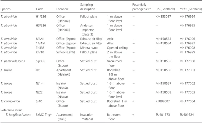
The Trichodermastrains were isolated from five buildings located in different Finnish cities where occupants reported indoor air-related symptoms and illnesses. Sam- pling details of the collected material, dust and air sam- ples are shown in Table 1.
Material samples from exhaust air filter and mineral wool (between inner and outer ceiling) were collected in sterile plastic bags. Pieces of material samples
(c.1 cm91 cm) were spread on malt extract agar (MEA) plates (15 g malt extract from Sharlab, Barcelona, Spain, and 12 g of agar from Amresco, Dallas, USA, in 500 ml of H2O). Dust samples were swept from surfaces (c.30930 cm2) above floor level (1–2 m) with a sterile paper tissue. Floor dust was collected with a vacuum clea- ner (Volta Equipt with Volta Equipt vacuum bags), the dust was removed from the vacuum cleaner bag with a sterile disposable spoon and placed into a sterile plastic bag. The dust (c.10 mg) was spread with a sterile cotton swab on MEA plates. Air samples were collected with six- stage Andersen Impactor on MEA plates during 10 min at 1 m above the floor level, and with MEA fallout plates kept open 1 h at 1–15 m above the floor level (Andersen 1958).
Malt extract agar culture plates were inoculated, sealed and cultivated at 22°C for 4 weeks. Fungal colonies sus- pected to belong to the genusTrichodermabased on colony morphology and the characteristic conidiophores visible in the light microscope were rapidly screened for toxicity and the toxic colonies were pure cultured on MEA plates.
Extraction of ethanol-soluble compounds from biomass and collection of guttation droplets from MEA-cultured Trichodermaisolates
Fungal biomass (c. 100 mg wet wt) containing hyphae and conidia (no guttation droplets visible under UV light in stereomicroscope, 1609 magnification) was extracted with ethanol, as described by Andersson et al. (2010), after 2 weeks of incubation at 22°C of the MEA plates.
Exudate vesicles fluorescent under UV light appeared on MEA plates after 1 week of incubation, at the begin- ning of sporulation. Exudates with a volume of 1–5ll were collected under UV light (360 nm), mixed with an equal volume of ethanol (96%, all the chemicals were purchased from local suppliers) and heated for 10 min at 80°C in a water bath. The exudates collected from MEA plates into glass ampules, 20–200 ll per plate, contained no hyphae or conidia when inspected with phase contrast microscope (Olympus CKX41, Tokyo, Japan; 4009 mag- nification). The ethanol-soluble compounds from bio- masses and exudate suspensions were used to expose the test cells in the toxicity assays.
Cultivation and extraction ofTrichoderma atroviride colonies grown on gypsum boards and paperboards Purchased pieces of gypsum boards and paperboards of 25 cm2 were autoclaved, saturated with sterile water and inoculated with conidia of T. atroviride strains 14/AM, H1/226 and H3/226 (200ll of phosphate buffered saline, PBS, containing c.106 conidia per ml). The inoculated paperboards and gypsum boards were incubated at room
temperature for 4 weeks inside Petri dishes sealed with gas-permeable tape. The Petri dishes were inspected weekly under the stereomicroscope and sterile water was added to maintain the moisture content of the gypsum boards and paperboards. Twenty to 50 mg (wet wt) of collected fungal material (including conidia, hyphae and guttation droplets) from theT. atroviride strains 14/AM, H1/226 and H3/226 cultivated on gypsum boards and paperboards for 2–4 weeks were extracted with ethanol as described by Anderssonet al.(2010).
Identification of fungal strains
The suspected Trichoderma strains were deposited in the Szeged Microbiology Collection (http://www.szmc.hu). Total DNA was extracted from the strains’ cultures grown on yeast extract—glucose agar medium (05 g l1yeast extract, 10 g l1 glucose and 20 g l1 agar) using the GenElute Plant Genomic DNA Miniprep Kit (Sigma-Aldrich, St.
Louis, MO). A nuclear rDNA region containing the internal transcribed spacers 1 and 2 (ITS 1 and 2) and the 58S rRNA gene was amplified with primers ITS1 (50- CCGTAGGTAACCTGCGG-30) and ITS4 (50-TCCTCCGC- TTATTGATATGC-30) (White et al. 1990; Naeimi et al.
2011), while a fragment of the translation elongation factor 1 alpha (tef1) gene was amplified with primers EF1-728F (50-CATCGAGAAGTTCGAGAAGG-30) and TEF-LLErev (50-AACTTGCAGGCAATGTGG-30) (Jaklitsch and Vogl- mayr 2015). PCR amplifications were carried out in a MJ Mini Personal Thermal Cycler (Bio-Rad, Hercules, CA) with the following temperature profiles: ITS—initial denaturation of 2 min at 94°C, 35 cycles of 30 s at 94°C, 40 s at 48°C, 40 s at 72°C and a final extension of 2 min at 72°C;tef1—
initial denaturation of 1 min at 94°C, 30 cycles of 1 min at 94°C, 1 min at 59°C, 50 s at 74°C and a final extension at 74°C for 7 min. The ITS and tef1 amplicons were sequenced by Sanger sequencing with the ITS4 and EF1- 728F primers, respectively, on a 3500 Series Genetic Ana- lyzer (Applied Biosystems, Foster City, CA). Sequence analy- sis was carried out with the aid of the programsTrichOkey 2.0 andTrichoMARK available online at http://www.isth.inf o/ (Druzhininaet al.2005; Kopchinskiyet al.2005).
Toxicity assays with resting and motile boar spermatozoa
Motility of boar sperm can be reversibly induced by warming to 37°C with oxygen availability (mimicking the
Table 1 Characterization of theTrichodermastrains isolated from five buildings in Finland
Species Code Location
Sampling description
Potentially
pathogenic?* ITS (GenBank) tef1a(GenBank)
T. atroviride H1/226 Office
(Helsinki)
Fallout plate 1 m above floor level
KM853017 MH176994
T. atroviride H3/226 Office
(Helsinki)
Andersen impactor (plate 3)
1 m above floor level
MH176995
T. atroviride 8/AM Office (Espoo) Exhaust air filter Attic MH158553 MH176996
T. atroviride 14/AM Office (Espoo) Exhaust air filter Attic MH158554 MH176997
T. atroviride Tri335 Office (Espoo) Mineral wool Opened ceiling MH176998
T. atroviride KIV10 School (Lahti) Fallout plate 2 m above
the floor
MH176999
T. paraviridescens Sip335 Office (Espoo)
Settled dust Vacuumed floor
MH158555 MH177000
T. trixiae LB1 Apartment
(Helsinki)
Settled dust Bookshelf 15 m above floor
MH158556 MH177001
T. trixiae NJ14 Ice rink
(Nivala)
Settled dust 15 m above floor
MH158557 MH177002
T. trixiae NJ22 Ice rink
(Nivala)
Settled dust 15 m above floor level
MH158558 MH177003
T. citrinoviride SJ40 Office
(Espoo)
Settled dust Bookshelf 1 m above floor
+ KP889007 MH177004
Reference strain
T. longibrachiatum SzMC Thg† Apartment‡ (Oulu)
Insulation material
Bathroom floor
+ EU401573 EU401624
*Pathogenic potential was tested at 37°C.+: potentially pathogenic.: nonpathogenic.
†The reference strain was identified in Druzhininaet al.(2008).
‡Mikkolaet al.(2012).
short-lasting behaviour of sperm cells during physiologi- cal condition inside the female)—or switched off by anoxia and cooling to room temperature (not induced to swim and rest, mimicking the long-lasting behaviour of sperm cells inside the male) (Kamp et al. 2003). Both resting (indicated with the subscript capital R) and motile (indicated with the subscript capital M) sperm cells were used in the toxicity assays.
Boar sperm motility inhibition assay with resting spermatozoa (BSMIR)
The boar sperm motility inhibition assay with resting spermatozoa (BSMIR) measuring motility inhibition, that is, inability to respond to induction of motility in resting sperm cells exposed for 1 day at room temperature, is described in Andersson et al. (1998). For testing the motility inhibition of the sperm cells, the test compounds were dissolved in ethanol. The ethanol solutions (05–
10ll) were dispensed in 2000 ll of extended boar semen (Figen Ltd, Tuomikyl€a, Finland; density of 279 106 sperms per ml) and motility of the sperms was inspected using the phase contrast microscope (4009 magnifica- tion) with a heated stage as described by Andersson et al.
(2004). The EC50 concentration for motility inhibition was concluded as the toxin concentration closest to that provoking a >50% decrease in the number of sperm cells exhibiting rapid tail beating, visible in microscope by the human eye as sperm cells with two tails, compared with the sperm cells in the solvent control as described in Bencsik et al. (2014). The EC50 was calculated from the equation of the straight line between EC50-40and EC80-90: Y =DY/DX 9X+C where Y is the motility closest to 50% of the motility of the solvent control, Xis the EC50
concentration and C is a constant between 100 and 60%.
All tests were run in triplicates and differences between replicate tests were within one dilution step (twofold).
The sperm assays were calibrated with triclosan and vali- nomycin.
Sperm membrane integrity disruption assay with resting spermatozoa (SMIDR)
The sperm membrane integrity disruption assay with resting spermatozoa (SMIDR), measuring intactness of the plasma membrane integrity in resting sperm cells, applies double staining with the DNA labelling stains propidium iodide (PI) and Hoechst 33342. PI cannot penetrate the intact plasma membrane of viable sperm cells but binds to dsDNA emitting red fluorescence in sperm cells with disrupted plasma membrane integrity.
Hoechst 33342 penetrates living cells with intact plasma membrane integrity, binds to intact dsDNA and emits
blue fluorescence. The staining protocol was as follows:
200 ll of extended boar semen containing 27 9106 sperm cells per ml was mixed with 200ll PBS containing 10lg ml1PI and 10lg ml1Hoechst 33342.
Mitochondrial membrane potential assay with resting spermatozoa (DΨmR)
The mitochondrial membrane potential assay with resting spermatozoa (DΨmR) monitored the mitochondrial membrane potential changes (DΨm) by staining with the lipophilic potentiometric stain JC-1 as described by Mik- kolaet al.(2015).
For the staining with PI plus Hoechst 33342 or JC-1, the sperm cells were incubated at 37°C for 15 min and 5 min, respectively, and inspected with the fluorescence microscope using 4009 magnification (Nikon Eclipse E600; Nikon Corporation, Tokyo Japan) with filters BP 330–380 nm per LP400 nm and BP 450–490 nm per LP 520. The EC50 concentration in these microscopic assays was defined as the lowest concentration where the ratio of cells similar to those in the solvent control was<50%.
This EC50 fitted between EC90 and EC10 observed in the microscope calculatingc. 100–120 sperm cells from three microscopic fields. The maximal difference between four parallel tests in each of the two methods was one dilution step. The assays were calibrated with triclosan.
Boar sperm motility inhibition assay with motile spermatozoa (BSMIM)
Boar sperm motility inhibition assay exposing motile sperm cells (BSMIM) to dilutions of the biomass extracts and exudates at 37°C for 20 min was performed as fol- lows: aliquots of 200 ll of extended boar semen were exposed to 05, 1 and 2ll of ethanol-soluble compounds from 10-fold dilutions of biomass extracts or exudates.
Estimation of the ratio of motile spermatozoa compared to the control and calculation of EC50was done as in the BSMIRassay described above.
Sperm membrane integrity disruption assay with motile spermatozoa (SMIDM)
Disruption of sperm cells membrane integrity in motile sperm cells exposed at 37°C for 2 h was assessed by stain- ing with PI as described by Bencsik et al. (2014) with modifications. Aliquots of 50ll PBS were pipetted into a microtitre plate. Ethanol-soluble compounds from bio- masses or guttation droplets (50ll) of Trichoderma strains were added to the first column of the microtitre plate, serially diluted to 29, and extended boar cell ali- quots (150 ll) were added to the wells. The possible
autofluorescence of the toxins was excluded by measur- ing no fluorescence emission of the crude extracts (50 ll of the crude extracts solved in 150ll of PBS).
PBS was used as a blank reagent. Three parallel dilu- tions were performed for each sample. Frozen-thawed semen only exposed to ethanol was used as a positive control (100% mortality) representing the maximal flu- orescence emitted by the cells permeable to PI. Sperm cells only exposed to ethanol were used as a negative control (viable cells). The microtitre plate was pre-incu- bated for 2 h at 37°C on an orbital shaker (Innova 5000 New Brunswick Scientific, Enfield, CT) at 160 rev min1. A volume of 100 ll PI solution (10 lg ml1) was added to each well of the microtitre plate. The plate was incubated for 15 min at 37°C in the dark. Fluorescence was measured with a microplate reader (Fluoroskan Ascent; Thermo Scientific, Vantaa, Finland) at excitation and emission wavelengths of 544 and 590 nm respectively.
Loss of viability, that is, mortality (permeability to PI) in the samples was calculated as described by Alm et al.
(2001) using the following equation:
Loss of viability of sample (%)
¼ fluorescence of samplebackground
fluorescence of dead controlbackground100 The toxicity reported as EC50 (the half maximal effec- tive concentration) corresponded to the concentration causing a 50% decrease in mortality compared to the positive control (=100% mortality). The lower the EC50
value is, the more toxic is the substance. The assay was calibrated with triclosan in five parallel tests, the EC50
was 2lg ml1(SD06).
Toxicity assay with somatic cell lines (ICP)
The inhibition of cell proliferation (ICP) assay with kid- ney tubular epithelial cells (PK-15) and feline fetus lung cells (FL) (FL and PK-15; Finnish Food Safety Authority, EVIRA, Finland) and the determination of EC50 concen- trations followed the methods described by Bencsiket al.
(2014).
Rapid toxicity screening of single colonies with boar sperm and somatic cell lines
For initial toxicity screening, 10–20 mg of biomass (wet wt) from each colony on the original culture plates was looped into 02 ml of ethanol and heated in a water bath for 10 min at 80°C (Anderssonet al.2004). Porcine sper- matozoa (BSMIM) and kidney tubular epithelial cells (ICP, PK-15) were exposed to the obtained ethanolic lysates, which were considered toxic when 25 vol%
inhibited boar sperm motility or 5 vol% inhibited prolif- eration of PK-15 cells.
Identification and purification of peptaibols
The ethanol-soluble toxic compounds from biomass and guttation droplets of theTrichodermaisolates were identi- fied with high-performance liquid chromatography/electro- spray ionization—tandem mass spectrometry (HPLC/ESI- MS/MS) performed with an Esquire ion trap mass spectrometer (Bruker Daltonik, Bremen, Germany) equipped with ESI source and Agilent 1100 series liquid chromatography (Agilent Technologies, Wilmington, DE).
The liquid chromatography column was SunFire C18, 25lm921 mm950 mm (Waters, Milford, MA).
Separation of the toxins was performed with gradient elu- tion using eluents A (01% formic acid) and B (methanol).
Gradient elution was from 60% A to 100% B in 30 min at a flow rate of 02 ml min1. Positive mode mass analyses were performed in the mass range ofm/z50–2000. Alame- thicin was used as a reference compound. HPLC fractions of the ethanol extract of T. atroviride H1/226 were col- lected as described in Mikkola et al. (2012). The toxicity of the fractions was tested using boar sperm assays.
Results
Species diversity ofTrichodermain the sampled buildings
Trichoderma atroviride was the most frequently isolated Trichoderma species (6 out of 11 strains) in the five buildings sampled in Finland (Table 1). The other iso- lated Trichoderma species were T. trixiae, T. paraviri- descens and T. citrinoviride. Strain T. citrinoviride SJ40 (and the reference strain T. longibrachiatum) grew at 37°C which suggests possible pathogenic potential.
Exudates and biomass extracts of MEA-cultured Trichodermacontained toxic metabolites
The presence of toxic metabolites in the biomass and exudate of selected MEA-cultured Trichoderma strains representing each species (Table 1) was tested by motility inhibition (BSMIM assay), disruption of sperm plasma membrane integrity (SMIDMassay) of motile boar sperm and ICP with feline fetus lung cells (FL) and porcine kid- ney cells (PK-15).
The ethanol-soluble compounds from biomasses (Table 2) and the exudates (Table 3) were over 50 times more toxic than the exudates and extracts from the non- toxic reference strains representing the upper limits of nonspecific response in the assays. The lowest EC50values
recorded in the BSMIMand the SMIDMassays were two to 10 times smaller, respectively, than in ICP (FL, PK-15) assays. Thus, the toxic metabolites were more toxic to sperm cells than somatic cells, inducing visible motility inhibition after 20 min (BSMIM assay) and rapid necrotic cell death in sperm cells exposed for 2 h (SMIDM assay).
The differentTrichodermaisolates exhibited uniform toxic- ity profiles in the three toxicity assays and similar responses were provoked by the ethanol-soluble com- pounds from biomasses and by the exudates. The toxicity profiles were comparable to the biomass extract of the tri- longin-producing reference strain ofT. longibrachiatum.
Toxigenic colonies ofT. atroviridecultured on paperboards and gypsum boards emitted airborne exudate vesicles and conidia
Colonies of T. atrovirideH1/226, H3/226 and 14/AM cul- tivated on building material substrates were visible after 2–4 weeks of incubation (e.g. T. atroviride 14/AM, Fig. 1). When cultured on paperboards and gypsum boards, the colonies of strain 14/AM contained big exu- date vesicles compared to cellular biomass (Fig. 1b,c).
The colonies emitted exudate vesicles and conidia capable to attach to the inner surface of the lid of the plastic Petri dish (Fig. 1d–f). Figure 1 shows that the potentially mycoparasitic T. atroviride 14/AM colonized paperboard without underlying fungal growth and colonies on paper- board were capable of airborne emission of exudate vesi- cles and conidia.
Biomass extracts ofTrichodermacultured on building materials and MEA revealed similar toxicity profiles Toxicity of the ethanol extracts (from hyphae, conidia and guttation droplets) of H1/226, H3/226 and 14/AM cultured on building materials was tested towards somatic cells (ICP, PK-15) and resting boar spermatozoa (motility induction: BSMIR, mitochondrial depolariza- tion: DΨmR and sperm plasma membrane integrity dis- ruption: SMIDRassays). The same protocol was applied for the MEA-cultured strains, the difference being that the extracts contained hyphae and conidia only (no exu- date visible under stereomicroscope; Leica M25, Leica Microsystems, Mannheim, Germany; from 50 to 1209 magnification).
Fluorescence micrographs of Fig. 2 illustrate the sperm cells exposed to ethanol control (Fig. 2a,b) and extracted compounds from T. atroviride14/AM grown on gypsum board (Fig. 2c,d) in the resting boar sperm assays. The ethanol-exposed sperm cells capable of motility induction after 1 day of exposure in nonmotile resting conditions exhibited a high DΨm indicated by the orange fluores- cence of the mitochondrial sheath in the midpiece of the sperm tail (Fig. 2a) and intact plasma membrane emit- ting blue fluorescence (impermeable to PI in the SMIDR
assay, Fig. 2b). At 4lg ml1 the ethanol extract from biomass of T. atroviride 14/AM grown on paperboard inhibited motility induction and the immobilized sperm cells exhibited depolarized mitochondria as indicated by the green fluorescing mitochondrial sheath (Fig. 2c) and
Table 2 Toxicity of the ethanol-soluble compounds from the biomass ofTrichodermastrains cultured on MEA
Species
EC50(lg ml1)
Identified peptaibol Motile sperm cells
(37°C) Somatic cell lines (ICP)
Exposure time 2 h 20 min 2 days 2 days
Code SMIDM BSMIM FL PK-15
T. atroviride H3/226 2 50 60 n.d. Trichorzianines
T. atroviride 14/AM 2 5 30 30 Trichorzianines
T. atroviride Tri335 2 5 30 60 Trichorzianines
T. paraviridescens Sip335 1 10 15 30 Trichostrigocins
T. trixiae LB1 2 25 60 60 Trichostrigocin-like
T. citrinoviride SJ40 1 5 15 15 Trilongins
Reference strain
T. longibrachiatum SzMC Thg 2 25 120 60 Trilongins
Reference toxin
Alamethicin* 06 5 8 8
Nontoxic reference strain
Penicilliumsp. TR 600 >100 500 n.d.
Aspergillussp. Hk2 600 >100 500 n.d.
n.d.—no data available.
*Forming potassium channel.
disrupted plasma membrane integrity permeable to PI (red fluorescence, Fig. 2d).
The toxicity endpoints obtained in the ICP assay (PK- 15) and the three resting sperm assays (BSMIR, SMIDR
and ΨmR) are summarized in Table 4. Sperm cells were still capable of motility induction, that is, exhibited motility and showed high DΨm and intact plasma
membrane integrity after exposure to 50lg ml1 etha- nol-extracted substances from biomass of the reference strainPenicilliumsp. TR grown on gypsum board, repre- senting the upper limits of nontoxic responses.
The EC50 values of the Trichoderma crude extracts from colonies grown on building materials and MEA were 10 times lower in the three resting sperm assays
Table 3 Toxicity of the exudates ofTrichodermastrains cultured on MEA
Species
EC50(ll ml1) Motile sperm cells
(37°C) Somatic cell lines (ICP)
Exposure time 2 h 20 min 2 days 2 days
Code SMIDM BSMIM FL PK-15 Identified peptaibol
T. atroviride H3/226 n.d. 10 >50 >50 Trichorzianines
T. atroviride 14/AM 1 25 >25 >50 Trichorzianines
T. atroviride Tri335 25 <10 >25 >50 Trichorzianines
T. paraviridescens Sip335 n.d. n.d. n.d. >25 Trichostrigocins
T. trixiae LB1 8 10 >25 n.d. Trichostrigocin-like
T. citrinoviride SJ40 05 25 >25 >50 Trilongins
Reference strain
T. longibrachiatum SzMC Thg n.d. n.d. n.d. n.d. Trilongins
Nontoxic reference exudate
Aspergillus calidoustus MH34 >50 >50 >50 >50
Aspergillus westerdijkiae PP2 >50 >50 >50 >50
Aspergillus versicolor SL3 >50 >50 >100 >100
n.d.—no data available.
1 cm 0·2 mm 0·4 mm
0·1 mm 0·1 mm
0·1 mm
(a) (b) (c)
(d) (e) (f)
Figure 1 Trichoderma atroviride14/AM (a) colonies were visible after 4 weeks of cultivation on paperboard. Stereomicroscope showed visible exudate (b) after 2 weeks of cultivation on gypsum board and (c) 3 weeks of cultivation on paperboard. (d) Empty dry membrane structures were frequently observed after 4 weeks of cultivation on gypsum board substrate. Stereomicroscopy revealed colonies with large exudates (e) on gyp- sum board and (f) on the inner surface of the lid of the Petri dish.
(BSMIR, SMIDRand ΨmR) than in the ICP assay and 10 times lower than for the reference strain TR. Thus, boar sperm cells were 10 times more sensitive to the toxins present in the extracted biomasses of T. atroviride H1/
226, H3/226 and 14/AM than the somatic cell lines (ICP, PK-15) (Table 4).
The compounds extracted from biomasses grown on MEA and building materials exhibited similar toxicity profiles in the ICP (PK-15) and resting boar sperm assays (BSMIR, SMIDRand ΨmR) as the reference toxins trilongin and alamethicin, concentrations inhibiting sperm motility also depolarized mitochondria and dis- rupted the integrity barrier of the plasma membrane (Table 4).
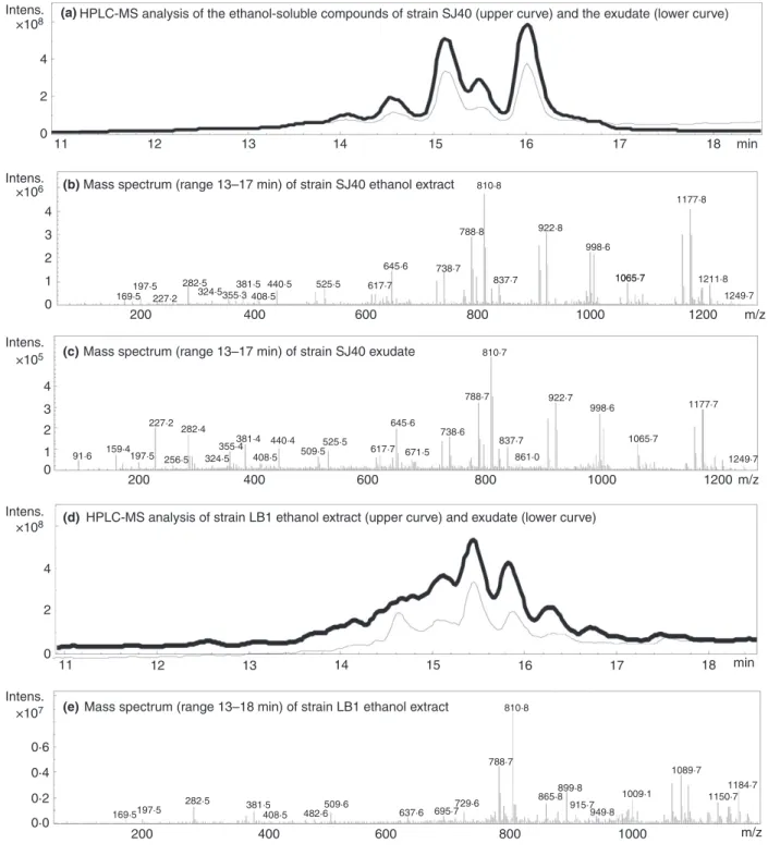
The toxic metabolites were identified as peptaibols The toxic metabolites produced by the indoor-isolated Trichoderma strains were identified as peptaibols with HPLC/ESI-MS/MS analysis (Fig. 3). The peptaibols of strains H1/226, H3/226, 14/AM and Tri335 present in the ethanol-soluble compounds from biomasses and exudated guttation droplets were identified as trichorzianines (Tables 2–4). Strain SJ40 produced trilongins in the ethanol-soluble compounds from biomass and guttation droplets (Tables 2 and 3). Ethanol-soluble compounds from biomass and exudate of strain LB1 contained trichostrigocin-like peptaibols which resembled tri- chostrigocins of strain Sip335 except that the C-terminus of the trichostrigocin-like peptaibols contained pheny- lalaninol, whereas the C-terminus of trichostrigocins con- tained leucinol (Tables 2 and 3).
HPLC/ESI-MS/MS analyses showed that peptaibols were present in the ethanol-soluble compounds from bio- masses and exudates of the indoor-isolated Trichoderma
strains. Moreover, the Trichodermaisolates produced the same peptaibols in the ethanol-soluble compounds from biomass, as in the corresponding exudate. Results in Tables 2 and 3 show that the crude extracts and the exu- dates, containing trilongins, trichorzianines and tri- chostrigocins were more toxic in the boar sperm assays BSMIM and SMIDM than in the ICP assays, exhibiting the same toxicity profile as the commercial peptaibol alamethicin.
Trichorzianine TA IIIc purified from biomass extract of T. atrovirideH1/226 exhibited similar toxicity profile as purified trilongin and alamethicin
Over 10 sperm-toxic HPLC fractions, identified as tri- chorzianine peptaibols, were found in the ethanol-soluble compounds from the biomass of T. atroviride H1/226.
The trichorzianines identified with MS/MS analysis were TA IIIb (MW=1948), TA IIIc (MW =1948), TA VII (MW =1923), TA IVb (MW =1962), TA VIb (MW =1909) and TA VIa (MW=1937), similar to the ones described earlier for T. atroviride by Stoppacher et al.(2007). The fraction containing the known voltage- dependent channel producer trichorzianine TA IIIc (MW =1948) reported by Molle et al. (1987) was selected for further toxicity assays (Table 4).
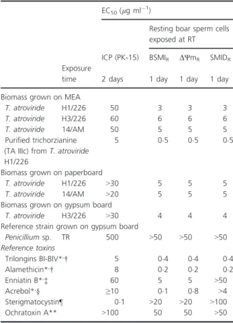
In the ICP (PK-15) and resting boar sperm assays (BSMIR, SMIDRandΨmR) the toxicity of the purified tri- chorzianine TA IIIc was 20- and 100-fold, respectively, of the toxicity of ochratoxin A (upper limit of nonspecific response), thus resting boar sperm assays were the most sensitive to detect the toxic trichorzianine TA IIIc. Concen- trations of 02, 04 and 05lg ml1 of alamethicin, tri- longin and trichorzianine TA IIIC, respectively, inhibited sperm motility, depolarized mitochondria and depleted the
(a) (b)
(c) (d)
Figure 2 Toxicity of boar sperm cells exposed one day to the ethanol extracts ofT.
atroviride14/AM cultivated on paper board (c and d, 5lg ml1) compared to the ethanol control (a,b). After exposure, the cells were stained with the membrane potential responsive stain JC-1 (a,c) and DNA vital staining with PI and Hoechst 33342 (b,d). In the ethanol control, motile sperms exhibited (a) high mitochondrial membrane potential, orange fluorescent mitochondrial sheath in the mid piece of the sperm tail and (b) intact plasma membrane of sperm cells,
impermeable to red fluorescent PI. Ethanol extract ofT. atroviride14/AM cultivated on paperboard showed depolarised mitochondria (c) and disrupted plasma membrane integrity (d). The size of the sperm head is 894lm.
plasma membrane integrity. These toxic responses differ from those provoked by the mitochondrial toxins enni- atin and acrebol which had no effect on plasma mem- brane at motility-inhibiting concentrations. The toxic response of TA IIIc also differed from that exhibited by sterigmatocystin which was 1000 times more toxic in the ICP (PK-15) assay than in the resting sperm assays (BSMIR, SMIDR and ΨmR). The toxicity actions of the purified trichorzianine TA IIIc were similar and compara- ble to the potassium channel-forming peptaibols trilon- gins and alamethicin, indicating that trichorzianine TA IIIc induces the same toxicity mechanism.
Discussion
According to our information, this is the first report of indoor-isolated Trichodermastrains producing peptaibols
in their exudates. They were identified by HPLC/ESI-MS/
MS as trichorzianines, trilongins, trichostrigocins and tri- chostrigocin-like peptaibols. Moreover, the same peptai- bol was present in the biomass extract (hyphae and conidia) and the exudate of the corresponding Tricho- dermaisolate (MEA-cultivated).
The trichorzianines produced by the T. atroviride strains were previously described from T. atroviride and T. harzianum (Stoppacher et al. 2007; Panizel et al.
2013). From a forest soil isolate ofT. strigosum, Degen- kolb et al. (2008) isolated and identified trichostrigocins similar to the ones detected in this study from T. par- aviridescens and T. trixiae. Mikkola et al. (2012) showed that trilongins were also produced by clinical and indoor isolates ofT. longibrachiatum.
Only a few studies have reported the presence of toxins in fungal exudates. Gareis and Gareis (2007) described the secretion of a high concentration of mycoxotins in the exudates ofPenicillium expansum(from a culture col- lection). Toxic trichothecenes were detected in the exu- dates of indoor Stachybotrys chartarum isolates, it was suggested that these toxins might be easily released into the environment due to the aerosolization of toxic gutta- tion droplets favoured by ventilation or air-conditioning systems (Gareis and Gottschalk 2014). Recently, Salo et al. (2015) showed that indoor-isolated Penicillium expansumproduced exudates containing toxic chaetoglo- bosins and communesins.
The exudates and biomass extracts (hyphae+conidia) of theTrichoderma isolates cultivated on MEA substrates were highly cytotoxic (Tables 2 and 3). Their toxicity pat- terns were similar to the biomass extract of T. longi- brachiatum SzMC Thg producing channel-forming trilongins and to alamethicin produced by T. arundi- naceum (Degenkolb et al. 2008; Mikkola et al. 2012).
Moreover, the biomass extract (hyphae+conidia +exu- date) of theTrichodermacultivated on building materials contained substances exhibiting the same toxicity profile as the Trichoderma cultivated on laboratory medium MEA (hyphae+conidia) and as the purified peptaibol trichorzianine TA IIIc (Table 4). As expected, the peptai- bol trichorzianine TA IIIc purified from strain H1/226 exhibited the same toxicity pattern as its peptaibol rela- tive’s alamethicin and trilongins and new peptaibols recently detected in forest-derived Trichoderma isolates from sectionLongibrachiatum(Mikkolaet al.2012; Marik et al. 2017). The resting and motile boar sperm assays were more sensitive for the screening of Trichoderma peptaibols than the ICP assay with somatic cells. The disruption of sperm cell membrane integrity assay (SMIDM) is very sensitive to detect and assess the expo- sure risk of mammalian cells to Trichodermapeptaibols (Peltola et al. 2004; Mikkola et al. 2012; Marik et al.
Table 4 Toxicity of the ethanol extracts from biomasses (including hyphae, conidia and exudate) ofTrichodermastrains cultured on building material substrates, and of the purified trichorzianine TA IIIc
EC50(lg ml1)
Resting boar sperm cells exposed at RT
ICP (PK-15) BSMIR DΨmR SMIDR
Exposure
time 2 days 1 day 1 day 1 day
Biomass grown on MEA
T. atroviride H1/226 50 3 3 3
T. atroviride H3/226 60 6 6 6
T. atroviride 14/AM 50 5 5 5
Purified trichorzianine (TA IIIc) fromT. atroviride H1/226
5 05 05 05
Biomass grown on paperboard
T. atroviride H1/226 >30 5 5 5
T. atroviride 14/AM >20 5 5 5
Biomass grown on gypsum board
T. atroviride H3/226 >30 4 4 4
Reference strain grown on gypsum board
Penicilliumsp. TR 500 >50 >50 >50
Reference toxins
Trilongins BI-BIV*,† 5 04 04 04
Alamethicin*,† 8 02 02 02
Enniatin B*,‡ 60 5 5 >50
Acrebol*,§ ≥10 01 08 >4
Sterigmatocystin¶ 01 >20 >20 >100
Ochratoxin A** >100 50 50 >50
*Bencsiket al.(2014).
†Forming potassium channel.
‡Potassium carrier ionophore and mitochondrial toxins.
§Blocking respiratory chain in mitochondria.
¶Inhibitor of protein synthesis.
**Upper limit of nonspecific response.
2017). McMullin et al. (2017) also reported membrane disruption of Fusarium sambucinum spores by tri- chorzianine-like peptaibols isolated from indoor T.
atroviride.
In Nordic countries, negative pressure is commonly used to prevent moisture damage of buildings. Airaksinen et al.(2004) reported that an indoor negative pressure of 5–20 Pa enables fungal spores below 4lm to cross Intens. HPLC-MS analysis of the ethanol-soluble compounds of strain SJ40 (upper curve) and the exudate (lower curve)
Mass spectrum (range 13–17 min) of strain SJ40 ethanol extract
Mass spectrum (range 13–17 min) of strain SJ40 exudate
HPLC-MS analysis of strain LB1 ethanol extract (upper curve) and exudate (lower curve)
Mass spectrum (range 13–18 min) of strain LB1 ethanol extract
×108 4
Intens.
Intens.
Intens.
Intens.
×105
×108
×107
×106 4 3 2 1
0 200
169·5 227·2 282·5
324·5355·3381·5 408·5
440·5 525·5 617·7
645·6 738·7 788·8
810·8
837·7 922·8
998·6 1065·7
1177·8
1211·8 1249·7 1065·7
1177·7
91·6 159·4 197·5
227·2
324·5 355·4
509·5 408·5
440·4
671·5
788·7
800 600
400
169·5197·5 282·5
408·5 482·6509·6 637·6 729·6
899·8
1009·1 949·8
1150·7 1184·7 1089·7
865·8 915·7 788·7
810·8
695·7 381·5
200 1000 1200 m/z
1065·7
1249·7 810·7
861·0
998·6 837·7
922·7
738·6 645·6
381·4 525·5
617·7 256·5
282·4 197·5
400 600 800 1000 1200 m/z
2
011 12 13 14 15 16 17 18 min
4
4 2
0 11 12
0·6 0·4 0·2
0·0 200 400 600 800 1000 m/z
13 14 15 16 17 18 min
3 2 1 0
(a)
(b)
(c)
(d)
(e)
Figure 3 HPLC/ESI-MS/MS analyses of the ethanol-soluble compounds from biomass and exudates of the indoor-isolatedTrichodermaspecies. a, d,g,j and m: total ion chromatograms of the ethanol-soluble compounds from biomass and exudate of strains SJ40, LB1, Tri335, 14/AM, and Sip335, respectively. Mass spectra (range 13-18 min) of the ethanol-soluble compounds from biomass (b,e,h,k,n) and exudate (c,f,i,l,o) of strains SJ40, LB1, Tri335, 14/AM and Sip335, respectively.
structures. Thus, the small-sized conidia of Trichoderma may more easily spread in the indoor air. In this study, we observed that T. atroviridegrown on building materi- als was capable of emitting conidia and exudate vesicles as airborne.
Immunocompromised patients exposed to fungal patho- gens via—for example, their conidia, can develop peritonitis or systemic infections (Kuhlset al.1999; Kredicset al.2004;
Kubicek et al. 2008; Druzhinina et al. 2011; Naeimi et al.
2011). Thus, humans may experience pulmonary mycoses or pathogenic infections triggered when exposed to potentially pathogenic strains likeT. citrinovirideSJ40, isolated from an indoor settled dust sample (Hoog 1996).
If Trichoderma grows inside a building structure, the risks of respiratory exposure due to the air leaks caused by negative pressure are larger. Although most of the Trichoderma isolates identified in this study were either T. atroviride or T. trixiae, the total number of isolates Intens.
Intens.
Mass spectrum (range 13–18 min) of strain LB1 exudate
Mass spectrum (range 13–17 min) of strain Tri335 ethanol extract
Mass spectrum (range 13–17 min) of strain Tri335 exudate HPLC-MS analysis of strain Tri335 ethanol extract (upper curve) and exudate (lower curve)
HPLC-MS analysis of strain 14/AM ethanol extract (upper curve) and exudate (lower curve) Intens.
Intens.
×105
×107
×105
Intens.
×108 2 1 0
×106
91·6 197·5
169·5199·5 227·2
282·5 355·5 395·5 454·5 523·6 582·6
667·6 752·7
802·8 863·8 824·8
924·7 985·1
1004·6
1122·8
1144·7 1194·7 159·4
243·2 227·2
282·4
381·4
408·5 509·5 695·6
788·7 810·7
899·7 865·7 915·7 992·0
1009·0
1150·7 1088·9
1184·7 751·6
729·6 637·6 575·6 531·5
91·6
199·5 159·4
243·2 227·2
289·2 325·2
363·1 395·4
425·1 454·5
493·1 523·5
707·0
863·7
924·6 985·0
1005·6 1059·6 1178·6 1122·7 802·7
824·7
752·6 667·6 547·5 591·6
5 4 3 2 1
0 200 400 600 800 1000 m/z
200 400 600 800 1000 m/z
200 400 600 800 1000 m/z
0 11 12 13 14 15 16 17 18 min
11 12 13 14 15 16 17 18 min
0
0 1 2 1 2 3 4 2 4
(j) (i) (h) (g) (f)
Figure 3 Continued.
was insufficient to conclude on species predominance in problematic buildings in Finland. Isolates identified as T. atroviride and T. citrinoviride have also earlier been reported from water-damaged buildings in Denmark and Canada (L€ubeck et al. 2000; McMullin et al. 2017).
The species T. paraviridescens and T. trixiae were described during the recent revision of the T. viridescens species complex (Jaklitsch et al. 2013), thus, even
though these species are widely distributed, they have rarely been reported under their new names and only from outdoor samples (Błaszczyket al.2016; Braithwaite et al.2017).
This is the first report of indoor Trichoderma isolates emitting toxic metabolites (peptaibols) in their exudated guttation droplets when growing on building materials or laboratory medium. Moreover, the same peptaibols Intens.
Mass spectrum (range 14–18 min) of strain 14/AM ethanol extract
Mass spectrum (range 13–18 min) of strain Sip335 ethanol extract
Mass spectrum (range 13–18 min) of strain Sip335 exudate Mass spectrum (range 13–17 min) of strain 14/AM exudate
HPLC-MS analysis of strain Sip335 ethanol extract (upper curve) and exudate (lower curve)
×106 4 2 0
200 400 600 800 1000 m/z
200 400 600 800 1000 m/z
200 400 600 800 1000 m/z
200 400 600 800 1000 m/z
Intens.
×106
0 Intens.
×108 4
1 2 3
0
11 12 13 14 15 16 17 18 min
Intens.
×106 4
1 2 3
0 2 1
Intens.
×105 4
1 2 3
0
169·5199·5 227·2
282·5 355·5 395·5 440·5
454·5 523·5
537·6 582·6
604·6
653·6667·6 738·6752·7 802·8
824·8
863·8 924·7
985·1 1004·6
1122·8
169·5
91·6 159·4
227·2
197·5 243·2
282·4 369·4
395·5 523·5 651·5 729·6
768·7
814·7 790·7
899·7
999·1
1184·7
1150·7 1206·6 197·5
227·2 282·5 369·5395·5
523·5 651·6
622·6 695·7 729·6
768·8
814·8 790·8
899·8
865·8 949·8 999·2
1089·0
1150·71206·6 1184·7 169·5 199·5227·2 282·5 355·5395·5
440·5 454·5
523·6 582·6
667·6 752·7 802·8
824·8
838·7
863·7 924·7
985·1 1004·6
1122·8
(m) (l) (k)
(n)
(o)
Figure 3 Continued.
were detected in the ethanol-soluble compounds from biomass and the exudate of the same cytotoxic Tricho- derma strain. Based on the results of this study we spec- ulate that the toxin productions of indoor fungi in guttation droplets may serve as a mechanism of aerosol formation from nonvolatile toxins in the indoor air. Fur- ther studies are needed to determine the chemical com- position and structure of the exudates, to examine their behaviour and to determine the possible indoor trans- port mechanisms.
Acknowledgements
The authors thank Riikka Holopainen at the Finnish Food Safety Authority (EVIRA) for providing the feline fetus lung and porcine kidney cells. Henri Gustavson, Liisa Hakamies-Blomqvist, Lauri Sipil€a and Johanna Salo are thanked for providing some of the indoor samples.
The authors warmly thank the Academy of Finland (TOXICPM 289161), Business Finland (grant 4098/31/
2015) and the National Research, Development and Inno- vation Office, Hungary (grant NKFI K-105972) for fund- ing this research. LK is grantee of the Janos Bolyai Research Scholarship (Hungarian Academy of Sciences).
Conflict of Interest
No conflict of interest declared.
References
Airaksinen, M., Kurnitski, J., Pasanen, P. and Seppanen, O.
(2004) Fungal spore transport through a building structure.Indoor Air14, 92–104. https://doi.org/10.1046/j.
1600-0668.2003.00215.x.
Alm, K., Taponen, J., Dahlbom, M., Tuunainen, E., Koskinen, E. and Andersson, M. (2001) A novel automated fluorometric assay to evaluate sperm viability and fertility in dairy bulls.Theriogenology56, 677–684. https://doi.org/
10.1016/S0093-691X(01)00599-4.
Andersen, A.A. (1958) New sampler for the collection, sizing and enumeration of viable airborne particles.J Bacteriol 76, 471–484.
Andersen, B., Frisvad, J.C., Sondergaard, I., Rasmussen, I.S.
and Larsen, L.S. (2011) Associations between fungal species and water-damaged building materials.Appl Environ Microbiol77, 4180–4188. https://doi.org/10.1128/
AEM.02513-10.
Andersson, M.A., Mikkola, R., Helin, J., Andersson, M.C. and Salkinoja-Salonen, M. (1998) A novel sensitive bioassay for detection ofBacillus cereusemetic toxin and related depsipeptide ionophores.Appl Environ Microbiol64, 1338– 1343.
Andersson, M.A., J€a€askel€ainen, E.L., Shaheen, R., Pirhonen, T., Wijnands, L.M. and Salkinoja-Salonen, M.S. (2004) Sperm bioassay for rapid detection of cereulide-producing Bacillus cereusin food and related environments.Int J Food Microbiol94, 175–183. https://doi.org/10.1016/j.ijf oodmicro.2004.01.018.
Andersson, M., Mikkola, R., Rasimus, S., Hoornstra, D., Salin, P., Rahkila, R., Heikkinen, M., Mattila, S.et al.(2010) Boar spermatozoa as a biosensor for detecting toxic substances in indoor dust and aerosols.Toxicol In Vitro 24, 2041–2052. https://doi.org/10.1016/j.tiv.2010.08.011.
Bencsik, O., Papp, T., Berta, M., Zana, A., Forgo, P., Dombi, G., Andersson, M., Salkinoja-Salonen, M.et al.(2014) Ophiobolin A fromBipolaris oryzaeperturbs motility and membrane integrities of porcine sperm and induces cell death on mammalian somatic cell lines.Toxins6, 2857– 2871. https://doi.org/10.3390/toxins6092857.
Błaszczyk, L., Strakowska, J., Chełkowski, J., Gazbka-Buszek, A.
and Kaczmarek, J. (2016)Trichodermaspecies occurring on wood with decay symptoms in mountain forests in Central Europe: genetic and enzymatic characterization.J Appl Genet 57, 397–407. https://doi.org/10.1007/s13353-015-0326-1.
Bohemen, A.-I.V., Zalouk-Vergnoux, A., Poirier, L., Phuong, N.N., Inguimbert, N., Salah, K.B.H., Ruiz, N. and Pouchus, Y.F. (2016) Development and validation of LC– MS methods for peptaibol quantification in fungal extracts according to their lengths.J Chromatogr B1009–
1010, 25–33. https://doi.org/10.1016/j.jchromb.2015.11.
039.
Braithwaite, M., Johnston, P.R., Ball, S.L., Nourozi, F., Hay, A.J., Shoukouhi, P., Chomic, A., Lange, C.et al.(2017) Trichodermadown under: species diversity and occurrence ofTrichodermain New Zealand.Australas Plant Pathol46, 11–30. https://doi.org/10.1007/s13313-016-0457-9.
Degenkolb, T., Dieckmann, R., Nielsen, K.F., Gr€afenhan, T., Theis, C., Zafari, D., Chaverri, P., Ismaiel, A.et al.(2008) TheTrichoderma brevicompactumclade: a separate lineage with new species, new peptaibiotics, and mycotoxins.
Mycol Prog7, 177–219. https://doi.org/10.1007/s11557- 008-0563-3.
Druzhinina, I.S., Kopchinskiy, A., Komon, M., Bissett, J., Szakacs, G. and Kubicek, C.P. (2005) An oligonucleotide barcode for species identification inTrichodermaand Hypocrea.Fungal Genet Biol42, 813–828. https://doi.org/
10.1016/j.fgb.2005.06.007.
Druzhinina, I.S., Komon-Zelazowsa, M., Kredics, L., Hatvani, L., Antal, Z., Belayneh, T. and Kubicek, C.P. (2008) Alternative reproductive strategies ofHypocrea orientalis and genetically close but clonalTrichoderma
longibrachiatum, both capable of causing invasive mycoses of humans.Microbiology154, 3447–3459. https://doi.org/
10.1099/mic.0.2008/021196-0.
Druzhinina, I.S., Seidl-Seiboth, V., Herrera-Estrella, A., Horwitz, B.A., Kenerley, C.M., Monte, E., Mukherjee, P.K., Zeilinger, S.et al.(2011)Trichoderma: the genomics
of opportunistic success.Nat Rev Microbiol9, 749–759.
https://doi.org/10.1038/nrmicro2637.
Gareis, M. and Gareis, E.-M. (2007) Guttation droplets of Penicillium nordicumandPenicillium verrucosumcontain high concentrations of the mycotoxins ochratoxin A and B.Mycopathologia163, 207–214. https://doi.org/10.1007/
s11046-007-9003-1.
Gareis, M. and Gottschalk, C. (2014)Stachybotrysspp. and the guttation phenomenon.Mycotoxin Res30, 151–159.
https://doi.org/10.1007/s12550-014-0193-3.
Gravesen, S., Nielsen, P.A., Iversen, R. and Nielsen, K.F.
(1999) Microfungal contamination of damp buildings— examples of risk constructions and risk materials.Environ Health Perspect107, 505–508.
Hatvani, L., Manczinger, L., Vagv€olgyi, C. and Kredics, L.
(2013) 17Trichodermaas a human pathogen. In
Trichoderma: Biology and Applicationsed. Mukherjee, P.K.
pp. 292–313. Boston, MA: CABI. https://doi.org/10.1079/
9781780642475.0292
Hermosa, R., Cardoza, R.E., Rubio, M.B., Gutierrez, S. and Monte, E. (2014) Secondary metabolism and
antimicrobial metabolites ofTrichoderma. In
Biotechnology and Biology of Trichodermaed. Gupta, V.K., Schmoll, M., Herrera-Estrella, A., Upadhyay, R.S., Druzhinina, I. and Tuohy, M. pp. 125–137. New York, NY: Elsevier B.V. https://doi.org/10.1016/b978-0-444- 59576-8.00010-2.
Hoog, G.S. (1996) Risk assessment of fungi reported from humans and animals.Mycoses39, 407–417. https://doi.org/
10.1111/j.1439-0507.1996.tb00089.x.
Hutwimmer, S., Wang, H., Strasser, H. and Burgstaller, W.
(2009) Formation of exudate droplets byMetarhizium anisopliaeand the presence of destruxins.Mycologia102, 1–10. https://doi.org/10.3852/09-079.
Jaklitsch, W.M., Samuels, G.J., Ismaiel, A. and Voglmayr, H.
(2013) Disentangling theTrichoderma viridescenscomplex.
Persoonia31, 112–146. https://doi.org/10.3767/
003158513X672234.
Jaklitsch, W.M. and Voglmayr, H. (2015) Biodiversity of Trichoderma(Hypocreaceae) in Southern Europe and Macaronesia.Stud Mycol.80, 1–87. https://doi.org/10.
1016/j.simyco.2014.11.001.
Kamp, G., Busselmann, G., Jones, N., Wiesner, B. and Lauterwein, J. (2003) Energy metabolism and intracellular pH in boar spermatozoa.Reproduction126, 517–525.
https://doi.org/10.1530/rep.0.1260517.
Kopchinskiy, A., Komon, M., Kubicek, C.P. and Druzhinina, I.S. (2005)TrichoBLAST: a multilocus database for TrichodermaandHypocreaidentifications.Mycol Res109, 658–660.
Kredics, L., Antal, Z., Szekeres, A., Manczinger, L., Doczi, I., Kevei, F. and Nagy, E. (2004) Production of extracellular proteases by human pathogenicTrichoderma
longibrachiatumstrains.Acta Microbiol Immunol Hung51, 283–295. https://doi.org/10.1556/AMicr. 51.2004.3.6.
Kubicek, C.P., Komon-Zelazowska, M. and Druzhinina, I.S.
(2008) Fungal genusHypocrea/Trichoderma: from barcodes to biodiversity.J Zhejiang Univ Sci B9, 753–763. https://d oi.org/10.1631/jzus.B0860015.
Kubicek, C.P., Herrera-Estrella, A., Seidl-Seiboth, V., Martinez, D.A., Druzhinina, I.S., Thon, M., Zeilinger, S., Casas- Flores, S.et al.(2011) Comparative genome sequence analysis underscores mycoparasitism as the ancestral life style ofTrichoderma.Genome Biol12, R40. https://doi.org/
10.1186/gb-2011-12-4-r40.
Kuhls, K., Lieckfeldt, E., B€orner, T. and Gueho, E. (1999) Molecular reidentification of human pathogenic Trichodermaisolates asTrichoderma longibrachiatumand Trichoderma citrinoviride.Med Mycol37, 25–33. https://d oi.org/10.1111/j.1365-280X.1999.00197.x.
Leitgeb, B., Szekeres, A., Manczinger, L., Vagv€olgyi, C. and Kredics, L. (2007) The history of alamethicin: a review of the most extensively studied peptaibol.Chem Biodivers4, 1027–1051. https://doi.org/10.1002/cbdv.200790095.
L€ubeck, M., Poulsen, S.K., L€ubeck, P.S., Jensen, D.F. and Thrane, U. (2000) Identification ofTrichodermastrains from building materials by ITS1 ribotyping, UP-PCR fingerprinting and UP-PCR cross hybridization.FEMS Microbiol Lett185, 129–134. https://doi.org/10.1111/j.1574- 6968.2000.tb09050.x.
Marik, T., Kredics, L., Szekeres, A., Vagv€olgyi, M., Buchner, R.€ and Vagv€olgyi, C. (2016) Effect of peptaibol extracts derived fromTrichodermastrains on mammalian cells. In 18th Danube-Kris-Mures-Tisza (DKMT) Euroregional Conference on environment and health, 2–4 June 2016, Novi Sad, Serbia.
Marik, T., Szekeres, A., Andersson, M.A., Salkinoja-Salonen, M., Tyagi, C., Leitgeb, B., Vagv€olgyi, C., Druzhinina, I.
et al.(2017) Bioactive peptaibols of forest-derived Trichodermaisolates from sectionLongibrachiatum, InSoil Biological Communities and Ecosystem Resilience in Sustainability in Plant and Crop Protectioned. Lukac, M., Grenni, P. and Gamboni, M. pp. 277–290. Switzerland:
Springer Nature.
McMullin, D.R., Renaud, J.B., Barasubiye, T., Sumarah, M.W.
and Miller, J.D. (2017) Metabolites ofTrichodermaspecies isolated from damp building materials.Can J Microbiol 63, 621–632. https://doi.org/10.1139/cjm-2017-0083.
Mikkola, R., Andersson, M.A., Kredics, L., Grigoriev, P.A., Sundell, N. and Salkinoja-Salonen, M.S. (2012) 20-Residue and 11-residue peptaibols from the fungus
Trichoderma longibrachiatumare synergistic in forming Na+/K+-permeable channels and adverse action towards mammalian cells.FEBS J279, 4172–4190. https://doi.org/
10.1111/febs.12010.
Mikkola, R., Andersson, M.A., Hautaniemi, M. and Salkinoja- Salonen, M.S. (2015) Toxic indole alkaloids
avrainvillamide and stephacidin B produced by a biocide tolerant indoor moldAspergillus westerdijkiae.Toxicon99, 58–67. https://doi.org/10.1016/j.toxicon.2015.03.011.