This article has been accepted for publication and undergone full peer review but has not been through the copyediting, typesetting, pagination and proofreading process, which may lead to differ- ences between this version and the Version of Record. Please cite this article as doi:
10.1111/pace.13576.
This article is protected by copyright. All rights reserved.
Unchanged QRS morphology with atrial pacing in a case of WPW syndrome.
What is the mechanism?
Cristina Tutuianu MD, Laszlo Saghy MD PhD, Robert Pap MD, PhD
University of Szeged, Hungary
Short title:Unchanged QRS in WPW syndrome.
Key Words: Unchanged QRS, WPW syndrome
Corresponding Autor:
Cristina Tutuianu, MD,
2nd Department of Medicine and Cardiology Center University of Szeged, Hungary
6725 Szeged, Semmelweis str. 6 Phone: +3662545220
Fax: +3662544568
Email: tutucris2008@gmail.com
Autor Disclosure Information: C. Tutuianu: None, L Saghy: None, R Pap: None,
This article is protected by copyright. All rights reserved.
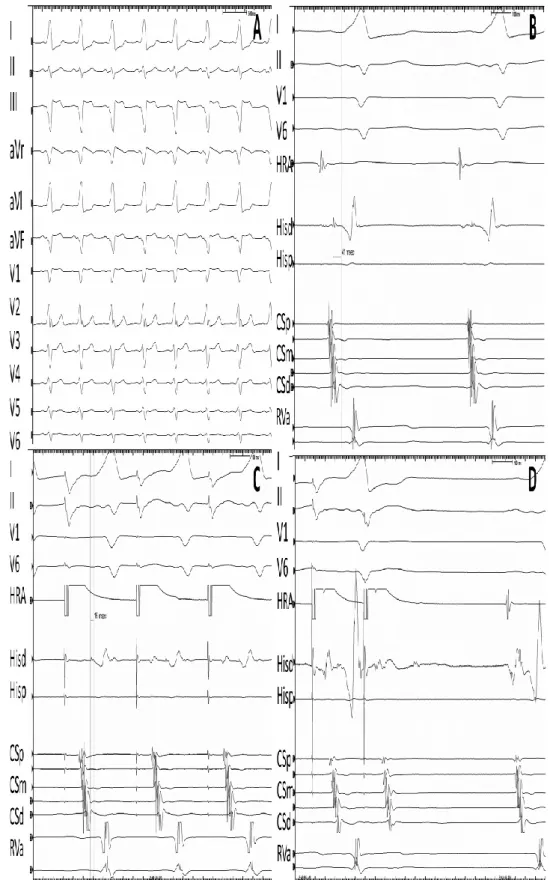
A 55 year-old woman presented to our clinic for short episodes of palpitation without doc- umented arrhythmia. Her family history was unremarkable except for an uncle implanted with a pacemaker for atrioventricular (AV) block. Her surface electrocardiogram showed sinus rhythm with wide QRS morphology and very short PR interval (Fig 1A) consistent with WPW syndrome. Echocardiography was normal. After informed consent she underwent an electrophysiological study. At baseline the His to ventricular (HV) interval was 41 ms (Fig 1B). There was no retrograde conduction. During incremental atrial pacing (Fig 1C) and ex- trastimulation the atrium to His (AH) interval progressively lengthened, the stimulus to QRS stayed constant, while the HV shortened, without any change in the morphology of the QRS, until the atrial extrastimulus failed to conduct to the ventricles (Fig 1D). What can be the explanation for the constant QRS morphology, despite shortening HV during atrial pacing?
Discussion
The HV interval was unusually long for ventricular pre-excitation, but progressively short- ened with atrial pacing, suggesting an AV accessory pathway (AP). In case of an AV AP ante- grade ventricular activation usually results from a fusion of conduction through the AV node and the AP. During atrial pacing progressive slowing of conduction in the AV node results in increasing pre-excitation and changing of QRS morphology as progressively larger part of the ventricles are activated through the AP. The lack of fusion during atrial pacing in case of an AV AP may be related to ventricular activation present only through the AP and lacking through the AV node (1).
Due to assumed high risk features of the AP (effective refractory period (ERP) < 240 ms on isoproterenol infusion) a decision was made to ablate it. The earliest ventricular activation was found on the right posteroseptal region close to the coronary sinus ostium, where no His potential was recorded. During the first radiofrequency application, after 3 seconds complete AV block occurred without any junctional rhythm (Fig 2A). Energy delivery was stopped immediately and an infra Hisian AV block was revealed but after 5 minutes AP con- duction recovered. At this point preexisting infra-His AV block in addition to the presence of the AV AP was realized. After discussion with the patient about the unpredictable conduc-
This article is protected by copyright. All rights reserved.
tion properties of the AP in the long term, a DDD pacemaker was implanted. The procedure was repeated after 3 months. After ablation of the AP, 1:1 AV conduction with left bundle branch block and a long HV interval of 60 msec was observed (Fig 2B). We hypothesize that during the first procedure complete AV block was related to preexisting left bundle branch block plus mechanical right bundle block due to catheter trauma in the His position.
In hindsight, several findings pointed to the absence or significant prolongation of antero- grade conduction over the normal AV conduction system even before the first ablation at- tempt. The QRS was maximally preexcited even in sinus rhythm and did not change with atrial pacing. The HV interval (a pseudointerval in this case) had no relation to AV conduc- tion: it was long at baseline , then progressively shortened with atrial pacing, but the stimu- lus to QRS remained constant, with constant QRS morphology verifying absence of the con- tribution of the normal AV conduction system to the QRS. Finally, there was no normal QRS after the His potential when the AP blocked (Fig. 1. D).
Whether the ablation of this AP and the implantation of the pacemaker was necessary can be debated. Guidelines state that ablation is reasonable of the AP capable of rapid conduc- tion (AP ERP < 240-250 ms), but observation is also a reasonable alternative in the asymp- tomatic adult (2). Whether this patient’s symptoms were related to the AP remains uncer- tain in view of the lack of retrograde conduction and inducible arrhythmia. The conduction capacity of an AP is thought to decrease with ageing (3) posing potential risk of AV block to a patient with impaired normal conduction system. These dilemmas were discussed thor- oughly with the patient before proceeding with ablation and implantation.
This case reminds us that when no change is observed in the amount of ventricular fusion during atrial pacing with an AV AP, occult AV block should be suspected.
This article is protected by copyright. All rights reserved.
References
1. Mossuti E, Elia F, Martello G, Brancati B. Coexistence of complete infra-Hisian block, WPW syndrome and Mobitz type II Kent Bundle block. Pacing Clin Electrophysiol. 1990 Dec;13(12 Pt 1):1563-6.
2. Page RL, Joglar JA, Caldwell MA, Calkins H, Conti JB, Deal BJ, Estes NAM 3rd, at al. 2015 ACC/AHA/HRS Guideline for the Management of Adult Patients With Supraventricular Tach- ycardia: A Report of the American College of Cardiology/American Heart Association Task Force on Clinical Practice Guidelines and the Heart Rhythm Society. J Am Coll Cardiol. 2016 Apr 5;67(13):e27-e115.
3. Fan W, Peter CT, Gang ES, Mandel W. Age-related changes in the clinical and electrophysio- logic characteristics of patients with Wolff-Parkinson-White syndrome: comparative study between young and elderly patients. Am Heart J. 1991 Sep;122(3 Pt 1):741-7.
Figures Legend
Figure 1. Twelve-lead ECG during sinus rhythm (Panel A) Paper speed 50 mm/s. Surface electrocar- diogram lead I, II, V1, V6 along with intracardiac recordings from high right atrium (HRA), coronary sinus (CS) and mapping catheter placed on the His region (His) during sinus rhythm (Panel B), during atrial incremental pacing (Panel C) and during atrial extrastimulation (Panel D). Paper speed 200 mm/s.
This article is protected by copyright. All rights reserved.
Figure 2. Surface electrocardiogram lead I, II, V1, V6 along with intracardiac recordings from high right atrium (HRA), coronary sinus (CS), right ventricular apex (RV) and ablation catheter (Abl) during
This article is protected by copyright. All rights reserved.
ablation in the first procedure (Panel A). Paper speed 50 mm/s, and during the successful ablation in the second procedure (Panel B). Paper speed 200 mm/s.