NON-PHARMACOLOGICAL TECHNIQUES OF ANTIARRHYTHMIC THERAPY
PhD thesis
Emin Evren Özcan, MD.
Doctoral School of Basic Medicine Semmelweis University
Supervisor: László Gellér, MD, med. habil.
Official reviewers: László Sághy, MD, PhD Pál Ábrahám, MD, PhD Head of the Final Examination Committee:
Prof. Katalin Darvas, MD, med. habil Members of the Final Examination Committee:
Prof. Zoltán Járai, MD, med. habil.
András Zsáry, MD, PhD
Budapest
2017
2
Table of Contents
List of Abbreviations 4
1. Introduction 5
2. Objectives 10
3. Methods 11
3.1. Evaluating the effects of endocardial vs. epicardial LV pacing 11
3.1.1. Patient population 11
3.1.2. Endocardial LV lead implantation 11
3.1.3. Electrocardiographic measurements 11
3.1.4. Statistical analysis 12
3.2. Evaluating the effects of bipolar vs. unipolar pacing 12
3.2.1. Patient population 12
3.2.2. Biventricular pacemaker implantation 13
3.2.3. Definition of LV lead electrode location 13
3.2.4. Device optimization and ECG measurements 13
3.2.5. Statistical analysis 15
3.3. Evaluating the effects of respiratory gating on AF ablation procedures 15
3.3.1. Study population 15
3.3.2. Electroanatomical map construction and respiratory gating 15 3.3.3. CT/MRI image acquisition and integration 16
3.3.4. Catheter Ablation 16
3.3.5. Statistical analysis 16
4. Results 17
4.1. Evaluating the effects of endocardial vs. epicardial LV pacing 17 4.2. Evaluating the effects of bipolar vs. unipolar and basal vs. non-basal LV
pacing 19
4.3. Evaluating the effects of respiratory gating on AF ablation procedures 22
5. Discussion 26
5.1. Discussion on effects of endocardial vs. epicardial LV pacing during CRT 26 5.2. Discussion on effects of pacing polarity and pacing site during CRT 29
3
5.3. Discussion on effects of respiratory gating on AF ablation procedures 32
5.3.1. EAM reconstruction times 33
5.3.2. Potential advantage of respiratory gating 33
5.3.3. Safety 34
6. Conclusion 35
7. Summary 36
8. Összefoglalás 37
9. References 38
10. Bibliography of the candidate’s publications 45
Publications related to the PhD thesis 45
Publications not related to the PhD thesis 46
11. Acknowledgements 49
12. Appendix 50
Publications I. – XI. 50
4
List of Abbreviations
AF Atrial fibrillation
CRT Cardiac resynchronization therapy
CS Coronary sinus
CT Computed tomography CTI Cavotricuspid isthmus DC Direct current
DM Diabetes mellitus
EAM Electroanatomic mapping ECG Electrocardiography FAM Fast anatomical mapping HF Heart failure
HT Hypertension
ICD Implantable cardioverter defibrillator ICE Intracardiac echocardiography LA Left atrium
LAO Left anterior oblique LBBB Left bundle branch block LV Left ventricular
MRI Magnetic resonance imaging PV Pulmonary vein
PVI Pulmonary-vein isolation RAO Right anterior oblique
RF Radiofrequency
RV Right ventricle SD Standard deviations
TDR Transmural dispersion of repolarization
5
1. Introduction
Historical background of cardiac electrophysiology and treatment of clinical arrhythmias has a short but fascinating history. Over the last fifty years, non- pharmacological treatment of clinical arrhythmias has shown a very rapid and enormous development. The development of electrocardiography was the key issue for understanding arrhythmias. The first electrogram of the heart in animals was recorded, using a capillary electrometer by Étienne-Jules Marey in 1876. Only few years later, Augustus D. Waller recorded the first human electrocardiogram in 1887. The most important achievement was the development of electrocardiography by Willem Einthoven in 1892. Development of string galvanometer permitted the widespread use of electrocardiogram. Furthermore, true understanding of cardiac rhythm disorders really began with using the string galvanometer invented by Einthoven.
Since that time, with the increasing knowledge about the arrhythmia mechanisms, antiarrhythmic drug therapy has progressed a lot. However, although novel antiarrhythmic drugs are quite effective in terminating the variety of tachyarrhythmias, the chronic use of the drugs is limited by a fear of adverse effects and possible life- threatening pro-arrhythmic complications. Furthermore, cardiac pacing is still the only alternative for chronic treatment of bradycardia. The past half century has witnessed a virtual explosion in non-pharmacological antiarrhythmic therapies. Throughout this period cardiac pacing, invasive electrophysiology and catheter ablation of cardiac arrhythmias gained widespread acceptance.
Hyman developed the first artificial pacemaker in 1930, which was operated by a hand crank and spring motor. Electrical impulses were directed into the patient's heart through a bipolar needle electrode introduced via an intercostal space. Advances in electronic technology helped the development of pacing systems. In 1952, Paul M. Zoll introduced the external defibrillator. The first implantable pacemaker has been developed by the surgeon Ake Senning and the physician inventor Rune Elmqvist and implanted in 1958. The first device lasted only a few hours but saved the patient’s life.
Actually, he survived as well as the scientists who had saved his life. He required five lead systems and 22 pulse generators of 11 different models until his death on 2001 aged 86 of a malignancy totally unrelated to pacemaker system. Over the last years,
6
with the tight collaboration between engineers and physicians simple and rough machines are developed to sophisticated technological wonders and helped countless patients all over the world. During this time period implantable cardioverter defibrillators (ICDs) were developed and besides bradyarrhythmias non- pharmacological treatment of tachyarrhythmias became possible. Michel Mirowski developed an implantable defibrillator after witnessing the sudden death of a colleague and the first implantation was successfully performed in 1980. About the time of these improvements, idea of synchronous pacing of heart chambers in heart failure patients with interventricular dyssynchrony was born and indication of cardiac pacing is not limited by arrhythmias anymore.
Despite all the advances in pharmacological treatment, heart failure remains a leading cause of morbidity and mortality (1). For this reason, cardiac resynchronization therapy (CRT) is an important therapeutic option in the management of patients with symptomatic systolic heart failure (HF) (2). Biventricular pacing significantly improves cardiac output, quality of life, and functional capacity in patients with congestive heart failure. However, the incidence of sudden cardiac death still remains high (3). Recent studies have shown that reversal of the normal myocardial activation sequence during epicardial pacing, as it occurs during conventional CRT, increases the transmural dispersion of repolarization (TDR) and causes ventricular arrhythmias (4,5).
Biventricular pacing or left ventricular (LV) epicardial pacing may increase the QT interval and TDR, which have the potential to increase the risk of ventricular arrhythmias (5-7). Increased TDR as measured by Tpeak-Tend (Tp-Te) and Tp-Te/QT is associated with a higher incidence of ventricular arrhythmias in CRT-D patients (8).
Therefore, it is important to determine CRT patients who are prone to have ventricular arrhythmias.
CRT with transseptal endocardial LV pacing is an alternative in patients where the conventional approach has failed (9, 10). Endocardial pacing also leads more physiological activation. Experimental observations in humans suggest that potential arrhythmias can be avoided by stimulation of the LV endocardium (6). A recently published small case control study suggests that permanent biventricular pacing with LV endocardial lead placement through the interatrial septum is associated with fewer
7
proarryhthmic ventricular characteristics when compared to CRT with a coronary sinus (CS) LV lead (11). Scott et al. evaluated seven patients with transseptal LV endocardial leads, 28 matched patients with CS LV leads, and eight patients with surgical LV epicardial leads. Significant postpacing reduction in Tp-Te and QT dispersion values were observed in the transseptal group compared to the CS group. In contrast, there were no differences between the surgical and CS groups in terms of the effect of CRT on these repolarization parameters. This small observational study with a case control design provides crucial information. However, finding a perfect match between small patient groups is not always possible and is a clear limitation. Differences in baseline characteristics between groups may have an impact on results.
In addition, different pacing configurations may also produce different vectorial activation and may affect ventricular repolarization patterns. Yang et al. reported a significant difference in the mechanical activation sequence between unipolar and bipolar LV pacing during CRT (12). They observed higher basal endocardial strain and more uniform global with bipolar pacing. The difference in the mechanical activation sequence between pacing polarities indicates differential activation of different layers of myocardium, which may have an impact on ventricular repolarization. There is an intrinsic repolarization difference among epicardium, mid-myocardial M cells and endocardium. Delayed activation and repolarization of mid-myocardial M cells during biventricular pacing leads to prominent increase in QT and TDR (5). These issues have raised concern as to whether the LV pacing polarity might play a role in the development of ventricular arrhythmias.
Although the impact of LV pacing polarity on contractile functions has been investigated, little is known about the role of pacing polarity on repolarization patterns (13). A novel quadripolar LV lead offers more pacing configurations, including six bipolar and four unipolar LV pacing options, and allows stimulation of the LV from different epicardial locations (14). The impact of pacing polarity and pacing sites from different perspectives (endocardial vs. epicardial and basal vs. non-basal) have not been investigated in the same patient group.
During the second half of the 20th century, while the cardiac device therapies were emerging, invasive cardiac electrophysiology was born in 1960s. Firstly, Benjamin J.
8
Scherlag described the intra-cardiac recordings of His bundle in 1968. Two scientists, Dirk Durrer in Netherlands and Philippe Coumel in France were the first to execute programmed stimulation in human. Another invaluable contribution was made by Hein J. Wellens combination of programmed electrical stimulation with multiple intra-cardiac recordings led to discern different arrhythmia mechanisms. The programmed stimulation technique has firstly used to induce ventricular tachycardia (VT) and to elucidate the mechanisms of supraventricular tachycardias (SVT), especially Wolf- Parkinson-White syndrome.
With increasing experience, endocardial catheter mapping techniques are developed.
Mapping multiple cardiac chambers allowed the identification of the location of accessory pathways and the sites of origin of VTs. All these advancements opened the gate of ablation era. Shortly after surgical ablations, percutaneous, catheter-based techniques were conceived. Over last two decades, radiofrequency catheter ablation has shown an explosive development and almost all kind of tachycardia including atrial fibrillation (AF) is now treatable by catheter ablation.
Michel Haissaguerre and colleagues have reported that AF is triggered from premature atrial beats, primarily originating in and around the pulmonary veins in 1998. They have also demonstrated that isolation of pulmonary veins can treat highly selected patients.
However, this non-pharmacological treatment option was not widely used until the development of advanced mapping and imaging techniques such as electroanatomic mapping (EAM) and cryo-ablation.
AF is the most common supraventricular arrhythmia in human and antiarrhythmic drugs used for rhythm control have limited success (15, 16). Therefore, in the last decade pulmonary-vein isolation (PVI) became a corner stone for non-pharmacological treatment of AF. Radiofrequency (RF) catheter ablation is an effective therapy for AF and electroanatomic mapping systems play important role on ablation procedures (15-19). Unfortunately, benefits of these tools are limited by biological factors.
Significant changes in left atrium (LA) and pulmonary vein (PV) anatomy due to respiration have been reported (20, 21). Beinart et al. recently demonstrated favorable effects of respiratory gating on electroanatomical map accuracy (22). Respiratory compensated electroanatomical maps showed better correlation with pre-acquired
9
computed tomography (CT) and magnetic resonance (MR) images. However, better correlation does not always mean better ablation results and impact of respiratory gating on AF ablation has not been studied yet.
AccuResp algorithm (Biosense Webster Inc, Diamond Bar, CA) automatically detects out-of-gate acquisition and exclude them from the reconstructed anatomy, thus it may provide accurate maps at the expense of longer mapping times. More reliable maps may shorten procedure times and may increase success rates. On the other hand, integration of reconstructed electroanatomical maps and pre-acquired static 3D images might compensate negative effects of non-gated maps. Relative changes in left atrial anatomy are most pronounced in the distal pulmonary veins and in the left atrium body near the mitral valve (23). Therefore, avoiding from these regions during image integration might have similar effects as respiratory gating.
10
2. Objectives
Based upon information mentioned above, the objective of the present work was the following:
A/ Recent studies have shown that reversal of the normal myocardial activation sequence during epicardial pacing, as it occurs during conventional CRT, increases the TDR. In the first study, we evaluated the proarryhthmic repolarisation characteristics of endocardial and epicardial biventricular pacing in the same CRT patient group.
B/ The impact of pacing polarity and pacing sites from different perspectives (endo- cardial vs. epicardial and basal vs. non-basal) have not been investigated in the same patient group. In the second study, we investigated the impact of LV pacing polarity and epicardial pacing site on repolarization parameters by using quadripolar LV lead.
C/ Electroanatomic mapping systems play an important role on ablation procedures.
However, impact of respiratory gating on electroanatomic mapping and image integration guided AF ablation procedures have not been investigated. Therefore, we assessed the impact of respiratory gating on electroanatomic mapping and image integration guided AF ablation procedures.
11
3. Methods
3.1. Evaluating the effects of endocardial vs. epicardial LV pacing
3.1.1. Patient population
All patients suitable for transseptal endocardial LV lead placement, in whom epicardial CRT had failed due to lead dislodgement after successful CS implantation, were admitted to the study. All patients included were required to meet European Society of Cardiology criteria for CRT implantation (left bundle branch block with QRS duration
>120 ms, LV ejection fraction ≤35%, NYHA functional class II, III and ambulatory IV despite adequate medical treatment) (2). After successful transseptal CRT implantation, all patients’ hospital recordings were evaluated. Patients who had previous successful conventional CRT implantation were selected. ECGs were recorded before and after successful conventional CRT, as were location of CS leads and pacing threshold values.
3.1.2. Endocardial LV lead implantation
Procedures started with transseptal puncture from the femoral vein, under guidance of intracardiac echocardiography (ICE). The puncture site was dilated with an 8 mm angioplasty balloon, and the guidewire was left across the septum as a marker.
Subsequently, a guidewire was passed through the same hole inserted from the subclavian vein through a guiding catheter produced for coronary sinus lead implantation. Once the guiding catheter was advanced to the left atrium, its tip was directed to the mitral valve and an active fixation lead was implanted in the lateral area of the LV. All patients were maintained on anticoagulation therapy with either warfarin or coumarin.
3.1.3. Electrocardiographic measurements
ECGs (25 mm/s, 10 mm/mV) were recorded on admission (pre-endo-CRT ECG) and before discharge (post-endo-CRT ECG) after successful LV endocardial lead implantation. The ECGs of the same patients before and after successful CRT with CS epicardial pacing were found from hospital records. These ECGs were defined as pre-epi-CRT and post-epi-CRT ECGs. All ECGs were digitally scanned and
12
measurements were made with digital calipers at 400% magnification. Analysis was performed by a blinded physician. Lead V5 was selected for analysis. If the T wave in V5 was not eligible, lead II was used. The QT interval was defined as the time interval between the initial deflection of the QRS and the point at which a tangent drawn on the steepest downslope of the T wave crossed the isoelectric line (24). Intervals were corrected for heart rate using Bazett’s formula (25). The Tp-Te interval was obtained from the difference between the QT interval and the QT peak interval. The QT peak interval was measured from the beginning of the QRS until the peak of a positive T wave or the nadir of a negative T wave (26). The Tp-Te/QT ratio was also calculated.
3.1.4. Statistical analysis
Mean values and standard deviations (SD) were used for descriptive statistics.
Categorical data were summarized as frequencies and percentages. Comparisons of repolarization parameters between pacing modes (baseline, epi-CRT, endo-CRT) were performed with paired two-tailed Student’s t-tests. In all analyses, p<0.05 was considered statistically significant.
3.2. Evaluating the effects of bipolar vs. unipolar and basal vs. non- basal LV pacing
3.2.1. Patient population
Between September 2013 and June 2014, 26 consecutive patients with a standard indication for CRT implantation (left bundle branch block with QRS duration >120 ms, LV ejection fraction ≤35%, NYHA functional class II, III and ambulatory IV despite adequate medical treatment) were enrolled in the study. None of the patients had a history of previous ventricular arrhythmic events. Primary prevention for sudden cardiac death was the only indication for CRT-D implantation.
13 3.2.2. Biventricular pacemaker implantation
Device implantation was performed in the cardiac catheterization laboratory following standard CRT implantation techniques. After an (right ventricle-RV shock lead was implanted in apical position, a quadripolar LV lead (The Quartet Model 1458Q, St Jude Medical Inc. St Paul, MN) and a right atrial lead were implanted, and capture thresholds from all electrodes were recorded. The four LV electrodes from distal tip electrode to proximal ring electrode were named D1, M2, M3 and P4 respectively.
3.2.3. Definition of LV lead electrode location
After successful implantation, the final LV lead electrode positions were recorded in the longitudinal axis view (right anterior oblique, RAO 20º–40º) and the short axis view (left anterior oblique, LAO 30º–40º). The LAO view was used to define the LV electrode positions in the short-axis view of the LV wall, which is divided into three equal parts: anterior, lateral and posterior. The RAO view, representing the long axis of the heart, was used to define the LV electrode positions as basal, mid-ventricular, or apical. Electrode locations in the long axis view were divided into basal and non-basal groups. In addition to the locations of the electrodes, the distances between the D1-P4 electrodes in the RAO views were measured.
3.2.4. Device optimization and ECG measurements
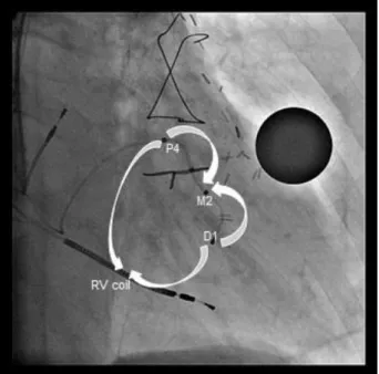
Subsequently, patients were brought to the ward and ECGs (25 mm/s, 10 mm/mV) were recorded under different biventricular pacing configurations. Four LV pacing configurations with the longest electrode distance were selected for comparison. Two bipolar pacing vectors (D1-M2, P4-M2) and two unipolar vectors, also called extended bipolar pacing vectors (D1-RVcoil, P4-RVcoil), were selected (Figure 1.). The LV pacing configurations with the longest inter-electrode distances were selected for comparison. The D1-M2 electrode pacing configuration was designated as bipolar LV1, the D1-RVcoil as unipolar LV1, P4-M2 as bipolar LV4, and the P4-RVcoil as unipolar LV4. A quick optimization module was used to program pacing parameters for each configuration. Threshold tests were performed under 12 lead ECG monitoring to avoid anodal capture. Both unipolar and bipolar pacing amplitude was programmed at the
14
same output and 0.5 mV above the threshold to minimize initial capture area. Twelve- lead QRS morphology was assessed to control stable capture at the programmed output.
Patients with high pacing thresholds (>3 mV) and patients who had capture problems in selected electrodes were excluded.
Figure 1. Right anterior oblique fluoroscopic view of seventh patient, representing the four analyzed pacing vectors between the quadripolar LV lead electrodes and right ventricular (RV) coil. Bipolar- non-basal, Distal 1 (D1) to mid 2 (M2); bipolar-basal, proximal 4 (P4) to mid 2 (M2); unipolar- non-basal, Distal 1 (D1) to RV coil; unipolar- basal, proximal 4 (P4) to RV coil.
All ECGs were digitally scanned and measurements were made with digital calipers at 400% magnification. Analysis was performed by a blinded physician. Lead V5 was selected for analysis. If the T wave in V5 was not eligible, lead II was used. The QT interval was defined as the time interval between the initial deflection of the QRS and the point at which a tangent drawn to the steepest downslope of the T wave crossed the isoelectric line (24). Intervals were corrected for heart rate using Bazett’s formula (25).
The Tp-Te interval was obtained from the difference between the QT interval and the QT peak interval. The QT peak interval was measured from the beginning of QRS until
15
the peak of a positive T wave or the nadir of a negative T wave (26). The Tp-Te/QT ratio was also calculated. Repolarization parameters during bipolar and unipolar LV pacing from basal and non-basal locations were compared.
3.2.5. Statistical analysis
Mean values and standard deviations (SD) were used for descriptive statistics.
Categorical data were summarized as frequencies and percentages. Comparisons of repolarization parameters between pacing modes were performed with paired, two- tailed Student’s t tests. In all analyses p<0.05 was considered statistically significant.
3.3. Evaluating the effects of respiratory gating on AF ablation procedures
3.3.1. Study population
One-hundred-forty consecutive patients who underwent catheter ablation of AF for the first time admitted to the study. Three experienced physicians who perform totally more than 400 AF ablations per year participated the study. All procedures were done by the same physicians in both groups under conscious sedation and spontaneous respiration.
All patients had symptomatic, drug refractory AF.
3.3.2. Electroanatomical map construction and respiratory gating
Left atrial geometry was constructed using fast anatomical mapping (FAM) feature of EAM system Carto 3 (Biosense Webster Inc., Diamond Bar, CA). Navistar 7F open irrigated-tip catheter (Biosense Webster Inc, Diamond Bar, CA) was used for mapping and ablation. In respiratory gated group AccuResp module (Biosense Webster Inc., Diamond Bar, CA) was enabled and respiration training was done. During the training phase, mapping catheter was placed in the left inferior pulmonary vein with a good contact. Gated threshold was set at 30% of the respiratory cycle and FAM of the LA was constructed. Mapping was started immediately in non-gated group. Time from the connection of mapping catheter to the first RF application was defined as electro-
16
anatomical map reconstruction time. Resolution thresholds were set at the same levels in both groups during FAM construction
3.3.3. CT/MRI image acquisition and integration
After completion of FAMs, they were integrated with pre-acquired CT or MRI images (125 patients CT, 15 patients MRI). CT was performed using 64 slice CT scanner (Philips Brilliance iCT 256) and MR angiography was performed using 1.5 T MR scanner (Philips Achieva 1.5 T Dual Nova HP R2.6.3p7). Carto Merge System (Biosense Webster Inc., Diamond Bar, CA) was used for segmentation and integration of LA model into electroanatomic maps. Since the respiratory changes are most prominent in distal PVs and anterior areas near the mitral valve, these regions were not used during landmark registration (23).
3.3.4. Catheter Ablation
Circumferential left atrial ablation lines were performed around the antrum of ipsilateral pulmonary veins with power limited to 30 W and temperature to 43 ºC. PVs were continuously assessed for electrical isolation using the circular mapping catheter. After completion of the lines, bidirectional block was verified with pacing maneuvers. When needed additional lines were added in patients with persistent AF. Ablation of complex fractionated electrograms was not performed. If AF persisted following the linear ablation, DC cardioversion was performed. Ablation time was determined from the beginning of first ablation to the end of last ablation. Each application duration was automatically calculated by electrophysiology recording system and total value was defined as total RF application duration.
3.3.5. Statistical analysis
All variables are represented as a mean±standard deviation or median and interquartile range as appropriate. Normality was evaluated using Kolmogorov-Smirnov test.
Comparisons between groups were performed with either an unpaired Student’s t-test or Mann-Whitney U test where a normal distribution could not be assumed. A p value
<0.05 was considered to be statistically significant.
17
4. Results
4.1. Evaluating the effects of endocardial vs. epicardial LV pacing
Between September 2007 and March 2014, 48 patients in our center had transseptal endocardial LV lead placement. Thirteen of them had a CS lead dislodgement history after successful conventional CRT implantation and epicardial pacing. Five patients who did not have analyzable CRT ECGs and one patient who had CRT upgrade from RV pacing were excluded. No patient was excluded due to uncontrolled heart rate (biventricular pacing rate <90%), procedure related complications, or death. The data of seven eligible patients were analyzed. Baseline characteristics are given in Table 1.
Table 1. Baseline characteristics of the patients. Values are mean SD or n (%). LVEF, left ventricle ejection fraction; NYHA, New York Heart Association; CRT-D, cardiac resynchronization therapy with defibrillator; CRT-P, cardiac resynchronization therapy with pacemaker; ACE-I, angiotensin-converting enzyme inhibitor; ARB, angiotensin II receptor blocker; LBBB, left bundle branch blockade.
Age (years) 63 3.7
Male 6 (85.7)
LVEF (%) 30 3.0
Etiology Ischemic Nonischemic
4 (57.1) 3 (42.9) NYHA functional class
II III
1 (14.3) 6 (85.7) Device
CRT-D CRT-P
4 (57.1) 3 (42.9)
Atrial Fibrillation 2 (28.6)
Diabetes Mellitus 1 (14.3)
Hypertension 5 (71.4)
Drugs
ACE-I/ARB Beta bloker Amiodarone
Other QT prolonging drug
7 (100) 5 (71.4) 1 (14.3) 0
QRS morphology (LBBB) 7 (100)
18
The mean age of the patients was 63±4 and the majority of them were male. Four of them (57.1%) had ischemic dilated cardiomyopathy. The positions of the CS leads were reported in a lateral location with stable pacing thresholds. Transseptal LV leads were implanted in lateral position in all patients. No thromboembolic complications were observed during the follow up.
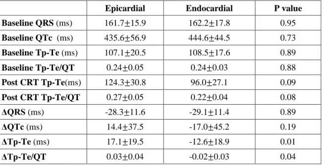
Baseline QRS durations (161.7±15.9 vs. 162.2±17.8 ms, p=0.95), QTc intervals (435.6±56.9 vs. 444.6±44.5 ms, p=0.73), Tp-Te values (107.1±20.5 vs. 108.5±17.6 ms, p=0.89) and Tp-Te/QT ratios (0.24±0.05 vs. 0.24±0.03, p=0.88) were similar before epi and endo CRT (Table 2.).
In all patients QRS intervals reduced significantly following both epi and endo CRT (161.71±16 vs. 133.42±15 ms, p<0.01; 162.28±18 vs. 133.14±8 ms, p<0.01 resp.).
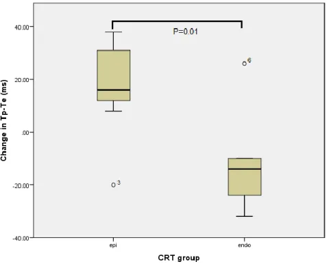
Although QRS interval reductions were similar (-28.3±11.6 vs.-29.1±11.4 ms, p=0.89), epi CRT was associated with a significant increase in Tp-Te values (17.1±19.5 vs.
-12.6±18.9 ms, p=0.01) and Tp-Te/QT ratios (0.03±0.04 vs. -0.02±0.03, p=0.04) compared to endo CRT (Figure 2.). Differences in QTc interval changes were not significant (14.4±37.5 vs. -17.0±45.2 ms, p=0.19) (Table 2.).
Table 2. Comparison of repolarization parameters before and after cardiac resynchronization therapy with endocardial and epicardial left ventricular pacing.
Epicardial Endocardial P value
Baseline QRS (ms) 161.7 15.9 162.2 17.8 0.95
Baseline QTc (ms) 435.6 56.9 444.6 44.5 0.73 Baseline Tp-Te (ms) 107.1 20.5 108.5 17.6 0.89
Baseline Tp-Te/QT 0.24 0.05 0.24 0.03 0.88
Post CRT Tp-Te(ms) 124.3 30.8 96.0 27.1 0.09
Post CRT Tp-Te/QT 0.27 0.05 0.22 0.04 0.08
ΔQRS (ms) -28.3 11.6 -29.1 11.4 0.89
ΔQTc (ms) 14.4 37.5 -17.0 45.2 0.19
ΔTp-Te (ms) 17.1 19.5 -12.6 18.9 0.01
ΔTp-Te/QT 0.03 0.04 -0.02 0.03 0.04
19
Figure 2. Box plot of change in Tp-Te following epicardial and endocardial cardiac resynchronization therapy. CRT, cardiac resynchronization therapy; epi, epicardial;
endo, endocardial.
4.2. Evaluating the effects of bipolar vs. unipolar and basal vs. non- basal LV pacing
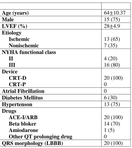
The protocol was successfully completed in 20 of the 26 patients. Two patients in whom the proximal LV electrode (P4) could not be inserted into the branch of the CS were excluded. Three patients with high pacing thresholds (>3 mV) and one patient with phrenic nerve capture in selected electrodes were also excluded. None of the subjects were excluded due to procedure-related complications. The baseline characteristics of the remaining 20 patients are given in Table 3. The mean age of the patients was 64±10, and a majority of them (65%) had ischemic dilated cardiomyopathy.
20
Table 3. Baseline characteristics of study patients. Values are mean SD or n (%).
LVEF, left ventricle ejection fraction; NYHA, New York Heart Association; CRT-D, cardiac resynchronization therapy with defibrillator; CRT-P, cardiac resynchronization therapy pacemaker; AF, atrial fibrillation; DM, diabetes mellitus; HT, hypertension; ACE-I, angiotensin-converting enzyme inhibitor; ARB, angiotensin II receptor blocker;
LBBB, left bundle branch block.
Age (years) 64 10.37
Male 15 (75)
LVEF (%) 28 4.9
Etiology Ischemic Nonischemic
13 (65) 7 (35) NYHA functional class
II III
4 (20) 16 (80) Device
CRT-D CRT-P
20 (100) 0
Atrial Fibrillation 0
Diabetes Mellitus 6 (30)
Hypertenson 13 (75)
Drugs
ACE-I/ARB Beta bloker Amiodarone
Other QT prolonging drug
20 (100) 14 (70) 1 (5) 0
QRS morphology (LBBB) 20 (100)
The quadripolar LV lead was placed in the lateral branch in 12 patients, in the posterolateral branch in four patients and in the anterolateral branch in four patients. In two patients, due to angulation of the CS branch, the D1 and P4 electrodes were located in different segments of the LV in the LAO view. In all other patients, the D1 and P4 electrodes were located in the same segments. In all patients, the P4 electrodes were located in basal segments and the D1 electrodes were located in non-basal segments of the LV (mid-ventricular location in 13 patients, apical location in seven patients). The mean distance between the D1 and P4 electrodes was 43.17±3 mm in the RAO view.
21
Table 4. shows the electrocardiographic parameters generated by unipolar and bipolar pacing from both basal and non-basal locations. Bipolar LV pacing was associated with a significantly better Tp-Te value than unipolar pacing from both sides of the LV.
(Basal, unipolar vs. bipolar, 119.1±36.7 vs. 97.6±27.9, p<0.05; non-basal, unipolar vs.
bipolar, 117.9±36.3 vs. 98.6±20.4, p<0.05). Pacing from basal and non-basal segments of LV had no differential effect on repolarization parameters (Bipolar Tp-Te, basal vs.
non-basal, 97.6±27.9 vs. 98.6±20.4, p=0.89; unipolar Tp-Te, basal vs. non-basal, 119.1±36.7 vs. 117.9±36.3, p=0.92). The mean baseline Tp-Te/QT ratio was 0.25±0.05.
Tp-Te/QT ratios were lower with bipolar pacing, the differences were not significant (Basal, unipolar vs. bipolar, 0.26±0.06 vs. 0.23±0.06, p=0.14; non-basal, unipolar vs.
bipolar, 0.28±0.10 vs. 0.23±0.03, p=0.06). There was no significant change between QTc intervals (Table 4.).
Table 4. Comparison of repolarization parameters and QRS intervals generated by unipolar and bipolar pacing from both basal and non-basal locations.
Unipolar Bipolar p Basal NonBasal p
QRS (ms)
Basal 135±18 119±15 <0.01 Unipolar 135±19 134±16 0.89 NonBasal 134±16 122±10 <0.01 Bipolar 119±15 122±10 0.53
Tp-Te (ms)
Basal 119±37 98±28 <0.05 Unipolar 119±37 118±36 0.92 NonBasal 118±36 99±20 <0.05 Bipolar 98±28 99±20 0.89
Tp- Te/QT
Basal 0.26±0.06 0.23±0.06 0.14 Unipolar 0.26±0.06 0.28±0.10 0.54 NonBasal 0.28±0.10 0.23±0.03 0.06 Bipolar 0.23±0.06 0.23±0.03 0.81
QTc (ms)
Basal 449±43 431±48 0.22 Unipolar 449±43 442±52 0.62 NonBasal 442±52 429±49 0.43 Bipolar 431±48 429±48 0.83
QRS intervals in all patients reduced significantly following both unipolar and bipolar CRT (p<0.01). However, QRS reduction was more prominent with bipolar pacing than with unipolar pacing (Basal, unipolar vs. bipolar, 135.1±17.8 vs. 119.3±14.5 ms, p<0.01; non-basal, unipolar vs. bipolar, 134.4±15.7 vs. 121.9±10.3 ms, p<0.01).
22
Interestingly, LV pacing location had no impact on QRS duration (Bipolar, basal vs.
non-basal, 119.3±14.5 vs. 121.9±10.3 ms, p=0.53; unipolar, basal vs. non-basal, 135.1±17.8 vs. 134.4±15.7 ms, p=0.89).
4.3. Evaluating the effects of respiratory gating on AF ablation procedures
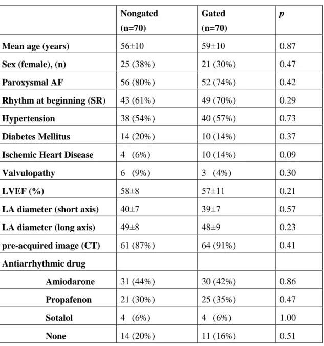
We enrolled 140 patients who underwent AF ablation for the first time. In respiratory gated group (70 patients, mean age 59±10 years, 21 female) underwent procedures after enabling AccuResp module. In nongated group (70 patients, mean age 56±10 years, 25 females) procedures were performed using the same EAM system without enabling the AccuResp module. Baseline clinical factors did not differ between the two groups (Table 5.).
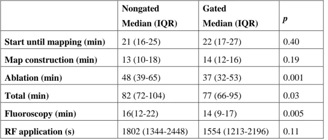
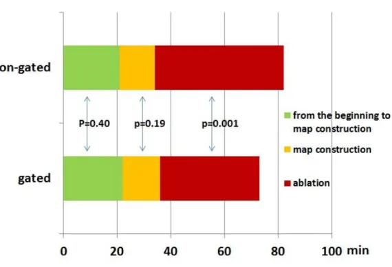
Durations from the beginning of the procedure to beginning of the EAM reconstruction were similar in both groups [median 22 (IQR 17-27) vs. median 21min (IQR 16-25), p=0.40, Table 6., Figure 3.).
In gated group electroanatomical maps reconstructed within median 14 (IQR 12-16) vs.
median 13 (IQR 10-18) min in non-gated group (p=0.19) (Table 6., Figure 3.).
Significantly shorter ablation times were observed in respiratory gated group [median 37 (IQR 32-53) min vs. median 48 (IQR 39-65) min, p<0.05]. However, total RF application durations were not different between two groups [median 1554 (IQR 1213- 2196) s vs. median 1802 (IQR 1344-2448) s, p=0.11, Table 6., Figure 3.].
23
Table 5. The demographics of the patients from the third study are summarized in this table. AF, atrial fibrillation; CT, computed tomography; EF, left ventricle ejection fraction; LA, left atrium; SR, sinus rhythm.
Nongated (n=70)
Gated (n=70)
p
Mean age (years) 56±10 59±10 0.87
Sex (female), (n) 25 (38%) 21 (30%) 0.47
Paroxysmal AF 56 (80%) 52 (74%) 0.42
Rhythm at beginning (SR) 43 (61%) 49 (70%) 0.29
Hypertension 38 (54%) 40 (57%) 0.73
Diabetes Mellitus 14 (20%) 10 (14%) 0.37
Ischemic Heart Disease 4 (6%) 10 (14%) 0.09
Valvulopathy 6 (9%) 3 (4%) 0.30
LVEF (%) 58±8 57±11 0.21
LA diameter (short axis) 40±7 39±7 0.57
LA diameter (long axis) 49±8 48±9 0.23
pre-acquired image (CT) 61 (87%) 64 (91%) 0.41 Antiarrhythmic drug
Amiodarone 31 (44%) 30 (42%) 0.86 Propafenon 21 (30%) 25 (35%) 0.47 Sotalol 4 (6%) 4 (6%) 1.00 None 14 (20%) 11 (16%) 0.51
24
Table 6. Durations of different steps of procedures are compared between non-gated vs.
gated groups. RF, radiofrequency.
Nongated Median (IQR)
Gated
Median (IQR) p Start until mapping (min) 21 (16-25) 22 (17-27) 0.40 Map construction (min) 13 (10-18) 14 (12-16) 0.19
Ablation (min) 48 (39-65) 37 (32-53) 0.001
Total (min) 82 (72-104) 77 (66-95) 0.03
Fluoroscopy (min) 16(12-22) 14 (9-17) 0.005
RF application (s) 1802 (1344-2448) 1554 (1213-2196) 0.11
A significant reduction in total procedure times [median 77 (IQR 66-95) min vs. median 82 (IQR 72-104) min, p<0.05] and fluoroscopy times [median 14 (IQR 9-17) min vs.
median 16 (IQR 12-22) min, p<0.05] were observed in the respiratory gated group.
One patient from each group had significant pericardial effusion and developed cardiac tamponade 24 hours after procedures. Pericardiocentesis was performed for both patients and surgery wasn’t needed. Vascular access complications rates were similar among two groups. Two hematoma developed in nongated group and 1 hematoma developed in gated group (p=0.56). None of them required intervention.
In nongated group, roof lines were performed in 3 patients and roof plus mitral isthmus lines were performed in 5 patients. In gated group, roof plus mitral isthmus lines were performed in 6 patients and box isolation was performed in one patient. Successful pulmonary vein isolation and sinus rhythm were obtained in all patients. DC cardioversion was performed in 46 patients (24 in nongated and 22 in gated group, p=0.71).
25
Figure 3. Comparison of different time intervals during AF ablation. Note that the intervals from the beginning of the procedure to the start of ablation are similar in both groups, but the ablation time is significantly shorter in respiratory gated group.
26
5. Discussion
5.1. Discussion on effects of endocardial vs. epicardial LV pacing during CRT
The main findings of our first study suggest that permanent LV endocardial CRT is associated with better ventricular repolarization characteristics compared to conventional epicardial CRT. Non-physiological activation of both ventricles with different transmural activation sequences might be responsible for an increase in TDR during epicardial CRT. Our study is the first to compare the effects of permanent endocardial and epicardial CRT in the same patient group. The effect of stimulating both sides of the same substrate (the lateral wall of the LV) was evaluated.
It has been shown that even in the absence of any difference in final repolarization time, reversing the direction of activation affects the action potential curve and T wave morphology (5, 6). Medina-Ravell et al. evaluated pacing site dependent changes in ventricular repolarization, and observed more significant prolongation in TDR with epicardial and biventricular pacing (4). They reported the development of torsade de pointes in a heart failure patient with epicardial or biventricular pacing, but not with endocardial pacing only. Monomorphic ventricular tachycardias induced by CRT are also reported (27, 28).
Recently, Scott et al. investigated the effect of permanent endocardial CRT on ventricular repolarization in humans (11). Tp-Te and QT dispersion values were significantly lower in a transseptal LV endocardial group than in a CS epicardial group.
However, this case control designed study had limitations due to the difficulty of finding a perfect match between the groups. Currently, LV endocardial CRT indication is limited to patients in whom conventional techniques have failed and who have a higher chance of responding. Patients scheduled for transseptal CRT implantation may have more severe heart failure than usual. Therefore, differences in baseline characteristics between two groups might have an impact on results.
Although our observations on repolarization patterns were consistent with Scott et al., we observed different depolarization changes. They reported significantly better post
27
CRT reduction in QRS duration in the transseptal group. However, we observed similar QRS reduction with epi and endo CRT in the same patient group.
The QRS interval during biventricular pacing is complicated because there are two activation vectors. The underlying heart disease and localizations of myocardial scars can make a contribution to the electrophysiological effects of biventricular pacing.
Therefore, differences between the LV endocardial patients and the control group might be responsible for this finding.
Factors that influence depolarization patterns may also affect repolarization patterns.
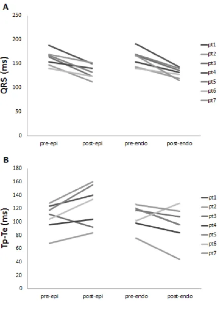
Nevertheless, our results corroborate animal experiments evaluated the effects of endocardial and epicardial pacing on transmural wedge preparations (4, 29). Despite a similar reduction in QRS intervals, we observed significant changes in Tp-Te and TpTe/QT values (Figure 4.). QTc and Tp-Te values were increased with epi CRT and decreased with endo CRT. Therefore, the main electrical benefits of LV endocardial CRT are attributable to the favorable effects of repolarization rather than to depolarization.
28
Figure 4. The effects of epicardial and endocardial cardiac resynchronization therapy (CRT) on QRS (A) and Tp-Te (B) in the same patient group. Please note that epicardial and endocardial CRT produced similar QRS interval reductions. However, epicardial CRT was associated with a significant increase in Tp-Te values compared to endocardial CRT. pre-epi, pre-epicardial CRT; post-epi, post-epicardial CRT, pre-endo, pre-endocardial CRT; post-endo, post-endocardial CRT.
A Tp-Te/QT ratio of 0.25 is a significant risk factor for appropriate ICD therapy in conventional CRT-D patients (8). In our patient group, baseline Tp-Te/QT ratios were similar for both epi and endo CRT. However, mean Tp-Te/QT ratios increased to 0.27±0.05 with epi CRT and decreased to 0.22±0.04 with endo CRT. Δ Tp-Te/QT values were significantly different (Table 2).
29
Our study focused on TDR response during epi and endo CRT, and was neither designed nor powered to evaluate the relevant clinical arrhythmias. However, there are previous studies indicating that TDR prolongation during epi CRT is associated with ventricular arrhythmias (4, 30). Better TDR characteristics with endo CRT may decrease clinical arrhythmias. Unfortunately, our patient group was too small to demonstrate a significant difference in terms of relevant clinical arrhythmias.
Although our study was limited by the small sample size, it was sufficient to show significant changes in TDR parameters during endo and epi CRT in a uniform pattern.
However, further multicenter studies with larger patient groups are warranted to confirm these findings.
We selected only lead V5 (or lead II if V5 was not eligible) for repolarization parameter measurements. Analysis of a single lead might influence the accuracy of ventricular repolarization. However, previous studies that showed the association of increased Tp-Te interval and Tp-Te/QT ratio and ventricular arrhythmias during CRT also used one-lead measurements, and these parameters are widely accepted (8, 30, 31).
In addition, only acute responses to CRT were examined in our study, but long term electrical and mechanical remodeling could modify the results (7, 32).
5.2. Discussion on effects of pacing polarity and pacing site during CRT
The main finding of the our second study, investigating the impact of LV pacing polarity and LV pacing site on repolarization parameters in the same patient group, can be summarized as follows:
(i) Bipolar LV pacing is associated with better ventricular repolarization characteristics than is unipolar LV pacing.
(ii) The LV pacing site has no differential effect on repolarization parameters from the perspective of basal and non-basal segments.
30
The spread of activation in the ventricle is different during unipolar and bipolar pacing.
An unipolar wave front attenuates with the square of the distance, and a bipolar wave front attenuates with the third power of the distance (33). The size and shape of the virtual electrode is also influenced by the pacing polarity (34, 35). The point of initial capture on the epicardium may be the same, but subepicardial layers captured by the virtual electrode may be different. Furthermore, the myocardium of patients with heart failure is electrically and mechanically heterogeneous. The presence of scars may lead to changes in conduction vectors and may change the transmural activation sequence.
Our observation on QRS duration is consistent with a previous study that evaluated the changes in electromechanical parameters during different pacing configurations using a quadripolar lead (36). It was observed in this study that the best configurations for QRS duration were most commonly bipolar pacing modes (D1-M2, P4-M2) and the worst configuration was most commonly the unipolar mode (P4-RV). In our study, QRS reduction was more prominent with bipolar pacing, and LV pacing location had no impact on QRS duration.
These findings collectively indicate that there are differences in the capture and activation of ventricles. Naturally, factors that influence depolarization patterns may also affect repolarization patterns. It has been shown that even in the absence of any difference in final repolarization time, reversing the direction of activation affects the action potential curve and T wave morphology (5). There is an intrinsic repolarization difference between epicardium, mid myocardial M cells and endocardium. Different vectoral activation of LV with different transmural activation during unipolar and bipolar pacing might be responsible for our findings. We observed a significant difference in Tp-Te values between unipolar and bipolar pacing from both basal and non-basal segments of the LV (Figure 5.). A Tp-Te/QT ratio of ≥0.25 has been reported as a significant risk factor for appropriate ICD therapy in CRT patients (8).
Although the difference between pacing configurations was not statistically significant, the mean Tp-Te/QT ratio was >0.25 with unipolar pacing, and <0.25 with bipolar pacing (Table 4).
31
Figure 5. Box plot of Tp-Te generated by unipolar and bipolar pacing from both basal and non-basal locations.
Data on the role of LV pacing site during conventional CRT is controversial. Kleemann et al. suggested that different LV lead positions were not associated with an increase in ventricular arrhythmias (37). Kutyifa et al. analyzed the association between the LV lead position and the risk of ventricular arrhythmias in patients enrolled in a MADIT- CRT trial (38), and found that posterior or lateral lead locations were associated with decreased risk of arrhythmic events compared with anterior LV lead positions.
Contrarily, patients with apical LV lead location had a similar incidence of ventricular arrhythmias to patients with non-apical lead location. The underlying heart disease and localizations of myocardial scars can make a contribution to the electrophysiological effects of biventricular pacing. Therefore, differences in baseline characteristics among the patient groups might be responsible for these controversial results. We assessed the impact of basal and non-basal pacing on repolarization patterns in the same patient group using a quadripolar LV lead. Consistent with the previous clinical study, we
32
observed no difference in terms of repolarization patterns between basal and non-basal pacing of the same substrate (38). We must acknowledge that our observation is limited by the longitudinal aspect of the LV. The impact of pacing locations along the short axes of the heart would be different and should be addressed in future investigations.
Another limitation of our study is its small sample and ischemic etiology bias. The majority of our patients (65%) had ischemic cardiomyopathy and 11 of them had a history of anterior myocardial infarction. As noted above, the presence of large ischemic scars and heterogeneity of the myocardial substrate may have had an impact on our results. Nevertheless, this population reflects the general patient population receiving CRT. Due to the relatively small number of patients, no subgroup analysis on ischemic and non-ischemic patients was performed.
All limitations related to ECG documentations and long term electrical and mechanical remodeling that we emphasized above, are also clear limitations for our second study.
However, as we mentioned before these parameters and techniques are widely accepted and commonly used (8).
5.3. Discussion on effects of respiratory gating on AF ablation procedures
Principal findings of our third study demonstrated that respiratory gating allows faster AF ablation procedures and reduces X-ray screening. However, the use of this technology was not able to reduce the total RF application durations.
During the past decade EAM systems achieved remarkable advances and became the cornerstone of AF ablations (15, 10, 11). Despite these advances, accuracy of maps are still affected by biological factors such as respiration (23, 41). During EAM reconstruction, catheter movements are affected by respiration and significant changes occur in LA-PV geometry. Moreover, deep inspiration during reconstruction could deform maps. PV ostiums are crucial landmarks of AF ablation and important changes in their positions, predominantly in inferior direction, were reported during respiration (23).
33
Respiratory gating based on the end expiration might provide accurate maps and compensate negative effects of respiration. Previous work by Beinart et al. has confirmed that more accurate electroanatomical maps can be obtained by respiratory gating (22). The only study investigated the impact of respiratory gating on RF ablation was about cavotricuspid isthmus (CTI) ablation procedures (42). In this study respiratory gated acquisition resulted in a better visualization of CTI and a significant reduction in fluoroscopy and RF times. However, LA anatomy is more complex then CTI and ablation lines in multiple dimensions are needed during PV isolations. To the best of our knowledge this is the first study investigated the impact of respiration gating on atrial fibrillation ablation and our results support its favorable effects.
5.3.1. EAM reconstruction times
AccuResp module automatically detects out-of-gate acquisition and excludes them from the created anatomy, thus it does not slows down the mapping procedure. Although reconstruction times were slightly longer in respiratory gated group, difference was not significant. However, all procedures in our study were performed with image integration and only crucial parts for landmark registration were mapped instead for detailed reconstruction of all LA. Therefore, our results may not be generalized for ablation procedures without image integration.
5.3.2. Potential advantage of respiratory gating
Significant reduction in procedural times is the result of shorter ablation times in respiratory gated group. Durations from the beginning of the procedure to the start of ablation were similar in both groups, but the ablation times were significantly shorter in respiratory gated group (Figure 3). On the other hand, total RF energy applied to isolate PVs was not influenced by respiratory gating. These data seems to be the result of uninterrupted ablation applications in respiratory gated group. Although this study was not designed to evaluate the accuracy of electroanatomical maps and compare them with pre-acquired CT or MRI images, reduction in ablation and fluoroscopy times supports superiority of gated maps.
34
Catheter-tissue contact force is also critically influenced by respiration (43, 44).
However, it seems to be directly related to the respiratory movement itself and it is hard to suggest that the respiratory gating had positive effects on contact force. Catheter positions are visualized real-time during ablation and respiratory gating does not have effect of catheter view, it only excludes out-of-gate ablation points and may affect shape of ablation lines. For these reasons, main advantage of respiratory gating is attributable to the realistic LA geometry construction.
In addition, merging electroanatomical maps and pre-acquired static 3D images did not compensate negative effects of respiration. Although the prominently mobile points were avoided during landmark registration, there were significant differences between two groups.
5.3.3. Safety
We observed no difference in cardiovascular complication rates among two groups.
However, extensive use of fluoroscopy during long ablation procedures increases the radiation risk for patients and medical staff (45-48). Moreover, radiation exposure during AF ablation is longer than simpler catheter ablation procedure (20, 21). EAM systems have been demonstrated to reduce fluoroscopy times during RF ablations (51- 53). Our results imply that, the respiratory gating provides further reduction in fluoroscopy time.
It should be remembered that all procedures in our study were performed with Carto3 system under guidance of image integration. Therefore, validation of our results is limited for this ablation technique. All patients ablated under conscious sedation and spontaneous respiration. Our results can’t be generalized for procedures performed under general anesthesia, where respiratory rate and tidal volumes are predictable. In addition, this study was not designed to assess long term clinical outcome after AF ablation, but was focused on procedural outcomes. Studies evaluate the long term effects of this technique are needed.
35
6. Conclusion
A/ Transseptal LV endocardial pacing is associated with better arrhythmogenic repolarization characteristics than epicardial pacing in CRT. Although large multicenter randomized studies have not reported any increased incidence of ventricular tachycardia (VT) or ventricular fibrillation (VF) episodes, it is important to determine CRT patients who are prone to have ventricular arrhythmias. Larger clinical studies are needed to determine whether these effects may contribute to reduction of arrhythmias.
B/ LV pacing polarity significantly affects repolarization patterns regardless of pacing site. Bipolar LV pacing is associated with better ventricular repolarization characteristics compared to unipolar LV pacing. The LV pacing site, from the perspective of basal and non-basal segments, has no differential effect on repolarization parameters. Quadripolar LV leads offer ten left ventricular pacing configurations, and unipolar (extended bipolar) LV pacing is widely used to overcome technical issues such as phrenic nerve capture and stimulation thresholds. Randomized controlled studies comparing the bipolar and unipolar LV pacing are needed to determine whether these changes are related to arrhythmic risk in patients with CRT.
C/ Respiratory gated electroanatomic mapping significantly improves ablation and fluoroscopy times during AF ablation. Beside these advantages, using automatic respiratory gating module does not prolong mapping times.
36
7. Summary
The aims of this work were to asses (A) the proarryhthmic repolarisation characteristics of endocardial and epicardial biventricular pacing in the same patient population; (B) to investigate the impact of LV pacing polarity and epicardial pacing site on repolarization parameters in the same patient group; and, (C) to determine impact of respiratory gating on procedural parameters in patients undergoing catheter ablation of AF.
To investigate the above fields 3 clinical studies were accomplished as follows:
A/ Seven patients who had transseptal endocardial left ventricle (LV) lead placement, in whom the standard epicardial CRT implantation had failed due to coronary sinus (CS) lead dislodgement after successful implantation, were admitted to the study. ECG markers of TDR (Tp-Te and Tp-Te/QT ratio) were measured and compared.
B/ Twenty patients, who had CRT-D implantation with quadripolar LV lead were enrolled to the study. ECG markers of TDR (Tp-Te and Tp-Te/QT ratio) were measured and compared.
C/ One-hundred-forty consecutive patients undergoing pulmonary vein isolation were selected to study. Total procedure times, fluoroscopy times, RF application durations and electroanatomical map reconstruction times were recorded and compared.
The results show that the transseptal LV endocardial pacing is associated with better arrhythmogenic repolarization characteristics than epicardial pacing in CRT (A); and, that the LV pacing polarity significantly affects repolarization patterns regardless of pacing site: bipolar LV pacing is associated with better ventricular repolarization characteristics compared to unipolar LV pacing. At the same time, the LV pacing site, from the perspective of basal and non-basal segments, has no differential effect on repolarization parameters (B); finally, respiratory gating significantly improves fluoroscopy and ablation times during electroanatomic mapping guided AF ablation.
Respiratory gated maps may provide uninterrupted continuous ablation applications.
Furthermore, using automatic respiratory gating module does not prolong mapping times (C).
As a conclusion endocardial biventricular pacing creates a more favourable repolariz- ation settings in CRT patients and respiration gating might be helpful in atrial fibrill- ation ablation.
37
8. Összefoglalás
A munka célja volt (A) az endokardiális és epikardiális biventrikuláris ingerlés proaritmiás repolarizációs tulajdonságainak vizsgálata ugyanabban a betegpopulá- cióban; (B) a bal kamrai (BK) ingerlés-polaritás és az epikardiális ingerlési hely hatásának vizsgálata a repolarizációs paraméterekre ugyanabban a betegpopulációban;
(C) a légzés-kompenzáció hatásának vizsgálata a beavatkozásra pitvarfibrilláció abláción áteső betegek esetében.
A fentiek vizsgálatát az alábbiak szerint végeztük:
A/ Hét, korábban sikeresen, a hagyományos úton (sinus coronarius oldalvéna) implantált elektróda kimozdulása miatt transszeptális endokardiális BK elektróda implantáción átesett CRT-s betegnél mértük és hasonlítottuk össze a repolarizáció transzmurális diszperziójának (TDR) EKG markereit (Tp-Te és Tp-Te/QT arány).
B/ Húsz, korábban kvadripoláris BK elektródával rendelkező CRT-D implantáción átesett betegnél mértük és hasonlítottuk össze a TDR EKG markereit (Tp-Te és Tp-Te/QT arány).
C/ Száznegyven pitvarfibrilláció abláción átesett betegnél vizsgáltuk a teljes procedúra időt, a fluoroszkópos időt, a radiofrekvenciás applikációk időtartamát és az elektro- anatómiai térképezés idejét.
Az eredményeink alapján a transszeptális endokardiális BK ingerlés jobb aritmogén repolarizációs karakterisztikával bír az epikardiális ingerléssel szemben CRT esetén (A); a BK ingerlés polaritása szignifikáns hatással van a repolarizációs mintázatra függet-lenül az ingerlés helyétől: a bipoláris BK ingerlés kedvezőbb kamrai repolarizációs karakterisztikával bír az unipolaris BK ingerléssel szemben. Ugyanakkor, a BK ingerlési helynek (bazális vs. non-bazális szegmensek) nincs jelentős hatása a repolarizációs paraméterekre (B); végül a légzés-kompenzáció szignifikánsan javítja a fluoroszkópiás és az ablációs időt az elektroanatómiai térképezéssel történő pitvarfibrilláció abláció során. Az automata légzés-kompenzációs modulok a térképezés idejét nem nyújtják meg, azonban jelentős segítséget nyújthatnak az ablációs pontok folyamatos elhelyezésében (C).
Összefoglalásként eredményeink alapján elmondható, hogy az endokardiális bipoláris BK ingerlés az unipolárissal szemben kedevezőbb repolarizációs tulajdonságokkal bír, valamint a légzés-kompenzáció segítséget jelenthet a pitvarfibrilláció abláció során.
38
9. References
1. McMurray JJ, Adamopoulos S, Anker SD, Auricchio A, Böhm M, Dickstein K, Falk V, Filippatos G, Fonseca C, Gomez-Sanchez MA, Jaarsma T, Køber L, Lip GY, Maggioni AP, Parkhomenko A, Pieske BM, Popescu BA, Rønnevik PK, Rutten FH, Schwitter J, Seferovic P, Stepinska J, Trindade PT, Voors AA, Zannad F, Zeiher A. (2012) ESC Committee for Practice Guidelines. ESC Guidelines for the diagnosis and treatment of acute and chronic heart failure 2012: The Task Force for the Diagnosis and Treatment of Acute and Chronic Heart Failure 2012 of the European Society of Cardiology. Developed in collaboration with the Heart Failure Association (HFA) of the ESC. Eur Heart J, 33: 1787-1847.
2. Brignole M, Auricchio A, Baron-Esquivias G, Bordachar P, Boriani G, Breithardt OA, Cleland J, Deharo JC, Delgado V, Elliott PM, Gorenek B, Israel CW, Leclercq C, Linde C, Mont L, Padeletti L, Sutton R, Vardas PE. (2013) 2013 ESC guidelines on cardiac pacing and cardiac resynchronization therapy: the task force on cardiac pacing and resynchronization therapy of the European Society of Cardiology (ESC).
Developed in collaboration with the European Heart Rhythm Association (EHRA).
Europace, 15: 1070-1118.
3. Cleland JG, Daubert JC, Erdmann E, Freemantle N, Gras D, Kappenberger L, Tavazzi L. (2006) Longer-term effects of cardiac resynchronization therapy on mortality in heart failure [the CArdiac REsynchronization-Heart Failure (CARE- HF) trial extension phase]. Eur Heart J, 27: 1928-1932.
4. Medina-Ravell VA, Lankipalli RS, Yan GX, Antzelevitch C, Medina-Malpica NA, Medina-Malpica OA, Droogan C, Kowey PR. (2003) Effect of epicardial or biventricular pacing to prolong QT interval and increase transmural dispersion of repolarization: Does resynchronization therapy pose a risk for patients predisposed to long QT or torsade de pointes? Circulation, 107: 740-746.
5. Fish JM, Di Diego JM, Nesterenko V, Antzelevitch C. (2004) Epicardial activation of left ventricular wall prolongs QT interval and transmural dispersion of repolarization: implications for biventricular pacing. Circulation, 109: 2136-2142.
6. Fish JM, Brugada J, Antzelevitch C. (2005) Potential proarrhythmic effects of biventricular pacing. J Am Coll Cardiol, 46: 2340–2347.
