Investigation of the human ABCB6 transporter
PhD thesis
Zsófia Rakvács
Semmelweis University
Doctoral School of Molecular Medicine
Supervisor: Gergely Szakács MD., Ph.D.
Official reviewers: Miklós Geiszt, MD., DSc.
Péter Lőw Ph.D.
Chairman of the examination committee: János Réthelyi MD., Ph.D.
Members of the examination committee: Gergely Papp Ph.D.
Ákos Sveiczer Ph.D.
Budapest 2019
2
Table of Contents
List of Abbreviations ... 4
1. Introduction ... 5
1.1. ABC protein family ... 5
1.1.1. Structural determinants of function ... 5
1.1.2. ABCB subfamily ... 8
1.2. ABCB6 ... 9
1.2.1. Identification of a new protein ... 10
1.2.2. ABCB6 and porphyrins ... 11
1.2.3. ABCB6 and resistance ... 13
1.2.4. ABCB6 – contrasting results on intracellular localization ... 16
1.2.5. Structure – Function relation ... 17
1.2.6. ABCB6 knockout mice ... 20
1.2.7. ABCB6 and pathology ... 21
1.2.8. ABCB6 and cadmium ... 23
1.2.9. ABCB6 and pigmentation ... 24
2. Aims ... 29
3. Methods ... 30
3.1. Cell culturing ... 30
3.2. Molecular cloning of ABCB6 and HMT-1 constructs ... 31
3.3. Generation of transgenic lines ... 32
3.4. Immunoblotting ... 34
3.5. Cytotoxicity assays ... 35
3.6. Determination of the vacuolar cadmium content ... 36
3.7. Measurement of melanin content... 38
3.8. Confocal microscopy ... 38
3.9. Genome editing by CRISPR/Cas9 ... 41
4. Results ... 45
4.1. The relation of ABCB6 function to the HMT-1 proteins ... 45
4.1.1. Heterologous expression of SpHMT-1 and ABCB6 transporter in S. pombe model organism ... 45
4.1.2. Heterologously expressed human ABCB6 localizes to the vacuolar membrane of S. pombe ... 46
3
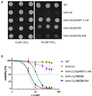
4.1.3. Heterologous expression of human ABCB6 restores cadmium tolerance of
S. pombe hmt-1Δ mutants ... 47
4.1.4. S. pombe functional assay ... 48
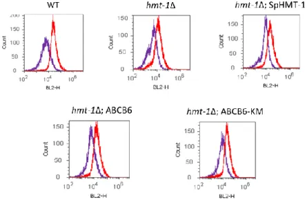
4.1.5. Determination of vacuolar cadmium contents... 50
4.1.6. Human ABCB6 rescues the Cd Hypersensitivity of hmt-1-deleted C. elegans ... 52
4.1.7. Heterologously expressed human ABCB6 localizes to endolysosomes of C. elegans. ... 54
4.1.8. Human ABCB6 confers Cd tolerance to SNB-19 glioblastoma cells ... 58
4.1.9. Human ABCB6 localizes tolysosomes of SNB-19 glioblastoma cells... 59
4.2. ABCB6 and melanogenesis ... 60
4.2.1. ABCB6 localizes to early melanosomes and lysosomes in the human melanocytic cell line MNT-1 ... 60
4.2.2. Downregulation of ABCB6 by siRNA perturbs early steps of PMEL amyloid formation without eliminating melanogenesis ... 63
4.2.3. ABCB6 mutations prevent the rescue of normal amyloid fibril formation .. ... 65
4.3. MNT-1 cell lines genome edited by CRISPR/Cas9 ... 70
4.3.1. Cytogenetic analysis of MNT-1 cells ... 70
4.3.2. Endogenous ABCB6 is located to lysosomes ... 71
4.3.3. ABCB6 KO cell line ... 75
5. Discussion ... 79
5.1. The role of ABCB6 in Cd detoxification ... 79
5.2. The role of ABCB6 in pigmentation ... 84
5.3. MNT-1 cell lines genome edited by CRISPR/Cas9 ... 89
6. Conclusion ... 91
7. Summary ... 93
8. Összefoglalás ... 94
9. References... 95
10. List of publications ... 117
11. Acknowledgement ... 118
12. Supplementary Data ... 119
4
List of Abbreviations
ABC ATP binding cassette
AIF Apoptosis inducing factor
ALA δ-aminolevulinic acid
AO Acridine-orange
BACE2 β-site APP-cleaving enzyme 2 CPIII Coproporphyrin III
CYP Cytochrome P450 oxygenase
DSB Double strand brake
DUH Dyschromatosis universalis hereditaria EEA1 Early endosome antigen 1
EGFP Enhanced green fluorescent protein
EM Electron microscope
FACS Fluorescence-activated cell sorting
FBS Fetal bovine serum
Fech Ferrochelatase
FISH Fluorescent in situ hybridization
FP Familial pseudohyperkalemia
GFAAS Graphite furnace atomic absorption spectrometry HDR Homology drived recombination
HMT-1 Heavy Metal Transporter factor-1
KO Knockout
LAMP1 Lysosomal-associated membrane protein 1
Lan Langereis blood group
LRO Lysosome-related organelle MDR Multidrug resistance
mtABC3 Mitochondrial ABC protein 3 NBD Nucleotide binding domain NHEJ Non-homologous end-joining
Ntrg Non-target
PC Phytochelatin
PCS Phytochelatin synthase
PMEL Pre-melanosomal protein
PPIX Protoporphyrin IX
PRP P-glycoprotein related protein Rab Ras-associated binding proteins
RCAS1 Receptor-binding cancer-associated surface antigen ROS Reactiv oxigene species
RPE Retinal pigment epithelium
RUSH Retention Using Selective Hooks
Sa Streptavidin
SA Succinyl-acetone
SBP Strepavidine-binding protein
TMD Trasmembrane domain
TRP-1 Tyrosinase-related protein-1
UMAT Ubiquitously expressed mammalian ABC half-transporter
Vctr Vector control
5
1. Introduction
1.1. ABC protein family
Transporter proteins relocate substances across biological membranes to provide the appropriate concentration of molecules. Active transport is catalyzed by one of three energy sources: electrochemical or osmotic gradients or the hydrolysis of ATP. ATP binding cassette (ABC) transporters use ATP to mediate the energy-dependent movement of structurally diverse compounds across membrane barriers. The ABC superfamily is one of the largest protein families, performing diverse functions in organisms such as bacteria, fungi, plants, and members of the animal kingdom. ABC genes are widely dispersed in eukaryotic genomes and are highly conserved between species. The human ABC transporter superfamily lists 48 members and based on sequence and structure homology they are distributed into seven subfamilies (A-G)1.
1.1.1. Structural determinants of function
A functional ABC protein typically contains two nucleotide binding domains (NBD) and two transmembrane domains (TMD) (Fig. 1A,B). The protein is a full transporter if the two NBDs and two TMDs are encoded in one polypeptide chain, but in case of half-transporters such as ABCB6, two polypeptide chains form a functional unit through dimerization. TMDs have low homology and are responsible for substrate binding and translocation, while NBDs are highly conserved and participate in ATP binding and hydrolysis. The NBD can be further divided into a RecA-like domain and a helical domain. RecA contains two characteristic motifs found in all ATP-binding proteins. The Walker motifs (A and B) are separated by ∼90–120 amino acids and are involved in nucleotide binding. Signature A (or P-loop: GXXGXFKS; X can represent any amino acid) is responsible for H-bond formation with the α, β and γ phosphates, while the aromatic residues of A-loop interact with the adenine ring of ATP. The A-loop is found about 25 amino acids upstream of the Walker A motif. In signature B (hhhhDE, h represents any hydrophobic amino acid) the aspartate residue coordinates magnesium ions, and the glutamate is essential for ATP hydrolysis. ABC transporters contain an extra element, the signature C motif (LSGGQ) is placed opposite to the walker A and B of the other NBD and helps in completing the ATP binding sites2 (Fig. 1B). The TMD
6
contains 6–11 membrane-spanning α-helices and provides the substrate specificity.
Contrary to the nucleotide binding domain, the transmembrane domain has significant sequential plasticity between different ABC proteins, but based on X-ray crystallization studies, the structures are remarkably similar. The two TMDs together form a cavity in the plane of the membrane, thus forming the translocation channel and the substrate binding pocket (Fig. 1). The crystal structures of full length ABC transporters have provided a plausible mechanism for coupling ATP hydrolysis to transport. Effective coupling requires the transmission of the molecular motion from the NBDs to the TMDs.
At this interface, architecturally conserved α-helices, which are part of the TMDs, are present in all reported crystal structures. These ‘coupling helices’ interact with grooves formed at the boundaries of the two sub-domains of the NBDs3.
Figure 1. Architecture of ABC transporters A Domain arrangement of ABC transporters. Two NBDs and two TMDs form a functional transporter. B Conserved and functionally critical motifs of a single NBD: residues of the P loop are responsible for H-bond formation with the α, β and γ phosphates; the A loop interacts with the purine ring of adenine; a Walker-B motif provides the catalytic glutamate; a signature LSGGQ motif (Walker C) orients ATP during hydrolysis; and D loop has a role in coupling hydrolysis to transport. A groove in the NBD surface forms the contact interface with the coupling helix of the TMD. These helices are the only architecturally conserved contact among distinct TMD folds and provide the majority of contacts between TMD and NBD4. In some cases, the basic structure is supplemented with additional membrane and cytoplasmic sections. Based on the TMD layout, ABC proteins can be divided into three structural families: Type I and Type II importers and exporters (Fig. 2). Prokaryotes have all three types of ABC proteins, while in eukaryotes – like in humans - only exporters occur (Fig. 2 red underline). In bacteria ABC pumps are generally involved in the uptake of essential compounds, which cannot diffuse into the cell (e.g., sugars, vitamins, metal
7
ions, etc.). In eukaryotes, most ABC proteins move compounds from the cytoplasm to the outside of the cell or into an intracellular compartment. Most human ABC proteins are active transporters, meaning they work against the electrochemical potential, to translocate substances. However, we also find an example of an ABC transporter acting as an ion channel (ABCC7/CFTR) or a regulator protein that defines the operation of other transporters (ABCC8)3,5,6.
Figure 2. ABC-transporter structures. ABC importers TMDs are expressed as separate protein subunits that belong to either type I or type II. Yellow and green, TMDs; red, periplasmic substrate-binding proteins; purple and blue, NBDs. The TMDs and NBDs are fused in B-family exporters. In eukaryotes only exporters occur (red underline)4.
Some ABC proteins exhibit a high degree of substrate specificity, while other ABC proteins can move a variety of significantly, even chemically different molecules (e.g.
ABCB1)7. Presumably the functional difference is a consequence of the sequential variance of TMD, though the relationship between amino acid sequence and degree of specificity have not yet been fully understood6.
Several ABC proteins are involved in detoxification (MDR1/ABCB1)8, endo- and xenobiotic protection, oxidative stress reduction (MRP1/ABCC1)9 Thus, ABC multidrug transporters are essential parts of an immune-like defense system, and their network is a major contributor to “chemoimmunity” in living organisms by transporting toxic molecules out of the cell10. They therefore can play a key role in development of multidrug resistance (MDR)11. MDR is a main cause of chemotherapy inefficacy. Some cancers exhibit significant primary resistance to cytostatic drugs, others acquire MDR phenotype after prolonged exposure to cytostatic drugs. The development of MDR makes
8
further treatment ineffectual11–13. Other ABC proteins play an important role in the lipid metabolism (ABCA1, ABCB4, ABCB11) 14,15 but also in the MHC I-type antigen presentation (TAP1/ABCB2, TAP2/ABCB3)16 or the ionic balance of the epithelium (CFTR/ABCC7)17. Their mutation, malfunction, or potential deficiency is responsible for many diseases.
1.1.2. ABCB subfamily
In my doctoral research, I studied ABCB6, member of ABCB subfamily. Although results have been controversial in recent years, first studies identified ABCB6 as a mitochondrial transporter, importing an intermediate of heme synthesis into the mitochondrial lumen.
The ABCB subfamily contains four full transporters and seven half transporters.
ABCB1 (MDR1/Pgp) was the first cloned human ABC transporter. It confers multidrug resistance phenotype to cancer cells. It has critical function in the blood–brain barrier and the liver8. ABCB11 (BSEP) exports bile salts from hepatocytes across the canalicular membrane. As a phospholipid translocator ABCB4 (MDR3), facilitates the incorporation of phosphatidylcholine into bile, so both proteins are involved in the secretion of bile acids18. ABCB2 and ABCB3 (TAP) are half transporters that form a heterodimer to transport peptides into the endoplasmic reticulum, which are presented as antigens by the Class I HLA molecules16. ABCB9/TAPL is a half transporter and shuttle cytosolic polypeptides into the lumen of lysosomes. Phylogenetic analysis suggests that TAPL was the common ancestor of the TAP family. TAPL orthologues are also found in Caenorhabditis elegans and in Agnates, as well in plants. None of these organisms has an adaptive immune system. Therefore, a more general function of TAPL can be assumed throughout multi-cellular organisms19.
The remaining three half-transporters ABCB7, ABCB8 and ABCB10 localize to the mitochondria, and are involved in iron metabolism and transport of Fe/S protein precursors20. It is known that all three mitochondrial transporters contain a targeting signal21. The gene coding ABCB7 protein is considered to be the orthologue of the atm1 gene found in yeast. The protein is responsible for mitochondrial iron homeostasis and plays a role in the maturation of iron-sulfur clusters. Its mutation causes X-linked sideroblastic anemia. Anemia is caused by the accumulation of a form of iron that cannot
9
be used to make heme molecules. The partial loss of the function of ABCB7 directly or indirectly inhibits hem biosynthesis thereby causing decreased amount of hemoglobin or red blood cells (RBCs) in the blood22. ABCB8 performs similar tasks in the body.
Additionally, mitochondrial iron accumulation has been observed in the study of abcb8- deleted mouse myocardium tissue. Mitochondrial injury, increased levels of reactive oxygen radicals, and death of cells were also detectable. In vitro silencing of ABCB8 decreased iron excretion from mitochondria and overexpression of the protein resulted in an opposite effect23. Studies in mice also revealed that higher levels of oxidative stress occurred in heterozygous animals after ischemic/reperfusion. The amount of reactive radicals and damaged lipids increased, resulting in reduced mitochondrial respiration.
Like ABCB7, ABCB8 is also essential for cytosolic maturation of the iron-sulfur cluster, its deletion in vivo and in vitro led to decreased activity of iron-sulfur-containing enzymes in the cell plasma20. ABCB10 is also localized in the inner membrane of the mitochondria, forming homodimers with its NBD orienting towards the mitochondrial matrix. During erythroid maturation, the protein is produced in large yields, which increases the hemoglobin synthesis in erythroid cells. ABCB10 is expressed not only in erythroid tissues but also in many other tissues, suggesting that its function is not directly related to hemoglobin synthesis. Although the dysfunction of the mitochondrial ABC transporters described above results in relatively well-detectable phenotypes, the specific mechanism of operation is not known in either case22.
1.2. ABCB6
ABCB6 is a half ABC transporter of 842 amino acids, containing a unique N- terminal region followed by the ABC-core consisting of a transmembrane domain and a cytoplasmic nucleotide binding domain24–26. ABCB6 forms homodimers and is widely expressed in many tissues, especially in the heart, liver, skeletal muscles27, red blood cells28,29, and skin30. The first publications identified the protein under different names (PRP, MTABC3, UMAT) but finally the transporter was officially named ABCB6, based on ABC nomenclature.
10 1.2.1. Identification of a new protein
Pgp (P-glycoprotein/ABCB1) is an ATP-dependent drug efflux pump for xenobiotic compounds with broad substrate specificity. It is responsible for decreased drug accumulation in multidrug-resistant cells and often mediates the development of resistance to anticancer drugs31. ABCB6 was identified in 1997 and based on sequential similarity, it was named P-glycoprotein related protein (PRP). The authors screened a normal rat liver cDNA library with probes corresponding to the coding sequence of the NBD of Pgp protein. This new transcript was detected in many normal tissues and was overexpressed in rat hepatocarcinoma cells. Already the first sequence analysis pointed out that ABCB6 may be related to Heavy Metal Transporter factor-1 (HMT-1). BLAST search of the non-redundant databases revealed that a cDNA isolated from Schizosaccharomyces pombe, encoding a heavy metal tolerance gene, was the most similar to PRP, with 61% similarity and 44% identity over the nucleic acid and amino acid sequence32. The first hypothesis about the physiological role of ABCB6 was based on Saccharomyces cerevisiae studies. To identify the human ortholog of mitochondrial yeast Atm1p, researchers screened the GenBank data base, and found two ABC proteins:
the already known ABCB733 and ‘EST 45597’. Mitsuhashi and his colleagues showed that the transcript was expressed widely in rat tissues and in different cell lines. This new gene, named MTABC3 (mitochondrial ABC protein 3), encodes 842 amino acids of a protein that has 31.1% identity to Atm1p. The authors noticed that it has 47.5 and 42.9%
similarity to Schizosaccharomyces pombe HMT-134 and human ABCB7, respectively.
Based on this relation between ABCB6 and ABCB7, the authors hypothesized mitochondrial localization. For immunoblot and microscopic analysis, flag tagged MTABC3 was expressed in CHO cells. For functional analysis, an Atm1p-deficient S.
cerevisiae strain was used that showed a 50-fold higher level of free iron accumulation then wild-type control cells. It was found that MTABC3 is mitochondrial and atm1p mutant cells can be rescued by the expression of MTABC3. The non-functional ABCB6 mutant variant surprisingly effectively rescued the mutant phenotype27.
Meantime another group also identified the protein and because it was expressed universally in all tissue UMAT (Ubiquitously expressed Mammalian ABC half- Transporter) name was given to it. Using arsenic resistant hepatocarcinoma cells, the authors presumed association between arsenic tolerance and mRNA levels of multidrug
11
resistance-associated proteins (MRP1,2, or 3) or P-glycoprotein related protein (ABCB6) but alteration could not be detected35.
As previously mentioned, Furuya and Schuetz identified rat Abcb6 in 199732, then they reported the isolation of the human ABCB6 gene in 200236. The protein was identical to the previously cloned MTABC3 human ABC transporter27. Emadi-Konjin and his colleagues found that ABCB6 was overexpressed in human hepatocellular carcinomas compared to surrounding non-malignant tissue (there was no difference in ABCB6 copy number between tissues). Screening a human liver genomic DNA library, a 14 kb genomic DNA fragment was isolated which contained the ABCB6 gene. Using luciferase assay the promoter region was predicted on the forward strand of ABCB6 beginning 3565 to 3315 bp from the translational start site. Based on bioinformatic analysis the 5’- flanking region contains a CpG island and a number of putative transcription factor binding sites. The predicted ABCB6 promoter region is GC-rich and contains six glucocorticoid response (GR) elements, three putative c-myc binding sites within the CpG island. In spite of different expression levels between normal and cancer liver cells, the function of ABCB6 remained unknown36.
1.2.2. ABCB6 and porphyrins
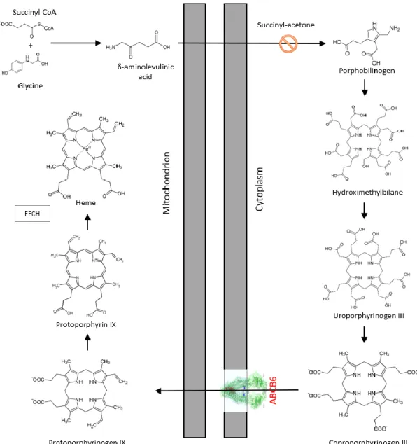
In 2006, Krishnamurthy and colleagues established the classification of ABCB6 as mitochondrial transporter. In a seminal Nature article they claimed that ABCB6 imports porphyrins into the mitochondria25. Porphyrins are important intermediates of the highly conserved heme synthesis pathway. In humans, during erythroid maturation, cells have a high demand of heme molecules to produce hemoglobin. In other species, the pathway also produces similar substances such as cobalamin (vitamin B12). The heme synthesis starts and ends in the mitochondrial matrix, but the intermediate steps occur in the cytosol.
The initial molecules are glycine and succinyl-CoA, they form δ-aminolevulinic acid (ALA) by ALA synthase, which is strictly regulated by intracellular iron levels and heme concentration. ALA is transformed into porphobilinogen in the cytoplasm, and four molecules of porphobilinogen form a linear tetrapyrrole, which is closed by enzymatic processes. The coproporphyrinogen III molecule (CPgenIII) enters the mitochondria and protoporphyrin IX (PPIX) is formed by oxidation steps. The closing step is made by ferrochelatase (Fech), the enzyme incorporates Fe2+ into the porphyrin ring in the
12
mitochondrial matrix (Fig 3). The heme synthetic pathway can be upregulated by ALA treatment and blocked by succinyl-acetone (SA)37. In 2006 Krishnamurthy and his colleagues observed elevated ABCB6 expression in human and murine fetal livers, which became upregulated during erythroid maturation. Treatment of MEL (myeloid erythroid leukemia) cells with ALA to induce heme biosynthesis caused a dose- and time- dependent increase in ABCB6 protein and mRNA levels. In contrast, blocking heme synthesis pathway by SA led to decreased intracellular PPIX levels and ABCB6 expression. Results from immunoblot and confocal microscopy experiments suggested that Flag-tagged ABCB6 is localized to the outer membrane of mitochondria. Using hemin-agarose affinity chromatography it was concluded that ABCB6 is a hemin binding protein and beside heme it could bind CPIII and PPIX molecules. Using isolated mitochondria, the ATPase activity of the ABCB6 protein seemed to be stimulated by CPIII. The authors found that PPIX fluorescence values were much greater in ABCB6- overexpressing K562, Saos-2 and MEL cells than in vector-control or ABCB6 non- functional mutant variant expressing cells (ABCB6-K629M). Every result therefore suggested that ABCB6 is located in the outer membrane of the mitochondria and it imports coproporphyrinogen III, thus acting as a main factor of heme biosynthesis25,38.
Overexpression of ABCB6 significantly increased intracellular (but not intramitochondrial) heme concentrations in K562 myeloid leukemia cells. ABCB6 overexpression also positively influenced expression and activity of hemoproteins, catalase and cytochrome c oxidase. It was suggested that ABCB6 plays a primary role in regulating hemoproteins by modulating intracellular heme concentration. Interestingly it was found that ABCB6 overexpressing cells were more resistant to H2O239.
13
Figure 3. The putative role of ABCB6 in heme synthesis. The initial step is the condensation of a succinyl coenzyme-A and glycine molecule, resulting in the formation of δ-aminolevulinic acid in the mitochondrial matrix. The synthesis continues in the cytosol, where four molecules of porphobilinogen form a linear tetrapyrrole. This linear molecule is closed by enzymatic processes then protoporphyrin is produced by oxidation steps, to which the ferrochelatase enzyme incorporates the divalent iron ion. The putative role of ABCB6 in this pathway is the mitochondrial porphyrin uptake.
1.2.3. ABCB6 and resistance
Other early studies took a different approach and suggested that ABCB6 may be involved in multidrug resistance (MDR). It was found that increased ABCB6 expression correlated with different cancer types. Increased resistance was observed against chemotherapeutic drugs, such as camptothecin or cisplatin in A549 lung cancer cells40
14
and paclitaxel/FEC (5-fluorouracil, epirubicin and cyclophosphamide) in breast cancer cells41. Expression levels of 45 genes from the ABC superfamily were analyzed in different drug resistant (doxorubicin/methotrexate/topotecan/paclitaxel/vincristine) ovarian carcinoma cell lines. Three transporters were significantly upregulated: ABCB1, ABCB4 and ABCG2. ABCA8 gene was significantly downregulated. ABCB6 was slightly upregulated in every cell line and paclitaxel caused the biggest deviation in expression levels42. In 2007 an article was published in which high ABCB6 mRNA expression level was found in primary melanoma and also in metastatic melanoma cells43. In 2009 an antioxidant response element was identified at –7575 base pair up-stream of the ABCB6 transcription site in human small airway epithelial cells44. Moreover, in 2011 it was reported that ABCB6 protect cells from oxidative stress and conferred arsenite resistance to HepG2 and Hep3B hepatocellular carcinoma cells45. In another arsenite- resistant cell line, in KAS myeloma cells ABCB6 overexpression enhanced resistance to 5-fluorouracil (5-FU), SN-38 and vincristine (Vcr) but not to arsenite. However ATO (arsenic-trioxide) resistant human epidermoid carcinoma cell line had higher ABCB6 expression level and accumulated less arsenic then parental cells46. The cellular differences between epidermoid and hepatocarcinoma cells may have led to different results47.
ABCB6 was also connected to drug resistance but not in a chemotherapeutic perspective: Gómez and colleagues found that increased intracellular survival of Leishmania panamensis in human macrophages treated with antimonial drug correlates with low expression of the transporter ABCB6. It seemed that antimony could induce ABCB6 expression in human macrophages. Furthermore, the protein was detected in the cell membrane and the phagolysosomal membrane surrounding the intracellular pathogen. Based on these results the authors speculated that ABCB6 acts as an efflux pump in the cell membrane and as an importer when localized to intracellular membranes48.
In the last few years several papers have been published about the diverse pharmacological relevance of ABCB6. A study showed that women younger than age 50 had significantly higher levels of mRNA expression for several drug transporters, including ABCB649. Analyzing Taxol resistant cells, ABCB6 was identified as an androgen receptor target gene50. Investigating patients with prostate cancer, it seemed that
15
expression levels of ABCB6, ABCC3 and ABCC10 can be valuable predictors of prostate cancer progression51. Strong correlation was also detected between ABCB6 expression levels and progression of human gliomas52. Another prostate cancer study showed elevated PPIX levels in dormant PC3 cells and upregulated ABCB6 expression53. Interestingly, in acute myeloid leukemia mRNA expression of ABCC3 and ABCB6 were significantly higher in daunorubicin resistant samples compared to sensitive ones54. Another observation occurred in D. melanogaster, where authors found that suppression of ABCB6 increased mortality when animals were treated with malathion insecticide55.
Researchers hypothesized several times that ABCB6 can mediate protection against cytosolic ROS45,56–58. It was mentioned above that Hübner and his colleagues identified an antioxidant response element in ABCB6 promoter region examining epithelial cells44. Association between ABCB6 and resistance to these diverse therapeutic compounds required explanation. The putative protective mechanism was to eliminate ROS by producing more heme to increase catalase activity38. Catalase enzyme converts toxic hydrogen peroxide to water using heme as a co-factor. In the recent years three studies supposed positive correlation between ABCB6 expression level and smoking59–61. In the latest article an in vitro model of oral keratinocyte cell line was developed by exposing them with different concentrations of cigarette smoke extract (CSE). CSE treatment increased cytotoxicity and cell death through ROS generation in keratinocyte cells.
Cigarette smoke exposure was associated with elevated ABCB6, while smoking cessation was associated with lower expression of ABCB6. In addition to toxic components, cadmium and arsenic can be found in tobacco products61.
In 2018 another puzzling result emerged about the connection of ABCB6 and a parasite. It was observed that erythrocytes from Lan null individuals are resistant to invasion by Plasmodium falciparum parasites. Cells from six unrelated donors with null alleles were examined and it was found that parasite invasion was impaired in all cases when ABCB6 was absent. But other Plasmodium species could invade Lan null erythrocytes, so this genetic background does not provide defense against all Plasmodium parasites. This suggests that P. falciparum may have special connection with ABCB6 as a host factor during invasion. Based on this observation, Lan null blood group can be considered as evolutionary advantage62.
16
1.2.4. ABCB6 – contrasting results on intracellular localization
Contrary to the suggested mitochondrial function25, in 2007 researchers observed extramitochondrial localization of ABCB6. Paterson and co-workers found that two distinct molecular weight forms of ABCB6 protein exist in SNB-19, U251 glioblastoma and KB-3-1 epidermoid carcinoma cells. Using novel antibodies, it seemed that the low weight form (79 kDa) was located to the mitochondria while high weightform (104 kDa) was located to the plasma membrane of the cells63. Tsuchida and his colleagues suggested other extramitochondrial localization for ABCB6. It was found that the transporter is located mainly in the Golgi system, and they presented confocal pictures of Cos-7 cells and proposed that the N-terminal extension of ABCB6 acts as an ER-targeting signal owing to its special hydrophobic nature64. A human colon adenocarcinoma line (LoVo) cell line was found devoid of endogenous ABCB6 mRNA. LoVo cells were used for heterologous expression of rat Abcb6. Performed distinct methods (subcellular fractionation, indirect immunofluorescence analyses, and determination of fluorescent rAbcb6-GFP distribution) authors concluded that rAbcb6 was localized to endolysosomal structures. Primary rat hepatocytes and liver tissue sections also corroborated this intracellular localization. Moreover, expression of rAbcb6 in LoVo cells increased the tolerance toward copper. The authors suggested that rAbcb6 may play a role in transition metal homeostasis56.
In our laboratory during her PhD work, Katalin Kiss tried to prove the mitochondrial localization of ABCB6. It was difficult to reconcile mitochondrial localization with studies suggesting that ABCB6 may play a role in increased resistance to chemotherapeutic agents in tumor cells. After years of thorough work our results24 questioned the reigning concept linking ABCB6 to mitochondrial porphyrin import25,65. During the study more approaches were used, first, we examined the intracellular location of ABCB6 in K562 human erythroleukemia and in HeLa cells. Intracellular organelles were separated from HeLa cells by differential centrifugation. Different membrane fractions were analyzed by immunoblot to characterize the localization of endogenous ABCB6. Because mitochondrial contamination was detected in the fractions, in further experiments, immunomagnetic separation was used. With the organelle markers we proved that lysosomes are devoid of mitochondrial contamination. The experiments unanimously demonstrated that ABCB6 is located in the lysosomal compartments and is
17
absent from the mitochondrial fractions. In the next step, it was shown that ABCB6 is expressed in human erythrocytes. The glycosylation of the protein was studied on erythrocyte ghosts. Glycosidase treatment resulted in a marked shift in molecular weight of the protein. This meant that B6 was indeed produced in the N-glycosylated form and was located to the plasma membrane of mature erythrocytes. Note, that mature red blood cells no longer contain other membrane structures (intracellular organelles), including mitochondria. Fukuda and his colleagues also observed that the protein undergoes glycosylationin the cells66. This posttranslational modification is predominantly specific for proteins in the plasma membrane and the endolysosomal continuum. The hypothesis proposed by the laboratory of J. Schuetz suggested that ABCB6 caused elevated mitochondrial uptake of porphyrin which led increased PPIX fluorescence in the cells.
The following experiments showed that however the level of ABCB6 increased during erythroid maturation of K562 cells, the attenuation of ABCB6 did not affect hemoglobin synthesis or erythroid maturation of the cells. We concluded that ABCB6 is not required for the synthesis of porphyrins. Fluorescently labeled ABCB6 was expressed and analyzed by confocal microscopy in K562 and HeLa cells. Results showed that endogenous protein clearly colocalized with lysosomal markers, which were in line with data from membrane fractionation. These results fundamentally call into question the mitochondrial localization and the role of ABCB6 in heme biosynthesis.24.
1.2.5. Structure – Function relation
Although ABCB6 was thought to be a mitochondrial protein, it is important to note that it does not contain a target signal like the other mtABCs24. Generally, the mitochondrial targeting signal is a 10-70 amino acid sequence forming an amphipathic helix at the N-terminal. Cleavable pre-sequences are the classical type of mitochondrial targeting signals. However, numerous types of non-cleavable targeting and sorting signals, which are located in the mature regions of mitochondrial proteins, have also been described21. Half-transporters in the B subfamily contain additional, relatively long and unique sequences, which have been implicated in protein–protein interactions and targeting. Topology prediction programs for transmembrane proteins (i.e., CCTOP, or HMMTOP) suggested that ABCB6 contains 11 transmembrane (TM) helices, of which five helices are not found on the basic ABC structure. This N-terminal extra domain
18
(TMD0) does not show sequence homology to any other protein. Kiss investigated the function of the TMD0 domain and created N-terminal truncated variants of ABCB6 in Sf9 (Spodoptera frugiperda) model cell lines. The expression level of ABCB6-core in Sf9 insect cells was comparable to full-length ABCB6, suggesting that it could fold properly and integrate into the membrane. After checking the expression of the truncated protein, functional assays were performed. Being an ABC transporter, ABCB6 cleaves ATP generating ADP and inorganic phosphate (Pi) which is linked to substrate translocation. This can be tested in an ATPase assay where Pi can be measured by a simple colorimetric reaction. The amount of released Pi is directly proportional to the activity of the transporter and transported substrates can increase baseline ATPase activity. On Sf9 vesicles containing ABCB6, basal or substrate-stimulated ATPase activity could not be detected. However, implementing azido-ATP and trapping assays, we showed that ABCB6-core variant was able to bind ATP and cleave the bound ATP molecule. The non-functional K629M mutant allowed ATP binding, but prevented ATP hydrolysis. For further experiments, ABCB6 protein variants were successfully expressed in human cell lines (HeLa and K562). Co-immunoprecipitation assay revealed that the TMD0 is not required for protein dimerization but it contains a glycosylation site67. This predicted glycosylation site was in agreement with the literature66. In the next step intracellular localization of the truncated variant was examined by confocal microscopy.
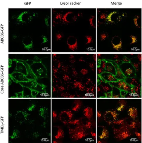
While the wild-type protein co-localized with lysosomal markers, ABCB6-core variant was detected in the plasma membrane. In contrast, the TMD0 fragment showed lysosomal localization similar to the wild-type protein. These results suggested that the N-terminal extension of the transporter is an independent folding domain with sufficient information for lysosomal targeting (Fig. 4). Electron microscopy analysis showed that ABCB6 was localized to the membranes of multivesicular bodies, multilamellar lysosomes and dense lysosomes67. Fukuda and co-workers studied intramolecular disulfide bonds and glycosylation of ABCB6. They showed that during maturation, ABCB6 is glycosylated at a conserved single atypical amino-terminal site (Cys-8). Results showed that mutation of highly conserved cysteines destroyed the disulfide bonds, leading ER retention of the protein. The data suggested that these cysteines are important for proper folding and stability of ABCB666.
19
Figure 4. Intracellular targeting of GFP-tagged ABCB6 variants in HeLa cells. Cells were transfected by GFP-fusion proteins. Localization of ABCB6–GFP, core–ABCB6–GFP and TMD0–GFP was revealed by GFP (green) in the context of the lysosomal marker LysoTracker (red)67.
In case of transporters a functional assay greatly facilitates the examination of the structure-function relation. Chavan and his colleagues developed an in vitro system with pure and active protein, where they could investigate the function of ABCB6. In the first step of the essay, the FLAG tagged protein was overexpressed in HEK293 cells. The tagged ABCB6 was purified from the mitochondrial fraction of the cells. In binding assay, they saw that ABCB6 retained its ability to bind ATP through the purification process. In ATPase assay the non-functional mutant (ABCB6-K629M) showed only background activity, while the purified ABCB6 was active in ATP hydrolysis. To analyze ABCB6 in its membrane-embedded state, purified mitochondrial ABCB6 (both WT and K629M) was reconstituted into preformed detergent-destabilized liposomes. Performing hemin- agarose affinity chromatography authors found that CPIII was able to displace liposome- reconstituted ABCB6 from hemin-agarose in a dose-dependent manner. In ATPase assay
20
lipid-reconstituted ABCB6 demonstrated basal ATPase activity that was comparable to the ATPase activity of the purified detergent-solubilized protein. Activity was strongly stimulated in the presence of CPIII, hemin, and protoporphyrin IX. CPIII uptake of WT- ABCB6 and transport incompetent ABCB6-KM liposomes were also measured, which authors concluded as an ATP-dependent substrate transport of ABCB626.
1.2.6. ABCB6 knockout mice
In spite of the putative essential mitochondrial porphyrin transport, Abcb6 knockout mice appeared phenotypically normal. However, evaluation of their hematological parameters revealed mild anemia. Phenylhydrazine (Phz) is a generally used molecule to induce acute hemolytic anemia in animal models. When Abcb6 KO mice were chemically stressed with Phz greater mortality and a more sustained anemia was observed. The authors concluded that ABCB6 is the sole mammalian ATP-dependent mitochondrial porphyrin importer, while they suggested that ABCB6 only mean benefit in stressful situations58. Next, Krishnamurthy and his co-workers also studied the physiological function of Abcb6 in knockout mice. It was found that deficiency of the protein in vivo does not have a significant impact on basal hepatic porphyrin or heme levels. Based on earlier results of the research group68 cytochrome P450 oxygenase (CYP) enzymes were investigated in Abcb6+/+ and Abcb6-/- mice. CYPs are hemoproteins, they use heme molecule as a cofactor. Mammalian hepatic CYPs are involved in the metabolism of endogenous compounds and xenobiotics69. Abcb6−/− liver microsomes showed significant decrease in midazolam biotransformation, suggesting decreased Cyp3a11 activity in Abcb6 KO mice. Several CYP enzymes were tested and results showed that Abcb6 deficiency in mice modified the activity of a specific set of hepatic P450s.
Measuring mRNA and protein levels of CYPs, it was found that altered P450 activity in Abcb6−/− mice is due to decreased expression of P450s. Though Abcb6−/− mice did not show any observable gross or histological alteration in liver, nor did they demonstrate any increase in serum biomarkers of liver injury. ABCB6 was attenuated in human hepatoma cell lines (Hep3B and Huh7) and expression of CYPs were monitored. Results showed decreased level of CYP1A2, CYP2B6, and CYP3A4 transcript. After accurate experiments with CYP enzymes authors concluded that ABCB6 deficiency could involve repression of P450 promoter activity70.
21 1.2.7. ABCB6 and pathology
In each specific porphyria the activity of specific enzymes in the heme biosynthetic pathway is defective. Porphyrias may also be classified as either erythropoietic or hepatic, depending on the principal site of accumulation of pathway intermediates71. In 2016 it was found that severely affected porphyria patients were heterozygous for ABCB6 alleles. In experiments using a mouse model, genetic deficiency of ABCB6 coupled with a Fech deficiency (a classic porphyria model) produced greater porphyrin elevation in red blood cells, hepatocytes, and increased liver damage72. Despite these results, in the well- controlled laboratory environment, Abcb6 KO mice have no pathological phenotype70. While researchers were hunting for a connection between ABCB6 and drug resistance45,58 or cell cycle regulatory components that are involved in carcinogenesis73,74, the first disease linked to a mutation in the ABCB6 gene was described. In ocular coloboma, which is an eye development disorder, two missense mutations were identified (L811V and A57T) in ABCB6 in Chinese population. Comparative analyses of ABCB6 genes in different species showed that both amino acids are highly conserved, presumably essential to normal biological function. In RPE cells wild type and L811V mutant ABCB6 showed colocalization with ER and Golgi markers. ABCB6 attenuation by morpholino oligos caused coloboma-like phenotype in zebra fish. Normal phenotype could be restored by the co-injection of wild type, but not by the mutant ABCB6 mRNA. In these experiments there was no detectable porphyrin accumulation that would suggest a role for ABCB6 in tetrapyrrole transport75.
The Langereis (Lan) blood group was first described in 196176 but scientists identified ABCB6 as the Lan blood group antigen only in 2012. First monoclonal antibody with Lan specificity (OSK43; IgG1κ) was generated from immortalized lymphocytes of a healthy Japanese Lan (-) woman who had developed anti-Lan antibody during pregnancy. This antibody proved that the protein is located at plasma membrane of RBC (which are devoid of mitochondria) and also hepatocellular carcinoma cells. 10 patients bearing ABCB6 null mutations were analyzed and they seemed asymptomatic, they did not show anemia or abnormal erythropoiesis28. Lan-negativity is believed to be very rare77,78 but recent population and genetic studies show that missense polymorphisms in ABCB6 gene can be more common than previously expected29.
22
While reports published continuously about porphyrin transport68,79,52 other clinically important mutations of ABCB6 appeared in the literature30,30,80–82. Genetic studies reveal that mutations in ABCB6 can cause two other phenotypes, familial pseudohyperkalemia (FP)81 and dyschromatosis universalis hereditaria (DUH)83. FP is a dominant red cell trait characterized by increased serum potassium concentration in whole blood stored below room temperature, without additional hematological abnormalities. DUH is a pigmentary genodermatosis characterized by a mixture of hyperpigmented and hypopigmented macules distributed randomly over the body. I shall return to this in more detail below. Through the years new heterozygous mutations were described: healthy Lan (-) individuals84,85,86, DUH8788–91, FP92,93 and coloboma patients94. We are not aware yet of the factor when ABCB6 protein expression provides obvious benefits to a cell or organism but it is clear that mutations in the ABCB6 gene can lead to hereditary forms of eye dysfunction, pigmentary defect and pseudohyperkalemia (Fig. 5).
Figure 5. Diverse observations about ABCB6
ABCB6
Drug response
Oxidative stress
DUH, Coloboma,
FP, Lan Detoxifi-
cation Porphyrin
transport Elevated expression
in tumors
23 1.2.8. ABCB6 and cadmium
Observation that ABCB6 exhibits topological and sequential similarities with a heavy metal transporter protein family has appeared earlier in several places in the literature32,65,95.
Detoxification capacity of organisms is extremely important, as heavy metals such as cadmium (Cd), mercury (Hg), lead (Pb), and arsenic (As), are constantly present in our environment. Contamination in the drinking water and food can cause serious damage to living organisms. Research summarizing statistical data suggests that the risk of developing cancer can increase in areas exposed to high Cd levels96. By using chelators, the intracellular concentration of free metal ions can be reduced, thereby lower their toxic effect. The natural defense is based on similar principle, meaning animals and fungi produce cysteine-rich proteins that bind heavy metals to form thiolate bonds97. On the other hand, plants and certain species of fungi, (i.e. Schizosaccharomyces pombe) produce small peptides, phytochelatins (PCs). Caenorhabditis elegans, which is an exception in the animal kingdom also use PCs in its detoxification pathways. PCs are characterized by the repetition of glutamic acid-cysteine amino acid residues followed by glycine. The formation is catalyzed by phytochelatin synthase (PCS) from glutathione98,99. Importance of PCs is proven by the fact that PCS-deleted fission yeast (S. pombe) strains become more sensitive to heavy metals100. After finding PC-mediated Cd tolerance, the goal was to identify the proteins involved in the transport process. As a first step, the genomic library of S. pombe was created, and then a coding region was identified, which could restore the Cd-tolerant phenotype. The identified gene was named Heavy metal tolerance factor-1 (hmt-1). The gene is located on chromosome 3 and it encodes a protein containing 830 amino acids with a molecular weight of 90.5 kDa.
Homologues were identified in fission yeast (Schizosaccharomyces pombe), nematode (Caenorhabditis elegans) and the fruit fly (Drosophila melanogaster) fulfill a conserved role in conferring resistance to heavy metals, specially cadmium34,101.
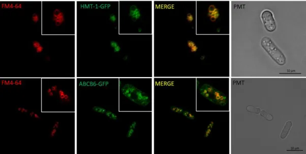
In S. pombe HMT-1 localizes to the vacuolar membrane and catalyzes the sequestration of cadmium complexes to this intracellular compartment102. Experiments with C. elegans surprisingly revealed that, unlike SpHMT-1, CeHMT1 is able to provide tolerance to cadmium, arsenic and copper as well. The Vatamaniuk Lab showed that CeHMT-1 is expressed in liver-like cells, the coelomocytes, head neurons and intestinal
24
cells103,104. In 2018 they identified the intracellular localization of CeHMT-1 in the intestinal cells. The transporter is located to the apical recycling endosomes and partly to early and late endosomes. Similar to ABCB667, CeHMT-1 lacking the NTE domain was targeted to the plasma membrane95. A deficiency in Ce-HMT-1 does not interfere with the phenotype associated with CePCS-1 deficiency and vice versa. It became clear that HMT1-deficient worms are more sensitive to Cd than PCS-deficient ones. It was concluded that although CeHMT-1 is required for Cd detoxification, it is not only PC- dependent105. This is important because, following the evolution of HMT-1, neither D.
melanogaster cells nor the human cells carry out the detoxification of heavy metals by conjugation with phytochelatins. Since D. melanogaster is devoid of PCs, further experiments were needed to determine whether Cd-PC complex is transported in yeast cells. Heterologously expressed DmHMT-1 suppressed the Cd hypersensitivity of S.
pombe hmt-1 mutants and localized to the vacuolar membrane. Despite this the vacuolar level of PCs were similar to the vacuoles from hmt-1-deleted cells. The authors assumed that SpHMT-1 is not the only Cd-PC carrier in the system. Several heavy metals were also tested in this rescue system. PCS-deficient S. pombe cells were hypersensitive to Cd, Hg and As, but SpHMT-1 does not cause tolerance to additional (heavy) metals Pb, Ag, As(III), Cu or Hg101. Experiments with C. elegans have revealed that, unlike SpHMT-1, CeHMT-1 can provide tolerance to cadmium, arsenic and copper106. These results indicated that the HMT-1-mediated detoxification of heavy metals is preserved during evolution, extending to some invertebrate species lacking the ability to synthesize PC101.
1.2.9. ABCB6 and pigmentation
Dyschromatosis universalis hereditaria (DUH) is a pigmentary genodermatosis characterized by diffuse symmetrically distributed hypopigmented macules mixed with hyperpigmentation. The molecular basis of DUH was unknown until Zhang and his colleagues reported ABCB6 as a causative gene30. Since then, more mutations of ABCB6 were identified relating to DUH phenotype30,83,87,88,91,107. Interesting fact, that Lan (-) individuals28 and ABCB6 knockout mice show no abnormal pigmentation phenotype70,72.
Melanin is a natural pigment widespread in most organisms. Specialized pigment cells, including melanocytes and retinal pigment epithelium, synthesize melanin.
Melanocytes are found in the basal layer of the epidermis. There are three basic types of
25
melanin: eumelanin, pheomelanin, and neuromelanin. Eumelanin is generally black or dark brown, pheomelanin is a yellow to reddish brown pigment108. Both pigments are polymeric and are derived via a series of redox reactions from a common precursor, dopaquinone, which is formed by the action of the enzyme tyrosinase on Tyr residue.
Pheomelanin synthesis starts from tyrosine and cystine and contains polybenzothiazine portions109,110. Melanosomes are specialized intracellular organelles of pigment cells in which melanin pigments are synthesized and stored. They are members of a family of cell-type-specific lysosome-related organelles (LROs) that coexist with traditional endosomes and lysosomes and are generated from them through a progressive series of membrane sorting steps87,111. Lysosomal-like organelles carry a number of common characteristics with lysosomes, while requiring special proteins, other additional elements, cellular organelles for their construction and function112,113. Lysosomes and LROs are involved in a number of processes, including cholesterol homeostasis, maintenance and repair of plasma membranes, bone and tissue regeneration, protection against pathogens, regulation of cell death and signaling processes (Fig. 6). There are four steps in the maturation of the melanosome (Fig. 7A). Melanosomes originate from endosomal precursors (pre-melanosomes). The best known of the structural components is the pre-melanosomal protein (PMEL), which is a fibrillar component of melanosomes.
Figure 6. Biogenesis of lysosomes and lysosome-related organelles (LROs) (https://mynotebook.labarchives.com) LROs comprise a group of functionally diverse compartments that share features with lysosomes but are distinct and harbor specific cargoes that confer their unique properties. There are four well-studied LROs: pigment cell melanosomes, endothelial lamellar bodies, platelet α-granules and natural killer cell lytic granules114.
26
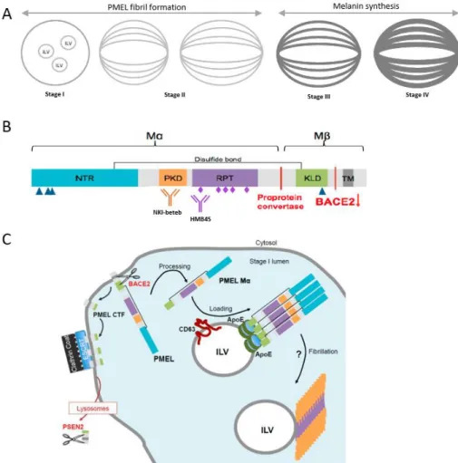
PMEL is processed into amyloid fibrils forming the intraluminal matrix of stage II melanosomes. PMEL-derived amyloid structures belong to the emerging category of physiological amyloids that have beneficial cellular functions115. During maturation, PMEL is cleaved by several proteases to release luminal amyloidogenic fragments116–118 (Fig 7). After translation, the full length peptide chain reaches the endosomal membrane where β-site APP-cleaving enzyme 2 (BACE2) generates the transmembrane M-β fragment118. This C-terminal polypeptide is processed by the gamma-secretase complex containing presenillin-2 in the lysosomes of pigment cells119. BACE2-mediated cleavage releases the M-α fragment, the amyloidogenic luminal domain of PMEL, into the melanosome lumen118. The M-α fragment is proteolytically processed to produce the amyloidogenic peptides that finally assemble into detergent insoluble protofibrils and fibrils recognized by the HMB45 and NKI-beteb antibodies (Fig. 7B,C)116. Melanin starts to be produced in stage III melanosomes by Tyrosinase (TRP-1) and stage IV melanosomes correspond to fully-melanized mature organelles. At the final step, melanin-filled, mature melanosomes enter the extracellular space, similar to exosomes and are passed on to the keratinocytes around the pigment cell. Stage I melanosomes are tyrosinase negative, whereas stage II melanosomes, including fibrillary elongated structures, are tyrosinase positive43,111,120,121.
27
Figure 7. Schematic picture of PMEL isoforms melanosomes and in different compartments. A Biogenesis of melanosomes. Stage I melanosomes, where PMEL fibrils start to assemble. In stage II melanosomes, PMEL fibrils give the melanosomes their characteristic ellipsoidal shape and striated appearance. Melanin starts to be produced in stage III melanosomes, to which melanin synthesizing enzymes, such as Tyrosinase (TRP-1), are transported. Melanin is sequestered on PMEL fibrils, which become completely masked by melanin in stage IV melanosomes. B Schematic representation of pre-melanosomal protein (PMEL) domain structure.
Triangles, pentagons and rhombuses represent N- and O-linked glycosylation. PMEL cleavage sites and the involved proteases are indicated in red. C Model for pre-melanosomal protein (PMEL) fibril formation in stage I melanosomes. The Mα fragment of PMEL is released into the lumen of stage I melanosomes by action of BACE2 (beta-site APP cleaving enzyme 2) protease.
This cleavage also produces a C-Terminal Fragment (CTF) that is sequestered at the limiting membrane of stage I melanosomes by the endosomal sorting complexes required for transport (ESCRT) machinery, to be further cleaved by the presenilin 2 (PSEN2) of the γ-secretase complex in lysosomes. The Mα fragment is then loaded onto intraluminal vesicles (ILVs). ILVs have been proposed to act as nucleators for PMEL fibril formation115.
Amyloid PMEL fragments can be cytotoxic, so mutations distracting PMEL trafficking or processing can be associated with melanocyte survival or melanin defects115,117,122. However, melanocytes devoid of PMEL expression still have normal pigment levels123.
28
Whereas melanosomes and lysosomes are distinct organelles in melanocytes124, recent studies have proposed that lysosomes are required for correct PMEL amyloid matrix formation125–127, suggesting a potential role for lysosomal proteins such as ABCB6 in early steps of melanosome biogenesis.
Genetic pigmentation disorders give new insights into the understanding of the pigmentation process, including melanosome biogenesis, melanin synthesis111. Mutations in the ABCB6 gene manifest in DUH, which is characterized by hyper- and hypopigmented areas over the body. Skin histological examination of a DUH proband showed a normal number of melanocytes in the basal layer in both hyper- and hypo pigmented areas. However, the number of mature melanosomes in normal control and hyperpigmented skin areas was considerably higher than in hypopigmented area.
Additionally, many immature melanosomes were observed in hypopigmented skin region. However, the influence of ABCB6 on the above described process of melanogenesis is not known and its intracellular localization and function are also not clear.
We can see that conflicting observations were described about ABCB6 in the last two decades. Based on healthy Lan (-) patients, healthy ABCB6-KO mice and studies about the extramitochondrial localization we supposed that the protein is not an essential mitochondrial porphyrin importer. In summary, the pathophysiological function of ABCB6 in the endo-lysosomal continuum remains to be clarified. Therefore, we thought that the answer must be sought in another direction.
29
2. Aims
ABCB6 is usually discussed in the context of mitochondrial ABC transporters based on early studies suggesting that it catalyzes the mitochondrial import of a heme synthesis intermediate. Recent studies have evidenced alternative localizations, but the physiological function of ABCB6 in these extramitochondrial compartments has remained elusive. My overall aim was to establish in vitro assays for the study of the function and intracellular localization of ABCB6. ABCB6 shows high amino acid sequence similarity to HMT-1, which is involved in the heavy metal tolerance of Schizosaccharomyces pombe, Caenorhabditis elegans and Drosophila melanogaster.
HMT-1 (heavy metal tolerance factor-1) localizes to the vacuolar/endosomal membrane of S. pombe and C. elegans where it catalyzes the sequestration of cadmium complexes.
To analyze the relation of ABCB6 function to the HMT-1 proteins, I pursued the following aims:
1. Investigation of the localization of heterologously expressed human ABCB6 in S.
pombe and C. elegans.
2. Pending successful expression in these model systems, the second aim was to analyze the function of ABCB6 in wild-type and knock-out strains.
3. Relevance of Cd detoxification in human cells
An additional aim was to use cellular models to decipher the role of ABCB6 in melanogenesis, and to suggest a functional model explaining the DUH phenotype.
4. The third aim was to set up an in vitro model system to study the localization of ABCB6 in a melanocytic cell line.
5. Given the link of ABCB6 mutation to aberrant pigmentation, the final aim was to set up an in vitro model system to characterize the role of ABCB6 variants melanogenesis in a human cell line.
30
3. Methods
3.1. Cell culturing
S. pombe culture conditions and strains — The S. pombe strains BG_00008 (ade6- M216, ura4-D18, leu1-32) and the hmt1-deleted mutant strain BG_H4691 (ade6-M216, ura4-D18, leu1-32) were a kind gift from R. Lill (Philipps-Universität Marburg). A common feature of these laboratory strains is adenine, uracil and leucine autotrophy.
EMM Broth, EMM agar and EMM without dextrose were purchased from Formedium (Hunstanton, UK). Depending on the experiment, medium was supplemented with 225 mg/l adenine-HCl, 225 mg/l L-leucine and 225 mg/l uracil.
C. elegans culture conditions and strains — C. elegans experiments were performed together with János Barna and Dániel Kovács (Department of Genetics, Eötvös Loránd University). The strains were grown on solid Nematode Growth Medium (NGM) at 20
°C containing a lawn of the bacterium Escherichia coli OP50128. The following strains were used: N2 C. elegans wild-type, var. Bristol; DP38 unc-119(ed3)III; VC287 hmt- 1(gk161)III; VF31 gfIs1[phmt-1::hmt-1::gfp, unc-119(+); VF12 hmt-1(gk161)III;
gfIs1[phmt-1::hmt-1::GFP, unc-119(+)]. For microscopic studies we used the following strains (generous gift from Dr. Xiaochen Wang (Institute of Biophysics, Chinese Academy of Sciences)): XW1957: qxIs110 (pges-1::mCHERRY::RAB-5); XW1962:
qxIs111 (pges-1::mCHERRY::RAB-7); XW9119: qxIs213 (pges-1::mCHERRY::RAB- 10).
Cell lines — HeLa cells were purchased from ATCC, the glioblastoma SNB-19 cells were from DSMZ (Germany), and the melanoma MNT-1 cell line was a kind gift of Guillaume van Neil. HeLa and SNB-19 cells were grown in high glucose DMEM (Gibco 521000-47) supplemented with 10% FBS, 2 mmol/l glutamine, and 100 units/ml penicillin and streptomycin (Life Technologies) at 37 °C in 5% CO2. MNT-1 cells were grown in DMEM high glucose medium (4.5 mg/ml Gibco, Grand Island, NY, USA) supplemented with 10% v/v AIM-V, 20% v/v FBS, 1% v/v sodium pyruvate, 1% v/v non- essential amino acids, 100 U/ml penicillin and 100 μg/ml streptomycin. MNT-1 cells were incubated at 37 °C with 5% CO2 and regularly passaged at a density of 80% (1:8 ratio). Cells were periodically tested for mycoplasma contamination with the MycoAlert mycoplasma detection Kit (Lonza, Basel, Switzerland).
31
3.2. Molecular cloning of ABCB6 and HMT-1 constructs
Plasmid constructs were amplified in E. coli strain Top10 (Invitrogen, Carlsbad, CA, USA), grown at 37 °C in liquid Luria-Bertani (LB) medium supplemented with appropriate antibiotics.
S. pombe — The hemagglutinin-tagged S. pombe hmt-1 cDNA (Z14055) was ordered from GenScript (Piscataway, NJ, USA). Human ABCB6 (NM_005689) cDNA was provided by Jill Paterson in pcDNA3.1-Flag vector63. My former colleague, Katalin Kiss generated the Walker-A region lysine mutant ABCB6 variant (K629M) by overlap extension PCR mutagenesis24. Melinda Gera generated various mutants of ABCB6 (Table S1). Hmt-1 and ABCB6 variants encoding cDNAs were subcloned into the pREP1 fission yeast expression vector. pEGFP-N1 (BD Biosciences, Franklin Lakes, NJ, USA) plasmid was used for the N-terminal GFP-tagging of the transporters by exchanging the ABCB6- GFP construct from a respective pAcUW plasmid129 HMT-1-GFP was assembled by PCR (Polymerase Chain Reaction) using a primer pair generating a new restriction site at the 3’end of the cDNA. First, HMT-1 C-terminal was cloned to pEGFP-N1, then the pREP1- HMT-1-GFP expression plasmid was created by exchanging the C-terminal region of HMT-1.
C. elegans — For experiments in C. elegans, codon-optimized ABCB6 cDNA was synthetized by GenScritpt (Piscataway, NJ, USA). To generate the phmt-1::ABCB6::gfp and phmt-1::ABCB6::mCherry reporter for localization studies, ABCB6 was subcloned in frame with the GFP sequence of the pPD95.75-GFP vector. The 5’ regulatory region of hmt-1 (2.8 kb immediately upstream of the start of the hmt-1 coding sequence) was PCR-amplified using primer pairs as previously104 and with another primer pair we introduced the appropriate restriction enzyme recognition sites at the 5′ and 3′ends (Table S1). The PCR-amplified hmt-1 promoter was subcloned into the pPD95.75-ABCB6-GFP vector. In the case of phmt-1::ABCB6::mCherry, the GFP reporter sequence of pPD95.75- ABCB6-EGFP was changed to the sequence encoding mCherry from plasmid pRH21.
Human cell lines — The lysine mutant ABCB6 variant of the Walker-A region (K629M) and DUH mutation G579E83 were generated by overlap extension PCR mutagenesis24,130. Constructs were cloned into lentiviral plasmid obtained via the Addgene Plasmid Repository. (Table S2).