Investigation of the human ABCB6 transporter PhD thesis
Zsófia Rakvács
Semmelweis University
Doctoral School of Molecular Medicine
Supervisor: Gergely Szakács MD., Ph.D.
Official reviewers: Miklós Geiszt MD., DSc.
Péter Lőw Ph.D.
Chairman of the examination committee: János Réthelyi MD., Ph.D.
Members of the examination committee: Gergely Papp Ph.D.
Ákos Sveiczer Ph.D.
Budapest 2019
2
Introduction
The ATP binding cassette (ABC) superfamily is one of the largest protein families, performing diverse functions in bacteria, fungi, plants, and the animal kingdom. ABC transporters use ATP to facilitate/mediate the energy-dependent movement of structurally diverse compounds across membrane barriers. A functional ABC protein typically contains two nucleotide binding domains (NBD) and two transmembrane domains (TMD). In case of half-transporters such as ABCB6, two polypeptide chains form a functional unit through dimerization. TMDs have low homology and are responsible for substrate binding and translocation, while NBDs are highly conserved and participate in ATP binding and hydrolysis.
Based on sequence homology, 48 different ABC proteins (grouped into seven subfamilies ranging from A to G) were defined in the human genome. In my PhD work I studied ABCB6, a member of the ABCB subfamily.
In 2006, it was reported that ABCB6 catalyzes the mitochondrial uptake of coproporphyrin III (CPIII) in mammalian cells, an oxidized derivative of the heme synthesis intermediate, thereby serving as an important regulator of cellular porphyrin biosynthesis.
However, several papers have questioned the mitochondrial localization of ABCB6. Prior to my PhD project our laboratory has provided extensive evidence based on various imaging techniques to support the endolysosomal localization of ABCB6. However, precise intracellular localization can only be established with the discovery of a matching physiological function. Unfortunately, the physiological function of ABCB6 in the endolysosomal compartment has remained elusive.
Genetic studies revealed that mutations in ABCB6 can cause distinct phenotypes: FP (Familial pseudohyperkalemia) is a dominant red cell trait characterized by increased serum potassium in whole blood stored below room temperature, OC (ocular coloboma) is a developmental defect of the eye. DUH (dyschromatosis universalis hereditaria) is a pigmentary genodermatosis characterized by a mixture of hyperpigmented and hypopigmented macules distributed randomly over the body. Interestingly, the absence of the protein in humans or mice does not lead any obvious phenotype.
ABCB6 shows high amino acid sequence similarity to HMT-1, which is involved in the heavy metal tolerance of Schizosaccharomyces pombe, Caenorhabditis elegans and Drosophila melanogaster. HMT-1 (heavy metal tolerance factor-1) localizes to the vacuolar/endosomal membrane of S. pombe and C. elegans where it catalyzes the sequestration of cadmium complexes.
3
Aims
My overall aim was to establish in vitro assays for the study of the function and intracellular localization of ABCB6. We hypothesized that the cadmium-sensitive phenotype of S. pombe and C. elegans strains defective of HMT-1 can be rescued by the human ABCB6 protein. In addition, my goal was to use cellular models to link ABCB6 function to melanogenesis and the DUH phenotype.
I pursued the following specific aims:
Investigation of the localization of heterologously expressed human ABCB6 in S.
pombe and C. elegans.
Analysis of the function of ABCB6 in wild-type and HMT-1 knock-out S. pombe and C. elegans strains.
Establishment of heterologous model systems to characterize the effect of disease- causing ABCB6 mutations.
Establish an in vitro model system to study the role of wild-type and DUH-variant ABCB6 in melanogenesis.
Methods
❖ ABCB6 cDNA constructs were designed and cloned in different expression plasmids (S.
pombe – pREP1, C. elegans – pPD95.75 and mammalian cells – EF1 lentiviral vector).
Expression of HMT-1-HA and the different ABCB6 constructs were detected by immunoblot.
❖ S. pombe cultures were treated with CdCl2, then spheroplasts were formed by cell wall digestion. Vacuoles were isolated by differential centrifugation. The integrity of the vacuoles was assessed by flow cytometry. Vacuolar Cd content was determined by graphite furnace atomic absorption spectrometry (GFAAS).
4
❖ To examine the effect of the heterologously expressed HMT-1 or ABCB6, the model organisms were grown in Cd containing media. Viability curves were fitted with Graph Pad Prism 7 software using the sigmoidal dose–response model.
❖ For determination of melanin content, MNT-1 cells were dissolved in NaOH and melanin contents were determined by measuring absorbance at a wavelength of 405 nm
❖ For the evaluation of intracellular localization of the transporters, hmt-1-deleted S. pombe was transformed with pREP1-HMT-1-GFP or ABCB6-GFP. Cells were stained with FM 4-64 then examined with a confocal microscope.
❖ To test the co-localization of ABCB6 and CeHMT-1, the phmt1::ABCB6::mCherry strain was crossed with phmt-1::hmt-1::gfp males and the F1 progeny co-expressing both transgenes was examined with a confocal microscope.
❖ To determine the subcellular localization of ABCB6 and CeHMT-1, phmt-1::hmt-1::gfp and phmt-1::abcb6::gfp worms were crossed with strains expressing different endosomal markers: RAB5-mCherry, RAB7-mCherry or RAB10-mCherry.
❖ HeLa, SNB-19 and MNT-1 cells expressing ABCB6 variants were immunolabeled with different monoclonal antibodies.
❖ For FISH analysis cells were labeled with a probe for ABCB6 and with a control probe for Chromosome II. Cells in interphase were analyzed for the FISH pattern by fluorescent microscope.
❖ CRISPR/Cas technology was used to modify the endogenous ABCB6 gene in the human MNT-1 cell line. First, we used HDR to modify ABCB6 alleles (ABCB6-GFP, ABCB6- SBP-GFP), while we used NHEJ to generate ABCB KO cells. Single cell clones were tested and selected by the following Sanger sequencing, Real Time-PCR, immunoblot, flow cytometry and confocal microscopy.
5
Results
The relation of ABCB6 function to the HMT-1 proteins
ABCB6 shows significant sequence identity to HMT-1 (heavy metal tolerance factor 1) proteins, whose evolutionarily conserved role is to confer tolerance to heavy metals through the intracellular sequestration of metal complexes.
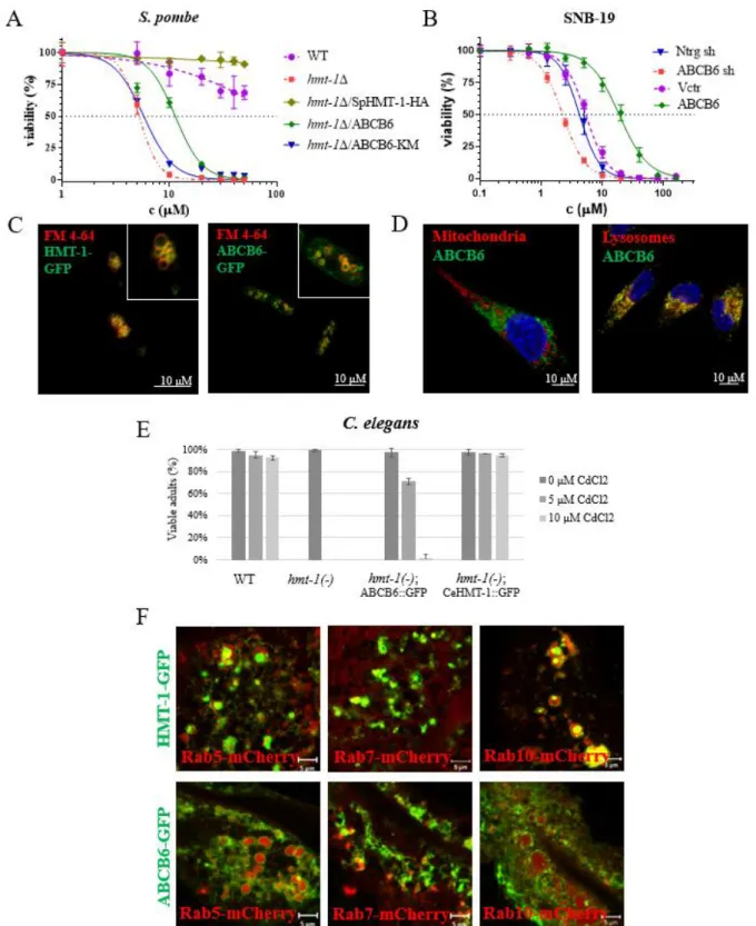
First, we showed that ABCB6 rescues the Cd-sensitive phenotype of HMT-1-deficient S.
pombe (Fig 1A). ABCB6 was localized to the vacuolar membrane of S. pombe cells (Fig 1C).
ABCB6 restored the Cd content of vacuoles indicating that the rescue of HMT-1 deficient yeast cells was based on the vacuolar sequestration of cadmium.
Second, we showed that expression of ABCB6 resulted in Cd tolerance of the HMT-1 deficient C. elegans strain (Fig 1E). ABCB6 partially colocalized with markers of the early, late and basolateral recycling endosomes, similar to CeHMT-1 (Fig 1F).
Third, we provided evidence that ABCB6 localizes in the lysosomes of human glioblastoma cells and overexpression of ABCB6 increased the cadmium resistance of the cells (Fig 1B).
6
Figure 1. A In cytotoxicity assay ABCB6 rescues the Cd-sensitive phenotype of HMT-1- deficient S. pombe. B ABCB6 is localized to the vacuolar membrane of yeast cells. Vacuolar membrane was stained with FM 4-64. C Overexpression of ABCB6 increased, but downregulation decreased the Cd tolerance of human SNB-19. D Lysosomal localization of ABCB6 in SNB-19 cells. ABCB6 was visualized using OSK43 ABCB6 antibody (green);
7
nuclei were labelled with Hoechst 33342 (blue); organelles were labelled with specific markers (red): mitochondria (AIF), lysosomes (LAMP1) E ABCB6 rescues the Cd-sensitive phenotype of HMT-1-deficient C. elegans. F CeHMT-1 and ABCB6 show identical localization in C.
elegans. CeHMT-1 and ABCB6 partially colocalize with markers of the early (Rab5), late (Rab7) and basolateral recycling endosomes (Rab10).
ABCB6 and melanogenesis
Genetic studies revealed mutations in ABCB6 causing the pigmentary disorder DUH.
However, the mechanism how ABCB6 affects skin pigmentation is still unknown. Therefore, we investigated the subcellular localization and function of ABCB6 in a human melanocytic cell line, MNT-1.
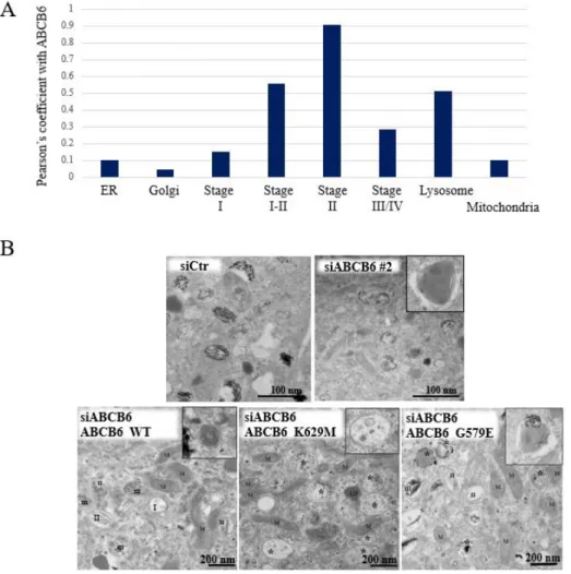
Consistent with our previous results we corroborated the localization of ABCB6 in the endolysosomal continuum. Cell fractionation, confocal and electron microscopic analysis revealed that the protein is localized to the lysosomes and early melanosomes of MNT-1 cells (Fig 2A).
Depletion of ABCB6 by different siRNA constructs did not change the melanin levels significantly, but melanin content of the cells was highly variable between siRNA constructs.
Whereas trafficking of melanosomal proteins such as was not affected. morphological analysis at the ultrastructural level revealed a unique phenotype associated with ABCB6 depletion, confirming a defect in fibril formation (Fig. 2B: siCtr, siABCB6). Downregulation of ABCB6 resulted in decreased amyloid formation, and the appearance of large aggregates displaying a hydrangea-like morphology in early and mature melanosomes.
Downregulation of ABCB6 allowed testing the rescue effect of ABCB6 variants.
Expression of the wild-type form of ABCB6 almost fully eliminated the aberrant structures and restored normal PMEL fibril formation (Fig 2B: WT). Expression of the functionally inactive K562M mutant resulted in the accumulation of aberrant compartments filled with protofibrils or small aggregates unable to form fully mature amyloid fibrils, but devoid of the abnormal structures observed in the case of ABCB6 depletion (Fig. 2B: K562M). In contrast, expression of the DUH mutant ABCB6 G579E resulted in the accumulation of aberrant early, and to a less extent late melanosomes (Fig. 2B: G579E). Overall, mutations induced the accumulation of
8
unpigmented aberrant melanosomes, showing signs of improper fibril formation, suggesting ABCB6 affects early stages of melanogenesis.
Figure 2. A Evaluation of the subcellular localization of endogenous ABCB6 by immunofluorescence labeling and laser-scanning confocal microscopy. Chart show the colocalization coefficient of ABCB6 with different organellar markers. B Ultrathin section images of MNT-1 cells embedded in resin. Images show control (siCtr), ABCB6-depleted (siABC6#2) or overexpressing (ABCB6 WT, ABCB6 K629M or ABCB6 G579E) MNT-1 cells with endogenous ABCB6 downregulation. Insets show individual melanosomes.
9
Genome editing of MNT-1 cell by CRISPR/Cas9
To evaluate the functional consequence of the deletion of ABCB6, we used the CRISPR/Cas9 based gene editing method.
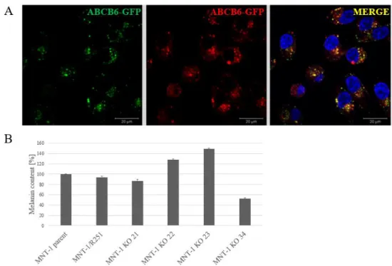
First, we used fluorescent in situ hybridization to establish that MNT-1 cells have four ABCB6 alleles. We engineered cells expressing GFP-tagged endogenous ABCB6, which showed the expected lysosomal localization pattern (Fig. 3A). Next, a SBP (Streptavidin- binding peptide) tag was added to C terminal end of ABCB6-GFP (ABCB6-SBP-GFP), to enable the monitoring of the intracellular trafficking of ABCB6 by the RUSH (Retention Using Selective Hooks) system (not shown).
Finally, we engineered ABCB6 knock-out cell lines. Our preliminary analyses suggest that ABCB6 KO MNT1 cells show variable melanin levels (Fig 3B).
Figure 3. A Endogenous ABCB6-GFP is localized to the lysosomes of MNT-1 cells. B ABCB6 affects the intracellular melanin content of MNT-1 cells. ABCB6 knock out cells show variable melanin levels.
10
Conclusions
The intracellular localization of ABCB6 has been a matter of debate, with conflicting reports suggesting mitochondrial or endolysosomal expression. ABCB6 shows significant sequence identity to HMT-1 (heavy metal tolerance factor 1) proteins, whose evolutionarily conserved role is to confer tolerance to heavy metals through the intracellular sequestration of metal complexes. We hypothesized that the cadmium-sensitive phenotype of S. pombe and C.
elegans strains defective for HMT-1 can be rescued by the human ABCB6 protein. It may be argued that the precise intracellular localization can only be established with the discovery of a matching physiological function.
We showed that ABCB6 localizes to the same intracellular compartment as HMT-1, performing an overlapping function linked to the intracellular sequestration of metal complexes. First, we showed that ABCB6 rescues the Cd-sensitive phenotype of HMT-1- deficient S. pombe and C. elegans strains. Second, we revealed that ABCB6 function is required for the sequestration of cadmium into HMT-1-deficient yeast vacuoles. Third, we provided evidence that ABCB6 modulates the cadmium sensitivity of human glioblastoma cells. Our results provide functional evidence linking ABCB6 to endolysosomal compartment, paving the way for further research to fully understand the pathophysiological role of this elusive membrane transporter. In an attempt to understand the link between ABCB6 mutations and the DUH phenotype, we investigated the subcellular localization and function of ABCB6 in a human melanocytic cell line MNT-1.
Consistent with our earlier results, we corroborated the localization of ABCB6 in the endolysosomal continuum of MNT-1 cells. Endogenous ABCB6 was found to localize to lysosomes and early melanosomes, which was further validated in MNT-1 cells engineered to express GFP-tagged endogenous ABCB6 by CRISPR/Cas9. ABCB6 depletion in melanoma cells resulted in decreased amyloid formation, and the appearance of large aggregates in early and maturing melanosomes. The DUH mutant G579E or the inactive K562M mutant variant also induced the accumulation of unpigmented aberrant melanosomes and could not restore normal biogenesis of PMEL fibrils. We hypothesize that ABCB6 affects the generation of amyloidogenic fragments or their aggregation into fibrils.
Overall, given the rare occurrence of Cd in mammalian cells, we believe that ABCB6 is involved in certain metal-complex transport in the lysosomal system. Since stage I
11
melanosomes are at the crossroad of melanosomal and endolysosomal pathways ABCB6 may also play a role in a cell specific endolysosomal function in pigment cells.
List of publications
Rakvács Z, Kucsma N, Gera M, Igriczi B, Kiss K, Barna J, Kovács D, Vellai T, Bencs L, Reisecker JM, Szoboszlai N, Szakács G.
The human ABCB6 protein is the functional homologue of HMT-1 proteins mediating cadmium detoxification.
Cellular and Molecular Life Sciences. 2019 May 3. doi: 10.1007/s00018-019-03105-5. PMID:
31053883
Impact Factor (2018): 7,014
Bergam P, Reisecker JM, Rakvács Z, Kucsma N, Raposo G, Szakacs G, van Niel G.
ABCB6 Resides in Melanosomes and Regulates Early Steps of Melanogenesis Required for PMEL Amyloid Matrix Formation.
Journal of Molecular Biology. 2018 Oct 12;430(20):3802-3818.
doi: 10.1016/j.jmb.2018.06.033. Epub 2018 Jun 22.
PMID: 29940187
Impact Factor (2018): 5,067