Interaction between Connexin 43 and nitric oxide synthase in mice heart mitochondria
M€ ucella Kirca
a, b*, Petra Kleinbongard
b, Daniel Soetkamp
a, Jacqueline Heger
a, Csaba Csonka
c, d, P eter Ferdinandy
d, e, Rainer Schulz
aa
Physiologisches Institut, Justus-Liebig-Universit€ at, Giessen, Germany
b
Institute for Pathophysiology, West German Heart and Vascular Center, University Schhool of Medicine Essen, Essen, Germany
c
Cardiovascular Research Group, Department of Biochemistry, University of Szeged, Szeged, Hungary
d
Pharmahungary Group, Szeged, Hungary
e
Department of Pharmacology and Pharmacotherapy, Semmelweis University, Budapest, Hungary Received: February 20, 2014; Accepted: October 22, 2014
Abstract
Connexin 43 (Cx43), which is highly expressed in the heart and especially in cardiomyocytes, interferes with the expression of nitric oxide syn- thase (NOS) isoforms. Conversely, Cx43 gene expression is down-regulated by nitric oxide derived from the inducible NOS. Thus, a complex interplay between Cx43 and NOS expression appears to exist. As cardiac mitochondria are supposed to contain a NOS, we now investigated the expression of NOS isoforms and the nitric oxide production rate in isolated mitochondria of wild-type and Cx43-deficient (Cx43
Cre-ER(T)/fl) mice hearts. Mitochondria were isolated from hearts using differential centrifugation and purified
viaPercoll gradient ultracentrifugation. Isolated mitochondria were stained with an antibody against the mitochondrial marker protein adenine-nucleotide-translocator (ANT) in combination with either a neuronal NOS (nNOS) or an inducible NOS (iNOS) antibody and analysed using confocal laser scanning microscopy. The nitric oxide formation was quantified in purified mitochondria using the oxyhaemoglobin assay. Co-localization of predominantly nNOS (nNOS:
93 4.1%; iNOS: 24.6 7.5%) with ANT was detected in isolated mitochondria of wild-type mice. In contrast, iNOS expression was increased in Cx43
Cre-ER(T)/flmitochondria (iNOS: 90.7 3.2%; nNOS: 53.8 17.5%). The mitochondrial nitric oxide formation was reduced in Cx43
Cre-ER(T)/flmitochondria (0.14 0.02 nmol/min.
/mg protein) in comparison to wild-type mitochondria (0.24 0.02 nmol/min.
/mg).
These are the first data demonstrating, that a reduced mitochondrial Cx43 content is associated with a switch of the mitochondrial NOS isoform and the respective mitochondrial rate of nitric oxide formation.
Keywords: connexin nitric oxide heart mitochondria
Introduction
Connexin 43 (Cx43) is a gap junction protein, which is composed of four transmembrane domains, two extracellular and one intracellular loop, as well as cytosolic amino- and carboxy-termini. Cx43 is the tar- get of different kinases and several phosphorylation sites are located within the carboxy-terminus. As the predominant protein of ventricu- lar gap junctions, Cx43 forms hemichannels at the plasma membrane and regulates electrical cell coupling via gap junctions in myocardium.
Cx43 is also present at the inner membrane of cardiomyocyte subsar- colemmal mitochondria (SSM), but not in interfibrillar mitochondria (IFM) [1]. Cx43 is involved in cardioprotective signalling pathways [2]
and reduction of mitochondrial Cx43 abrogates cardioprotection by ischaemic preconditioning [3]. Nitric oxide is involved in cardiopro- tective signalling pathways and plays a central role in cardioprotection by ischaemic preconditioning [2, 4, 5]. In the myocardium, nitric oxide is produced by different nitric oxide synthase (NOS) enzymes:
neuronal NOS (nNOS), endothelial NOS (eNOS), inducible NOS (iNOS) and mitochondrial NOS (mtNOS) [6
–8]. The nNOS, eNOS and mtNOS are calcium sensitive and constitutively active, while iNOS is calcium insensitive and inducible. Nitric oxide is involved in the regulation of myocardial contractility, which is enhanced by low concentrations [9, 10] and depressed by high concentrations [11]. The mtNOS- dependent nitric oxide serves as mediator in the regulation of the
*Correspondence to: M€ucella KIRCA,
Institut f€ur Pathophysiologie, Universit€atsklinikum Essen, Hufelandstr. 55, 45122 Essen, Germany
Tel.: +49-201-723-4058 Fax: +49-201-723-4481
E-mail: muecella.kirca@uk-essen.de
ª2015 The Authors.
doi: 10.1111/jcmm.12499
energy metabolism, whereas it is notably associated with complex I (NADH dehydrogenase) and complex IV (cytochrome c oxidase) of the respiratory chain [12]. In isolated mitochondria, mtNOS- dependent nitric oxide reversibly inhibits the cytochrome c oxidase [13, 14], reduces the mitochondrial oxygen consumption and consequently ATP synthesis [15]. After myocardial infarction, the expression, the activity and the localization of nNOS is changed [16
–18]. In cardiomyocytes, sarcoplasmatic reticulum localized nNOS translocates to the sarcolemma and into mitochondria [19, 20].
The activated cardioprotective nitric oxide signalling pathway results in increased production of S-nitrosothiols (SNO). The S-nitrosy- lation is a modification of protein sulfhydryl residues by nitric oxide.
This reaction changes the structure and activity of mitochondrial pro- teins in cardiomyocytes and protects proteins against irreversible oxi- dative injury during reperfusion [21, 22]. One of these proteins is mitochondrial complex I: the reversible inhibition of mitochondrial complex I by S-nitrosylation is cardioprotective by limiting reactive oxygen species (ROS) formation during reperfusion [23
–25]. Reduc- tion of NOS-dependent nitric oxide formation through NOS inhibition abolishes the cardioprotection [26
–28].
As yet, an interaction between nitric oxide and Cx43 is only described at the cellular level. Gene expression of Cx43 is down-regu- lated by iNOS-mediated nitric oxide formation [29
–31]. Conversely, a decreased nNOS expression by a reduction in Cx43 expression was demonstrated in mice bladder cells [32], in cultured cardiomyocytes [33] and astrocytes [34]. How in detail Cx43 expression alters nNOS expression remains to be established, but the carboxy-terminus of Cx43 functions as transcription factor [35] and thus might bind to the promoter region of the nNOS gene. Interactions between Cx43 and nitric oxide in mitochondria are still completely unclear.
Therefore, the aim of this study was to investigate a Cx43-depen- dent alteration of NOS-mediated nitric oxide formation in cardiac mitochondria. As mitochondria are supposed to contain a NOS, an important way by which more insight into this mechanism of action could be obtained, is investigation of mitochondrial NOS isoform expression and measurement of mitochondrial nitric oxide formation rate in isolated mitochondria of wild-type and Cx43-deficient (Cx43
Cre-ER(T)/fl) mouse hearts.
Materials and methods
Animal model
The present study was in accordance with the German laws for animal welfare and approved by the local review committee. It conforms to the Guide for the Care and Use of Laboratory Animalspublished by the US National Institutes of Health (NIH publication No. 85-23, revised 1996).
For experiments, 12–24-week-old male C57BL/6J wild-type (Charles River Laboratories) and heterozygous Cx43Cre-ER(T)/fl mice (B6.129- Gja1tm1Kdr, JAX mice; Bar Harbor, ME) were used. Heterozygous Cx43Cre-ER(T)/flmice have the same phenotype as wild-type mice. The het- erozygous knockout mice for Cx43 were generated by replacing exon-2 of the Cx43 gene by neomycin resistance gene [36]. The Cx43 expression in
mitochondria was characterized by Western blot. Cx43Cre-ER(T)/fl mice showed lower mitochondrial Cx43 levels than wild-type mice (Fig. 2A and B). As negative control served nNOS / mice, which were provided by Dr.
Martin Szibor from Bad Nauheim, Germany as a gift. The right ventricles were used as positive controls in Western blot analyses. Left ventricles (LV) were used for the isolation of mitochondria.
Mitochondrial isolation
The mouse hearts were rapidly removed after cervical dislocation and SSM were isolated from ventricles as previously described [37] using a modification of the protocol described by Holmuhamedov et al. [38]
and IFM were isolated as previously described [1] according to a modi- fied protocol by Judgeet al.[39]. Isolated SSM were used for immuno- cytochemical analyses. For Western blot analysis and nitric oxide measurements, SSM and IFM were further purified by Percoll gradient (30%) ultracentrifugation (35,0009g, 30 min.) for enrichment of mitochondrial proteins and elimination of other cellular elements. The protein concentration of the supernatant was determined using the Dc protein assay (Bio-Rad, Hercules, CA, USA).
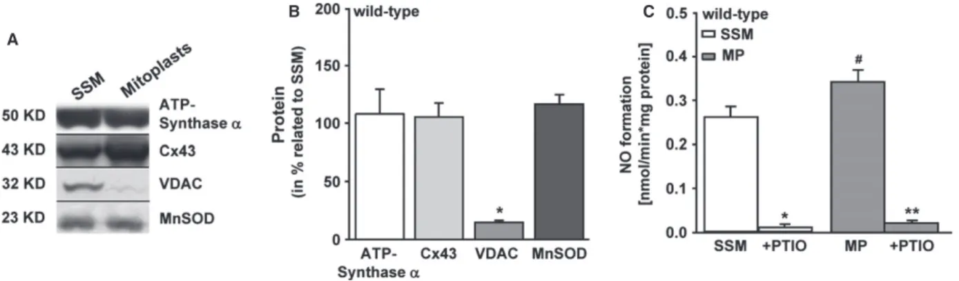
To rule out putative mitochondrial contamination with NOS isoforms attached to the outer membrane of mitochondria, mitoplasts were pre- pared by removing the outer membrane using the digitonin detergent (2 mmol/l). The difference between mitochondria and mitoplasts was shown by the content of voltage-dependent anion channel (VDAC), a marker for the outer mitochondrial membrane. The presence of ATP- synthase a, manganese superoxide dismutase (MnSOD) and Cx43 proved the intactness of mitoplasts.
Western blot analysis
For Western blot analysis, 100lg of mitochondrial proteins were elec- trophoretically separated on 10% SDS-PAGE and transferred to nitrocel- lulose membranes. After blocking, the membranes were incubated with the following antibodies: rabbit polyclonal anti-rat Cx43 (Invitrogen, Carlsbad, CA, USA), ATP-synthasea (BD Transduction, San Jose, CA, USA), rabbit polyclonal anti-human MnSOD (Upstate, Lake Placid, NY, USA), rabbit polyclonal anti-human VDAC (Abcam, Cambridge, UK) or mouse monoclonal anti-rabbit Na+/K+-ATPase (Upstate), mouse mono- clonal anti-dog sarcoplasmatic calcium (SERCA2)-ATPase (Sigma- Aldrich, Saint Louis, MO, USA). Immunoreactive signals were detected by chemiluminescence (SuperSignal West Femto Maximum Sensitivity Substrate, Pierce, Rockford, Il, USA) and quantified with the Scion Image software (Frederick, ML, USA).
Immunocytochemistry
For immunocytochemistry, purified SSM were incubated simultaneously with the following primary antibodies: anti-Cx43 (rabbit polyclonal anti- rat Cx43, Zytomed, Berlin, Germany) with anti-cyclophilin D (mouse monoclonal anti-rat, MitoSciences, Eugene, OR, USA), anti-nNOS (BD Transduction) or anti-iNOS (BD Transduction) with anti-ANT (Santa Cruz, Heidelberg, Germany) respectively. After washing, SSM were incubated with corresponding secondary antibodies (TRITC donkey anti- rabbit and FITC goat anti-rabbit for Cx43, TRITC donkey anti-rabbit and FITC goat antimouse for nNOS, TRITC bovine antimouse and FITC
bovine anti-rabbit for iNOS). SSM were examined by confocal laser scan microscopy (Pascal, Zeiss, Jena, Germany, or Leica TCSSP2AOBS, Wetzlar, Germany) at 6309magnification.
Measurement of cardiac nitric oxide
Oxyhaemoglobin assay
The nitric oxide formation in SSM (0.86 mg/ml) of wild-type mouse hearts was measured by the oxyhaemoglobin assay. The oxyhaemoglo- bin assay was modified to Feelischet al. [40]. The nitric oxide forma- tion was evaluated as the change in the absorbance at 401 and 590 nm using a diode array spectrophotometer (HP 8453; Hewlett Packard, Waldbronn, Germany). SSM, IFM or mitoplasts were incubated in incu- bation buffer (pH 7.4) with L-arginine (1 mmol/l), BH4 (10lmol/l), CaCl2(100lmol/l) and oxyhaemoglobin (HbO2, 50lmol/l) for 60 min.
at 37°C. SOD (1 U/ml) and catalase (50 U/ml) were also present in the reaction medium to avoid unspecific reactions. SOD catalysed superox- ide to oxygen and hydrogen peroxide. Catalase dissipated the hydrogen peroxide in oxygen and water.
Every 10 min., an aliquot was diluted in isolation buffer (pH 7.4) and the oxidation of HbO2to methaemoglobin by nitric oxide was measured.
The nitric oxide scavenger carboxy-PTIO (2-(4-Carboxyphenyl)-4,4,5,5- tetramethylimidazoline-1-oxyl-3-oxide) was used to determine the speci- ficity of the nitric oxide signal. NOS-dependent nitric oxide formation was confirmed by the selective NOS inhibitors SMTC and 1400 W (selective for nNOS/iNOS) [41, 42].
Electron spin resonance spectroscopy
Mitochondrial nitric oxide content was also measured using electron spin resonance spectroscopy (ESR) in isolated SSM. An aqueous spin trapping solution Fe2+–(MGD)2was prepared before each experiment as described [4, 43]. GSNO was resolved in phosphate buffer (35 mg/ml) and used as positive control. Isolated SSM and GSNO (200lmol/l) were treated with L-arginine (1 mmol/l), BH4(10lmol/l), CaCl2(100lmol/l), SOD (1 U/ml) and catalase (50 U/ml) and Fe2+–(MGD)2and incubated for 10 min. at 37°C. The prepared samples were then placed into quartz ESR tubes and frozen immediately in liquid nitrogen. Samples were assayed for ESR spectra of the relatively stable nitric oxide–Fe2+–(MGD)2 adduct. ESR spectra were recorded with a ECS106 spectrometer (Bruker, Rheinstetten, Germany) operating at X band with 100 kHz modulation frequency at a temperature of 160 K, using 10 mW microwave power to avoid satura- tion. Scans were traced with 2.85 G modulation amplitude, 340 G sweep width, and 3356 G central field as described [44, 45]. Analysis of nitric oxide content was performed with double integration of the nitric oxide signal as described previously [46].
Statistics
Data are reported as meanSEM. Confocal laser scanning microscopy data for Cx43, nNOS and iNOS were compared by two-wayANOVA, sub- sequent Bonferroni test. Quantitative Western blot data for Cx43 and ATP-synthaseawere compared by one-wayANOVA. The nitric oxide for- mation by the oxyhaemoglobin assay and the ESR spectroscopy in SSM of wild-type, Cx43Cre-ER(T)/fl
and nNOS / mice was compared by one- wayANOVArespectively. AP<0.05 was considered to indicate a signifi- cant difference.
Results
NOS expression in SSM of Cx43-deficient mice
Confocal laser scanning microscopy demonstrated mainly nNOS immunoreactivity and a weak expression of iNOS in isolated SSM from wild-type mice (Fig. 1A). Conversely, in SSM of Cx43
Cre-ER(T)/flmice, iNOS immunoreactivity was increased while nNOS expression was reduced (Fig. 1A). As marker for mitochondria, the inner mito- chondrial membrane protein ANT was detected. The statistical evalua- tion of NOS expression showed a significant difference between mitochondria expressing nNOS and iNOS in wild-type mice (93 4.1%
versus24.6 7.5% co-localization of NOS with ANT,
n=7 individual preparations, Fig. 1B). The nNOS expression in SSM of Cx43
Cre-ER(T)/flmice (53.8 17.5% co-localization of NOS with ANT,
n=7 individual preparations) was also significantly reduced compared to wild-type mice. In contrast, the iNOS expression (90.7 3.2% co-localization of NOS with ANT,
n=7 individual prep- arations, Fig. 1B) in SSM of Cx43
Cre-ER(T)/flmice was significantly increased compared to iNOS in wild-type mice.
To verify the immunocytochemical results by Western blot analy- sis in the mitochondrial samples of wild-type and Cx43
Cre-ER(T)/flmice, immunoblotting with anti-nNOS antibody against the amino-terminus showed no distinctive band at 160 kD compared to the positive con- trol (right ventricle, Fig. 2A). Only an unspecific band at 140 kD, which was also seen in mitochondria of nNOS
/mice (negative con- trol), was present (Fig. 2A). Antibodies against the iNOS isoform showed no visible band. Mitochondria were not contaminated with proteins of sarcolemma and with sarcoplasmatic reticulum as shown by the absence of Na
+/K
+-ATPase and SERCA immunoreactivity (Fig. 2A). Cx43 protein content was normalized to mitochondrial mar- ker protein ATP-synthase
a(Fig. 2B). Immunoprecipitation analysis also showed no detectable signal of the NOS isoforms. By definition, mitochondrial Cx43 expression in Cx43
Cre-ER(T)/flmice was signifi- cantly reduced compared to wild-type mice.
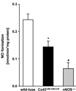
Nitric oxide formation in Cx43-deficient mice
Nitric oxide formation was measured by the oxyhaemoglobin assay in SSM of wild-type mice (Fig. 3). The basal NOS activity resulted in a nitric oxide formation of 0.24 0.02 nmol/min.
/mg protein (n
=15). The specificity of the nitric oxide signal was shown by the nitric oxide scavenger PTIO. Inhibition of nNOS using the non-selec- tive (W7) or the selective nNOS inhibitor (SMTC) resulted in a signifi- cant reduction of the mitochondrial nitric oxide formation.
Digitonin treatment of mitochondria significantly reduced the con- tent of the outer mitochondrial membrane protein VDAC to 14 2.6% (n
=6) of the signal of untreated mitochondria (set as 100%, Fig. 4A). The unchanged level of ATP-synthase
a(93 27%
protein content of mitoplasts compared to mitochondria set as 100%,
n=6), MnSOD (116 18%,
n=6) and mitochondrial Cx43
(105 27%,
n=6) confirmed an intact inner membrane of
mitoplasts (Fig. 4B). The nitric oxide production in mitoplasts was
A B
Fig. 2Expression of nNOS in subsarcolemmal mitochondria. (A) The expression of nNOS is presented in isolated subsarcolemmal mitochondria (SSM) of Cx43Cre-ER(T)/fl(n=9) and wild-type mice (n=7). Right ventricles (RV) from wild-type and nNOS / mice served as positive and negative control. Na+/K+-ATPase and SERCA served for the purity of the mitochondrial preparation. The Cx43 content showed the difference between the two mice strains. (B) The statistical evaluation of the Cx43 signal in SSM as related to the ATP-synthaseasignal is present as meanSEM (*signifi- cance withP<0.05 between wild-type and Cx43Cre-ER(T)/flmice).
A B
Fig. 1Mitochondrial NOS expression in subsarcolemmal mitochondria of wild-type mice and Cx43Cre-ER(T)/flmice. (A) Subsarcolemmal mitochondria (SSM) isolated from the ventricles of Cx43Cre-ER(T)/fland wild-type mice were stained with antibodies against nNOS or iNOS (red) and the mitochon- drial marker cytochrome c (green) and analysed by confocal laser scanning microscopy. Merged image shows co-localization pixels in yellow. (B) Amount in per cent of nNOS- and iNOS-positive SSM were referred to 100%. Individual preparations ofn=7.§P<0.003 nNOS of wild-typeversus nNOS of Cx43Cre-ER(T)/fl
,*P<0.001 nNOS of wild-typeversusiNOS of wild-type,#P<0.02 iNOS of Cx43Cre-ER(T)/fl
versusnNOS of Cx43Cre-ER(T)/fl
,
$P<0.002 iNOS of Cx43Cre-ER(T)/fl
versusiNOS of wild-type.
comparable with the nitric oxide production in SSM of wild-type mice (Fig. 4C). Therefore, a contamination of mitochondria with cellular NOS isoforms attached to the outer mitochondrial membrane as explanation for the measured nitric oxide formation appeared unlikely and an existence of nNOS-dependent mitochondrial nitric oxide pro- duction was confirmed. Again the nitric oxide specificity of the signal was shown by PTIO.
The NOS-dependent nitric oxide formation in Cx43
Cre-ER(T)/flmice (0.14 0.02 nmol/min.
/mg,
n=12) was reduced in comparison to wild-type mice (0.24 0.02 nmol/min.
/mg; Fig. 5). Inhibition of nNOS by SMTC in SSM of wild-type mice strongly reduced the nitric oxide formation (0.05 0.01 nmol/min.
/mg; Table 1). SMTC
showed a lower effect of reducing of nitric oxide formation in SSM of Cx43
Cre-ER(T)/flmice (0.06 0.02 nmol/min.
/mg). In contrast, 1400 W reduced the nitric oxide formation in SSM of Cx43
Cre-ER(T)/flto 62%. A similar result was detectable by iNOS inhibition in SSM of wild-type mice (58% nitric oxide formation). The negative control showed a residual NOS activity of 0.06 0.02 nmol/min.
/mg (n
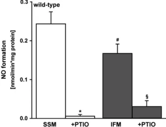
=4). The enzymatic residual NOS activity was completely reduced by SMTC and 1400 W. To confirm the Cx43 dependence of the NOS shift, we used isolated IFM of wild-type mice, the mitochondrial sub- population without Cx43, as internal control [1]. The purity of IFM preparation was controlled by Western blot analysis. IFM were not contaminated with proteins of sarcolemma and sarcoplasmatic reticu- lum as shown by the absence of Na
+/K
+-ATPase and SERCA immuno- reactivity (Fig. 6A). Cx43 was also detected exclusively in SSM (Fig. 6B).
Interfibrillar mitochondria showed a significantly reduced nitric oxide formation (0.17 0.02 nmol/min.
/mg,
n=9) in contrast to SSM (0.24 0.03 nmol/min.
/mg,
n=10; Fig. 7). In presence of the nitric oxide scavenger PTIO, the nitric oxide formation was almost undetectable in both mitochondrial preparations (0.007 0.006 nmol/min.
/mg in SSM; 0.03 0.01 nmol/min.
/mg IFM;
Fig. 7).
Another independent method for quantifying nitric oxide formation is the ESR technique. The ESR spectrum of the positive control (GSNO) showed a prominent nitric oxide
–Fe
2+–MGD triplet (Fig. 8A).
In the SSM of wild-type mice, a basal spectrum of nitric oxide
–Fe
2+–MGD triplet was detected (Fig. 8B). In the SSM of Cx43
Cre-ER(T)/flmice, the intensity of the specific spectra of nitric oxide
–Fe
2+–MGD complex was reduced compared to the wild-type mice (Fig. 8C). Peak three is an indicator of the nitric oxide content in SSM of wild-type and Cx43
Cre-ER(T)/flmice obtained from relative signal amplitudes of nitric oxide
–Fe
2+–MGD complex. The nitric oxide content of peak three in SSM of Cx43
Cre-ER(T)/flmice was significantly reduced com- pared to wild-type mice (n
=6 preparations, Fig. 8D).
A
B C
Fig. 4Nitric oxide formation in mitoplasts of wild-type mice. (A) Representative Western blot shows the difference between subsarcolemmal mito- chondria (SSM,n=5) and mitoplast (MP,n=6) preparation by the absence of the outer mitochondrial membrane protein voltage-dependent anion channel (VDAC).#P<0.05 indicates the significant difference between MP and mitochondria. (B) Statistical evaluation presents the meanSEM of MP as related to SSM (in %),*significance withP<0.05 MP and mitochondria. (C) Nitric oxide formation in MP (n=11) and mitochondria (n=10). PTIO scavenged the accumulated nitric oxide. *Significance between SSMPTIO (P<0.001),**significance between MPPTIO (P<0.001).
Fig. 3Basal nitric oxide formation in subsarcolemmal mitochondria of wild-type mice. PTIO (n=7) reduced the nitric oxide formation. The enzymatic NOS inhibition by the inhibitors W7 (non-selective,n=5) and SMTC (nNOS selective, n=7) reduced nitric oxide formation.
*P<0.001 indicates significant difference after treatment with PTIO, W7 or SMTC. Wild-type mice (n=13).
Discussion
In the present study, Cx43 expression correlated with the NOS iso- form mainly present in SSM. While in SSM from wild-type hearts nNOS was the main isoform, in SSM from Cx43-deficient mice hearts iNOS dominated.
The constitutive mitochondrial NOS isoform expressed differs among species. The iNOS was present in porcine and rat heart mito- chondria [47, 48], while nNOS was mainly found to be expressed in mitochondria from mice hearts [49]. The latter finding was confirmed in the present study in SSM isolated from wild-type C57BL/6J mice.
In previous studies, also eNOS protein was localized to mitochon- dria [50]; however, in the present study while we could detect an eNOS signal in the vascular endothelium, there was no signal detected in mitochondria (data not shown). More recently, increased
translocation of eNOS protein (located in caveolae) to the outer mito- chondrial membrane was demonstrated upon short periods of ischae- mia/reperfusion [51]. Thus, some of the differences observed in mitochondrial eNOS expression might relate to the presence or absence of attached sarcolemmal proteins which was excluded in our mitochondrial preparation.
The mtNOS protein was mainly detected by immunohistochemical [8, 52] and biochemical methods [48, 53
–55]. Similarly, the presence of mtNOS in the present study could only be confirmed by immuno- histochemistry and direct measurement of nitric oxide formation from isolated mitochondria; no specific NOS signal was detected by Wes- tern blot using commercially available antibodies. One explanation for the lack of detection of NOS expression in the Western blot analysis is the limited sensitivity (1/10) in comparison to laser scan micros- copy [56]. This assumption is supported by the fact that with massive overexpression of nNOS in mice, a mitochondrial signal was detected also by Western blot [57].
Given the lack of NOS detection in mitochondria by a method like Western blot, the existence of NOS in mitochondria is still under dis- cussion [58]. In different studies, mitochondrial NOS was attributed to a contamination of the preparation or the attachment of NOS to the outer mitochondrial membrane [59
–61]. To circumvent this problem, we generated mitoplasts [1] and the rate of nitric oxide formation of mitoplasts was similar to that of intact SSM.
An array of methods exists for the detection of mtNOS-depen- dent nitric oxide formation including photometrical (oxyhaemoglo- bin assay, DAF-2 assay) [58, 62
–64], electrochemical (porphyrinic microsensor) [49] and radioactive (citrulline assay) techniques [7].
The sensitivity and specificity of the oxyhaemoglobin assay is suf- ficient to detect mtNOS-dependent changes in nitric oxide forma- tion [48, 49, 58, 65] and allows real-time detection of generated nitric oxide [66]. In the present study, we were able to confirm the rate of NOS-derived basal nitric oxide formation in the SSM from wild-type hearts. The nitric oxide formation of about 0.24 nmol/min. mg and was thus close to the range of NOS- derived nitric oxide formation rates from rat heart mitochondria (0.37 nmol/min.
/mg and 0.69 nmol/min.
/mg) [48, 65, 67]. Compo- sition of the buffer, the temperature and the mitochondrial meta- bolic state could cause the small differences in basal nitric oxide formation. However, the specificity of the detected nitric oxide sig- nal was proven by scavenging nitric oxide through addition of
Table 1Nitric oxide formation after nitric oxide synthase inhibition in all mice strains
Mice strains
Nitric oxide formation (nmol/min./mg)
Basal After nNOS inhibition (SMTC) After iNOS inhibition (1400 W)
Wild-type 0.240.02 0.050.01* 0.130.01*
Cx43Cre-ER(T)/fl 0.140.02 0.060.01† 0.050.02†
nNOS / 0.060.02 0.030.01‡ 0.0090.009‡
*Wild-typeSMTC (P<0.001) and1400 W (P<0.001).
†Cx43Cre-ER(T)/fl
SMTC (P<0.001) and1400 W (P<0.001).
‡nNOS / miceSMTC (P<0.005) and1400 W (P<0.001).
SMTC, S-methyl-thiocitrulline, iNOS, inducible nitric oxide synthase; nNOS, neuronal nitric oxide synthase.
Fig. 5Mitochondrial nitric oxide formation and connexin 43 deficiency.
Comparison of nitric oxide formation in subsarcolemmal mitochondria (SSM) of wild-type (n=15) and Cx43Cre-ER(T)/fl mice (n=12,
*P<0.001). NNOS / mice served as negative controls (n=4). # indicates the significant difference between wild-type and nNOS / mice (P<0.001).
PTIO. The strong reduction of the nitric oxide formation by selec- tive nNOS inhibition confirmed a mainly expressed mitochondrial nNOS in wild-type mice. The slight reduction of the nitric oxide formation by the selective iNOS inhibitor approved a small amount of active mitochondrial iNOS in wild-type mice.
Concomitant with an enhanced mitochondrial iNOS expression, an increased nitric oxide formation rate was expected. However, and to our surprise, the enhanced mitochondrial iNOS and reduced mito- chondrial nNOS expression in Cx43-deficient hearts was associated with a reduced rate of nitric oxide formation in mitochondria derived from Cx43-deficient mice hearts. These data from the oxyhaemoglo- bin assay were confirmed by a second independent technique, namely ESR, which also shows a high nitric oxide sensitivity [68].
Indeed, iNOS may be present in two forms either as uncoupled monomer and/or coupled dimer. Zhang
et al., found in a stress-induced myocardium of wild-type mice, both iNOS monomer and iNOS dimer [69]. One can speculate that the reduced nitric oxide for- mation in mitochondria from Cx43-deficient mice relates to the pres- ence of an enhanced monomer iNOS isoform and coincident low dimer iNOS isoform.
Mitochondrial nitric oxide formation in nNOS
/mice (negative control) was because of an enzymatic residual activity of the conceiv- ably available splice variants nNOS-
bund nNOS-
c[70, 71], which were inhibited to a great extent by the selective nNOS inhibitor, and some contribution of iNOS. The remaining nitric oxide formation might represent non-enzymatically released nitric oxide from nitrite [72], which have been suggested to contribute to the cardioprotective effect of nitrite [73, 74].
Nitrosylation of several respiratory complexes including complex I are supposed to impair respiration [25]. Indeed, IFM which have a lower nitric oxide production as compared to SSM have a higher respiratory rate. Although SSM of Cx43-deficient mice also have a reduced nitric oxide production as compared to SSM of wild-type mice, they nevertheless have a reduced respiratory rate and ATP pro- duction [75]. One potential explanation for this contradictory finding might relate to decreased potassium flux in Cx43-deficient mice upon ADP stimulation, which impairs matrix volume recovery and thus mitochondrial oxygen consumption [76].
Indeed, we have recently shown that increased S-nitrosation of Cx43 enhanced potassium influx into the mitochondrial matrix as well as the generation of reactive oxygen species [77], the latter being an important signalling molecule for endogenous cardioprotection.
Importantly, in the above study, ischaemic preconditioning increased S-nitrosation of mitochondrial Cx43. The present study adds one more facet to the interaction of nitric oxide and Cx43 in that the mtNOS isoform and subsequently the mitochondrial nitric oxide pro- duction depends on the level of Cx43 expression (Fig. 9). An overall reduction of Cx43 expression in Cx43-deficient mice not only reduced mitochondrial Cx43 but also mitochondrial nNOS expression with a subsequent decrease in mitochondrial nitric oxide production and overall protein S-nitrosation (Data S1), both contributing to a reduction
A B
Fig. 6Content of connexin 43 in subsarcolemmal and interfibrillar mitochondria. (A) Representative Western blot of Cx43 in isolated subsarcolem- mal (SSM,n=5) and interfibrillar (IFM,n=5) mitochondria from left ventricles (LV). SERCA and Na+/K+-ATPase served as marker protein for the purity of the mitochondrial preparation. Ponceau S staining shows equal protein loading. (B) Quantification of the Cx43 content in SSM and IFM nor- malized to Ponceau S staining (n=5 individual preparations,*P<0.001).
Fig. 7Nitric oxide formation in subsarcolemmal and interfibrillar mito- chondria. Nitric oxide formation was compared between subsarcolem- mal (SSM,n=10 individual experiments) and interfibrillar mitochondria (IFM, n=9 individual experiments) of wild-type mice (#P<0.04 between SSM and IFM). Isolated IFM served as internal control. Nitric oxide specificity was shown by PTIO. *P<0.001 SSMPTIO,
§P<0.001 IFMPTIO.
of mitochondrial matrix potassium influx and reactive oxygen species formation upon an external stimulus such as ischaemic or pharmaco- logical preconditioning thereby contributing to the lack of endogenous cardioprotection seen in Cx43-deficient mice [78, 79].
In conclusion, the present study showed that reduced mitochondrial Cx43 content is associated with a switch of the mitochondrial NOS isoform and the respective mitochondrial nitric oxide production rate.
Acknowledgements
We thank Mrs. Anna Reis and Elvira Ungefug for her technical support. This work was supported by the German Research Foundation (Schu 843/7-2 grant to RS). CC was funded by the Janos Bolyai Research Scholarship of the Hun- garian Academy of Sciences. PF holds a Szentagothai professorship of the National Excellence Program.
Conflicts of interest
The authors confirmed that there are no conflicts of interest.
Author contribution
PK, MK and CC performed the research and analysed the data, RS designed the research study; PF contributed essential reagents or tools, MK and RS wrote the paper. The data of the present paper are part of the doctorate thesis of MK.
Supporting information
Additional Supporting Information may be found in the online version of this article:
Data S1
Quantification of S-nitrosated protein of Cx43 in heart.
A B C D
Fig. 8Formation of nitric oxide by electron spin resonance spectroscopy after spin trapping with Fe2+–MGD complex. Representative electron spin resonance (ESR) spectra of nitric oxide–Fe2+–MGD adduct in SSM of (A) wild-type and (B) Cx43Cre-ER(T)/flmice. (C) GSNO served as positive control.
ESR parameters: X band, 100 kHz modulation frequency, 160 K, 10 mW microwave power, 2.85 G modulation amplitude, 340 G sweep width, and 3356 G central field. (D) The nitric oxide content in subsarcolemmal mitochondria (in arbitrary units) obtained from the relative signal amplitude of the nitric oxide–Fe2+–MGD complex in peak three of wild-type and Cx43Cre-ER(T)/flmice (n=6 preparations,*P<0.04).
Fig. 9Scheme of nitric oxide and Connexin 43 (Cx43) in the context of cardioprotection. The mitochondrial NOS isoform expression and subse- quently the mitochondrial nitric oxide production depends on the level of Cx43 expression. An overall reduction of Cx43 expression not only reduced mitochondrial Cx43 but also mitochondrial nNOS expression with a subsequent decrease in mitochondrial nitric oxide production and overall protein S-nitrosation, both contributing to a reduction of mito- chondrial matrix potassium influx and reactive oxygen species formation upon an external stimulus such as ischaemic or pharmacological pre- conditioning, thereby contributing to the lack of endogenous cardiopro- tection seen in Cx43-deficient mice.
References
1. Boengler K, Stahlhofen S, van de Sand A, et al.Presence of connexin 43 in subsarco- lemmal but not in interfibrillar cardiomyo- cyte mitochondria.Basic Res Cardiol. 2009;
104: 141–7.
2. Heusch G, Boengler K, Schulz R.Cardiopro- tection: nitric oxide, protein kinases, and mitochondria.Circulation. 2008; 118: 1915– 9.
3. Boengler K, Konietzka I, Buechert A,et al.
Loss of ischemic preconditioning’s cardio- protection in aged mouse hearts is associ- ated with reduced gap junctional and mitochondrial levels of connexin 43.Am J Physiol Heart Circ Physiol. 2007; 292:
H1764–9.
4. Csonka C, Szilvassy Z, F€ul€op F,et al.Clas- sic preconditioning decreases the harmful accumulation of nitric oxide during ischemia and reperfusion in rat hearts. Circulation.
1999; 100: 2260–6.
5. Ferdinandy P, Schulz R. Nitric oxide, superoxide, and peroxynitrite in myocar- dial ischaemia-reperfusion injury and preconditioning.Br J Pharmacol. 2003; 138:
532–43.
6. Moncada S, Higgs A.The L-arginine-nitric oxide pathway.N Engl J Med. 1993; 329:
2002–12.
7. Elfering SL, Sarkela TM, Giulivi C. Bio- chemistry of mitochondrial nitric-oxide synthase.J Biol Chem. 2002; 277: 38079– 86.
8. Bates TE, Loesch A, Burnstock G, et al.
Immunocytochemical evidence for a mitoc- hondrially located nitric oxide synthase in brain and liver.Biochem Biophys Res Com- mun. 1995; 213: 896–900.
9. Rassaf T, Poll LW, Brouzos P,et al.Posi- tive effects of nitric oxide on left ventricular function in humans.Eur Heart J. 2006; 27:
1699–705.
10. Heusch G, Post H, Michel MC, et al.
Endogenous nitric oxide and myocardial adaptation to ischemia.Circ Res. 2000; 87:
146–52.
11. Rastaldo R, Pagliaro P, Cappello S,et al.
Nitric oxide and cardiac function.Life Sci.
2007; 81: 779–93.
12. Parihar MS, Nazarewicz RR, Kincaid E, et al. Association of mitochondrial nitric oxide synthase activity with respiratory chain complex I. Biochem Biophys Res Commun. 2008; 366: 23–8.
13. Antunes F, Boveris A, Cadenas E.On the mechanism and biology of cytochrome
oxidase inhibition ny nitric oxide. PNAS.
2004; 101: 16774–9.
14. Dedkova EN, Blatter LA. Modulation of mitochonrial Ca2+ by nitric oxide in cultured bovine vascular endothelial cells.
Am J Physiol Cell Physiol. 2005; 289: C836– 45.
15. Gutierrez J, Ballinger SW, Darley-Usmar VM,et al.Free radicals, mitochondria, and oxidized lipids: the emerging role in signal transduction in vascular cells. Circ Res.
2006; 99: 924–32.
16. Damy T, Ratajczak P, Robidel E,et al.Up- regulation of cardiac nitric oxide synthase 1-derived nitric oxide after myocardial infarction in senescent rats.FASEB J. 2003;
17: 1934–6.
17. Dawson D, Lygate CA, Zhang M-H,et al.
nNOS gene deletion exacerbates pathologi- cal left ventricular remodeling and funcitonal deterioration after myocardial infarction.Cir- culation. 2005; 112: 3729–37.
18. Sun J, Steenbergen C, Murphy E.S-nitrosy- lation: NO-related redox signaling to protect against oxidative stress.Antioxid Redox Sig- nal. 2006; 8: 1693–705.
19. Xu KY, Huso DL, Dawson TM,et al.Nitric oxide synthase in cardiac sarcoplasmic reticulum. Proc Natl Acad Sci USA. 1999;
96: 657–62.
20. Burkard N, Rokita AG, Kaufmann SG, et al. Conditional neuronal nitric oxide synthase overexpression impairs myocar- dial contractility. Circ Res. 2007; 100:
e32–44.
21. Sun J, Murphy E.Protein S-nitrosylation and cardioprotection.Circ Res. 2010; 106: 285–
96.
22. Sun J, Morgan M, Shen RF,et al.Precondi- tioning results in S-nitrosylation of proteins involved in regulation of mitochondrial ener- getics and calcium transport. Circ Res.
2007; 101: 1155–63.
23. Burwell LS, Brookes PS. Mitochondria as a target for the cardioprotective effects of nitric oxide in ischemia-reperfusion injury. Antioxid Redox Signal. 2008; 10:
579–99.
24. Prime TA, Blaikie FH, Evans C, et al. A mitochondria-targeted S-nitrosothiol modu- lates respiration, nitrosates thiols, and pro- tects against ischemia-reperfusion injury.
Proc Natl Acad Sci USA. 2009; 106: 10764–
9.
25. Chouchani ET, Methner C, Nadtochiy SM, et al.Cardioprotection by S-nitrosation of a
cysteine switch on mitochondrial complex I.
Nat Med. 2013; 19: 753–9.
26. Andelova E, Bartekova M, Pancza D,et al.
The role of NO in ischemia/reperfusion injury in isolated rat heart. Gen Physiol Biophys.
2005; 24: 411–26.
27. Lochner A, Marais E, Du TE, et al.Nitric oxide triggers classic ischemic precondition- ing.Ann N Y Acad Sci. 2002; 962: 402–14.
28. Tong G, Aponte AM, Kohr MJ,et al.Post- conditioning leads to an increase in protein S-nitrosylation. Am J Physiol Heart Circ Physiol. 2014; 306: H825–H832.
29. Radosinska J, Bacova B, Bernatova I,et al.
Myocardial NOS activity and connexin-43 expression in untreated and omega-3 fatty acids-treated spontaneously hypertensive and hereditary hypertriglyceridemic rats.
Mol Cell Biochem. 2011; 347: 163–73.
30. Roh CR, Heo JH, Yang SH,et al.Regulation of connexin 43 by nitric oxide in primary uterine myocytes from term pregnant women. Am J Obstet Gynecol. 2002; 187:
434–40.
31. Yao J, Hiramatsu N, Zhu Y, et al.Nitric oxide-mediated regulation of connexin43 expression and gap junctional intercellular communication in mesangial cells.J Am Soc Nephrol. 2005; 16: 58–67.
32. Li K, Yao J, Shi L,et al.Reciprocal Regula- tion between Proinflammatory Cytokine- induced Inducible NO Synthase (iNOS) and Connexin43 in Bladder Smooth Muscle Cells.
J Biol Chem. 2011; 286: 41552–62.
33. Jackson PE, Feng QP, Jones DL.Nitric oxide depresses connexin 43 after myocardial infarction in mice.Acta Physiol. 2008; 194:
23–33.
34. Retamal MA, Cortes CJ, Reuss L, et al.
S-nitrosylation and permeation through conexin 43 hemichannels in astrocytes:
induction by oxidant stress and reversal by reducing agents. PNAS. 2006; 103:
4475–80.
35. Dang X, Doble BW, Kardami E. The car- boxy-tail of connexin-43 localizes to the nucleus and inhibits cell growth. Mol Cell Biochem. 2003; 242: 35–8.
36. Eckardt D, Theis M, Degen J,et al.Func- tional role of connexin 43 gap junction chan- nels in adult mouse heart assessed by inducible gene deletion.J Mol Cell Cardiol.
2004; 36: 101–10.
37. Boengler K, Dodoni G, Rodriguez-Sinovas A, et al. Connexin 43 in cardiomyocyte mitochondria and its increase by ischemic
preconditioning.Cardiovasc Res. 2005; 67:
234–44.
38. Holmuhamedov EL, Jovanovic S, Dzeja PP, et al. Mitochondrial ATP-sensitive K+ channels modulate cardiac mitochondrial function.Am J Physiol. 1998; 44: H1567–
76.
39. Judge S, Jang YM, Smith A, et al. Age- associated increases in oxidative stress and antioxidant enzyme activities in cardiac inter- fibrillar mitochondria: implications for the mitochondrial theory of aging. FASEB J.
2005; 19: 419–21.
40. Feelisch M, Stamler JS.Methods in nitric oxide research. 1st ed. New York: John Wiley & Sons Ltd; 1996.
41. Garvey EP, Oplinger JA, Furfine ES,et al.
1400 W is a slow, tight binding, and highly selective inhibitor of inducible nitric oxide synthasein vitroandin vivo.J Biol Chem.
1997; 272: 4959–63.
42. Furfine ES, Harmon MF, Paith JE, et al.
Potent and selective inhibition of human nitric oxide synthases. Selective inhibition of neuronal nitric oxide synthase by S-methyl-L-thiocitrulline and S-ethyl-L-thio- citrulline.J Biol Chem. 1994; 269: 26677–
83.
43. Csont T, Szilvassy Z, F€ul€op F, et al.
Direct myocardial anti-ischaemic effect of GTN in both nitrate-tolerant and nontoler- ant rats: a cyclic GMP-independent activa- tion of KATP. Br J Pharmacol. 1999; 128:
1427–34.
44. Ferdinandy P, Szilvassy Z, Horvath LI, et al.Loss of pacing-induced precondition- ing in rat hearts: role of nitric oxide and cho- lesterol enriched diet. J Mol Cell Cardiol.
1997; 29: 3321–33.
45. Mulsch A, Vanin A, Mordvintcev P,et al.
NO accounts completely for the oxygenated nitrogen species generated by enzymic L- arginine oxygenation.Biochem J. 1992; 288:
597–603.
46. Csont T, Pali T, Szilvassy Z,et al.Lack of correlation between myocardial nitric oxide and cyclic guanosine monophosphate con- tent in both nitrate-tolerant and -nontolerant rats.Biochem Pharmacol. 1998; 56: 1139– 44.
47. French S, Giulivi C, Balaban RS. Nitric oxide synthase in porcine heart mitochon- dria: evidence for low physiological activity.
Am J Physiol Heart Circ Physiol. 2001; 280:
H2863–7.
48. Fellet AL, Balaszczuk AM, Arranz C,et al.
Autonomic regulation of pacemaker activity:
role of heart nitric oxide synthases.Am J Physiol Heart Circ Physiol. 2006; 291:
H1246–54.
49. Kanai AJ, Pearce LL, Clemens PR,et al.
Identification of a neuronal nitric oxide syn- thase in isolated cardiac mitochondria using electrochemical detection.PNAS. 2001; 98:
14126–31.
50. Zaobornyj T, Ghafourifar P.Strategic locali- zation of heart mitochondrial NOS: a review of the evidence. Am J Physiol Heart Circ Physiol. 2012; 303: H1283–93.
51. Sun J, Kohr MJ, Nguyen T,et al.Disruption of caveolae blocks ischemic precondition- ing-mediated S-nitrosylation of mitochon- drial proteins.Antioxid Redox Signal. 2012;
16: 45–56.
52. Bates TE, Loesch A, Burnstock G, et al.
Mitochondrial nitric oxide synthase: a ubiq- uitous regulator of oxidative phosphoryla- tion?Biochem Biophys Res Commun. 1996;
218: 40–4.
53. Zanella B, Giordano E, Muscari C, et al.
Nitric oxide synthase activity in rat cardiac mitochondria.Basic Res Cardiol. 2004; 99:
159–64.
54. La PP, Bustamante J, Czerniczyniec A, et al.Time course of regression of the pro- tection conferred by simulated high altitude to rat myocardium: correlation with mtNOS.
J Appl Physiol. 2008; 105: 951–7.
55. Hotta Y, Otsuku-Murakami H, Fujita M, et al.Protective role of nitric oxide synthase against ischemia-reperfusion injury in gui- nea pig myocardial mitochondria. Eur J Pharmacol. 1999; 380: 37–48.
56. Bieschke J, Giese A, Schulz-Schaeffer W, et al.Ultrasensitive detection of pathological prion protein aggregates by dual-color scan- ning for intensely fluorescent targets.Proc Natl Acad Sci USA. 2000; 97: 5468–73.
57. Burkard N, Williams T, Czolbe M, et al.
Conditional overexpression of neuronal nitric oxide synthase is cardioprotective in ische- mia/reperfusion. Circulation. 2010; 122:
1588–603.
58. Ghafourifar P, Richter C.Nitric oxide syn- thase activity in mitochondria. FEBS Lett.
1997; 418: 291–6.
59. Lacza Z, Pankotai E, Csordas A,et al.Mito- chondrial NO and reactive nitrogen species production: does mtNOS exist?Nitric Oxide.
2006; 14: 162–8.
60. Tay YM, Lim KS, Sheu FS,et al.Do mito- chondria make nitric oxide? no?Free Radic Res. 2004; 38: 591–9.
61. Csordas A, Pankotai E, Snipes JA, et al.
Human heart mitochondria do not produce physiologically relevant quantities of nitric oxide.Life Sci. 2007; 80: 633–7.
62. Giulivi C, Poderoso JJ, Boveris A.Produc- tion of nitric oxide by mitochondria.J Biol Chem. 1998; 273: 11038–43.
63. Dedkova EN, Ji X, Lipsius SL,et al.Mito- chondrial calcium uptake stimulates nitric oxide production in mitochondria of bovine vascular endothelial cells.Am J Physiol Cell Physiol. 2004; 286: C406–15.
64. Tao Y, Zhang P, Girdler F, et al.
Enhancement of radiation response in p53- deficient cancer cells by the Aurora-B kinase inhibitor AZD1152. Oncogene. 2008; 27:
3244–55.
65. Boveris A, D’Amico G, Lores-Arnaiz S, et al. Enalapril increases mitochondrial nitric oxide synthase activity in heart and liver. Antioxid Redox Signal. 2003; 5:
691–7.
66. Ghafourifar P, Parihar MS, Nazarewicz R, et al.Detection assays for determination of mitochondrial nitric oxide synthase activity;
advantages and limitations.Methods Enzy- mol. 2008; 440: 317–34.
67. Gonzales GF, Chung FA, Miranda S,et al.
Heart mitochondrial nitric oxide synthase is upregulated in male rats exposed to high altitude (4,340 m).Am J Physiol Heart Circ Physiol. 2005; 288: H2568–73.
68. Saito K, Kohno M. Application of electron spin resonance spin-trapping technique for evaluation of substrates and inhibitors of nitric oxide synthase.Anal Biochem. 2006;
349: 16–24.
69. Zhang P, Xu X, Hu X,et al.Inducible nitric oxide synthase deficiency protects the heart from systolic overload-induced ventricular hypertrophy and congestive heart failure.
Circ Res. 2007; 100: 1089–98.
70. Huang PL, Dawson TM, Bredt DS, et al.
Targeted disruption of the neuronal nitric oxide synthase gene.Cell. 1993; 75: 1273– 86.
71. Gyurko R, Leupen S, Huang PL.Deletion of exon 6 of the neuronal nitric oxide synthase gene in mice results in hypogonadism and infertility.Endocrinology. 2002; 143: 2767–
74.
72. Cosby K, Partovi KS, Crawford JH, et al.
Nitrite reduction to nitric oxide by deoxyhe- moglobin vasodilates the human circulation.
Nat Med. 2003; 9: 1498–505.
73. Shiva S, Huang Z, Grubina R, et al.
Deoxymyoglobin is a nitrite reductase that generates nitric oxide and regulates mito- chondrial respiration.Circ Res. 2007; 100:
654–61.
74. Shiva S, Gladwin MT.Nitrite mediates cyto- protection after ischemia/reperfusion by modulating mitochondrial function. Basic Res Cardiol. 2009; 104: 113–9.
75. Boengler K, Ruiz-Meana M, Gent S,et al.
Mitochondrial connexin 43 impacts on respi- ratory complex I activity and mitochondrial
oxygen consumption.J Cell Mol Med. 2012;
16: 1649–55.
76. Korge P, Honda HM, Weiss JN.K+-depen- dent regulation of matrix volume improves mitochondrial funciton under conditions mimicking ischemia-reperfusion.Am J Phys- iol Heart Circ Physiol. 2005; 289: H66–77.
77. Soetkamp D, Nguyen TT, Menazza S,et al.
S-nitrosation of mitochondrial connexin 43 regulates mitochondrial function.Basic Res Cardiol. 2014; 109: 433.
78. Schwanke U, Konietzka I, Duschin A, et al.No ischemic preconditioning in het- erozygous connexin43-deficient mice. Am
J Physiol Heart Circ Physiol. 2002; 283:
H1740–2.
79. Heinzel FR, Luo Y, Li X,et al.Impairment of diazoxide-induced formation of reactive oxygen species and loss of cardioprotection in connexin 43 deficient mice. Circ Res.
2005; 97: 583–6.