University of Szeged Faculty of Science and Informatics
Doctoral School of Biology
APPLICATION OF EPITHELIAL CELL CULTURE MODELS TO STUDY THE EFFECTS OF BIOLOGICALLY ACTIVE PEPTIDES AND
ADJUVANT THERAPEUTIC AGENTS
Summary of the Ph.D. Thesis
Ilona Gróf
Supervisors
Dr. Mária Deli, scientific adviser Dr. Alexandra Bocsik, research associate
Biological Barriers Research Group, Institute of Biophysics Biological Research Centre
Szeged 2021
1. INTRODUCTION
The main function of epithelial biological barriers is to separate the organism from the external environment. The respiratory and intestinal epithelia represent two of the major barrier systems, which pose two problems related to pathological conditions: their dysfunction can promote the development diseases and aggravate their symptoms, and the physical defense systems of epithelial cells can inhibit the absorption and the entry of drugs into organs. In these two biological barriers, similarly to others, the intercellular junctional proteins compose the physical barrier and restrict the paracellular permeability. An additional system of physical protection is the mucus covering the epithelial surface in the airways and intestines, which is involved in many diseases, including cystic fibrosis. Adjuvant therapies play an important role in the treatment of diseases, which can help alleviate the symptoms of the disease, otherwise they can increase the targeting of the active ingredients by reversibly opening the barrier systems. However, the study of these adjuvants requires model systems that adequately reflect the complexity of a particular organ in the body.
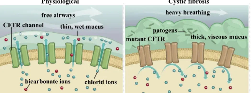
Cystic fibrosis is a genetic disorder caused by a mutation in the gene encoding cystic fibrosis transmembrane regulatory (CFTR) channels. CFTR is a cyclic AMP activated anion channel responsible for the transport of chloride and bicarbonate ions. In the absence of a functional CFTR channel, ion secretion decreases, Na+ and water reabsorption increases, leading to thickening and acidification of the mucus covering the cell surface (Fig. 1). This, in addition to breathing difficulties, provides an ideal environment for pathogens to multiply, leading to persistent inflammation in the patients’ lungs.
Figure 1. Airway epithelial CFTR channels in physiological conditions and in cystic fibrosis.
Sodium bicarbonate can be a promising agent in adjunctive therapies of lung diseases.
Its beneficial effects have been observed, primarily as a mucolytic in various respiratory diseases, and its bacteriostatic effects have also been described for the most common pathogen
in cystic fibrosis, Pseudomonas aeruginosa. In a recent clinical study sodium bicarbonate decreased mucus density and increased the airway fluid pH in cystic fibrosis patients. However, the direct effect of sodium bicarbonate on airway epithelial cells has not been studied yet.
The tightness of biological barriers is determined by the intercellular proteins forming tight junctions between the plasma membranes of adjacent epithelial cells. These structures block the so-called paracellular pathway between cells, thereby inhibiting the passage of ions, molecules and cells, as well as certain drugs, through the barrier. The restricted drug absorption makes the pharmacological treatment of diseases difficult. One of the ways to increase the entry of drugs into individual organs can be achieved by reversibly opening cell-cell connections.
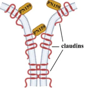
Figure 2. The PN159 peptide binds to the extracellular domain of claudin proteins and opens the paracellular pathway.
The synthetic PN159 peptide was first described as an amphipathic cell penetrating peptide. It was later demonstrated that PN159 can reversibly open tight junctions in culture models of biological barriers and effectively increase cell layer permeability (Fig. 2). Due to its α-helical secondary protein structure and amphipathic properties, the PN159 peptide interacts strongly with biological membranes and may therefore be a promising alternative in the treatment of multidrug-resistant bacterial strains.
2. AIMS
Despite its empirical use as adjuvant, the direct effect of sodium bicarbonate on bronchial epithelial cells has not been studied. Our aim was therefore to (i) establish a co-culture model for the study of cystic fibrosis, (ii) examine the effect of different culture conditions and (iii) the effect of sodium bicarbonate on these cells. The other main goal of our study was the investigation of the dual action of the PN159 peptide. Our aim was to test the effect of the peptide on intercellular junctions (iv) in bronchial and intestinal epithelial cultures, furthermore to investigate the membrane permeabilizing effect in (v) the culture model of the intestinal epithelium and (vi) on antibiotic-resistant ESKAPE pathogens.
3. MATERIALS AND METHODS 3.1. Cell cultures
3.1.1. The human cystic fibrosis bronchial epithelial cell lines
The CFBE41o- cell line was transfected with a plasmid carrying the wild type CFTR channel (WT-CFTR) and the most common CFTR channel mutation (ΔF508-CFTR). CFBE cells were cultured in Minimum Eagle Medium (MEM) supplemented with 10% serum (FBS, Pan-Biotech GmbH, Germany), glutamine (Glutamax, 2 mM) and gentamicin (50 µg/ml) in an incubator at 37°C with 5% CO2. To keep the transgene level high in the cell lines the culture medium was supplemented with puromicin (2 µg/ml) during growth. Culture surfaces were coated with collagen. For permeability experiments bronchial epithelial cells were grown in cell culture inserts placed in 12-well culture dishes (Transwell, 0.4 μm pore size, 1.1 cm2 membrane surface, Corning Costar Co., USA). Epithelial cells were grown for 10 days in monoculture or in co-culture with endothelial cells.
3.1.2. The human endothelial cells
Endothelial cells derived from human hematopoietic stem cells were used to establish the co-culture. Cells were cultured in endothelial cell medium (ECM-NG, Sciencell, Carlsbad, USA) supplemented with 5% serum, 1% endothelial growth factor (ECGS, Sciencell) and 0.5%
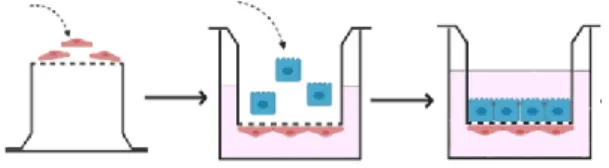
gentamicin. The establishment of the co-culture model is illustrated in Fig. 3.
Figure 3. The assembly of the human bronchial epithelial cell and endothelial cell co-culture model. First, endothelial cells were seeded on the lower surface of the membrane of the cell culture inserts, and then the cells were kept in an incubator for 3 hours. After cell adhesion, cell culture inserts were placed in 12-well culture dishes containing 1.5 ml of endothelial medium, and bronchial epithelial cells were pipetted onto the upper surface of the membrane.
3.1.2. The human Caco-2 intestinal epithelial cell line
The Caco-2 human intestinal epithelial cell line was obtained from ATCC (Cat. no.
HTB-37). Cells were grown in DMEM/F-12 (Gibco, Life Technologies) supplemented with 10% serum (Gibco, Life Technologies, USA) and 0.5% gentamicin in an incubator at 37°C with 5% CO2.
3.2. Treatments
3.2.1. Treatments of the CFBE cells
The CFTR channel activator cyclic AMP stock solution was diluted to a concentration of 250 μM for the treatment solutions. To avoid cyclic AMP degradation in the cells, cyclic AMP-specific phosphodiesterase 4 inhibitor (RO 201724, 17.5 μM) was added to the treatment solution. The stock solution of the CFTR inhibitor CFTRinh-172 was diluted to a concentration of 10 μM in medium for treatment. To model the inflammation, characteristic in patients' lungs, cells were treated with a combination of TNF-α (50 ng/ml) and IL-1β (25 ng/ml) cytokines on both sides of the cell culture insert. After the treatments, the integrity of the cell layers was examined by electrical resistance and permeability measurements. Sodium bicarbonate (NaHCO3) was diluted from a freshly prepared 500 mM stock solution. Treatment solutions were prepared taking into account that MEM medium contains 26 mM NaHCO3.
3.2.1. Cell viability measurements of CFBE cells
The biological status of cell cultures was monitored by impedance measurement (RTCA-SP, ACEA Biosciences, USA). CFBE cells were cultured in 96-well plates with gold microelectrodes for impedance measurement at the bottom (E-plate, ACEA Biosciences). The measured impedance, expressed as the cell index (CI), increases in proportion to the growth of the cells. The cell index is automatically calculated using the formula (Rn-Rb) / 15, where Rn is the impedance measured between the cells and the electrode and Rb is the background impedance. To determine the background impedance 50 μl of culture medium was pipetted into the wells. Subsequently, 50 μl of cell suspension was added to the wells of the plate. The measuring unit of the device with the E-plate was placed into the incubator. Epithelial cells were cultured until the cell layers became confluent in the wells (plateau phase). Impedance values were normalized to the values at the last time point before treatment to determine the effect of NaHCO3 on cells, which was plotted as a function of time.
3.2.3. Barrier integrity measurements of CFBE cells
The value of transepithelial electrical resistance (TEER) shows the permeability of the intercellular tight junctions to ions. Resistance values of human CFBE cell layers were measured with an EVOM resistance meter and STX-2 electrodes (World Precision Instruments, USA) every other day before medium change. The resistance values measured on cell-free culture inserts (background) were subtracted from the average of TEER values which were
expressed relative to the surface of the culture membrane (Ω×cm2). Resistance measurements were performed before and immediately after the experiments.
To determine cell layer tightness to hydrophilic molecules sodium fluorescein (SF; 376 Da) and Evans blue and bovine serum albumin complex (EBA; 67 kDa) were used. After treatment the culture inserts were placed into 12-well culture plates containing 1.5 ml of Ringer- Hepes solution per well. Ringer-Hepes solution (500 µl) containing SF (10 µg/ml) and EBA (1 mg/ml BSA + 167.5 μg/ml Evans blue) was pipetted into the upper compartment. The culture dishes were kept in an incubator on a shaker (120 rpm; OS10 shaker, BioSan, Latvia). Samples were collected from both compartments and the tracer concentrations were determined with a multiwell plate reader (BMG Fluostar Optima, BMG Labtech, Germany), then the apparent permeability coefficients (Papp) were calculated.
3.3. Resting intracellular pH measurement of CFBE cells
The internal resting pH (pHi) of CFBE cells were measured in cells grown on glass coverslips. To examine the effect of NaHCO3, CFBE cells were treated in an incubator with medium containing 100 mM bicarbonate for one hour. After washing, CFBE cells were loaded with HEPES buffer (pH 7.4) containing a pH-sensitive fluorescent dye (BCECF-AM; 2 μM;
Biotium Inc., USA). The cells were then incubated in a humid chamber at 37°C for 25 minutes.
Glass coverslips were placed with the cells in a perfusion chamber (QE-1, Warner Instruments, USA) mounted on a fluorescence microscope (Carl Zeiss Microscopy GmbH, Germany). Cells were excited with light at 495 nm and 436 nm, and the 495/436 fluorescence emission ratio was measured at 540 nm. Cells were selected on the microscope images, where fluorescence intensities were measured using the ZEN software (Carl Zeiss Microscopy GmbH). During the measurement, the quotients of the dye emission values excited at 495 and 436 nm were recorded. To determine the resting pH of the cells, the above-mentioned ratio was first recorded in the incubation solution, and then these values were also measured by passing a calibration buffer of different pH supplemented with nigericin sodium salt (10 μM; Tocris Bioscience, UK). By nigericin, the internal pH of the cells is equal to the pH of the solution, and a calibration line can be constructed from this data to calculate the pHi from the resting ratio of the cells.
3.4. PN159 peptide treatments
3.4.1. Treatment of the CFBE cell lines
To test the permeability enhancing effect of PN159/KLAL peptide on CFBE cell lines the peptide stock solution was diluted to a concentration of 10 µM in medium for the treatments.
The CFBE monoculture was treated with the peptide for 30 minutes. After removal of the treatment solution, TEER and permeability values were measured at two time points:
immediately after peptide exposure and 24 hours later, at the end of the cell layer recovery period.
3.4.2. Treatment of the Caco-2 cell line: colocalization of claudin-4 protein and the peptide To examine the colocalization of claudin-4 junctional protein and the peptide, cells were treated with Bodipy FL maleimide-labeled PN159 peptide (10 μM) for 5 minutes. Following claudin-4 immunostaining and nucleus labeling, images were taken by a confocal microscope.
3.5. Determination of the antimicrobial activity of the PN159 peptide
The spectrum of antimicrobial activity of the PN159 peptide was studied on five ESKAPE pathogens. The minimum inhibitory concentration (MIC) of the PN159 peptide in the ESKAPE pathogen strains was determined in Mueller-Hinton broth, except for Enterococcus faecium, which grows in Brain-Heart-Infusion broth. MIC values were determined using the general serial dilution technique. Cell growth was monitored by measuring optical density (OD600, Synergy 2 microplate reader BioTek Instruments Inc., USA). MIC values were defined as total growth inhibition (OD600 < 0.05). The antimicrobial activity of three different bactericidal agents, cefoxitin (100 µg/ml), gentamicin (100 µg/ml) and ciprofloxacin (10 µg/ml), were tested as reference compounds.
4. Statistical analysis
GraphPad Prism 5.0 (GraphPad Software Inc., USA) was used for statistical evaluation of the data. Experimental data are presented as mean ± SD. Differences between treatment groups were analyzed by Bonferroni test following two-way ANOVA. P < 0.05 values were considered statistically significant. The experiments were repeated at least three times and at least three biological replicates were used in each experiment.
4. RESULTS
4.1. Characterization of the barrier properties of the CFBE mono- and co-culture models:
transepithelial electrical resistance and permeability measurements
CFBE cell lines formed a tight cell layer in both models by day 10thof growth. The presence of human vascular endothelial cells further enhanced the biological barrier properties of bronchial epithelial cells, as reflected by high resistance values (Fig. 4).
Figure 4. Transepithelial electrical resistance (A) and permeability values (B) of cystic fibrosis bronchial epithelial cells (CFBE) grown in mono- and co-culture. Values are expressed as mean ± SD, n = 4/group. Statistical analysis: two-way Anova and Bonferroni test. ** p < 0.01, *** p < 0.001 compared to monoculture; # p < 0.05, ## p < 0.01, ### p < 0.001 compared to the given wild type group.
The presence of endothelial cells resulted in lower permeability values (Fig. 4B) for both low molecular weight fluorescein and high molecular weight albumin marker molecules.
These results were also confirmed by immunostaining and analysis of ZO-1 and E-cadherin junctional proteins.
4.2. Investigation of CFTR channel activation and inhibition on the CFBE cell pair:
resistance and permeability measurements
Activation of the CFTR anion channel with the cyclic AMP analog entering the cells resulted in reduced resistance values in wild type cells (Fig. 5A). However, the decrease in WT- CFTR CFBE cell layer resistance values was not associated with increased tracer permeation.
From this, it can be concluded that the decreased TEER values observed in wild type cells show increased ion flux through the cyclic AMP-activated CFTR channel.
Figure 5. Effect of 1 h treatment of cyclic AMP analog (250 µM) and CFTR channel inhibitor CFTRinh- 172 (inh; 10 µM) on cell layer electrical resistance (A) and permeability (B). Values are expressed as a percentage of the control group (mean ± SD, n = 4/group). Statistical analysis: two-way Anova and Bonferroni test. ** p < 0.01, *** p < 0.001 compared to the control group, ### p < 0.001 compared to the given wild type group.
CFTR inhibitor treatment showed different effect on WT-CFTR bronchial epithelial cells compared to the effect of the activator on TEER values (Fig. 5A). In contrast, the resistance values of epithelial cells expressing the mutant CFTR channel did not change, supporting that the CFTR channel is not functional in these cells.
4.3. The effect of cytokines on the barrier integrity of CFBE cell lines
To model the inflammation observed in cystic fibrosis, CFBE cells were treated with the combination of TNFα and IL-1β cytokines. In co-culture models the CFBE cell layers were damaged by cytokine treatment based on the barrier integrity measurements.
Figure 6. Transepithelial electrical resistance (A) and permeability values (B) of CFBE co-cultures after 6-hour cytokine treatment. Values are expressed as a percentage of the control group (mean ± SD, n = 4/group). Statistical analysis: two-way Anova and Bonferroni test. * p < 0.05, *** p < 0.001 compared to control group.
TEER values decreased to the half of the control groups (Fig. 6A). At the same time, the permeability values of the different marker molecules increased several fold compared to the control groups (Fig. 6B). The effect of cytokines in the presence of vascular endothelial cells was stronger than in monocultures where barrier function was slightly altered. Our results suggest that CFBE co-culture models respond more strongly to cytokine treatment, therefore they are more suitable models to study inflammatory conditions than monocultures.
4.4. The effect of sodium bicarbonate on CFBE cell cultures 4.4.1. Cell viability measurements
Sodium bicarbonate showed a time- and concentration-dependent effect on CFBE cell viability based on real-time cell analysis measurements (Fig. 7). Concentrations of 50 and 100 mM sodium bicarbonate altered the viability of WT-CFTR CFBE cells after the first hour of treatment (Fig. 7), indicating their increased sensitivity to sodium bicarbonate.
Figure 7. Real-time impedance measurement of WT-CFTR CFBE (A) and ΔF508-CFTR CFBE (B) cells after treatment with different concentrations (50, 100, 200 mM) of NaHCO3. The effect of sodium bicarbonate on impedance values was plotted as a normalized cell index (mean ± SD, n = 11-13/group).
In contrast, the 50 mM concentration did not change the cell impedance of cells expressing the mutant CFTR channel, whereas at the 100 mM concentration, impedance values returned to control levels after a transient decrease (Fig. 7A). The highest, 200 mM concentration of sodium bicarbonate caused cell death in both cell lines. Based on the viability assay the 100 mM concentration of NaHCO3 was chosen for further experiments.
4.4.2. The effect of sodium bicarbonate on CFBE barrier integrity: transepithelial electrical resistance and permeability measurements
To study the effect of NaHCO3 on the barrier integrity of CFBE cells, monocultures and co-cultures were treated with medium containing 100 mM sodium bicarbonate for 24 h. TEER values were also measured at the 1-hour treatment time point.
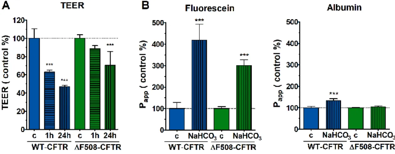
Figure 8. Effect of 100 mM NaHCO3 on barrier integrity of CFBE cells grown in co-culture.
Transepithelial electrical resistance values after 1 and 24 hours (A) and permeability values (B) after 24 hours of treatment. Values are expressed as a percentage of the control group (mean ± SD, n = 4/group).
Statistical analysis: two-way Anova and Bonferroni test. * p < 0.05, ** p < 0.01, *** p < 0.001 compared to the control group.
The WT-CFTR CFBE co-cultures responded more sensitively to sodium bicarbonate treatment at both treatment time points, as shown by lower TEER values (Fig. 8A). Consistent with this, there was an increase in the passage of both marker molecules through the cell layers in wild type cells compared to the control group (Fig. 8B). The 100 mM NaHCO3 treatment did not change the resistance of ΔF508-CFTR CFBE co-culture cell layers in the first hour of treatment. TEER values decreased only after 24 h of treatment (Fig. 8A), in parallel, higher fluorescein permeability was measured compared to the control. Albumin permeation remained unchanged (Fig. 8B), indicating still closed barrier function.
4.4.2. The effect of bicarbonate on the resting intracellular pH of CFBE cells
Elevated intracellular pH (7.74 ± 0.06) was measured in CFBE cells expressing the mutant CFTR channel, which significantly decreased (7.55 ± 0.05) after one-hour treatment with 100 mM NaHCO3, close to the level of pHi measured in wild type cells (Fig. 9).
Figure 9. Effect of sodium bicarbonate treatment (100 mM, 1 h) on the intracellular pH of CFBE cells.
Mean ± SD, n = 47-67/group. Statistical analysis: two-way Anova and Bonferroni test. ### p < 0.001 compared to wild type epithelial cells, *** p < 0.001 compared to control group.
Based on the results of intracellular pH measurements, we demonstrated a direct beneficial effect of 100 mM sodium bicarbonate treatment on bronchial epithelial cells expressing the mutant CFTR channel.
4.5. The effect of PN159 peptide on epithelial barrier models 4.5.1. The effect of PN159 peptide on CFBE barrier function
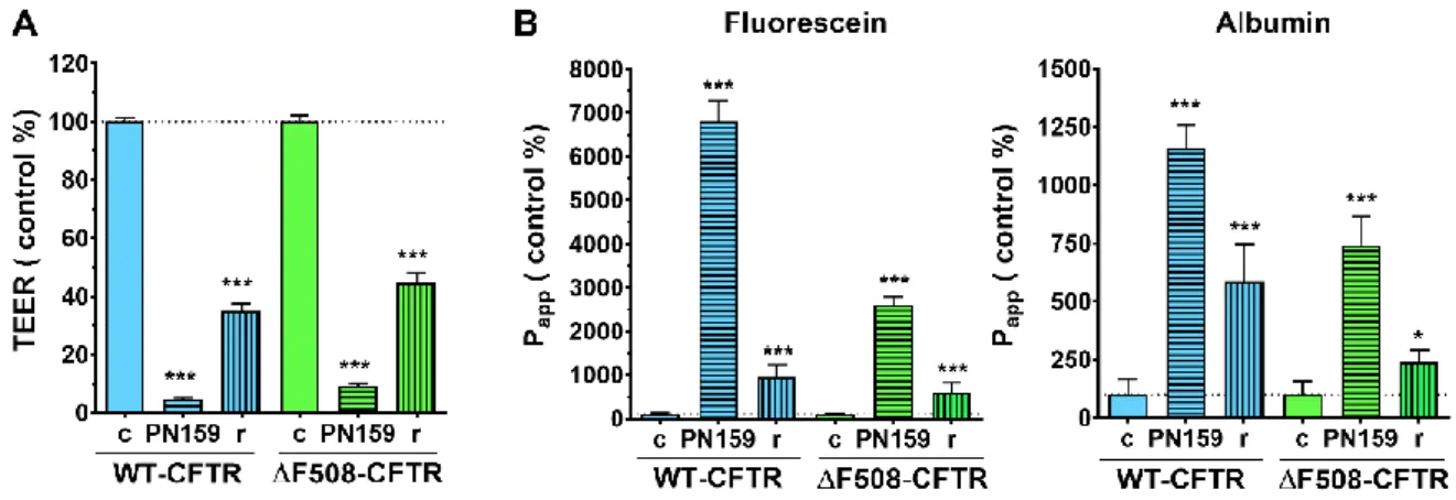
The permeability enhancing effect of PN159 peptide was also examined in the CFBE cell lines. As a result of the peptide treatment, the TEER of CFBE cells decreased to 10% of the values measured in the control groups (Fig. 10A).
Figure 10. TEER (A) and permeability (B) values of the CFBE cell layers after PN159 peptide treatment (10 μM, 30 min), followed by peptide removal and cell layer recovery (r) (24 h). Values are expressed as a percentage of the control groups (mean ± SD, n = 4/group). Statistical analysis: two-way Anova and Bonferroni test. * p < 0.05, *** p < 0.001 compared to control group (c).
In parallel, the passage of various markers through the cell layer increased several fold, indicating opening of the barrier (Fig. 10B). After peptide removal and cell layer recovery (24 h), TEER values increased but did not reach the level of control groups (Fig. 10A). Twenty- four hours after peptide removal, permeability data returned to near baseline, however, these values were higher in the untreated groups, consistent with the lower TEER values (Fig. 10B).
4.5.2. The colocalization of claudin-4 protein and PN159 peptide
Using molecular modeling of the PN159 peptide, our group showed an energetically favorable relationship between the claudin-4 epithelial junctional protein and the peptide. This result was also confirmed by visualization of the peptide in Caco-2 epithelial cells (Fig. 11).
Figure 11. Immunostaining of Caco-2 cells treated with Bodipy FL-labeled PN159 peptide (10 μM) for claudin-4 cell junctional protein. Red: claudin-4. Green: Bodipy FL labeled PN159 peptide. Blue:
nucleus. Scale bar: 10 µm. Arrow: colocalization of claudin-4 and labeled peptide.
In addition to the cytoplasm, the labeled PN159 peptide was also detectable in the cell membrane and showed colocalization with claudin-4 immunostaining at the site of junctional disruption (Fig. 11).
4.6. Antimicrobial activity of PN159 peptide
The antimicrobial effect of the membrane penetrating PN159 peptide was tested on six ESKAPE pathogenic bacteria in a wide concentration range (0.8–70 µM). Acinetobacter baumannii and Enterococcus faecium were the two most sensitive strains with MIC values below 5 µM. In the case of Staphylococcus aureus and Klebsiella pneumoniae, the growth inhibitory concentration of PN159 was around 10 µM, which also had an effect on human epithelial cells. Gentamicin and ciprofloxacin showed strong antimicrobial activity against all ESKAPE pathogens except for Enterococcus faecium, which was resistant to all antibiotics tested, however, the PN159 peptide was able to inhibit its growth.
5. SUMMARY
Adjuvant therapies play an important role in the treatment of many diseases, so there is a great need to search for novel potential adjuvant agents and procedures. These agents on the one hand can help alleviate the symptoms of diseases, on the other hand they can increase the absorption and effect of drugs by reversibly opening biological barriers. In our studies, we examined in culture models of human epithelial barriers sodium bicarbonate, an adjuvant therapeutic agent for cystic fibrosis, and the PN159/KLAL peptide with dual-action. The beneficial effects of sodium bicarbonate have been demonstrated in a clinical study and in experiments on microbial pathogens related to cystic fibrosis, however, its direct effects on bronchial epithelial cells have not been studied. As a first step, a suitable culture model is needed for these studies. To bring the model closer to the complexity of in vivo systems, co- culture of multiple cell types is essential. We are the first to establish and characterize a co- culture model for cystic fibrosis by culturing bronchial epithelial cell lines expressing wild type or ΔF508 mutant CFTR channel and human vascular endothelial cells together. In the presence of endothelial cells, bronchial epithelial cells formed a tighter cell layer. The strengthening of intercellular junctions was confirmed by analysis of the intensity of the immunostaining pattern.
Based on the results of CFTR channel function modifying treatments, we verified the reliability of the models and confirmed the presence of a functional CFTR channel in wild type cells and its absence in mutant cells. Cultures were treated with cytokines to model chronic inflammation characteristic of the respiratory system in patients with cystic fibrosis. A significant response to cytokine treatment was obtained only in co-cultures, suggesting that co-culture models may be more suitable for in vitro studies of inflammatory conditions. We are the first to investigate the direct effect of different concentrations of sodium bicarbonate on bronchial epithelial cells.
The CFBE cell line pair gave a different response to sodium bicarbonate treatment. Based on
viability and cell layer integrity studies, cells expressing the wild type CFTR channel were found to be more sensitive, while the beneficial effect of NaHCO3 was shown on mutant cells.
In our experiments the highest but still safe sodium bicarbonate concentration was 100 mM.
The NaHCO3 treatment decreased the higher intracellular pH of mutant CFBE cells, close to the level of wild type cells.
The PN159 peptide has a dual effect, it is known to have both cell penetrating and junction opening properties. In our studies we demonstrated the tight junction opening effect of the PN159 peptide on the human bronchial epithelial cell models. On Caco-2 cells the membrane-penetrating effect of the peptide was shown: the labeled peptide entered the living cells in a concentration-dependent manner. We observed the colocalization of the peptide with claudin-4 tight junction protein. Furthermore, the antimicrobial effect of the PN159 peptide on clinically relevant ESKAPE pathogens was detected. Compared with reference antibiotics, PN159 peptide was more effective in inhibiting the growth of the bacterial strain Pseudomonas aeruginosa, which is responsible for the high mortality in cystic fibrosis patients due to chronic respiratory infection and inflammation.
Our results confirm that sodium bicarbonate as an adjuvant can be safely used in the treatment of cystic fibrosis and may help alleviate the symptoms affecting the lungs. The PN159 peptide effectively opens the junctions between epithelial cells, making it a suitable candidate for the delivery of drugs through biological barrier systems, and a promising candidate for the treatment of multidrug-resistant bacterial strains due to its antimicrobial activity.
6. PUBLICATIONS RELATED TO THE SUBJECT OF THE THESIS
I. Bocsik A, Gróf I, Kiss L, Ötvös F, Zsíros O, Daruka L, Fülöp L, Vastag M, Kittel Á, Imre N, Martinek TA, Pál C, Szabó-Révész P, Deli MA.
Dual action of the PN159/KLAL/MAP peptide: increase of drug penetration across Caco-2 intestinal barrier model by modulation of tight junctions and plasma membrane permeability.
Pharmaceutics. 11(2). pii: E73. (2019) IF: 4.773
II. Gróf I, Bocsik A, Harazin A, Santa-Maria AR, Vizsnyiczai G, Barna L, Kiss L, Fűr G, Rakonczay Z Jr, Ambrus R, Szabó-Révész P, Gosselet F, Jaikumpun P, Szabó H, Zsembery Á, Deli MA.
The effect of sodium bicarbonate, a beneficial adjuvant molecule in cystic fibrosis, on bronchial epithelial cells expressing a wild type or mutant CFTR channel.
International Journal of Molecular Science. 21(11):4024. (2020) IF: 4.556
OTHER PUBLICATIONS
I. Bartos C, Jójárt-Laczkovich O, Katona G, Budai-Szűcs M, Ambrus R, Bocsik A, Gróf I, Deli MA, Szabó-Révész P.
Optimization of a combined wet milling process in order to produce poly(vinyl alcohol) stabilized nanosuspension.
Drug Design, Development and Therapy. 12:1567-1580. (2018) IF: 3.208
II. Veszelka S, Tóth A, Walter FR, Tóth AE, Gróf I, Mészáros M, Bocsik A, Hellinger É, Vastag M, Rákhely G, Deli MA.
Comparison of a rat primary cell-based blood-brain barrier model with epithelial and brain endothelial cell lines: gene expression and drug transport.
Frontiers in Molecular Neuroscience. 11:166. (2018) IF: 3.720
III. Kiss EL, Berkó S, Gácsi A, Kovács A, Katona G, Soós J, Csányi E, Gróf I, Harazin A, Deli MA, Budai-Szűcs M.
Design and optimization of nanostructured lipid carrier containing dexamethasone for ophthalmic use.
Pharmaceutics. 11(12). pii: E679. (2019) IF: 4.773
IV. Ismail R, Bocsik A, Katona G, Gróf I, Deli MA, Csóka I.
Encapsulation in polymeric nanoparticles enhances the enzymatic stability and the permeability of the GLP-1 analog, liraglutide, across a culture model of intestinal permeability.
Pharmaceutics. 11(11). pii: E599. (2019) IF: 4.773
V. Imre N, Hetényi A, Szabó E, Bodnár B, Szkalisity A, Gróf I, Bocsik A, Deli MA, Horvath P, Czibula Á, Monostori É, Martinek TA.
Routing nanomolar protein cargoes to lipid raft‐mediated/caveolar endocytosis through a ganglioside GM1‐specific recognition tag.
Advanced Science. 7(4):1902621. (2020) IF: 15.804
VI. Kiss EL, Berkó S, Gácsi A, Kovács A, Katona G, Soós J, Csányi E, Gróf I, Harazin A, Deli MA, Balogh GT, Budai-Szűcs M.
Development and characterization of potential ocular mucoadhesive nano lipid carriers using full factorial design.
Pharmaceutics. 12(7):682. (2020) IF: 4.421
VII. Bíró T, Bocsik A, Jurišić Dukovski B, Gróf I, Lovrić J, Csóka I, Deli MA, Aigner Z.
New approach in ocular drug delivery: in vitro and ex vivo investigation of cyclodextrin containing, mucoadhesive eye drop formulations
Drug Design, Development and Therapy. 15:351-360. (2021) IF: 3.216
ACKNOWLEDGEMENT
First of all, I am grateful to my supervisors, Dr. Mária Deli and Dr. Alexandra Bocsik, for their support, help and guidance during my work.
I would like to thank all the current and former colleagues of the Biological Barriers Research Group: Dr. Szilvia Veszelka, Dr. Zsófia Hoyk, Dr. Fruzsina Walter, Dr. András Harazin, Dr. Mária Mészáros, Dr. Ana Raquel Pato Santa Maria, Lilla Barna, Vigh Judit, Gergő Porkoláb, Dr. Petra Sántha, Dr. Lóránd Kiss, and our students.I am thankful for the friendly atmosphere and their support and help. Special thanks to Dr. Lóránd Kiss and Dr. Alexandra Bocsik for teaching me the basic experimental techniques needed for my research.
I am grateful to Dr. Pál Ormos and Dr. Ferenc Nagy, general directors of BRC, to Dr.
László Zimányi, director of Institute of Biophysics, and to Dr. László Siklós, head of Molecular Neurobiology Research Unit, for their support. I would like to thank all the colleagues of the research unit, the Institute of Biophysics and the whole research centre for their support, highlighting the help of Dr. Gaszton Vizsnyiczai in performing the image analysis.
I would like to thank our cooperating partners who contributed to the publications. We are grateful to Dr. Zsuzsanna Bebők for providing the CFBE cell line and to Dr. Ákos Zsembery for his professional support during the experiments with bicarbonate. I thank Dr. Zoltán Rakonczay, Dr. Lóránd Kiss and Gabriella Fűr for their help in the intracellular pH measurements. We thank Dr. Lívia Fülöp and Andrea Gyebrovszki for the synthesis of PN159 peptide, and Dr. Leila Daruka from Dr. Csaba Pál's group for testing it on bacteria.
Finally, I am deeply grateful to my family, my Mother, my Father, my Brother and Sister, and my friends, for their unconditional love and support.