For Peer Review
Characterization of auxin transporter PIN6 plasma membrane targeting reveals a function for PIN6 in plant
bolting
Journal: New Phytologist Manuscript ID NPH-MS-2017-24689 Manuscript Type: MS - Regular Manuscript Date Submitted by the Author: 13-Jun-2017
Complete List of Authors: Ditengou, Franck; Albert-Ludwigs-Universitat Freiburg Fakultat fur Biologie, Molecularpflanzenbiologie
Gomes, Dulceneia ; Albert-Ludwigs-Universitat Freiburg Fakultat fur Biologie, Molecularpflanzenbiologie
Nziengui, Hugues; Albert-Ludwigs-Universitat Freiburg Fakultat fur Biologie, Molecularpflanzenbiologie
Kochersperger, Philip; Albert-Ludwigs-Universitat Freiburg Fakultat fur Biologie, Molecularpflanzenbiologie
Lasok, Hanna; Albert-Ludwigs-Universitat Freiburg Fakultat fur Biologie, Molecularpflanzenbiologie
Medeiros, Violante; Albert-Ludwigs-Universitat Freiburg Fakultat fur Biologie, Molecularpflanzenbiologie
Paponov, Ivan; Albert-Ludwigs-Universitat Freiburg Fakultat fur Biologie, Molecularpflanzenbiologie; Norsk Institutt for Biookonomi, Division for Food Production and Society. Horticulture.
Nagy, Szilvia ; Semmelweis University, Department of Medical Chemistry, Molecular Biology and Pathobiochemistry
Nádai, Tímea ; Centre for Agricultural Research of the Hungarian Academy of Sciences, Department of Plant Cell Biology
Mészáros, Tamás; Semmelweis University, Department of Medical Chemistry, Molecular Biology and Pathobiochemistry
Barnabas, Beata; Centre for Agricultural Research of the Hungarian Academy of Sciences, Department of Plant Cell Biology
Rapp, Katja; Albert-Ludwigs-Universitat Freiburg Fakultat fur Biologie, Molecularpflanzenbiologie
Qi, Linlin; VIB-UGent, Center For Plant systems biology
Li, Xugang; Universität Freiburg, Institut für Biologie II und Zentrum für Angewandte Biowissenschaften
Becker, Claude; Max-Planck-Institut fur Entwicklungsbiologie, Molecular Biology; Gregor Mendel Institute of Molecular Plant Biology GmbH, Genomics and Epigenomics
Li, Chuanyou; Chinese Academy of Sciences, Institute of Genetics &
Developmental Biology;
Doczi, Robert; Royal Holloway, University of London, School of Biological Sciences;
Palme, Klaus; Albert-Ludwigs-Universität, Institut für Biologie II /
For Peer Review
Molecular Plant Physiology
Key Words: Arabidopsis thaliana, Auxin, inflorescence, Stem, Bolting
For Peer Review
Characterization of auxin transporter PIN6 plasma membrane targeting reveals 1
a function for PIN6 in plant bolting 2
3
Franck Anicet Ditengou1¥, Dulceneia Gomes1, Hugues Nziengui1, Philip 4
Kochersperger1, Hanna Lasok1, Violante Medeiros1, Ivan A. Paponov1,3, Szilvia K.
5
Nagy4, Tímea V. Nádai2, Tamás Mészáros4,5, Beáta Barnabás2, Katja Rapp1, Linlin 6
Qi6, Xugang Li1,7, Claude Becker1,8, Chuanyou Li6, Róbert Dóczi2 and Klaus 7
Palme1,8,9,10,11¥
8
9
1Institute of Biology II, Faculty of Biology, University of Freiburg, Schänzlestrasse 1, 10
D-79104 Freiburg, Germany.
11
2Department of Plant Cell Biology, Centre for Agricultural Research of the Hungarian 12
Academy of Sciences, H-2462 Martonvásár, Brunszvik u. 2, Hungary.
13
3NIBIO, Norwegian Institute for Bioeconomy Research, Postvegen 213, 4353 Klepp 14
Stasjon, Norway.
15
4Department of Medical Chemistry, Molecular Biology and Pathobiochemistry, 16
Semmelweis University, H-1094 Budapest, Tűzoltó u. 37-47, Hungary.5 Research 17
Group for Technical Analytical Chemistry, Hungarian Academy of Sciences - 18
Budapest University of Technology and Economics, H-1111 Budapest, Szt. Gellért 19
tér 4, Hungary.
20
6State Key Laboratory of Plant Genomics, National Centre for Plant Gene Research 21
(Beijing), Institute of Genetics and Developmental Biology, Chinese Academy of 22
Sciences, Beijing 100101, China.
23
7School of Biological Science and Technology, University of Jinan, 336, West Road 24
of Nan Xinzhuang, Jinan 250022, China 25
8Max Planck Institute for Developmental Biology, Department of Molecular Biology, 26
72076 Tuebingen, Germany.
27
9Centre for Biological Systems Analysis, Albert-Ludwigs-University of Freiburg, 28
Habsburgerstrasse 49, 79104 Freiburg, Germany.
29
10Freiburg Institute for Advanced Sciences (FRIAS), Albert-Ludwigs-University of 30
Freiburg, Albertstrasse 19, 79104 Freiburg, Germany.
31
11BIOSS Centre for Biological Signalling Studies, Albert-Ludwigs-University of 32
Freiburg, Schänzlestrasse 18, 79104 Freiburg, Germany.
33
¥Correspondence and request for material should be addressed to:
34
For Peer Review
Franck A. Ditengou (franck.ditengou@biologie.uni-freiburg.de; Tel. +49 761 203 97 35
636) and Klaus Palme (klaus.palme@biologie.uni-freiburg.de; Tel. +49 761 203 29 36
54) 37
38
Word counts 39
Introduction: 697 40
Material and methods: 869 41
Results: 3151 42
Discussion: 1424 43
Acknowledgments: 118 44
Total Word count: 6259 45
46
Figures 47
Fig. 1- Color 48
Fig. 2- Color 49
Fig. 3- Color 50
Fig. 4- Color 51
Fig. 5 52
Fig. 6- Color 53
Fig. 7- Color 54
55 56 57
For Peer Review
Summary 58
• Auxin gradients are sustained by series of influx and efflux carriers whose 59
subcellular localization is sensitive to both exogenous and endogenous factors.
60
Recently the localization of the Arabidopsis thaliana auxin efflux carrier PIN- 61
FORMED (PIN) 6 was reported to be tissue specific and regulated though 62
unknown mechanisms.
63
• Here, we used genetic, molecular and pharmacological approaches to 64
characterize the molecular mechanism(s) controlling the subcellular localization of 65
PIN6.
66
• PIN6 localizes to endomembrane domains in tissues with low PIN6 expression 67
levels such as roots, but localizes at the plasma membrane (PM) in tissues with 68
increased PIN6 expression such as the inflorescence stem and nectary glands.
69
We provide evidence that this dual localization is controlled by PIN6 70
phosphorylation and demonstrate that PIN6 is phosphorylated by mitogen- 71
activated protein kinases (MAPKs) MPK4 and MPK6. The analysis of transgenic 72
plants expressing PIN6 at PM or in endomembrane domains reveals that PIN6 73
subcellular localization is critical for Arabidopsis inflorescence stem elongation 74
post-flowering (bolting). In line with a role for PIN6 in plant bolting, inflorescence 75
stems elongate faster in pin6 mutant plants than in wild-type plants.
76
• We propose that PIN6 subcellular localization is under the control of 77
developmental signals acting on tissue specific determinants controlling PIN6- 78
expression levels and PIN6 phosphorylation.
79 80
Key words: Arabidopsis thaliana, auxin, bolting, inflorescence, stem 81
82 83
For Peer Review
Introduction 84
Plant developmental plasticity involves the activity of series of plant hormones which 85
modulate stem cell fate activity during plant development (Wolters & Jurgens, 2009;
86
Rodriguez et al., 2010). The plant hormone auxin plays a crucial role in this process 87
as it coordinates the patterning of the plant body plan, including the establishment of 88
apical–basal (Friml et al., 2003), radial (Bjorklund et al., 2007; Suer et al., 2011;
89
Ameres & Zamore, 2013), and proximal–distal axes (Sabatini et al., 1999; Cai et al., 90
2014) and the determination of cell fate by positional information (Ditengou et al., 91
2008; Finet & Jaillais, 2012). Auxin is polarly transported by auxin influx and efflux 92
carriers [(AUXIN RESISTANT 1/ Like AUX1 (AUX/LAX)) family (Ugartechea-Chirino 93
et al., 2010), ABCB/multi-drug resistance/P-glycoprotein (ABCB/MDR/PGP)(Paponov 94
et al., 2005; Geisler & Murphy, 2006), and PIN-FORMED (PIN) proteins (Paponov et 95
al., 2005)]. These proteins have been suggested to coordinate the patterning of the 96
plant body plan (Sabatini et al., 1999; Friml et al., 2003; Bjorklund et al., 2007;
97
Ditengou et al., 2008; Cai et al., 2014).
98
PAT relies on the proper subcellular localization of PIN proteins. PIN1, PIN2, PIN3, 99
PIN4 and PIN7 are targeted to the plasma membrane (PM) and they cycle between 100
the PM and endosomal compartments (Geldner et al., 2001). PIN8 localizes to the 101
endoplasmic reticulum (ER) membranes (Mravec et al., 2009; Dal Bosco et al., 2012;
102
Ding et al., 2012; Simon et al., 2016), while PIN5 and PIN6 localize to both the ER 103
and PM (Ganguly et al., 2014; Simon et al., 2016). PIN5 was proposed to mediate 104
auxin flow from the ER lumen to the cytosol (Mravec et al., 2009), while PIN8 and 105
PIN6 were proposed to export auxin in the opposite direction (Ganguly et al., 2010;
106
Dal Bosco et al., 2012; Ding et al., 2012). Together these studies suggest that PM- 107
targeting of PIN-proteins probably depends on some tissue and/or cell specific 108
determinants. Although it is unclear which mechanisms regulate PIN5 and PIN6 dual 109
localization, it can be envisaged that PIN5 and PIN6 may be post-translationally 110
modified prior their ultimate subcellular targeting, suggesting that these proteins are 111
no longer recognized by the sorting machinery responsible for their retention in 112
endomembrane domains. Phosphorylation is the most common post-translational 113
modification involved in signal transduction. Three protein kinase families have been 114
shown to phosphorylate PIN proteins: (i) D6 PROTEIN KINASE (D6PK) regulates 115
auxin transport by phosphorylation of PIN1, PIN2, PIN3, PIN4 and PIN7 (Shen et al., 116
2015); (ii) PINOID (PID) kinase and SERINE/THREONINE PROTEIN 117
For Peer Review
PHOSPHATASE 2A (PP2A) antagonistically affect phosphorylation of the PIN 118
hydrophilic loop, which is important for polar targeting of PM-located PIN proteins 119
(Michniewicz et al., 2007); and (iii) the recently characterized mitogen-activated 120
protein kinase (MAPK) pathway, which consists of the MKK7-MPK6 complex that 121
phosphorylates PIN1 serine 337 (S337) and impacts the polar localization of PIN1, 122
thereby modifying shoot branching (Jia et al., 2016).
123
In the present study, we aimed at characterizing the molecular mechanism(s) 124
controlling PIN6 subcellular localization. Our study reveals that both PIN6 gene 125
expression level and PIN6 phosphorylation modulate PIN6 subcellular localization in 126
Arabidopsis. Functional analysis of two phosphorylation sites, T392 and T393, which 127
were reported to be phosphorylated in vivo in Arabidopsis suspension cells treated 128
with bacterial elicitor flagellin (Benschop et al., 2007), reveals that these sites play a 129
key role in PIN6-ER exit and regulate root and root hairs growth, as well as 130
inflorescence stem development. We demonstrate that PIN6 is phosphorylated by 131
both MPK4 and MPK6 in vitro, although T393 is not phosphorylated by these 132
kinases. Finally, the analysis of transgenic plants expressing PIN6 predominantly at 133
PM or in ER reveals a critical role for PIN6 subcellular localization on inflorescence 134
stem elongation post flowering. Hence, over-expressing a PIN6 mutant protein 135
(T392V-T393V) that is retained in the ER-PIN6 does not affect inflorescence stem 136
growth, while over-expressing native PIN6 or its PM-localized phosphomimetic 137
mutant (T392E-T393E) drastically repressed plant growth and delayed bolting. In line 138
with a role for PIN6 in plant bolting, the inflorescence stems elongated faster in pin6 139
mutant plants than in wild-type plants. We conclude that PIN6 may act as a gate 140
keeper ensuring that Arabidopsis plants efficiently develop the inflorescence stem at 141
the appropriate, possibly environmentally determined time.
142 143
Materials and Methods 144
Materials and growth conditions 145
Arabidopsis thaliana (L.) Heynh. Columbia (Col-0) and Landsberg erecta (Ler) 146
ecotypes were used. All T-DNA insertion lines as well as transgenic lines are 147
described in Methods S1. Seeds were surface sterilized and sown on solid 148
Arabidopsis medium (AM) (2.3 g/L MS salts, 1% sucrose, 1.6% agar–agar, 5 mM 2- 149
(N-morpholino)ethanesulfonic acid (MES) sodium salt (Sigma, Steinheim, Germany), 150
For Peer Review
pH 6.0, adjusted with HCl). After vernalization for 2 days at 4°C, seeds were 151
germinated under a long-day period (16 h light, 8 h darkness) at 22°C. The same 152
growth conditions were applied in a phytochamber when plants were grown in soil.
153 154
Pharmacological treatments 155
For vesicular trafficking experiments, BFA (Invitrogen B7450) was used as previously 156
described (Geldner et al., 2001) with slight modifications: plants were incubated in 25 157
µM BFA (60 min). β-Estradiol was dissolved in 100% ethanol and added to AM 158
without exceeding an ethanol concentration of 0.1%.
159
Free IAA level determination 160
Approximately 15 mg (fresh weight) of 5 mm root sections was homogenized and 161
extracted for 16 h in methanol (Methods S2) 162
163
Generation of the PIN6 antibody 164
AtPIN6 cDNA (nucleotides 177-396) corresponding to the antigen peptide was 165
inserted into the pET-28a(+) expression vector (Novagen). After expression in the 166
Escherichia coli Rosetta strain (Novagen), the His6-tagged recombinant protein was 167
affinity purified according to the Qiagen manual (Qiagen) and confirmed by SDS- 168
polyacrylamide gel electrophoresis (PAGE). The antigen peptide included in the 169
PAGE slice was used to immunize a rabbit (Eurogentec). The polyclonal antiserum 170
was affinity purified against the recombinant AtPIN6 peptide as previously described 171
(Hasumura et al., 2005).
172 173
Detection of PIN6 by western blot 174
Proteins were extracted from 10, 3-week-old flower buds using extraction buffer 175
containing 50 mM Tris-HCl, 10 mM EDTA, 2 mM EGTA, 0.1% SDS, 1 mM DTT, 10 176
µM protease inhibitor cocktail, 0.01 mM MG132 and 0.1 mM PMSF. After 177
centrifugation at 10,000 rpm at 4°C for 15 min, the supernatant containing total 178
protein was collected, and the protein concentration was measured using the Thermo 179
Scientific Pierce Micro BCA Assay according to the manufacturer’s instructions. After 180
protein denaturation at 42°C in 5x Laemmli buffer (1:4), 7.4 mg/ml protein samples 181
were separated on a 10% SDS-PAGE gel and then transferred to a nitrocellulose 182
membrane. Blots were probed with a rabbit anti-PIN6 polyclonal antibody (1:1200), 183
and PIN6 signal was detected with an HRP-conjugated anti-rabbit antibody (1:5000) 184
For Peer Review
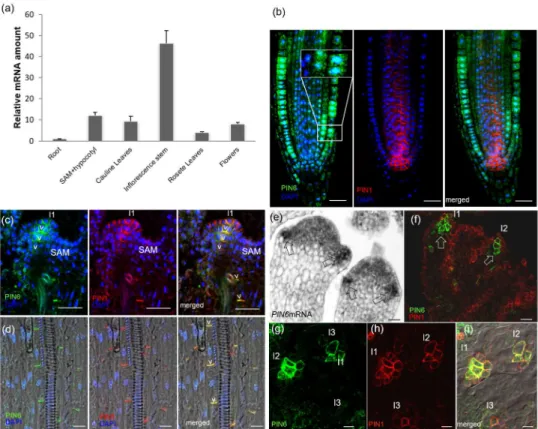
(Agrisera). Plasma membrane H+-ATPase was used as a control for equal loading.
185
Signal detection was performed with a Fujifilm ImageQuantTM LAS 4000 CCD 186
camera using Super Signal West Pico Chemiluminescent substrate.
187 188
Immunolocalization 189
Plants were fixed with 4% paraformaldehyde in PBS (pH 7.3) and used for whole- 190
mount in situ immunolocalization as previously described (Friml et al., 2004).
191 192
Whole mount in situ hybridization 193
In situ detection of PIN6 mRNA in Arabidopsis seedling root tips was performed as 194
previously described (Riegler et al., 2008; Begheldo et al., 2013).
195 196
Quantitative RNA analysis 197
PIN6 expression was assessed using semi-quantitative RT-PCR (PIN6 mRNA in 6- 198
day-old PIN6ox plants) or qRT-PCR for PIN6 expression throughout the plant 199
lifespan (Methods S4).
200 201
Microscopy and image post processing 202
Histological detection of β-glucuronidase (GUS) activity was performed as previously 203
described (Scarpella et al., 2004). Fluorescent proteins were analyzed as described 204
in Methods S4. All images were assembled using Microsoft PowerPoint 2013.
205 206
Kinase assay 207
To test whether PIN6 in phosphorylated by MPK4 or MPK6, the hydrophilic loop (HL:
208
residues 156-430) of PIN6 cDNA was amplified and cloned into pGEM-T Easy vector 209
(Promega), and the sequence was verified. A non-phosphorylatable mutant HL 210
version (T226A, T242A, S286A, T304A, T320A, S326A, S337A, and T393A;
211
positions according to the full-length PIN6 protein) was synthetized by GenScript.
212
The variant where T393 of the putative MAPK phosphosites is wild type was 213
generated by inserting the sequence corresponding to A156-D352 from the mutant 214
clone (including T226A, T242A, S286A, T304A, T320A, S326A and S337A) into the 215
WT construct. For in vitro transcription/translation, the HL sequence variants were 216
subcloned into the pEU3-NII-GLICNot vector with ligation-independent cloning 217
(Bardoczy et al., 2008). In vitro mRNA synthesis was carried out using a 218
For Peer Review
TranscriptAid T7 High Yield Transcription Kit (Thermo Scientific) according to the 219
manufacturer’s instructions. Cell-free translation was completed using a 220
WEPRO7240H Expression Kit (Cell Free Sciences, Japan). To activate His-tagged 221
MPK4 and MPK6 when included in the phosphorylation assay solution, mRNA 222
encoding constitutively active myc:MKK1 and myc:MKK4, respectively, were also 223
added to the translation mixture as described (Nagy & Meszaros, 2014).
224
In vitro-translated His6-AtMPK4 and His6-AtMPK6 proteins were purified by affinity 225
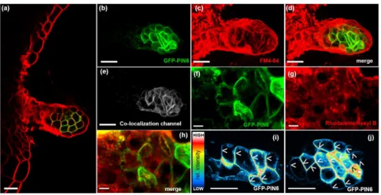
chromatography using TALON Magnetic Beads (Clontech), while in vitro-translated 226
wild-type and mutant GST-PIN6loop variants were purified by affinity chromatography 227
using Glutathione Magnetic Beads (Thermo Scientific)(Nagy & Meszaros, 2014). For 228
kinase assays, 300 and 100 ng of in vitro-translated, affinity-purified substrate and 229
kinase were used, respectively. As an activity control, 10 µg myelin basic protein 230
(MBP) was used as a generic MAPK substrate (not shown). The assay was carried 231
out in 20 mM HEPES, pH 7.5, 100 µM ATP, 1 mM DTT, 15 mM MgCl2, 5 mM EGTA 232
and 5 µCi [γ-32P]ATP with bead-bound GST-PIN6loop variants as substrates for 30 233
min at room temperature and then stopped by the addition of Laemmli SDS buffer.
234
Samples were fractionated by SDS-PAGE. The gel was fixed, stained with 235
Coomassie blue, dried and analyzed by autoradiography.
236 237
Results 238
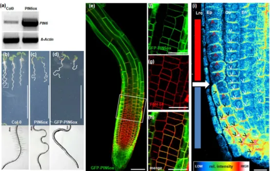
PIN6 expression level is variable and increases highly during plant bolting 239
PIN6 was reported to be located at the plasma membrane and in the ER in different 240
cells and organs (Simon et al., 2016), suggesting this dual localization may depend 241
on some tissue and/or cell specific determinants. To gain insight on regulation of 242
PIN6-localization, we first performed a quantitative RT-PCR analysis to ascertain 243
PIN6 expression throughout the Arabidopsis lifespan. As previously shown by 244
qualitative pPIN6:GUS analysis (Nisar et al., 2014), PIN6 is expressed during both 245
vegetative and reproductive plant growth phases (Fig. 1a) with highest PIN6 mRNA 246
levels in developing inflorescence stems (Fig. 1a). This observation was confirmed by 247
analysis of pPIN6::GUS plants where PIN6 expression was present in elongating 248
inflorescence stems (Fig.Fig. S1b). These results are in agreement with publicly 249
available data sets (Ismagul et al., 2014; Ivanova et al., 2014) and indicate that PIN6 250
expression is under the control of developmental signals during bolting. Cross- 251
sections from the inflorescence stem of pPIN6:GUS plants showed PIN6 expression 252
For Peer Review
in xylem parenchyma cells (Xpc), the fascicular cambium (Fc) and the interfascicular 253
fiber tissues (IF) (Fig. S1f). Altogether, these observations show that PIN6 is 254
ubiquitously expressed during Arabidopsis life span. The fact that PIN6 expression is 255
increasing during plant bolting and stays relatively high in the stem, suggests a role 256
of PIN6 in processes controlling both longitudinal and radial differentiation, 257
particularly during bolting.
258 259
PIN6 is localized at the PM in the shoot apical meristem, hypocotyl and 260
inflorescence stem 261
To visualize PIN6 subcellular localization, we generated a polyclonal anti-PIN6 262
antibody. In agreement with a recent report (Simon et al., 2016), PIN6 displayed dual 263
localization in endomembrane domains and at the PM. We used the anti-PIN6 264
antibody to detect PIN6 at the root tip, the plant organ with the lowest PIN6 265
expression levels. PIN6 was visible in the endomembrane compartments of the 266
epidermis and cortex cell files (Fig. 1B), the tissues in which PIN6 mRNA were low 267
(Fig. 1a, Fig. S1E). However, in other tissues such as inflorescence stem vascular 268
cells (Nisar et al., 2014), vegetative leaves and flower primordia, which displayed 269
higher PIN6 mRNA levels, PIN6 was detected at the PM co-localized with PIN1 (Fig.
270
1a,c,d,f,g). To test the quality of the anti-PIN6 antibody, we performed both western 271
blot analysis using the pin6-5 mutant and immunolocalization using both the pin6-5 272
and pin6-6 mutants (Fig. S2b). PIN6 was recognized by the anti-PIN6 antibody in WT 273
plants but as expected not in the mutants. This suggests that pin6-5 and pin6-6, 274
which were previously described as knock-down mutants at the mRNA level, are 275
indeed null mutants at the protein level. This confirms that this antibody can be 276
considered specific for PIN6. However, additional higher and lower molecular weight 277
proteins were detected in both WT and pin6-5 knock-out plants, which probably 278
resulted in the background signals which were observed in the immunolocalization 279
images (Fig. S2d-f, non-specific nuclear signals are indicated with an asterisk in WT 280
and pin6 knock-outs).
281
These data show that tissues with low PIN6 expression display PIN6 in 282
endomembrane domains, while PIN6 is at the PM in tissues with higher PIN6 283
expression. This suggests that the dual localization of PIN6 may be dependent on 284
PIN6 expression level. To substantiate this correlation, we extended our analysis to 285
other plant tissues reported to have strong PIN6 expression, such as nectary glands 286
For Peer Review
(Ludwig-Muller, 2014; Turi et al., 2014). Observation of plants expressing the 287
pPIN6:GFP-PIN6 construct revealed that the GFP signal co-localized almost perfectly 288
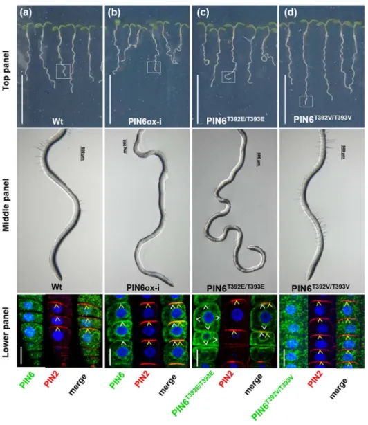
at the PM with the endosome tracker/PM-marker FM4-64 (Pearson’s correlation 289
coefficient in co-localized volume (PCCCV)=0.97, with a value of 1 representing a 290
perfect correlation) (Fig. 2a-e). In contrast, very limited co-localization with the ER 291
marker Rhodamine B (Zhang et al., 2014) (PCCCV=0.26) was observed in both the 292
median and lateral nectary glands (Fig. 2f-h). Taken together, these data support our 293
hypothesis that the PM localization of PIN6 most likely depends on the PIN6 294
expression level. Analysis of the PIN6-subcellular localization in nectary glands also 295
revealed basally (toward the root) localized GFP-PIN6, presumably exporting auxin 296
out of nectary glands, thus supporting the idea that nectary glands could be potential 297
sources of auxin (Aloni et al., 2006) (Fig. 2i,j).
298 299
PIN6 is targeted to the PM upon PIN6 overexpression 300
To confirm the relationship between PIN6 expression level and PIN6 subcellular 301
localization, we generated transgenic lines overexpressing the PIN6 genomic 302
sequence with (GFP-PIN6ox) or without (PIN6ox) a GFP tag and driven by the 303
constitutively active CaMV35S promoter. The non-tagged construct was used to 304
confirm that GFP does not affect PIN6 sub-cellular localization. We visualized PIN6 305
subcellular localization in GFP-PIN6ox and PIN6ox plants and analyzed its impact on 306
root and shoot growth. PIN6 overexpression increased the PIN6 mRNA level and 307
thus the PIN6 protein level (Fig.3a and Fig. S2b); both GFP-tagged and non-tagged 308
PIN6 were detected at the PM, where they co-localized with FM4-64 (PCCCV=0.7) 309
(Fig. 3e-h). In line with recent studies (Ganguly et al., 2010; Cazzonelli et al., 2013;
310
Simon et al., 2016), the roots of both GFP-PIN6ox and PIN6ox plants were hairless 311
and displayed a strong waving phenotype, suggesting that GFP insertion did not 312
affect PIN6 functionality (Fig. 3b-d). More thorough inspection of the PIN6 sub- 313
cellular localization showed that PM-located PIN6 (PM-PIN6) exhibited polar 314
localization in root cells similar to our observations in nectary glands (see above). In 315
the cortex and stele cells, PIN6 co-localized basally with PIN1 (Fig. 3i and Fig. S3a).
316
The PIN6 PM-localization was most striking in the epidermis For clarity, the epidermis 317
cell file of the root meristematic zone was divided into two tiers (see Fig. 3i).
318
Epidermal cell tier 1 represents cells located in the upper part of the meristematic 319
zone, while tier 2 represents the lower part. The oldest cell of the most recent lateral 320
For Peer Review
root cap cells (LRC) (solid white forward arrow in Fig. 3i) marks the limit between the 321
two tiers. In tier 1, PIN6 localized apically (toward the shoot) similarly to PIN2, with a 322
well-defined polarity in cells destined to elongate (Fig. 3i and Fig. S3b). In contrast, in 323
tier 2, PIN6 localized laterally and basally, pointing progressively towards the 324
quiescent centre (QC) and columella cells and presumably channeling auxin towards 325
this region (Fig. 3i and Fig. S3a). Indeed, PM-localized PIN6 perturbed auxin 326
distribution in PIN6ox roots as confirmed by both quantification of auxin levels and 327
the expression of the auxin-sensitive DR5::GUS reporter fusion protein at the PIN6ox 328
root tip (Fig. S3e-g) (Cazzonelli et al., 2013).
329 330
PID regulates PIN6 polarity despite altered phosphosite occurrence 331
The unique localization dynamics of PIN6 suggests that it is under the control of a 332
unique regulatory mechanism. Phosphorylation has been already shown to be a key 333
determinant of PIN localization, the best characterized mechanism is the regulation of 334
PIN polarity by members of the AGCVIII protein kinase subfamily, PID and D6PK and 335
their close paralogues. The main D6PK site of PIN1 (S271) is conserved in all long- 336
HL PINs, including PIN6 (S291) (Zourelidou et al., 2014). The three PID 337
phosphorylation sites are similarly well conserved in long-HL PINs with the exception 338
of PIN6, where the site corresponding to S252 of PIN1 is missing. Although PID is 339
known to regulate polarity, not localization to the PM, we first tested whether this 340
differential PID site composition can be associated with a differential regulation of 341
PIN6 by PID. In PIDox plants, PIN1 polarity was shifted from the basal to the apical 342
side in the stele (Friml et al., 2004), whereas the cytoplasmic localization of PIN6 in 343
epidermal cells remained unchanged, implying that PID does not bring about PM 344
translocation of PIN6 (Fig. S5a-c) (Friml et al., 2004; Rasmussen et al., 2015).
345
Furthermore, we crossed PIN6 overexpression (PIN6ox) and PINOID-overexpression 346
(PIDox) plant materials. Similarly to PIN6ox plants, PIN6 was localized to PM in 347
PIDoxPIN6ox plants, but the PM-PIN6 basal localization in the stele shifted, similarly 348
to PIN1 (Fig. S5g-i). Moreover, the basal localization of PM-PIN6 in tier 2 epidermal 349
cells also shifted, whereas the apical localization of PIN6 in tier 1 epidermal cells did 350
not (in 100% of plants tested, n>15; compare Fig. 3i and Fig. S5d-f with Fig. S5g-i).
351
These results imply that similarly to other PINs, PID plays a role in polarity regulation 352
of PIN6, however its ER to PM translocation is regulated by other means.
353
For Peer Review
Threonine-phosphorylation sites 392 (T392) and 393 (T393) are involved in PIN6 354
localization at the PM 355
A phosphoproteomic assay in Arabidopsis yielded PIN6 phosphopeptides from 356
suspension-cell-derived PM vesicles (Benschop et al., 2007). In that study, PM- 357
localised PIN6 protein was shown to be phosphorylated at both or either of the two 358
adjacent threonine residues at positions T392 and T393 in the hydrophilic loop 359
(Benschop et al., 2007). These phosphorylation sites are partially conserved among 360
PIN6-like proteins in other species (Fig. S6b). Remarkably, these residues are not 361
conserved in other members of the PIN family (Fig. S6b), raising the possibility of a 362
PIN6-specific regulation through their phophorylation. In order to test this hypothesis 363
both T392 and T393 were converted by site-directed mutagenesis to valine, an amino 364
acid that cannot be phosphorylated (PIN6T392EVT393V) or to glutamic acid, which 365
mimics constitutive phosphorylation (PIN6T392E/T393E
). Driven by a β-estradiol- 366
inducible promoter, the intact PIN6 gene (PIN6ox-i) and the mutated versions were 367
separately introduced into WT plants. When grown in the presence of β-estradiol, the 368
roots of PIN6ox-i and PIN6T392E/T393E
plants were dramatically affected in comparison 369
to the roots of WT and PIN6T392V/T393V
plants (Fig. 4a-d, top panel). PIN6ox-i and 370
PIN6T392E/T393E plants developed hairless agravitropic roots (Fig. 4a-d, middle panel).
371
Consistent with this, confocal microscope images revealed that, as for PIN6ox plants 372
(Fig. 3i), PIN6 localized basally at the PM in PIN6ox-i root tip epidermal cells (Fig. 4b, 373
lower panel). In PIN6T392E/T393E
plants, the GFP signal also localized to the PM but 374
was non-polar (Fig. 4c, lower panel; Video S1). Altogether, these data indicate that 375
the presence of PIN6 at the PM of epidermal cells, and not necessarily its polarity, is 376
sufficient to perturb both the root gravity response and root hair development. In 377
contrast, PIN6T392V/T393V
plants were indistinguishable from WT plants, and the GFP 378
signal was mainly visible in the ER, where it co-localized with the ER marker 379
rhodamine B hexyl (PCCCV=0.65) (Fig. 4d and Fig. S7a; Video S2). It is known that 380
PM-localized PINs are sensitive to the fungal toxin brefeldin A (BFA), which blocks 381
PIN protein recycling, while ER-PINs are BFA resistant (Mravec et al., 2009).
382
Accordingly, upon BFA treatment, PIN6T392E/T393E
co-localized with FM4-64 in BFA 383
compartments, while PIN6T392V/T393V
localization was not affected, thus confirming 384
their respective subcellular localizations (Fig. S7b-c). These data demonstrate that 385
T392 and T393 phosphorylation sites are crucial the translocation of PIN6 to the PM.
386 387
For Peer Review
PIN6 hydrophilic loop is phosphorylated by MPK4 and MPK6 but T393 is not a 388
MAP kinase phosphorylation site 389
In order to predict putative kinase(s), which may mediate T392/T393 phosphorylation, 390
we screened the PIN6 protein sequence by using the Eukaryotic Linear Motif (ELM) 391
database (Dinkel et al., 2016). This search resulted in a total of 59 predicted 392
phosphorylation sites targeted by eight types of kinases. In the region of interest 393
T393 was identified as a MAPK phosphorylation site, while no putative kinase was 394
associated with T292. T393 is one of eight potential proline-directed MAPK 395
phosphorylation sites in PIN6 HL (T226, T242, S286, T304, T320, S326, S337, and 396
T393) suggesting that PIN6 may be phosphorylated by MAP-kinase(s). In plants, 397
MAPK pathways are central regulators of various stress responses and 398
developmental processes (Rodriguez et al., 2010; Xu & Zhang, 2015). In particular, 399
MPK6 of Arabidopsis, the best studied plant MAP kinase, has been reported to 400
specifically regulate developmental processes such as lateral root development and 401
plant height (Jia et al., 2016), processes also shown to be altered in pin6 mutants 402
(Cazzonelli et al., 2013; Simon et al., 2016). MPK4, another well-characterized plant 403
MAPK is involved in defense regulation, with mpk4 mutants displaying severe 404
dwarfism and altered cell division and microtubule dynamics. Based on this 405
information we first tested whether PIN6 is phosphorylated by MPK4 and MPK6 using 406
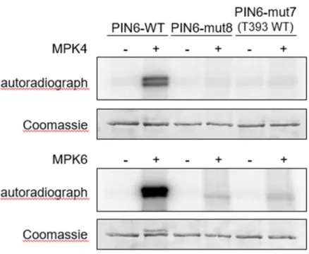
in vitro kinase assays (Fig. 5).
407
The incorporation of radiolabeled phosphate in the hydrophilic loop (HL) of PIN6 in 408
the presence of activated MPK4 or MPK6 indicates that this protein is phosphorylated 409
by both kinases (Fig. 5). As a negative control, all eight MAPK phosphorylation 410
residues (S or T) were replaced with the non-phosphorylatable amino acid alanine 411
(T226A, T242A, S286A, T304A, T320A, S326A, S337A, and T393A in PIN6-mut8), 412
which results in the loss of MAPK-mediated phosphorylation (Fig. 5). Faint, residual 413
phosphorylation of the mutant proteins by MPK6 indicates weak, unspecific 414
phosphorylation on non-cognate residues, probably related to the strong activity of 415
MPK6. These results confirm MAPK-mediated phosphorylation of PIN6. In order to 416
test whether T393 is one of the residues actually phosphorylated by these MAPKs we 417
tested specific phosphorylation of T393 by using a septuple mutant, where T393 is 418
wild type (i.e. T226A, T242A, S286A, T304A, T320A, S326A, S337A; PIN6-mut7).
419
Neither MPK4 nor MPK6 phosphorylated PIN6 on T393 (Fig. 5), suggesting that this 420
residue is not a genuine MAP kinase site. These data reveal complex regulation of 421
For Peer Review
PIN6 by at least two MAPK pathways, but also indicate that the modification of 422
T392/T393 regulating PIN6 PM translocation is probably brought about by yet 423
another type of protein kinase.
424 425
PM-localized PIN6 represses plant growth 426
To investigate the importance of PIN6 subcellular localization on plant development, 427
we analyzed the phenotype of PIN6T392E/T393E and PIN6T392V/T393V plants grown in soil.
428
Spraying plants with 100 µM ß-estradiol affected weakly the growth of Col-0 and 429
PIN6T392E/T393E
plants, but strongly retarded the growth of PIN6T392E/T393E
and PIN6ox-i 430
plants (Fig. S8a, b). Plants sprayed with water showed a similar elongation profile for 431
the inflorescence stems of Col-0 and PIN6T392V/T393V
, while PIN6T392E/T393E
plants 432
developed significantly smaller inflorescences. This result confirms the reported 433
leakiness of the estradiol-inducible promoter (Kubo et al., 2013), as indicated by the 434
GFP signal observed in the leaves of non-induced PIN6T392V/T393V
and PIN6T392E/T393E
435
plants (Fig. S8c). ß-estradiol also affected weakly the growth of Col-0 and 436
PIN6T392V/T393V
plants, but it strongly delayed the growth of PIN6T392E/T393E
437
inflorescence stems and completely suppressed the bolting of PIN6ox-i plants (Fig.
438
S8d). Taken together, these data demonstrate the importance of T392 and T393 for 439
the regulation of plant inflorescence stem elongation and indicate that the 440
phosphorylation-dependent PM targeting of PIN6 negatively controls inflorescence 441
stem development, while preventing this phosphorylation results in the ER retention 442
of PIN6. These data also suggested a role for PIN6-dependent auxin distribution 443
during the elongation of the inflorescence stem.
444 445
Inflorescence stem elongation is accelerated in pin6 loss-of-function mutants 446
In Arabidopsis thaliana, the floral transition (formation of an inflorescence meristem) 447
marks the transition from the vegetative to the reproductive phase followed by 448
elongation of the first internode, known as the bolting transition (Pritchard et al., 449
2012). This process is influenced by hormones but underlying mechanisms still 450
remain poorly understood. To explore how PIN6-dependent PAT modulates 451
inflorescence stem development, we analyzed the pin6-5 (GK-430B01) and pin6-6 452
(GK-711C09) T-DNA insertion lines (Supporting Information Fig. S2A; (Cazzonelli et 453
al., 2013). We also analyzed two additional novel knock-out lines isolated from the 454
Arabidopsis Genetrap collection in the Landsberg erecta (Ler) background 455
For Peer Review
(Sundaresan et al., 1995) (referred to here as pin6-7 (GT7129) and pin6-8 456
(GT6906)), in which T-DNA is inserted in the 4th and 5th introns, respectively 457
(Supporting Information Fig. S2C). All pin6 mutants developed inflorescence stems 3- 458
5 times longer than wild-type (WT) inflorescence stems while having the same 459
number of leaves. This phenotype suggests faster inflorescence stem elongation 460
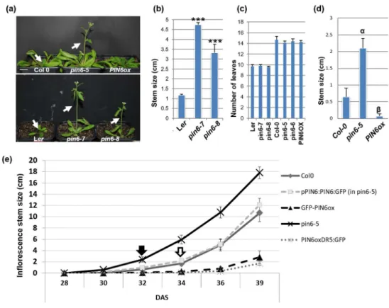
rather than early flowering (Fig. 6a-d).
461 462
PIN6 overexpression delays inflorescence stem elongation 463
In contrast to pin6 mutants and in line with previously published phenotypes 464
(Cazzonelli et al., 2013), plants overexpressing PIN6 developed significantly shorter 465
inflorescence stems (Fig. 6d). Compared to WT plants, these plants developed 466
siliques relatively close to the base of the inflorescence stem (indicated with an arrow 467
in Fig. S9a), suggesting that these plants were mature and capable of seed 468
production but that inflorescence stem elongation was arrested (Fig. 6a-d). To 469
confirm these results, we quantified the flowering time and the growth rates of WT, 470
pin6-5 and pin6-5 complemented with GFP-PIN6 driven by the PIN6 native promoter 471
(pPIN6:PIN6:GFP). In our conditions, all plants (WT, pPIN6:PIN6-GFP, pin6-5, and 472
PIN6ox) flowered around the 28th day after sowing (DAS). However, although the 473
apical flower was visible in PIN6ox and GFP-PIN6ox plants, inflorescence stem 474
elongation required four additional days (Fig. 6e). WT and pin6-5 pPIN6:PIN6-GFP 475
plants displayed accelerated growth starting on the 34th day (open arrow in Fig. 6e), 476
but this acceleration occurred earlier in pin6-5 plants (closed arrow in Fig. 6e). Thus, 477
six days after bolting, the growth rate of pin6-5 mutant inflorescences was 478
approximately 2.42 cm/day versus 1.65 and 1.43 cm/day for WT and pin6-5 479
pPIN6:PIN6-GFP plants, respectively (Fig. S9b), demonstrating the rapid growth of 480
pin6-5 plants and mutant complementation by the pPIN6:PIN6-GFP construct.
481
Conversely, GFP-PIN6ox and PIN6ox inflorescence stem elongation was slower 482
(only 0.25 and 0.13 cm/day, respectively (Fig. S9b)). The data also indicated that the 483
accelerated growth of pin6 mutants is transient. Later, the WT and pin6-6 mutant (a 484
trend visible in all mutants; not shown) growth rates became equal, while the PIN6ox 485
growth rate remained approximately 50% lower (Fig. S9c,d). Taken together, these 486
data demonstrate that PIN6 activity regulates inflorescence stem elongation and 487
strongly suggest a role for auxin transport during plant bolting.
488 489
For Peer Review
490 491
Auxin response is reduced in pin6 mutants 492
Auxin transport capacities and auxin levels are crucial for shoot branching and 493
vascular tissue development (Muller & Leyser, 2011; Peer et al., 2011; Bennett et al., 494
2014). For example, a cambial auxin peak resulting from basipetally derived shoot- 495
apex auxin is essential for both primary and secondary growth (Bjorklund et al., 2007;
496
Suer et al., 2011; Ameres & Zamore, 2013). To determine how PIN6 activity 497
modulates auxin distribution and levels in bolting inflorescences, we used a 498
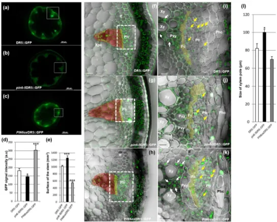
pDR5:GFP reporter (Ottenschlager et al., 2003) to visualize auxin distribution and 499
activity in inflorescence stems six days after bolting. In transverse sections 50 mm 500
above the uppermost rosette leaf of the inflorescence stem of WT plants, the 501
DR5:GFP signal was mainly visible in phloem and xylem parenchyma cells (Fig.
502
7a,f,i). The DR5 signal was significantly lower in the pin6-5 mutant (T-test, P<0.05, 503
n=10); in contrast, DR5 expression was significantly greater in PIN6ox plants (T-test, 504
P<0.001, n=10) (Fig. 7b-d). In addition, radial development was reduced in PIN6ox 505
plants but increased in pin6-5 plants (Fig. 7b-e). Consequently, pin6-5 DR5:GFP 506
plants displayed a greater transversal stem surface area than DR5:GFP and 507
PIN6oxDR5:GFP plants (T-test, P<0.001, n=10). In PIN6ox DR5:GFP plants, the 508
number of xylem elements was significantly reduced and accompanied by ectopic 509
development of xylem parenchyma cells (Fig. 7f-l; xylem parenchyma cells are 510
indicated with a white arrow in Fig. 7i and k). Altogether, these data establish PIN6 as 511
a key regulator of both the primary and secondary growth of Arabidopsis 512
inflorescence stems and show that impaired PIN6 activity strongly affects the auxin 513
response necessary for cambium proliferation and xylem differentiation (Hildebrandt 514
& Nellen, 1992; Nina Theis & Manuel Lerdau, 2003; Suer et al., 2011).
515 516
Discussion 517
Previous studies revealed the importance of the auxin transporter PIN6 in several 518
developmental processes, such as nectary development (Bender et al., 2013), leaf 519
vein patterning (Sawchuk et al., 2013), and root and lateral root development 520
(Cazzonelli et al., 2013; Simon et al., 2016). Recently, Simon and colleagues 521
described the PM and ER subcellular localization of PIN6 and suggested its role in 522
controlling auxin transport and homeostasis in auxin-mediated development (Simon 523
For Peer Review
et al., 2016). In the present study, using genetic, molecular and pharmacological 524
approaches, we characterized the mechanisms controlling PIN6 dual localization and 525
uncovered a role for PIN6 during Arabidopsis inflorescence stem development.
526 527
PIN6 subcellular localization is regulated by PIN6 gene expression levels 528
It was reported that auxin can induce the PIN6 promoter in a tissue-specific manner 529
(Cazzonelli et al., 2013). Hence when combined to the finding that high PIN6 530
expression results in PM-targeting of PIN6, it is not surprising to find PIN6 expressed 531
at the PM in tissues reported to contain high levels of auxin such as the tip of young 532
leaves, young flowers, vascular tissue of the stem and nectary glands (Muller et al., 533
2002; Benkova et al., 2003; Aloni et al., 2006; Cheng et al., 2006). We did not 534
observe PIN6 at the PM of root cells, despite the fact that auxin is synthesized and 535
accumulates at the root tip. This suggests that at the root tip, besides auxin levels 536
other cell determinants probably modulate PIN6 abundance and therefore its 537
localization. In contradiction with our data, GFP-tagged PIN6 driven by the PIN6 538
native promoter was recently reported to localize at the PM in the Arabidopsis root tip 539
(Simon et al., 2016), although whether this construct could rescue the described 540
mutant phenotypes is not presented in that paper.
541 542
T392/T393 phosphorylation sites modulate PIN6 subcellular localization 543
PINOID (PID) kinase and SERINE/THREONINE PROTEIN PHOSPHATASE 2A 544
(PP2A), were reported to be involved in the reversible phosphorylation of the PIN 545
hydrophilic loop (Michniewicz et al., 2007), although it is still unclear how PID 546
regulates PIN trafficking (New ref), Nevertheless. our data indicate that despite loss 547
of a PID site PIN6 retains a similar sensitivity to PID-dependent phosphorylation as 548
PIN1, i.e. PIN6 basal localization is flipped to apical localization. However, we have 549
no evidence that PID activity is responsible for PIN6 exit from the ER, since PIN6 is 550
not targeted to PM in PIDox plants, since PIN6 is not targeted to the PM in 35S::PID 551
plants; instead this observation indicates a role for other kinases.
552
Data mining retrieved in vivo phosphorylation at a tandem threonine pattern 553
(T392/T393) unique to PIN6. Accordingly, functional analyses confirmed that the 554
T392 and T393 residues are crucial for PIN6 ER exit and subsequent PM 555
localization. In line with this, genetic modifications preventing phosphorylation of 556
these residues resulted in PIN6 retention in the ER. A similar mechanism was 557
For Peer Review
described for the PM-targeting of Arabidopsis PHOSPHATE TRANSPORTER1 558
(PHT1) (Bayle et al., 2011), the nitrate transporter and auxin facilitator NRT1.1 559
(Krouk et al., 2010; Habets & Offringa, 2014) and the aquaporin PIP2;1 (Weiste &
560
Droge-Laser, 2014). Phosphorylation of these proteins was shown to modulate their 561
targeting to the PM.
562 563
PIN6 is phosphorylated by MPK4 and MPK6 564
Phosphorylation of PIN6 at T392/T393 represents a novel regulatory mechanism, 565
thus we set out to identify the corresponding kinase(s). As there are 942 kinases 566
encoded in the Arabidopsis genome (Zulawski et al., 2014), in silico pattern 567
screening appeared to be the feasible approach to predict the kinase(s) 568
phosphorylating T392 and/or T393. Accordingly, T393 is one of eight putative MAP 569
kinase phosphorylation sites in PIN6 HL. Here we provide evidence that PIN6 is 570
phosphorylated by both MPK4 and MPK6, thus revealing a novel regulatory 571
mechanism, although T393 is not phosphorylated by MPK4 or MPK6. Preference of 572
MAP kinases towards specific residues within a set of potential phosphosites has 573
been precedented in case of other substrates, e.g (Furlan et al., 2017). Thus the 574
identity of the kinase(s) phosphorylating T392 and/or T393 remains elusive in light of 575
current kinase analysis tools. MAP kinases are involved in several adaptive and 576
developmental processes controlled by environmental stress (Pitzschke et al., 2009;
577
Rodriguez et al., 2010; Xu & Zhang, 2015). In particular, MKK7 is a repressor of PAT 578
(Dai et al., 2006), and the MKK7-MPK6 cascade was recently shown to be involved 579
in PAT and to have a direct impact on auxin distribution in inflorescence stems (Jia et 580
al., 2016). MPK4 is known to modulate plant defense and development (Petersen et 581
al., 2000; Gawronski et al., 2014). In light of MAPK-mediated phosphorylation of two 582
PINs and the involvement of two MAPK pathways [(Jia et al., 2016); this study], a 583
complex regulatory network is emerging, which suggests an adaptive growth 584
mechanism allowing plants to rapidly respond to environmental or developmental 585
changes and fits well with the central role of MAPK pathways in adaptive regulation.
586
For this respect, in vivo analysis of PIN6 phosphorylation by MPK4 or MPK6 in 587
response to various stresses will be very informative.
588 589
PIN6-depedent auxin transport regulates inflorescence stem elongation 590
For Peer Review
Our data show that PIN6 expression is regulated by both developmental and tissue- 591
specific determinants throughout the entire plant lifespan. PIN6 expression does not 592
regulate the transition to flowering, as flowering time in terms of leaf number at the 593
onset of bolting is unaltered in both pin6 and PIN6ox genotypes. Therefore, PIN6- 594
dependent auxin transport is crucial for inflorescence stem elongation after floral 595
initiation. The significance of PIN6-mediated auxin transport in inflorescence stem 596
development is related to the degree of PIN6 expression. Ectopic expression of PIN6 597
in PIN6ox plants causes the auxin accumulation (Cazzonelli et al., 2013) responsible 598
for inhibiting inflorescence stem elongation, while lower auxin levels in the pin6 599
mutant promote both radial extension and faster inflorescence stem elongation. This 600
is in line with the well-accepted result that perturbing PAT alters the normal 601
development of Arabidopsis inflorescence stems (Okada et al., 1991; Wilson et al., 602
2013) and with the reported auxin concentration-dependent effect on stem 603
elongation, in which the application of high concentrations of auxin directly inhibits 604
the growth of shoots, while lowering auxin concentrations promotes growth (Thimann, 605
1939). It is possible that PIN6-dependent auxin gradients differentially regulate the 606
genes controlling cell expansion, thus inhibiting cell growth when auxin levels are 607
high, such as occurs in PIN6ox plants.
608
By delaying elongation of the inflorescence stem, PIN6-dependent auxin transport 609
allows the plant to optimally mature, hence optimizing seed yields. On the other 610
hand, lowering PIN6 function appears to be a relevant tool for accelerating or 611
delaying inflorescence stem development. We propose that PIN6 acts as a gate 612
keeper, ensuring that Arabidopsis plants efficiently develop the inflorescence stem at 613
the appropriate, environmentally determined time and that inflorescence stem 614
development is timed in accordance with environmental conditions. The underlying 615
regulatory mechanisms probably involve upstream factors that sense environmental 616
changes and activate the kinases that phosphorylate PIN6, thereby stimulating its 617
exit from the ER. In this respect, it is remarkable that MAPK signaling is activated by 618
various environmental signals.
619
The fact that inflorescence stem elongation is repressed in plants overexpressing 620
PIN6 [this study; (Cazzonelli et al., 2013)] and in PIN6T392E/T393E
plants, where PIN6 621
localizes at the PM, suggests the existence of a correlation between PIN6 622
phosphorylation status, the PM-localization of PIN6 and the elongation of the 623
inflorescence stem during plant bolting. PM localization of PIN6 is crucial, as it may 624
For Peer Review
contribute to the fine tuning of the tissue-specific auxin amounts necessary for the 625
optimal development of the Arabidopsis inflorescence stem. Furthermore, although 626
PIN6 is an auxin efflux carrier (Petrasek et al., 2006; Simon et al., 2016), its activity 627
once targeted to the PM is quite intriguing, particularly in relation to the other PM- 628
localized PIN proteins. The pin1 mutant displays several developmental defects such 629
as naked, pin-shaped inflorescences (Galweiler et al., 1998) and delayed bolting 630
(Okada et al., 1991; Galweiler et al., 1998), whereas 35S::PIN1 plants bolt similarly to 631
WT (Benkova et al., 2003). In comparison, pin6 mutants bolt faster, while PIN6ox 632
plants are severely delayed. Since PIN6 and PIN1 both localize to the PM in WT 633
stems, it is logical that their combined basipetal auxin transport activities are required 634
for inflorescence stem development. Interestingly, PAT was shown to be increased in 635
35S::PIN1 plants but significantly reduced in plants overexpressing PIN6 (Cazzonelli 636
et al., 2013). Altogether, these observations suggest that PIN6 and PIN1 probably 637
have distinct activities during inflorescence stem development.
638
Taken together, our data suggest a mechanism in which 639
environmental/developmental cues act at both the transcriptional and 640
posttranscriptional levels by stimulating PIN6 expression and inducing the 641
phosphorylation and subsequent translocation of PIN6 protein to the PM (Fig. S10).
642 643
Acknowledgements 644
This work could not have been accomplished without the help of colleagues, 645
collaborators and friends who provided support, suggestions and materials. We 646
gratefully acknowledge the excellent technical support from Beata Ditengou, 647
Khushbu Singh and Jean Hubschwerlin. This work was supported by the Baden- 648
Württemberg Stiftung, Deutsche Forschungsgemeinschaft (SFB 746, INST 649
39/839,840,841), the Excellence Initiative of the German Federal and State 650
Governments (EXC 294), Bundesministerium für Forschung und Technik (BMBF 651
0315329B, 0315690A, 0316185B), Deutsches Zentrum für Luft und Raumfahrt (DLR 652
50WB1022), the Hungarian Research Fund (OTKA K101250, NN114511, 653
NN111085), the National Natural Science Foundation of China (31320103910 and 654
31570291) and the National Basic Research Program of China (2015CB942900). RD 655
is a Bolyai Fellow of the Hungarian Academy of Sciences.
656 657
Author contributions 658
For Peer Review
F.A.D., H.N., P.K., C.L., T. M., R.D. and K.P. conceived and designed the 659
experiments. F.A.D., D.G., H.N., P.K., H.L., V.M., I.P., K.R., L.Q., X.L., C.B., S.N., 660
and T.V.N. performed the experiments. F.A.D., H.N., P.K., C.L., T. M., B.B., R.D. and 661
K.P. analyzed the data. F.A.D. wrote the paper. All authors discussed the results and 662
commented on the manuscript.
663 664
References 665
Aloni R, Aloni E, Langhans M, Ullrich C. 2006. Role of auxin in regulating Arabidopsis 666
flower development. Planta 223: 315 - 328.
667
Ameres SL, Zamore PD. 2013. Nature Rev. Mol. Cell Biol. 14: 475-488.
668
Bardoczy V, Geczi V, Sawasaki T, Endo Y, Meszaros T. 2008. A set of ligation- 669
independent in vitro translation vectors for eukaryotic protein production. Bmc 670
Biotechnology 8.
671
Bayle V, Arrighi JF, Creff A, Nespoulous C, Vialaret J, Rossignol M, Gonzalez E, Paz- 672
Ares J, Nussaume L. 2011. Arabidopsis thaliana High-Affinity Phosphate 673
Transporters Exhibit Multiple Levels of Posttranslational Regulation. Plant Cell 23(4):
674
1523-1535.
675
Begheldo M, Ditengou FA, Cimoli G, Trevisan S, Quaggiotti S, Nonis A, Palme K, 676
Ruperti B. 2013. Whole-mount in situ detection of microRNAs on Arabidopsis 677
tissues using Zip Nucleic Acid probes. Anal Biochem 434(1): 60-66.
678
Bender RL, Fekete ML, Klinkenberg PM, Hampton M, Bauer B, Malecha M, Lindgren 679
K, A. Maki J, Perera MADN, Nikolau BJ, et al. 2013. PIN6 is required for nectary 680
auxin response and short stamen development. The Plant Journal 74(6): 893-904.
681
Benkova E, Michniewicz M, Sauer M, Teichmann T, Seifertova D, Jurgens G, Friml J.
682
2003. Local, efflux-dependent auxin gradients as a common module for plant organ 683
formation. Cell 115(5): 591-602.
684
Bennett T, Hines G, Leyser O. 2014. Canalization: what the flux? Trends in Genetics 30(2):
685
41-48.
686
Benschop JJ, Mohammed S, O'Flaherty M, Heck AJ, Slijper M, Menke FL. 2007.
687
Quantitative phosphoproteomics of early elicitor signaling in Arabidopsis. Mol Cell 688
Proteomics 6(7): 1198-1214.
689
Bjorklund S, Antti H, Uddestrand I, Moritz T, Sundberg B. 2007. Cross-talk between 690
gibberellin and auxin in development of Populus wood: gibberellin stimulates polar 691
auxin transport and has a common transcriptome with auxin. Plant Journal 52(3): 499- 692
511.
693
Cai XT, Xu P, Zhao PX, Liu R, Yu LH, Xiang CB. 2014. Arabidopsis ERF109 mediates 694
cross-talk between jasmonic acid and auxin biosynthesis during lateral root formation.
695
Nature Communications 5: 5833.
696
Cazzonelli CI, Vanstraelen M, Simon S, Yin K, Carron-Arthur A, Nisar N, Tarle G, 697
Cuttriss AJ, Searle IR, Benkova E, et al. 2013. Role of the Arabidopsis PIN6 Auxin 698
Transporter in Auxin Homeostasis and Auxin-Mediated Development. PLoS One 8(7):
699
e70069.
700
Cheng Y, Dai X, Zhao Y. 2006. Auxin biosynthesis by the YUCCA flavin monooxygenases 701
controls the formation of floral organs and vascular tissues in Arabidopsis. Genes Dev 702
20(13): 1790-1799.
703
For Peer Review
Dai Y, Wang HZ, Li BH, Huang J, Liu XF, Zhou YH, Mou ZL, Li JY. 2006. Increased 704
expression of MAP KINASE KINASE7 causes deficiency in polar auxin transport and 705
leads to plant architectural abnormality in Arabidopsis. Plant Cell 18(2): 308-320.
706
Dal Bosco C, Dovzhenko A, Liu X, Woerner N, Rensch T, Eismann M, Eimer S, 707
Hegermann J, Paponov IA, Ruperti B, et al. 2012. The endoplasmic reticulum 708
localized PIN8 is a pollen-specific auxin carrier involved in intracellular auxin 709
homeostasis. Plant Journal 71(5): 860-870.
710
Ding Z, Wang B, Moreno I, Duplakova N, Simon S, Carraro N, Reemmer J, Pencik A, 711
Chen X, Tejos R, et al. 2012. ER-localized auxin transporter PIN8 regulates auxin 712
homeostasis and male gametophyte development in Arabidopsis. Nature 713
Communications 3: 941.
714
Dinkel H, Van Roey K, Michael S, Kumar M, Uyar B, Altenberg B, Milchevskaya V, 715
Schneider M, Kuhn H, Behrendt A, et al. 2016. ELM 2016--data update and new 716
functionality of the eukaryotic linear motif resource. Nucleic Acids Res 44(D1): D294- 717
300.
718
Ditengou FA, Tealea WD, Kochersperger P, Flittner KA, Kneuper I, van der Graaff E, 719
Nziengui H, Pinosa F, Li X, Nitschke R, et al. 2008. Mechanical induction of lateral 720
root initiation in Arabidopsis thaliana. Proceedings of the National Academy of 721
Sciences of the United States of America 105(48): 18818-18823.
722
Finet C, Jaillais Y. 2012. AUXOLOGY: When auxin meets plant evo-devo. Developmental 723
Biology 369(1): 19-31.
724
Friml J, Vieten A, Sauer M, Weijers D, Schwarz H, Hamann T, Offringa R, Jurgens G.
725
2003. Efflux-dependent auxin gradients establish the apical-basal axis of Arabidopsis.
726
Nature 426(6963): 147-153.
727
Friml J, Yang X, Michniewicz M, Weijers D, Quint A, Tietz O, Benjamins R, 728
Ouwerkerk PB, Ljung K, Sandberg G, et al. 2004. A PINOID-dependent binary 729
switch in apical-basal PIN polar targeting directs auxin efflux. Science 306(5697):
730
862-865.
731
Furlan G, Nakagami H, Eschen-Lippold L, Jiang X, Majovsky P, Kowarschik K, 732
Hoehenwarter W, Lee J, Trujillo M. 2017. Changes in PUB22 Ubiquitination 733
Modes Triggered by MITOGEN-ACTIVATED PROTEIN KINASE3 Dampen the 734
Immune Response. Plant Cell.
735
Galweiler L, Guan C, Muller A, Wisman E, Mendgen K, Yephremov A, Palme K. 1998.
736
Regulation of polar auxin transport by AtPIN1 in Arabidopsis vascular tissue. Science 737
282: 2226-2230.
738
Ganguly A, Lee SH, Cho M, Lee OR, Yoo H, Cho H-T. 2010. Differential Auxin- 739
Transporting Activities of PIN-FORMED Proteins in Arabidopsis Root Hair Cells.
740
Plant Physiology (Rockville) 153(3): 1046-1061.
741
Ganguly A, Park M, Kesawat MS, Cho HT. 2014. Functional Analysis of the Hydrophilic 742
Loop in Intracellular Trafficking of Arabidopsis PIN-FORMED Proteins. Plant Cell 743
26(4): 1570-1585.
744
Gawronski P, Witon D, Vashutina K, Bederska M, Betlinski B, Rusaczonek A, 745
Karpinski S. 2014. Mitogen-activated protein kinase 4 is a salicylic acid-independent 746
regulator of growth but not of photosynthesis in Arabidopsis. Mol Plant 7(7): 1151- 747
1166.
748
Geisler M, Murphy AS. 2006. The ABC of auxin transport: the role of p-glycoproteins in 749
plant development. FEBS Lett 580(4): 1094-1102.
750
Geldner N, Friml J, Stierhof YD, Jurgens G, Palme K. 2001. Auxin transport inhibitors 751
block PIN1 cycling and vesicle trafficking. Nature 413(6854): 425-428.
752