The Arabidopsis glutathione transferases, AtGSTF8 and AtGSTU19 are involved in the maintenance of root redox homeostasis affecting meristem size and salt stress sensitivity Edit Horváth1*, Krisztina Bela2, Botond Holinka2, Riyazuddin Riyazuddin2,3, Ágnes Gallé2, Ádám Hajnal2, Ágnes Hurton2, Attila Fehér1,2, Jolán Csiszár2
1Institute of Plant Biology, Biological Research Centre of HAS, Temesvári krt. 62., H-6726 Szeged, Hungary
2Department of Plant Biology, Faculty of Science and Informatics, University of Szeged, Közép fasor 52., H-6726 Szeged, Hungary
3Doctoral School in Biology, Faculty of Science and Informatics, University of Szeged, Szeged, Hungary
* Corresponding author:
Edit Horváth
horvathedo@yahoo.com
2 Abstract
The tau (U) and phi (F) classes of glutathione transferase (GST) enzymes reduce the glutathione (GSH) pool using GSH as a co-substrate, thus influence numerous redox-dependent processes including hormonal and stress responses. We performed detailed analysis of the redox potential and reactive oxygen species levels in longitudinal zones of 7-day-old roots of Arabidopsis thaliana L. Col-0 wild type and Atsgtf8 and Atgstu19 insertional mutants. Using redox-sensitive cytosolic green fluorescent protein (roGFP2) the redox status of the meristematic, transition, and elongation zones was determined under control and salt stress (3-hour of 75 or 150 mM NaCl treatment) conditions. The Atgstu19 mutant had the most oxidized redox status in all root zones throughout the experiments. Using fluorescent dyes significantly higher superoxide radical (O2•-) levels was detected in both Atgst mutants than in the Col-0 control. Salt treatment resulted in the highest O2•- increase in the Atgstf8 root, while the amount of H2O2 elevated most in the case of Atgstu19. Moreover, vitality decreased in Atgstu19 roots more than in wild type under salt stress. Our results indicate that AtGSTF8 and especially the AtGSTU19 proteins function in the root fine-tuning the redox homeostasis both under control and salt stress conditions.
Keywords: antioxidative mechanisms; Arabidopsis thaliana; glutathione transferases; reactive oxygen species; redox homeostasis; roGFP2
1 Introduction
Glutathione transferases (GSTs, EC 2.5.1.18) constitute a very ancient protein superfamily that participate in a broad network of catalytic and regulatory functions. Their most known role is the detoxification of exogenous and endogenous harmful compounds, including herbicides, xenobiotics and endogenous stress metabolites. They are also involved in numerous redox, hormone and stress responses. Mediating cross-talks between these signaling pathways, they have important roles in various developmental processes such as apoptosis or growth regulation [1, 2, 3, 4]. Plant GSTs are grouped into ten different classes, among them the tau (GSTU), phi (GSTF), lambda (GSTL) and dehydroascorbate reductase (DHAR) are specific to plants. The tau and phi classes are largely responsible for catalyzing conjugation of reduced glutathione (GSH; γ-glu-cys-gly) with wide range of electrophyl substrates [5, 6]. That is why their catalytic activity may reduce the GSH pool. These isoenzymes and a significant portion of other GSTs also has glutathione peroxidase (GPX) activity and can convert lipid peroxides and other peroxides to less harmful compounds [7]. DHAR and GSTL enzymes catalyse redox reactions, even deglutathionylation, and participate in the recycling of antioxidants, such as ascorbate (ASC) and flavonols [8, 9].
The total amounts of non-enzymatic antioxidants (e. g. GSH, ASC and flavonoids) and their reduced-oxidized status are essential elements of the redox homeostasis of cells [10]. They are linked with the production or enhanced availability of reactive oxygen species (ROS), among them superoxide radical (O2•-), hydrogen peroxide (H2O2), and hydroxyl radical (OH•). ROS generation accompanies the normal aerobic metabolism, but their level typically increases in plants exposed to different stresses [11]. ROS production induces detrimental oxidation of macromolecules including DNA, proteins, and lipids. On the other hand, endogenous change in oxidant levels can fulfill signaling functions and play a positive role in adaptation to the changed environmental conditions [12, 13, 14]. In addition, they regulate many developmental processes [15, 16]. The amount and distribution of ROS have been shown to be crucial in
3
maintaining the root meristem size, together with the antagonistic cytokinin/auxin interaction [17].
Plants maintain a high cellular ratio of GSH to its oxidized glutathione disulfide (GSSG) form, but GSH reacts with oxidants during environmental stress and becomes converted into GSSG.
Shifts in the cellular glutathione redox status may reversibly modify redox-sensitive thiol groups in target proteins either through glutathionylation or formation of cysteine crossbridges.
Many reports indicate that the [GSH]:[GSSG] ratio and the glutathione half-cell reduction potential (EGSSG/2GSH), which depends on the absolute glutathione concentration and the ratio of GSH and GSSG, can be effective markers of the overall redox homeostasis [18, 19, 20, 21, 22].
A non-destructive technology to detect changes in redox potential has been developed by the introduction of redox-sensitive green fluorescent protein (roGFP) imaging [23, 24, 21, 25, 26].
Engineering of two surface-exposed cysteines into the GFP allows reversible disulfide formation. The thiol-disulfide status of the roGFP can be equilibrated with that of glutathione in the cells. The fluorescence image can be obtained by confocal microscope. Determination of the fluorescent intensity of the fully reduced and fully oxidized form of the probe enables quantitative monitoring of EGSH without destroying the cell [21, 25]. Different roGFPs (roGFP1-4, roGFP-iX) have been used as ratiometric redox sensors [27, 28, 29, 30, 31, 32].
Jiang et al. [33] analysed the redox potential profile of the primary root tip of 5-day-old Arabidopsis seedlings applying roGFP1 as redox sensor and demonstrated that E values may differ according to root zones. They found that in seedlings grown on agar the most reduced redox status is at, or nearly at, the quiescent center (QC), and moving to proximal direction the redox status becomes more oxidized. The terminal 300 µm including the root cap initials, the QC and the most distal portion of the proximal meristem (PM) exhibited the highest redox difference of 5-10 mV under different growth condition. Treating roots with 50, 100 or 150 mM NaCl resulted in marked changes in root meristem structure and development, and also the redox profile [33].
It was reported that glutathione is specifically required to activate and maintain the cell division cycle in the root apical cells [34, 35, 36, 37]. Severe GSH depletion specifically inhibited root meristem development, while low root GSH levels decreased lateral root densities [37]. Low level of GSH significantly increased the redox potentials and caused arrest of the cell cycle in roots but not shoots. Applying the GSH-synthesis-inhibitor buthionine sulphoximine (BSO) resulted in significantly more oxidized redox potential in the root tip cells (the values were ca.
50 mV less negative both in the cytosol and nuclei). Vernoux and his colleagues performed a transcript profiling analysis of an Arabidopsis thaliana rootmeristemless1 (rml1) mutant, which is severly limited in GSH synthesis capacity. Expression of several hundred genes encoding transcription factors and proteins particularly involved in hormone-dependent regulation of plant growth and development had changed [35]. Although low GSH level affects mostly the hormonal homeostasis of plants modulating developmental responses, several results showed that it is also linked to stress responses [22]. Among the genes regulated by low GSH, numerous redox-related proteins were found, such as glutaredoxins (GRXs), h-type thioredoxins (TRXs), glutathione peroxidases (GPXs), DHARs and GSTs [37].
In A. thaliana the tau and phi GSTs are the two most numerous GST classes: they have 28 and 13 members, respectively [38]. Comprehensive expression analysis indicated overlapping and specific roles of GST genes during development and stress responses [39]. Overexpression of several GSTs have been reported to increase the chilling, osmotic stress, salinity and/or herbicide tolerance, and in most cases even the growth of transgenic plants were altered [40,
4
41, 42, 43, 44, 45, 46, 47, 48, 49]. Recently, Bielach et al. [50] suggested that the auxin- inducible AtGSTU5 may represent a general component of the environmental stress response, because it was upregulated by both auxin and cytokinin hormones and all the investigated abiotic stresses.
Analysis of the expression pattern of Arabidopsis GST-coding genes using the Genevestigator online tool revealed that AtGSTF8 and AtGSTU19 express at a very high level especially in roots [51]. The expression of AtGSTF8 was induced by short-term salicylic acid treatment [52, 53], ethylene, and H2O2 [54], while that of AtGSTU19 was induced by biotic stress [54], salicylic acid (SA) and H2O2 [55]. Xu et al. [45] demonstrated that the AtGSTU19- overexpressing plants showed not only enhanced tolerance to different abiotic stresses, increased percentage of seed germination and cotyledon emergence, but also the expression levels of several stress-regulated genes were altered. The AtGSTU19 overexpressing plants exhibited increased activities of antioxidant enzymes and had enhanced amount of proline along with decreased malondialdehyde level under stress conditions [45]. Interestingly, although the catalytic activities of GST proteins may reduce the GSH pool by using reduced glutathione as a co-substrate, it was also suggested that GST enzymes may participate in the maintenance of the redox status of cells [8, 9]. For instance, in our earlier investigations we found that Atgstf9 mutants accumulated more ASC and GSH than the wild type (WT) plants, and had altered redox homeostasis [56].
Here we report detailed analysis of the redox status across longitudinal zones of roots in one- week-old Col-0 and Atgstf8, Atgstu19 insertional mutant plants expressing the GRX1-roGFP2 fluorescent protein. Our main aim was the comparison of the redox state and ROS levels in different zones of roots under control conditions and after applying salt stress. According to our results, the redox status of un-treated Atgstu19 roots was more oxidized, than that of the Col-0 or Atgstf8, and the size of the mutant’s meristem proved to be shorter compared to the wild type. The redox potential showed the biggest differences in the proximal meristem of the roots.
Treatment with 75 or 150 mM NaCl for 3 hours resulted in more oxidized redox state generally in all studied zones of the roots of the investigated genotypes, but the highest redox potential values (most oxidized redox status) were detected in the transition zone of the Atgstu19 mutant.
2 Materials and methods
2.1 Plant material and growth conditions
Arabidopsis thaliana (L.) ecotype Columbia (Col-0) as a wild-type control and the mutants, Atgstf8 and Atgstu19, related to the At2g47730 and At1g78380 genes, respectively, were used in all experiments. The T-DNA insertional lines, Atgstf8 (N859808) and Atgstu19 (N541942) were obtained from the NASC [57], and tested for homogenisity using gene-specific PCR primers (Fig. S1, Table S1). The seedlings were grown in vitro at a photon flux density of 100 μmol m-2 s-1 (12/12 day/night period), at a relative humidity of 70 % and 21 °C. Stress treatments were carried out on 7-day-old plants germinated on solid half strength Murashige and Skoog (½ MS, Duchefa Biochemie; [58]) and placed into liquid ½ MS medium supplemented with 75 mM or 150 mM NaCl. Fluorescent and confocal microscopic (CM) analyses were performed after 3 h NaCl treatment [33]. The experiments were repeated at least twice, the measurements were performed in ten replicates (n=10) unless indicated otherwise.
5
2.2 Identification of the root zones and investigation of the root anatomy
Root zonation was determined according to Jiang et al. [33]. For the identification of the main regions, the landmarks of the cortical files were taken into consideration [59]. The meristem (proximal meristem) [60] includes cells between the quiescent center (QC) and the first isodiametric cortical cell. The transition zone (TZ) region includes all isodiametric cortical cells, elongation zone (EZ) is defined as originating at the site at which cells lose their isodiametric shape and begin to elongate [33] (Fig. S2 and S3). The length of the zones was determined using CM images of propidium iodide- (PI) stained roots as described earlier [61]
and measured using the ImageJ software [62].
2.3 Detection of the cell viability, superoxide radical and H2O2 levels in roots
Fluorescein diacetate (FDA) was used to determine cell vitality [56]. Arabidopsis seedlings (7- day-old) were incubated in 2 mL of 10 μM FDA staining solution (prepared in 10/50 mM MES/KCl buffer, pH 6.15) for 15 min, then washed four times with MES/KCl.
Dihydroethidium (DHE) in Tris–HCl (10 mM, pH 7.4) buffer was used to visualise superoxide (O2•-) radical in plants. Seedlings were incubated at 37 °C in darkness for 30 min with 10 μM DHE and then the samples were washed twice in the same buffer for 15 min [63].
For hydrogen peroxide (H2O2) detection, 10-acetyl-3,7-dihydroxyphenoxazine (ADHP, Amplex Red or resorufin) fluorescent dye was used. Seedlings were incubated in 50 μM ADHP/Amplex Red dye solution (prepared in 50 mM sodium phosphate buffer, pH 7.5) for 30 min and washed twice with the buffer [63].
Zeiss Axiowert 200M microscope (Carl Zeiss, Jena, Germany) equipped with a high resolution digital camera (Axiocam HR, HQ CCD, Carl Zeiss, Jena, Germany) and filter set 10 (excitation 535–585 nm, emission 600–655 nm) for FDA, filter set 9 (exc.: 450–490 nm, em.: 515–∞ nm) for DHE, or filter set 20HE (exc.: 546/12, em.: 607/80) for Amplex Red was used to detect fluorescence in the roots.
The intensity of fluorescence was quantified on digital images using Axiovision Rel. 4.8 software in the proximal meristem (PM), transition- and elongation zones, in a circle with 50 μm radius. The measurements were performed in two independent experiments (n=10) with the same microscopic settings.
2.4 Introducing GRX1-roGFP2 fluorescent marker into the wild type and mutant plants
To determine the glutathione redox potential in roots, we expressed the cytoplasmic GRX1- roGFP2 redox sensor protein in Col-0, Atgstf8 and Atgstu19 mutants. The vector construction expressing the human glutaredoxin1 (GRX1) and roGFP2 fusion protein was used [30]. Binary vectors were introduced into Agrobacterium GV3101 carrying pMP90 Ti-helper plasmid as described by Koncz et al. [64]. Stable transformation of wild type (Col-0) and Atgstf8 and Atgstu19 mutant Arabidopsis plants was achieved by the floral dip method [65]. As preculture, the Agrobacterium cells were incubated in 30 mL selective lysogeny broth (LB) medium (50 μg mL-1 rifampicin and 50 μg mL-1 kanamycin) at 28 °C and 180 rpm for 24 h to reach an OD600 of ~1.0. Subsequently, 300 mL of selective LB medium was inoculated with this preculture and incubated for a further 24 h under the same conditions. The cells were harvested by centrifugation at 5000 g for 10 min at 4 °C and resuspended in the floral-dip medium containing 5 % (w/v) sucrose and 0.02 % (v/v) Silwet L-77 to OD600 of 1-1.5. The floral plant parts were dipped into the suspension for 5 s. The dipping procedure was repeated after one
6
week to increase the transformation rate [66]. To identify the transformants UV stereomicroscope (Olympus SZX12, Hamburg, Germany) was used to visualize the expression of the roGFP2 protein (GFP filter was set at 470 ± 40 nm excitation and at 525 ± 50 nm emission).
2.5 Analysis of the redox potential by ratiometric measurements of the fluorescent probe
Fluorescence measurements were performed with a confocal laser scanning microscope (Olympus Fluoview FV1000, Olympus Life Science Europe GmbH, Hamburg, Germany).
Excitation wavelengths were set at 405 nm and 485 nm, and fluorescence values were detected between the 505-530 nm emission wavelength.
The redox potential was calculated using the formula of Schwarzländer et al. [25]. The degree of oxidation (OxD) of the roGFP2 was:
𝑂𝑥𝐷𝑟𝑜𝐺𝐹𝑃2 = 𝑅 − 𝑅𝑟𝑒𝑑
(𝐼488𝑟𝑒𝑑
𝐼488𝑜𝑥) ∗ (𝑅𝑜𝑥− 𝑅) + (𝑅 − 𝑅𝑟𝑒𝑑)
where: R is the ratio of excitation at 405/485 nm; Rred: the ratio of fully reduced form, using 10 mM dithiothreitol; Rox: the ratio of the fully oxidized form, using 5 mM AldrithiolTM-4;
I485ox: the intensity at 485 nm for the fully oxidized form; I485red: the intensity at 485 nm for the fully reduced form.
The redox potential was determined:
𝐸𝐺𝑆𝐻 = 𝐸𝑟𝑜𝐺𝐹𝑃20 − (2,303 ∗ 𝑅 ∗ 𝑇
𝑧 ∗ 𝐹 ) ∗ 𝑙𝑜𝑔101 − 𝑂𝑥𝐷𝑟𝑜𝐺𝐹𝑃2 𝑂𝑥𝐷𝑟𝑜𝐺𝐹𝑃2
where: E0roGFP2, the midpoint potential of roGFP2 is -272 mV at 30◦C pH 7; R: the gas constant (8.315 J K−1 mol−1); T: the absolute temperature (298.15 K); z: the number of transferred electrons (2); F: the Faraday constant (96485 C mol−1).
Redox potential of the roots was calculated in the PM, TZ and EZ. Sites of analysis are shown in Fig. S2.
2.6 In silico analysis of cis-regulatory sequences in the 5’ regulatory regions of AtGST genes
Sequences related to the 5’ region of the AtGSTF8 and AtGSTU19 were retrieved from The Arabidopsis Information Resource (TAIR, http://www.arabidopsis.org) database and the putative cis-acting regulatory elements were identified in the 1.5 kb upstream region from the translational start site (ATG) using the PlantCARE database (http://bioinformatics.psb.ugent.be/webtools/plantcare/html/).
2.7 Statistical analysis
Statistical analysis was carried out using SigmaPlot 11.0 software (SigmaPlot, Milano, Italy) by Duncan’s test and differences were considered at significant difference with 5% confidence (p ≤ 0.05). Values presented here represent mean with standard error (±SE), n=10 roots, unless indicated otherwise.
7 3 Results
3.1 AtGSTF8 and AtGSTU19 differently determines ROS levels and redox status Our aim was to investigate the involvement of AtGSTF8 and AtGSTU19 in the control of the redox status and ROS homeostasis in the root meristem. For this, the redox-dependent fluorescence of GRX1-roGFP2-expressing roots of the Atgstf8 and Atgstu19 mutants was monitored and compared to that of the Col-0 control. The redox potential values (EGSH) were calculated in the proximal meristem (PM), transition zone (TZ) and elongation zone (EZ).
The most reduced, -301.02 mV, redox potential was detected in the proximal meristem of the Col-0, while in the TZ and the EZ we measured -300.66, and -295.25 mV, respectively. The redox potential of the Atgstf8 roots increased by 5.41, 5.35 and 3.07 mV in the PM, TZ and EZ regions, respectively, when compared to the control, indicating more oxidized redox status in the whole root. In the Atgstu19 roots, the EGSH values shifted up by 12.19, 18.0 and 12.57 mV comparing to the respective regions of Col-0, showing an even higher oxidized redox environment in this mutant (Fig. 1A).
Applying different fluorescent dyes, ROS levels were measured in Col-0 and both mutants. In general, comparing the ROS level in roots, we found the same tendency in the investigated lines (Fig. 1B, C); the highest ROS level was measured in the proximal meristem, after which it decreased towards the elongation zone. However, as Figure 1B shows, the measured DHE fluorescence values were significantly higher in the mutant lines when compared the relevant zones in the control. Interestingly, the difference was more pronounced in the Atgstf8 line, indicating elevated O2•- levels in the studied regions of the mutant roots. The proximal meristem of both mutants had 20-25 % higher superoxide radical level than that of the Col-0 (Fig. 1B).
The level of the resorufin fluorescence was comparable in the mutant, Atgstf8 or Atgstu19 and wild type roots, indicating similar H2O2 content in the primary roots of the investigated lines (Fig. 1C).
3.2 The redox status, metabolic activity and ROS levels of roots show region-specific changes in response to salt treatment
To compare salt stress sensitivity of Col-0, Atgstf8 and Atgstu19 roots, we used the transgenic lines, Col-0, Atgstf8 and Atgstu19 carrying cytosolic roGFP2 protein. After three hours treatment of 7 days-old seedlings with 75 and 150 mM NaCl, we studied the redox status, the metabolic activity (vitality) and ROS levels in primary roots. Representative images upon treatment are shown in Fig. 2. The fluorescent analysis (taking the ratio of the fluorescence intensity at 405 and 488 nm, Olympus Fluoview FV1000) revealed an overall higher oxidized status in all treated genotypes compared to non-treated controls. The vitality, superoxide radical and H2O2 levels analysed using fluorescent dyes (FDA, DHE, resorufin, respectively) by Zeiss Axiowert 200M microscope changed differently along the roots in the three genotypes upon salt stress.
3.3 Salt treatments affected the redox status of the proximal meristem in all investigated lines dependent on their original redox potentials
Treatment with 75 or 150 mM NaCl shifted the redox potential of the proximal meristem in all three investigated lines (Fig. 3A). However, no significant change was detected between the two treatments. In Col-0, the redox potential decreased by 9 - 11 mV reaching -291.32 and - 289.85 mV (Fig. 3A). The changes in the mutant Atgstf8 were comparable to the control, though as the non-treated control had a higher oxidized status, the relative changes were smaller. In
8
contrast, in the Atgstu19, which had already under control conditions higher redox potential (- 288.83 mV) in the PM, only the treatment upon 150 mM NaCl resulted in a significant change (-283.93 mV).
Under normal growth conditions, the vitality of control and mutant roots in the PMs showed no difference (Fig. 3B). In Col-0, low (75 mM) concentration of salt has not, while high (150 mM) significantly reduced the FDA fluorescence in the PM. In contrast, both, the lack of AtGSTF8 and AtGSTU19 led to decrease in vitality already at low concentration of NaCl, with a stronger response in the case of AtGSTU19. We detected 20 % reduction in Atgstf8 and 55 % in Atgstu19 upon 75 mM, while 60 % and 70 %, in response to 150 mM NaCl, respectively (Fig. 3B).
Salt treatments did not effect the level of superoxide radical in the wild type PM but drastically altered it in the mutants. The already elevated level of O2•- in the mutant PMs further increased due to NaCl treatments (Fig. 3C), and reached almost 2-fold higher O2•- level in the Atgstf8 meristem (150 mM NaCl) compared to the Col-0 control (Fig. 3C). The analysis of H2O2 levels showed a different pattern (Fig 3D). Under normal growth conditions, the mutant Atgstu19 had higher H2O2 level, both compared to Col-0 and Atgstf8. In the wild type roots, the H2O2 level increased only upon 150 mM, while in Atgstf8, only at 75 mM NaCl. In contrast, in the meristem of Atgstu19, the H2O2 level decreased upon both NaCl treatments (Fig 3D).
3.4 The TZ of Atgstu19 had the highest redox potential and the lowest vitality but its ROS levels hardly changed in response to salt
The redox potential in the transition zone (TZ) changed in the salt-treated roots similarly to the PM, although the level of oxidization was lower (Fig. 3A vs 3E). In the mutants, the TZ had a higher oxidized status than in Col-0, and upon salt treatment comparable changes were detected in Col-0 and Atgstf8 (Fig. 3E). The Atgstu19 roots, however showed an even higher oxidized redox status under control conditions, that further decreased only in response to high (150 mM) NaCl concentration (Fig. 3E). The metabolic activity (vitality) was effected stronger and decreased more by the salt stress in this zone than in the PM in any of the investigated lines (Fig. 3B vs 3F). The O2•- level was generally higher in the TZ of the mutant roots when compared to the wild type, especially in the case of Atgstf8 (Fig. 3G). This level increased further upon salt treatment but only in the case of Atgstf8. Changes in the H2O2 level resulted in great variability among the studied lines (Fig. 3H), it increased in the Col-0, but in Atgstf8 similar values were detected even upon low NaCl concentration. In Atgstu19, the H2O2 level was as high already under control conditions as in the salt-treated roots of the Col-0 (150 mM) and Atgstf8 (75 or 150 mM). In this mutant, salt did not influence further the H2O2 level.
3.5 The salt treatments increased the EGSH in the EZ of Col-0, but the already elevated values of the mutants were not altered
The redox potential of the EZ of the Col-0 increased by 5.28 and 6.25 mV, reaching a higher oxidized state, upon 75 and 150 mM NaCl, respectively. As in the mutants, the E’ values were already less negative, salt treatments did not cause significant changes (Fig. 3I). The relatively low metabolic activity of cells in this root region decreased further due to salt stress; the FDA fluorescence indicated the lowest vitality in the EZ of Atgstu19 roots (Fig. 3J). The used salt treatments did not increase the O2•- level in the EZ of wild type roots. In the Atgstf8 mutant, where about 2-fold greater DHE fluorescence intensity was detected in the EZ of non-treated root than in the wild type, the 150 mM NaCl could further increase the superoxide level. The treatments caused less change in the O2•- level in the EZ of Atgstu19 (Fig. 3K). Based on the
9
detection of resorufin fluorescence, the H2O2 level was increased after the 150 mM NaCl treatment in the EZ of the roots of all genotypes (Fig. 3L).
3.6 The size of the proximal meristem was smaller in the 7-day-old mutants’ roots than in the wild type
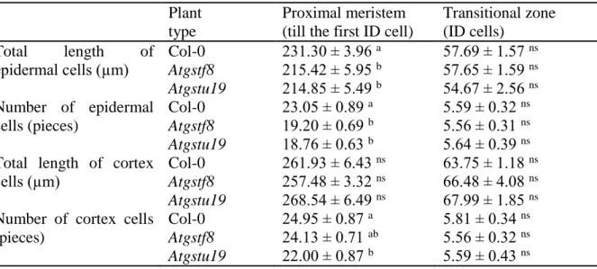
Identification of the root zones and determination of their size in one-week-old seedlings revealed that the total length of short epidermal cells and their number in the PM of mutants was lower compared to the wild type (Table 1). Determination of these parameters in the cortex cells of the Col-0 and mutant roots did not show differences in total length between the Col-0 and Atgstf8 roots, however the number of cells was a little less in the PM of Atgstu19 compared to other genotypes (Table 1). Identification of the two cell lines belonging to different zones are shown on Fig. S3.
3.7 In silico analysis of the 5’ regulatory regions of AtGSTF8 and AtGSTU19 indicates the presence of both stress- and hormone-responsive elements
To predict cis-acting regulatory sequences in the 5’ promoter regions of the selected AtGST genes we performed in silico analysis. The list and description of the main nucleotide motifs discovered in the 5’ regulatory regions of the investigated genes are shown in Table S2. Beside some common regulatory elements involved in defense and abiotic/biotic stress responses (e.g.
ARE, TC-rich repeats, MBS motif, ABRE elements), motifs involved in hormone regulation (auxin, gibberellin, methyl jasmonate, ethylene, salicylic acid) were identifed in the investigated regions (1.5 kb). For example, the auxin-responsive TGA-element (AACGAC) can be found in the 5’ regulatory region of AtGSTU19, while TGA-box (TGACGTGGC) sequences are present in both genes. Interestingly, numerous other cis-acting regulatory sequences associated with light responsiveness, the control of leaf morphology and seed development were also identified in the 5’ upstream regions of one or both genes indicating the complex interaction between different signaling inputs controlling their expression.
4 Discussion
4.1 The AtGSTF8 and AtGSTU19 enzymes are differentially involved in the maintenance of the redox homeostasis of root meristem zones
ROS modulate cell division, differentiation, and expansion via functioning as second messengers and/or affecting the cell wall structure [67, 68, 69, 16]. The responses of cells to cellular oxidation due to abiotic stress or the action of defense phytohormones depend on cell identity [70, 71]. Jiang et al. [33] demonstrated using the roGFP1 probe that the redox potentials of various regions of 3-9 day-old Arabidopsis roots differ significantly. In accordance with the above reports, we found that the redox potential of the Col-0 plant’s proximal meristem (cell division) and the elongation zone (cell differentiation) differed by circa 6 mV.
Hundreds of genes, including several ones coding for AtGSTUs, are differentially expressed in radial and/or longitudinal zones of the Arabidopsis root [72]. Dixon et al. [51]
reported that the relative expression of AtGSTU19 and AtGSTF8 genes is higher in roots than in shoots especially in specific parts and tissues of the root, such as the stele, endodermis, epidermal atrichoblasts and the lateral root cap or root hair zone. Our investigations of the meristematic region of the roots of the Atgstf8 and Atgstu19 mutants
10
revealed elevated O2•- level in comparison to the control. This indicates that both GSTs have important role in the ROS processing systems even in the root meristem.
Among the different redox active compounds determining the cellular redox status of plant cells [73], especially the ascorbate and GSH were reported to determine growth and development of plants by modulating processes, such as mitosis, cell elongation, senescence, cell death and stress responses [50]. The key role of the GSH redox potential in determining the “biological activity” of cells was suggested by Schafer and Buettner [18]. By analysis of the cellular redox environment through the life of different cell lines they reported that the proliferating state was connected with the calculated EGSH ranging -260 mV to -210 mV, but the differentiation was associated with a more positive, -210 to -180 mV potential [18]. Although the exact EGSH values of plant cells proved to be lower by using redox active fluorescent probes even by 50-60 mV [21], the correlation between the GSH redox status and different physiological processes in plants was proved [74]. Detection of a more oxidized redox status across longitudinal zones of mutants compared to wild type suggests that both isoenzymes may be involved in the maintenance of the redox homeostasis. The shift in the redox potential of the PM, TZ and EZ regions in Atgstu19 was 12-18 mV; 2-4 fold higher than in Atgstf8. According to these results, AtGSTU19 has more important role controlling the redox homeostasis in the investigated root zones than AtGSTF8.
Characterization of Arabidopsis rml mutants revealed that glutathione is specifically required to activate and maintain the cell division cycle in the root apical cells [34, 35, 37] however, it was also suggested that redox status by itself does not determine the cellular proliferation.
Berndt et al. [75] proposed the concept that redox regulation by glutathione needs enzymes. In the rml1 mutants, the post-embryonic root meristem does not form because of the defects of GSH biosynthesis and depletion of important antioxidants [35]. In these mutants, several transcripts encoding proteins involved in oxidative stress responses were decreased, indicating that low GSH is accompanied by a decrease in oxidative stress signaling. In contrast, overexpression of BcGSTU in A. thaliana resulted in higher glutathione level and the promotion of root and shoot growth as well as better performance of the plants under abiotic and biotic stresses [4]. In our study, the requirement of AtGSTF8 and AtGSTU19 for proper root meristem organisation and function was revealed. It was found that the size of the PM and the TZ as well as the number of epidermal and cortical cells was decreased in the mutants. This showed correlation with the decreased redox potantial in the meristem.
4.2 The more positive redox potential of the Atgstf8- and Atgstu19 mutants influences the salt response of their roots
Noctor et al. suggested that the same signaling pathways are involved in ROS-mediated acclimation in response to stress than in the control of normal growth and development [76].
Therefore, the potential involvement of AtGSTF8 and AtGSTU19 in the salt stress response of the root was tested. The vitality, redox potential, and ROS level of wild type and mutant seedling roots were compared after 3 hours treatments with 75 or 150 mM NaCl. Root cells of the Atgstf8 mutant exhibited similar FDA fluorescence (vitality) than that of the wild type. In contrast, 75 mM NaCl resulted in significantly increased salt sensitivity in all root zones of the Atgstu19 mutant in comparison to those of the wild type. Redox potential changes were in agreement with this difference. In the meristematic zones (PM and TZ), the redox potential of the Atgstf8 mutant increased in a similar manner in response to salt than that of the wild type although the values, including that of the control sample, were somewhat more positive in the
11
mutant. The meristematic regions of the Atgstu19 mutant exhibited a similar redox potential under control conditions than that of the salt-treated Atgstf8 and Col-0 roots. It was not increased further by the 75 mM only the 150 mM salt concentration in this mutant. In the elongation zone, both mutants had more positive redox potential than the wild type and this value could not be increased further by salt. The Atgstu19 mutant exhibited a significantly higher redox value in this region as well as in the transition zone in comparison to the other two lines.
In general, one can state that the relative redox potential changes in the mutant roots were lower than in the wild type likely due to their already more positive value under control conditions.
Their increased salt sensitivity can be associated with this decreased redox potential response.
It is supported by the observation that the highest sensitivity towards 75 mM salt was shown by the Atgstu19 mutant exhibiting no redox potential change in response to this salt concentration.
4.3 The determined ROS levels are not directly linked to the observed redox potential and vitality differences
Enhanced ROS production is suggested to temporarily shift the redox potential to more oxidized values that will alter the operational controls of many redox-sensitive proteins [69, 77]. A relative small global shift in the EGSH to values of about -260 mV is associated with a very large change in gene expression and plant development [32, 37]. Our data did not reveal direct correlation between the EGSH values and the level of investigated ROS or between the EGSH and cell vitality. However, it is possible, that the mutations in these AtGST genes triggered changes in those mechanisms which are common in salt stress-induced responses. For example, activation of different enzymes with peroxidase activity (such as Class III peroxidases and GST isoenzymes) may account for the smaller increase of the H2O2 level observed in Atgstf8/u19 roots compared to the wild type after applying salt stress (Fig. 3). This may result in the mutant roots in a different O2•-/H2O2 ratio than in the control. Tsukagoshi and his co-workers [78]
reported that the Arabidopsis UPBEAT1 (UPB1) transcription factor directly regulates the expression of root peroxidases that modulate the ratio between the O2•- and H2O2 levels [78].
Moreover, the ability of cells to switch between ROS-processing pathways may restrict ROS- accumulation and detection [76]. Using the membrane-anchored HyPer genetically encoded H2O2 sensor indicated the existence of discrete ROS maxima and a limited ROS diffusion distance [79]. Such spatial distribution of ROS might be explained by local downregulation of cellular antioxidant mechanisms that enables controlled accumulation of ROS, triggering downstream signaling processes [80].
4.4 AtGSTF8 and AtGSTU19 may integrate redox regulation with stress and hormonal responses
It was suggested that the function of certain antioxidative enzymes is not only to keep ROS levels low, but also to allow the cells to sense and signal ROS availability and redox perturbations [76]. Because several GSH-related enzymes were reported playing role in root architecture and lateral root development, they can be among the components linking redox signaling to growth [81, 50].
Detailed investigations of the Arabidopsis GSTU17 revealed that it has a strong impact on GSH:GSSG ratio and thus on redox status of the cells, moreover its expression pattern appeared to be associated with auxin and ABA signaling [82]. The AtGSTU5 expression was induced also by several different stressors and by auxin [50]. It was hypothesized that these genes represent general components of the environmental stress response. They could constitute
12
central components of the auxin-cytokinin regulatory transcriptional network, functioning as regulatory hubs between growth and stress-tolerance pathways. Jiang et al. already demonstrated that salt-induced changes in redox status in roots can influence root meristem maintenance, at least in part, by auxin transport [33]. The importance of the GSH/GSSG ratio and the expression of GSTs were also indicated in biotic stress responses [83]. For example, they were suggested as early indicators of the esca disease in grapevine [84, 85].
AtGSTF8 was proposed to be a marker gene for early stress and defense responses too, because its expression was induced rapidly by diverse biotic and abiotic stresses including pathogen attack, phytohormones, herbicides, heat and high-light [86].
Our analysis on possible regulatory sequences in the 5’ regulatory regions of AtGSTF8 and AtGSTU19 also suggests that these isoenzymes can be part of a complex signaling network.
Regulatory elements involved in defense, abiotic/biotic stress responses, hormone (auxin, gibberellin, methyl jasmonate, ethylene, salicylic acid) and light responsiveness, as well as in developmental pathways were identifed in the investigated promoter regions of their genes.
Both proteins might represent ROS-processing enzymes fine-tuning and maintening redox homeostasis under plant development especially under stress conditions.
5 Conflict of Interest
The authors declare that the research was conducted in the absence of any commercial or financial relationships that could be construed as a potential conflict of interest.
6 Author Contributions
JC contributed in planning the experiments, wrote and edited the manuscript. EH designed and coordinated the study, analyzed the data, wrote and edited the manuscript. KB, BH, RR, ÁH, ÁBH and EH performed the experiments. AF, KB and ÁG discussed the results and contributed to the writing of the manuscript.
7 Acknowledgement
This study was supported by the Hungarian National Research, Development and Innovation Office [Grant Numbers: NKFI-6 K 125265 and NKFI-1 PD 121027]. We thank Dr. F. Ayaydin and I. Kelemen for their technical assistance in confocal microscopy observations in the Cellular Imaging Laboratory of BRC, Szeged. We would like to thank Prof. Dr. A. Meyer for the c-GRX1-roGFP2-harbouring plasmids and Dr. G. Rigó for GV3101 Agrobacterium strain.
The authors would like to thank Mrs. Erzsébet Porkoláb for her excellent technical assistance.
We are grateful to Dr. Beatrix M. Horvath for critical reading and linguistic corrections of the manuscript.
13 8 References
1 R. Desikan, A. Reynolds, J.T. Hancock, S.J. Neill, Harpin and hydrogen peroxide both initiate programmed cell death but have differential effects on defence gene expression in Arabidopsis suspension cultures, Biochem. J. 330 (1998) 115–120. doi:
10.1042/bj3300115
2 D.P. Dixon, B.G. Davis, R. Edwards, Functional divergence in the glutathione transferase superfamily in plants: Identification of two classes with putative functions in redox homeostasis in Arabidopsis thaliana, J. Biol. Chem. 277 (2002) 30859–30869. doi:
10.1074/jbc.M202919200
3 A. Moons, Regulatory and functional interactions of plant growth regulators and plant glutathione S-transferases (GSTs), Vitam. Horm. 72 (2005) 155–202. doi: 10.1016/S0083- 6729(05)72005-7
4 C.W. Kao, M. Bakshi, I. Sherameti, S. Dong, M. Reichelt, R. Oelmüller, K.W. Yeh, A Chinese cabbage (Brassica campetris subsp. Chinensis) τ-type glutathione-S-transferase stimulates Arabidopsis development and primes against abiotic and biotic stress, Plant Mol. Biol. 92(6) (2016) 643–659. doi: 10.1007/s11103-016-0531-2
5 K.A. Marrs, The functions and regulation of glutathione S-transferases in plants, Annu.
Rev. Plant Physiol. 47 (1996) 127–158. doi: 10.1146/annurev.arplant.47.1.127
6 I. Cummins, D.P. Dixon, S. Freitag-Pohl, M. Skipsey, R. Edwards, Multiple roles for plant glutathione transferases in xenobiotic detoxification, Drug. Metab. Rev. 43(2) (2011) 266–
280. doi: 10.3109/03602532.2011.552910
7 R. Edwards, D.P. Dixon, V. Walbot, Plant glutathione S-transferases: enzymes with multiple functions in sickness and in health, Trends Plant Sci. 5 (2000) 193–198. doi:
10.1016/S1360-1385(00)01601-0
8 D.P. Dixon, R. Edwards, Roles for stress-inducible lambda glutathione transferases in flavonoid metabolism in plants as identified by ligand fishing, J. Biol. Chem. 285(47) (2010) 36322–36329. doi: 10.1074/jbc.M110.164806
9 P.A. Lallement, B. Brouwer, O. Keech, A. Hecker, N. Rouhier, The still mysterious roles of cysteine-containing glutathione transferases in plants, Front. Pharmacol. 5 (2014) 192.
doi: 10.3389/fphar.2014.00192
10 G. Potters, N. Horemans, M.A. Jansen, The cellular redox state in plant stress biology--a charging concept, Plant Physiol. Biochem. 48(5) (2010) 292–300. doi:
10.1016/j.plaphy.2009.12.007
11 C.H. Foyer, G. Noctor, Redox homeostasis and antioxidant signaling: a metabolic interface between stress perception and physiological responses, Plant Cell 17 (2005) 1866–1875.
doi: 10.1105/tpc.105.033589
12 D. Boguszewska, B. Zagdańska, ROS as signaling molecules and enzymes of plant response to unfavorable environmental conditions, in: V. Lushchak (Ed.), Oxidative stress—molecular mechanisms and biological effcts, InTech, Rijeka, 2012, pp. 341–362.
http:// www.intechopen.com, ISBN: 978-953-51-0554-1
14
13 A. Baxter, R. Mittler, N. Suzuki, ROS as key players in plant stress signalling, J. Exp. Bot.
65 (2014) 1229–1240. doi: 10.1093/jxb/ert375
14 W. Czarnocka, S. Karpiński, Friend or foe? Reactive oxygen species production, scavenging and signaling in plant response to environmental stresses, Free Radical Bio. Med. 122 (2018) 4-20. doi: 10.1016/j.freeradbiomed.2018.01.011
15 J. Dat, S. Vandenabeele, E. Vranova, M. Van Montagu, D. Inze, F. Van Breusegem, Dual action of the active oxygen species during plant stress responses, Cell Mol. Life Sci. 57 (2000) 779–795. doi: 10.1007/s000180050041
16 A. Mhamdi, F. Van Breusegem, Reactive oxygen species in plant development, Development 145(15) (2018) dev164376. doi: 10.1242/dev.164376
17 S. Perilli, R. Di Mambro, S. Sabatini, Growth and development of the root apical meristem, Curr. Opin. Plant Biol. 15 (2012) 17–23. doi: 10.1016/j.pbi.2011.10.006
18 F.Q. Schafer, G.R. Buettner, Redox environment of the cell as viewed through the redox state of the glutathione disulfide/glutathione couple, Free Radical Bio. Med. 30 (2001) 1191–1212. doi: 10.1016/S0891-5849(01)00480-4
19 A.J. Meyer, R. Hell, Glutathione homeostasis and redox-regulation by sulfhydryl groups, Photosynth. Res. 86 (2005) 435–457. doi: 10.1007/s11120-005-8425-1
20 I. Kranner, S. Birtic, K.M. Anderson, H.W. Pritchard, Glutathione half-cell reduction potential: a universal stress marker and modulator of programmed cell death?, Free Radical Bio. Med. 40 (2006) 2155–2165. doi: 10.1016/j.freeradbiomed.2006.02.013
21 A.J. Meyer, T. Brach, L. Marty, S. Kreye, N. Rouhier, J.P. Jacquot, R. Hell, Redox- sensitive GFP in Arabidopsis thaliana is a quantitative biosensor for the redox potential of the cellular glutathione redox buffer, Plant J. 52(5) (2007) 973–986. doi: 10.1111/j.1365- 313X.2007.03280.x
22 G. Szalai, T. Kellos, G. Galiba, G. Kocsy, Glutathione as an antioxidant and regulatory molecule in plants under abiotic stress conditions, J. Plant Growth Regul. 28 (2009) 66–
80. doi: 10.1007/s00344-008-9075-2
23 G.T. Hanson, R. Aggeler, D. Oglesbee, M. Cannon, R.A. Capaldi, R.Y. Tsien, S.J.
Remington, Investigating mitochondrial redox potential with redox-sensitive green florescent protein indicators, J. Biol. Chem. 279 (2004) 13044–13053. doi:
10.1074/jbc.M312846200
24 K. Jiang, C. Schwarzer, E. Lally, S. Zhang, S. Ruzin, T. Machen, S.J. Remington, L.
Feldman, Expression and characterization of a redox-sensing green florescent protein (reduction-oxidation-sensitive green florescent protein) in Arabidopsis, Plant Physiol. 141 (2006) 397–403. doi: 10.1104/pp.106.078246
25 M. Schwarzländer, M.D. Fricker, C. Müller, L. Marty, T. Brach, J. Novak, L.J. Sweetlove, R. Hell, A.J. Meyer, Confocal imaging of glutathione redox potential in living plant cells, J. Microsc. 231 (2008) 299–316. doi: 10.1111/j.1365-2818.2008.02030.x
15
26 M. Schwarzländer, M.D. Fricker, L.J. Sweetlove, Monitoring the in vivo redox state of plant mitochondria: effect of respiratory inhibitors, abiotic stress and assessment of recovery from oxidative challenge, Biochim. Biophys. Acta 1787 (2009) 468–475. doi:
10.1016/j.bbabio.2009.01.020
27 C.T. Dooley, T.M. Dore, G.T. Hanson, W.C. Jackson, S.J. Remington, R.Y. Tsien, Imaging dynamic redox changes in mammalian cells with green fluorescent protein indicators, J.
Biol. Chem. 279 (2004) 22284–22293. doi: 10.1074/jbc.M312847200
28 M.B. Cannon, J. Remington, Re-engineering redox-sensitive green fluorescent protein for improved response rate, Prot. Sci. 15 (2005) 45–57. doi: 10.1110/ps.051734306
29 J.R. Lohman, S.J. Remington, Development of a family of redox sensitive green fluorescent protein indicators for use in relatively oxidized subcellular environments, Biochemistry 47 (2008) 8678–8688. doi: 10.1021/bi800498g
30 M. Gutscher, A.L. Pauleau, L. Marty, T. Brach, G.H. Wabnitz, Y. Samstag, A.J. Meyer, T.P. Dick, Real-time imaging of the intracellular glutathione redox potential, Nat. Methods 6 (2008) 553–559. doi: 10.1038/nmeth.1212
31 A.J. Meyer, T.P. Dick, Fluorescent protein-based redox probes, Antioxid. Redox. Signal.
13 (2010) 621–650. doi: 10.1089/ars.2009.2948
32 I. Aller, N. Rouhier, A.J. Meyer, Development of roGFP2-derived redox probes for measurement of the glutathione redox potential in the cytosol of severely glutathione- deficient rml1 seedlings, Front. Plant Sci. 4 (2013) 506. doi: 10.3389/fpls.2013.00506 33 K. Jiang, J. Moe-Lange, L. Hennet, L.J. Feldman, Salt stress affects the redox status of
Arabidopsis root meristems, Front. Plant Sci. 7 (2016) 81. doi: 10.3389/fpls.2016.00081 34 J.C. Cheng, K. Seeley, Z.R. Sung, RML1 and RML2, Arabidopsis genes required for cell
proliferation at the root tip, Plant Physiol. 107 (1995) 365–376. doi: 10.1104/pp.107.2.365 35 T. Vernoux, R.C. Wilson, K.A. Seeley, J.P. Reichheld, S. Muroy, S. Brown, S.C. Maughan, C.S. Cobbett, M. Van Montagu, D. Inzé, M.J. May, Z.R. Sung, The ROOT MERISTEMLESS1/CADMIUM SENSI-TIVE2 gene defines a glutathione-dependent pathway involved in initiationand maintenance of cell division during postembryonic root development, Plant Cell 12 (2000) 97–110. doi: 10.1105/tpc.12.1.97
36 P. Frendo, J. Harrison, C. Norman, M.J.H. Jiménez, G. Van de Sype, A. Gilabert, A. Puppo, Glutathione and homoglutathione play a critical role in the nodulation process of Medicago truncatula, Mol. Plant Microbe Interact. 18(3) (2005) 254–259. doi: 10.1094/MPMI-18- 0254
37 D. Schnaubelt, G. Queval, Y. Dong, P. Diaz-Vivancos, M.E. Makgopa, G. Howell, A. De Simone, J. Bai, C.H. Foyer, Low glutathione regulates gene expression and the redox potentials of the nucleus and cytosol in Arabidopsis thaliana, Plant Cell Environ. 38 (2015) 266–279. doi: 10.1111/pce.12252
38 R. Edwards, D.P. Dixon, M. Skipsey, Roles for glutathione transferases in plant secondary metabolism, Phytochemistry 71 (2010) 338–350. doi: 10.1016/j.phytochem.2009.12.012
16
39 M. Jain, C. Ghanashyam, A. Bhattacharjee, Comprehensive expression analysis suggests overlapping and specific roles of rice glutathione S-transferase genes during development and stress responses, BMC Genomics. 11 (2010) 73. doi: 10.1186/1471-2164-11-73 40 V.P. Roxas, R.K. Smith Jr, E.R. Allen, R.D. Allen, Overexpression of glutathione S-
transferase/glutathioneperoxidase enhances the growth of transgenic tobacco seedlings during stress, Nat. Biotechnol. 15 (1997) 988–991. doi : 10.1038/nbt1097-988
41 V.P. Roxas, S.A. Lodhi, D.K. Garrett, J.R. Mahan, R.D. Allen, Stress tolerance in transgenic tobacco seedlings that overexpress glutathione S-transferase/glutathione peroxidase, Plant Cell. Physiol. 41 (2000) 1229–1234. doi: 10.1093/pcp/pcd051
42 K. Benekos, C. Kissoudis, I. Nianiou-Obeidat, N. Labrou, P. Madesis, M. Kalamaki, A.
Makris, A. Tsaftaris, Overexpression of a specific soybean GmGSTU4 isoenzyme improves diphenyl ether and chloroacetanilide herbicide tolerance of transgenic tobacco plants, J. Biotechnol. 150 (2010) 195–201. doi: 10.1016/j.jbiotec.2010.07.011
43 J. Xu, X.J. Xing, Y.S. Tian, R.H. Peng, Y. Xue, W. Zhao, Q.H. Yao, Transgenic Arabidopsis plants expressing tomato glutathione S-transferase showed enhanced resistance to salt and drought stress, PLoS One 10 (2015) e0136960. doi:
10.1371/journal.pone.0136960
44 B. Jia, M. Sun, X. Sun, R. Li, Z. Wang, J. Wu, Z. Wei, H. DuanMu, J. Xiao, Y. Zhu, Overexpression of GsGSTU13 and SCMRP in Medicago sativa confers increased salt–alkaline tolerance and methionine content. Physiol. Plantarum 156 (2016) 176- 189. doi: 10.1111/ppl.12350
45 J. Xu, Y.S. Tian, X.J. Xing, R.H. Peng, B. Zhu, J.J. Gao, Q. H. Yao, Over‐expression of AtGSTU19 provides tolerance to salt, drought and methyl viologen stresses in Arabidopsis, Physiol. Plant. 156(2) (2016) 164–175. doi: 10.1111/ppl.12347
46 G. Yang, Z. Xu, S. Pen, Y. Sun, C. Jia, M. Zhai, In planta characterization of a tau class glutathione S-transferase gene from Juglans regia (JrGSTTau1) involved in chilling tolerance, Plant Cell Rep. 35 (2016) 681–692. doi: 10.1007/s00299-015-1912-8
47 L. Lo Cicero, V. Catara, C.P. Strano, P. Bella, P. Madesis, E.R. Lo Piero, Over- expression of CsGSTU promotes tolerance to the herbicide alachlor and resistance to Pseudomonas syringae pv. tabaci in transgenic tobacco. Biol. Plant. 61(2017) 169–177.
doi: 10.1007/s10535-016-0659-6
48 I. Nianiou-Obeidat, P. Madesis, C. Kissoudis, G. Voulgari, E. Chronopoulou, A.
Tsaftaris, N.E. Labrou, Plant glutathione transferase-mediated stress tolerance:
functions and biotechnological applications. Plant Cell Rep. 36 (2017) 791-805. doi:
10.1007/s00299-017-2139-7
49 Q. Yang, Y.J. Liu, Q.Y. Zeng, Overexpression of three orthologous glutathione S- transferases from Populus increased salt and drought resistance in Arabidopsis.
Biochem. Syst. Ecol. 83 (2019) 57-61. doi: 10.1016/j.bse.2019.01.001
50 A. Bielach, M. Hrtyan, V. Tognetti, Plants under stress: Involvement of auxin and cytokinin, Int. J. Mol. Sci. 18(7) (2017) 1427. doi: 10.3390/ijms18071427
17
51 D.P. Dixon, M. Skipsey, R. Edwards, Roles for glutathione transferases in plant secondary metabolism, Phytochem. 71 (2010) 338–350. doi: 10.1016/j.phytochem.2009.12.012 52 P.G. Sappl, L. Onate-Sanchez, K.B. Singh, A.H. Millar, Proteomic analysis of glutathione
S-transferases of Arabidopsis thaliana reveals differential salicylic acid-induced expression of the plant-specific phi and tau classes, Plant Mol. Biol. 54 (2004) 205–219.
doi: 10.1023/B:PLAN.0000028786.57439.b3
53 C. Uquillas, I. Letelier, F. Blanco, X. Jordana, L. Holuigue, NPR1-independent activation of immediate early salicylic acid-responsive genes in Arabidopsis, Mol. Plant Microbe Interact. 17(1) (2004) 34–42. doi: 10.1094/MPMI.2004.17.1.34
54 U. Wagner, R. Edwards, D.P. Dixon, F. Mauch, Probing the diversity of the Arabidopsis glutathione S-transferase gene family, Plant Mol. Biol. 49 (2002) 515–532. doi:
10.1023/A:1015557300450
55 P.G. Sappl, A.J. Carroll, R. Clifton, R. Lister, J. Whelan, A.H. Millar, K.B. Singh, The Arabidopsis glutathione transferase gene family displays complex stress regulation and co- silencing multiple genes results in altered metabolic sensitivity to oxidative stress, Plant J.
58 (2009) 53–68. doi: 10.1111/j.1365-313X.2008.03761.x
56 E. Horváth, K. Bela, C. Papdi, Á. Gallé, L. Szabados, I. Tari, J. Csiszár, The role of Arabidopsis glutathione transferase F9 gene under oxidative stress in seedlings, Acta Biol.
Hung. 66(4) (2015) 406–418. doi: 10.1556/018.66.2015.4.5
57 R.L. Scholl, S.T. May, D.H. Ware, Seed and molecular resources for Arabidopsis, Plant Physiol. 124(4) (2000) 1477–1480. doi: 10.1104/pp.124.4.1477
58 T. Murashige, F. Skoog, A revised medium for rapid growth and bio assays with tobacco tissue cultures, Physiol. Plant. 15(3) (1962) 473–497. doi: 10.1111/j.1399- 3054.1962.tb08052.x
59 S. Perilli, S. Sabatini, Analysis of root meristem size and development, in: L. Henning, C.
Köhler (Eds.), Plant Developmental Biology, Methods in Molecular Biology, Springer,New York, 2010, pp. 177–187. doi: 10.1007/978-1-60761-765-5_12
60 J.P. Verbelen, T.D. Condder, J. Le, K. Vissenberg, F. Baluska, The root apex of Arabidopsis thaliana consists of four distinct zones of growth activities-meristematic zone, transition zone, fast elongation zone and growth terminating zone, Plant Signal. Behav. 1 (2006) 296–304. doi: 10.4161/psb.1.6.3511
61 N. Lehotai, G. Feigl, Á. Koós, Á. Molnár, A. Ördög, A. Pető, L. Erdei, Zs. Kolbert, Nitric oxide–cytokinin interplay influences selenite sensitivity in Arabidopsis, Plant Cell Rep.
35(10) (2016) 2181–2195. doi: 10.1007/s00299-016-2028-5
62 J. Schindelin, I. Arganda-Carreras, E. Frise, V. Kaynig, M. Longair, T. Pietzsch, S.
Preibisch, C. Rueden, S. Saalfeld, B. Schmid, J.Y. Tinevez, D.J. White, V. Hartenstein, K.
Eliceiri, P. Tomancak, A. Cardona, Fiji: an open-source platform for biological-image analysis, Nat. Methods 9(7) (2012) 676–682. doi: 10.1038/nmeth.2019
18
63 A. Pető, N. Lehotai, G. Feigl, N. Tugyi, A. Ördög, K. Gémes, I. Tari, L. Erdei, Nitric oxide contributes to copper tolerance by influencing ROS metabolism in Arabidopsis, Plant Cell Rep. 32(12) (2013) 1913–1923. doi: 10.1007/s00299-013-1503-5
64 C. Koncz, N. Martini, L. Szabados, M. Hrouda, A. Bachmair, J. Schell, Specialized vectors for gene tagging and expression studies, In: S. Gelvin, B. Schilperoort (Eds.) Plant Molecular Biology Manual, Kluwer Academic Publishers, Dordrecht-Boston-London, 1994, pp. 1–22. doi: 10.1007/978-94-011-0511-8_4
65 S.J. Clough, A.F. Bent, Floral dip: a simplified method for Agrobacterium-mediated transformation of Arabidopsis thaliana, Plant J. 16 (1998) 735–743. doi: 10.1046/j.1365- 313x.1998.00343.x
66 S. Attacha, D. Solbach, K. Bela, A. Moseler, S. Wagner, M. Schwarzländer, I. Aller, S.J.
Müller, A.J. Meyer, Glutathione peroxidase‐like enzymes cover five distinct cell compartments and membrane surfaces in Arabidopsis thaliana, Plant Cell Environ. 40(8) (2017) 1281–1295. doi: 10.1111/pce.12919
67 H. Sauer, M. Wartenberg, J. Hescheler, Reactive oxygen species as intracellular messengers during cell growth and differentiation, Cell Physiol. Biochem. 11 (2001) 173–
186. doi: 10.1159/000047804
68 A. Fehér, K. Ötvös, T.P. Pasternak, A.P. Szandtner, The involvement of reactive oxygen species (ROS) in the cell cycle activation (G0-to-G1 transition) of plant cells, Plant Signal.
Behav. 3 (2008) 823–826. doi: 10.4161/psb.3.10.5908
69 R. Schmidt, J.H. Schippers, ROS-mediated redox signaling during cell differentiation in plants. Biochim. Biophys, Acta Gen. Subj. 1850(8) (2015) 1497–1508. doi:
10.1016/j.bbagen.2014.12.020
70 J.R. Dinneny, P.N. Benfey, Plant stem cell niches: standing the test of time, Cell 132(4) (2008) 553–557. doi: 10.1016/j.cell.2008.02.001
71 G. Wachsman, E.E. Sparks, P.N. Benfey, Genes and networks regulating root anatomy and architecture, New Phytol. 208(1) (2015) 26–38. doi: 10.1111/nph.13469
72 J.R. Dinneny, T.A. Long, J.Y. Wang, J.W. Jung, D. Mace, S. Pointer, C. Barron, S.M.
Brady, J. Schiefelbei, P.N. Benfey, Cell identity mediates the response of Arabidopsis roots to abiotic stress, Science 320(5878) (2008) 942–945. doi: 10.1126/science.1153795 73 K.J. Dietz, Redox regulation of transcription factors in plant stress acclimation and development, Antioxid. Redox Signal. 21(9) (2013) 1356–1372. doi:
10.1089/ars.2013.5672
74 T. Jubany-Mari, L. Alegre-Batlle, K. Jiang, L.J. Feldman, Use of a redox‐sensing GFP (c‐
roGFP1) for real‐time monitoring of cytosol redox status in Arabidopsis thaliana water‐
stressed plants, FEBS lett. 584(5) (2010) 889–897. doi: 10.1016/j.febslet.2010.01.014 75 C. Berndt, C.H. Lillig, L. Flohé, Redox regulation by glutathione needs enzymes, Front.
Pharmacol. 5 (2014) 168. doi: 10.3389/fphar.2014.00168
19
76 G. Noctor, J.P. Reichheld, C.H. Foyer, ROS-related redox regulation and signaling in plants, Cell Dev. Biol. 80 (2018) 3–12. doi: 10.1016/j.semcdb.2017.07.013
77 C.H. Foyer, G. Noctor, Stress-triggered redox signalling: what's in pROSpect?, Plant Cell Environ. 39(5) (2016) 951–964. doi: 10.1111/pce.12621
78 H. Tsukagoshi, W. Busch, P.N. Benfey, Transcriptional regulation of ROS controls transition from proliferation to differentiation in the root, Cell 143(4) (2010) 606–616. doi:
10.1016/j.cell.2010.10.020
79 N.M. Mishina NM, P.A. Tyurin-Kuzmin PA, K.N. Markvicheva KN, A.V. Vorotnikov AV, V.A. Tkachuk VAV. Laketa, C. Schultz, S. Lukyanov, V.V. Belousov, Does cellular hydrogen peroxide diffuse or act locally? Antioxid. Redox Signal. 14 (2011) 1–7. doi: 10.1089=ars.2010.3539
80 C. Waszczak, M. Carmody, J. Kangasjärvi, Reactive oxygen species in plant signaling. Annu. Rev.Plant Boil. 69 (2018) 209-236. doi: 10.1146/annurev-arplant- 042817-040322
81 K. Bela, S.A.K. Bangash, J. Csiszár, Plant glutathione peroxidases: structural and functional characterization, their roles in plant development, in: M. Hossain, M. Mostofa, P. Diaz-Vivancos, D. Burritt, M. Fujita and L.S. Tran (Eds.), Glutathione in Plant Growth, Development, and Stress Tolerance, Springer, Cham, 2017, pp. 99–111. doi: 10.1007/978- 3-319-66682-2_4
82 H.W. Jiang, M.J. Liu, C. Chen, C.H. Huang, L.Y. Chao, H.L. Hsieh, A glutathione S- transferase regulated by light and hormones participates in the modulation of Arabidopsis seedling development, Plant Physiol. 154(4) (2010) 1646–1658. doi:
10.1104/pp.110.159152
83 G. Gullner, T. Komives, L. Király, P. Schröder, Glutathione S-Transferase Enzymes in Plant-Pathogen Interactions. Fron. Plant Sci. 9 (2018). doi: 10.3389/fpls.2018.01836 84 C. Valtaud, C.H. Foyer, P. Fleurat-Lessard, A. Bourbouloux, Systemic effects on leaf glutathione metabolism and defence protein expression caused by esca infection in grapevines. Funct. Plant Biol. 36 (2009) 260–279. doi: 10.1071/FP08293
85 M. Magnin-Robert, M. Adrian, S. Trouvelot, A. Spagnolo, L. Jacquens, P. Letousey, F. Rabenoelina, M. Harir, C. Roullier-Gall, C. Clément, P. Schmitt-Kopplin, A.
Vallat, E. Abou-Mansour, F. Fontaine, Alterations in grapevine leaf metabolism occur prior to esca apoplexy appearance. Mol. Plant Microbe Interact. 30 (2017) 946–
959. doi: 10.1094/MPMI-02-17-0036-R
86 L.F. Thatcher, L.G. Kamphuis, J.K. Hane, L. Oñate-Sánchez, K.B. Singh, The Arabidopsis KH-domain RNA-binding protein ESR1 functions in components of jasmonate signalling, unlinking growth restraint and resistance to stress. PLoS One, 10 (2015) e0126978. doi: 10.1371/journal.pone.0126978
Figure legends
Fig. 1 Calculated redox potential values of roots expressing cytosolic GRX1-roGFP2 (A), the pixel intensity of dihydroethidium (DHE) indicating the superoxide level (B), and that of 10- acetyl-3,7-dihydroxyphenoxazine (resorufin fluorescence) indicating the hydrogen peroxide level (C) of primary roots of 7-day-old Arabidopsis thaliana Col-0 and Atgstf8, Atgstu19 insertional mutants. Scale bar = 100 μm. Data are means±SE, n=10. Columns with different letters are significantly different at p≤0.05, determined by Duncan’s test. PM: proximal meristem, TZ: transition zone, EZ: elongation zone.
Fig. 2 Representative images of the fluorescent analysis of the redox status, the vitality, superoxide radical (O2•-) and hydrogen peroxide (H2O2) levels in the 7-day-old Arabidopsis thaliana Col-0 and Atgstf8, Atgstu19 insertional mutants after three hours of treatment with 75 and 150 mM NaCl. (See more details in Materials and methods). Scale bars = 100 μm.
Fig. 3 The redox status (A, E, I), vitality (B, F, J), superoxide radical (DHE fluorescence, C, G, K) and hydrogen peroxide (resorufin fluorescence, D, H, L) levels in the proximal meristem (A-D), transition- (E-H) and elongation zones (I-L) of 7-day-old Arabidopsis thaliana Col-0 and Atgstf8, Atgstu19 insertional mutants after three hours of treatment with 75 or 150 mM NaCl. EGSH: glutathione redox potential; FDA: fluorescein diacetate;
DHE: dihydroethidium; Resorufin: 10-acetyl-3,7-dihydroxyphenoxazine. Data are means±SE, n=10. Columns with different letters are significantly different at p≤0.05, determined by Duncan’s test.
21
Table 1 The total length of epidermis and cortex cells in the proximal meristem and transitional zone (isodiametric cells, ID) in the one-week-old Arabidopsis thaliana Col-0 and Atgstf8, Atgstu19 insertional mutants. Data are means±SE, n=10. Data with different letters are significantly different at p≤0.05, determined by Duncan’s test. ns= not significant
Plant type
Proximal meristem (till the first ID cell)
Transitional zone (ID cells)
Total length of epidermal cells (µm)
Col-0 Atgstf8 Atgstu19
231.30 ± 3.96 a 215.42 ± 5.95 b 214.85 ± 5.49 b
57.69 ± 1.57 ns 57.65 ± 1.59 ns 54.67 ± 2.56 ns Number of epidermal
cells (pieces)
Col-0 Atgstf8 Atgstu19
23.05 ± 0.89 a 19.20 ± 0.69 b 18.76 ± 0.63 b
5.59 ± 0.32 ns 5.56 ± 0.31 ns 5.64 ± 0.39 ns Total length of cortex
cells (µm)
Col-0 Atgstf8 Atgstu19
261.93 ± 6.43 ns 257.48 ± 3.32 ns 268.54 ± 6.49 ns
63.75 ± 1.18 ns 66.48 ± 4.08 ns 67.99 ± 1.85 ns Number of cortex cells
(pieces)
Col-0 Atgstf8 Atgstu19
24.95 ± 0.87 a 24.13 ± 0.71 ab 22.00 ± 0.87 b
5.81 ± 0.34 ns 5.56 ± 0.32 ns 5.59 ± 0.43 ns
22
23
24
25
26