Transactions
PAPER
Cite this:Dalton Trans., 2020,49, 7977
Received 4th April 2020, Accepted 14th May 2020 DOI: 10.1039/d0dt01256d rsc.li/dalton
An 8-hydroxyquinoline – proline hybrid with multidrug resistance reversal activity and the solution chemistry of its half-sandwich
organometallic Ru and Rh complexes †
János P. Mészáros, a,bJelena M. Poljarević, a,cIstván Szatmári,dOszkár Csuvik,d Ferenc Fülöp,dNorbert Szoboszlai,eGabriella Spenglerb,fand Éva A. Enyedy *a,b
Herein the design and synthesis of a new 8-hydroxyquinoline derivative, (S)-5-chloro-7-((proline-1-yl)methyl)8- hydroxyquinoline (HQCl-Pro), with good water solubility and multidrug resistance reversal activity are reported.
In this work the proton dissociation processes of HQCl-Pro and its complex formation with [Rh(η5-C5Me5) (H2O)3]2+, [Ru(η6-p-cymene)(H2O)3]2+and [Ru(η6-toluene)(H2O)3]2+were investigated by the combined use of pH-potentiometry, UV-visible spectrometry and 1H NMR spectroscopy. Our results revealed the prominent solution stability of the complexes in all cases. The lipophilicity of the complexes increased with the chloride ion concentration, and the complexes showed moderate logDvalues (−0.8 to +0.4) at pH 7.4 at all tested Cl−con- centrations. The formation of mixed hydroxido complexes from the aqua complexes was characterized by rela- tively high pKavalues (8.45–9.62 in chloride-free medium). Complexation processes are much slower with the Ru(η6-arene) triaqua cations than with [Rh(η5-C5Me5)(H2O)3]2+. Both the pKavalues and H2O/Cl−exchange con- stants of the Ru-complexes are lower by 0.5–1.0 orders of magnitude than those of the Rh analogue. Arene loss (p-cymene and toluene) and oxidation were found in the case of Ru-complexes when an excess of HQCl- Pro and aromatic (N,N) bidentate ligands was added. The cytotoxicity and antiproliferative effect of HQCl-Pro and its complexes were assayedin vitro. In contrast to the structurally familiar 8-hydroxyquinoline, HQCl-Pro and its Rh(η5-C5Me5) complex were somewhat more effective against drug resistant Colo 320 adenocarcinoma human cells compared to the drug sensitive Colo 205 cells. The Ru- and Rh-complexes showed a similar metal uptake level after 4 h, while a longer incubation time resulted in higher cellular Rh concentration.
Introduction
During the administration of conventional chemotherapeutic agents, multidrug resistant (MDR) cancer phenotypes are often
developed manifesting resistance to related and unrelated classes of compounds, which is one of the major impediments to suc- cessful treatment.1 The ATP-binding cassette transporter P-glycoprotein (P-gp, ABCB1) is the most known transmembrane transporter that is associated with this resistance phenomenon;
however, multidrug resistance protein 1 and 2 (MRP1 and MRP2) and breast cancer resistance protein (BCRP) also cause an elev- ated efflux of toxic compounds.1 Potent P-gp inhibitors were explored for overcoming the MDR (e.g.verapamil and cyclospor- ine A), although their use is accompanied by undesirable side effects.1Szakácset al.reported a group of anticancer compounds which are more cytotoxic against MDR cancer cells than drug sen- sitive ones.2These molecules are not the inhibitors of P-gp, and their mechanism of action is still not revealed in detail.
8-Hydroxyquinolines were found to be an important family of MDR-selective compounds. It was shown that the CH2–N subunit at position 7 plays an important role in the collateral selectivity, which is present in compounds e.g. NSC1014, NSC297366, NSC693871 and NSC57969.2 The 8-hydroxyquinoline (HQ) scaffold itself has the potential for various structural modifi-
†Electronic supplementary information (ESI) available: Characterization, additional UV-Vis spectra of kinetic measurements and titrations, and pH- dependent1H NMR spectra of compounds. See DOI: 10.1039/D0DT01256D
aDepartment of Inorganic and Analytical Chemistry, Interdisciplinary Excellence Centre, University of Szeged, Dóm tér 7, H-6720 Szeged, Hungary.
E-mail: enyedy@chem.u-szeged.hu
bMTA-SZTE Lendület Functional Metal Complexes Research Group, University of Szeged, Dóm tér 7, H-6720 Szeged, Hungary
cFaculty of Chemistry, University of Belgrade, Studentski trg. 12-16, 11000 Belgrade, Serbia
dInstitute of Pharmaceutical Chemistry and Stereochemistry Research Group of Hungarian Academy of Sciences, University of Szeged, Eötvös u. 6, H-6720 Szeged, Hungary
eLaboratory for Environmental Chemistry and Bioanalytics, Institute of Chemistry, Eötvös Lóránd University, Pázmány Péter stny. 1/A, H-1117 Budapest, Hungary
fDepartment of Medical Microbiology and Immunobiology, University of Szeged, Dóm tér 10, H-6720 Szeged, Hungary
Open Access Article. Published on 14 May 2020. Downloaded on 9/9/2020 8:21:42 AM. This article is licensed under a Creative Commons Attribution-NonCommercial 3.0 Unported Licence.
View Article Online
View Journal | View Issue
cations including changes due to pharmaceutical purposes.3–5 HQ and its numerous derivatives are well-known compounds and have been used in analytical chemistry for many decades due to their simple chemical structure and strong coordination ability to metal ions. E.g. clioquinol (5-chloro-7-iodo-8-hydroxyquinoline) was used as an antiprotozoal and antifungal drug; moreover, it was explored as a treatment for Alzheimer’s and Parkinson’s diseases.3,5
Compounds with the 8-hydroxyquinoline moiety were also reported to exhibit anti-inflammatory, antiviral and antiparasi- tic effects and numerous HQ derivatives with various substitu- ents have been developed and tested as anticancer agents in the last few decades.3–5Among them halogenated compounds were often found to be more efficient.5,6
Not only 8-hydroxyquinolines but their particular metal complexes show anticancer activity as well. The most promi- nent example is the orally active tris(8-hydroxy-quinolato) gallium(III) (KP46), which is now under clinical trials and was successfully tested in phase I on renal cancer.7,8
Other examples are the complexes of platinum group organometallic cations bearing an aromatic ligand in a half- sandwich configuration (usuallyp-cymene (p-cym) or 1,2,3,4,5- pentamethyl-cyclopentadienyl (C5Me5) ligand). The complexes of these cations usually have various physico-chemical pro- perties: the low oxidation state of the metal center (e.g. Ru2+) was stabilized and the lipophilicity was increased by the arene ligand; their kinetic lability can be associated with the strong π-donor/acceptor ability of the arene ligand as well.9,10 Moreover, the fine-tuning of the 3D structure and electronic properties can be easily achieved by functionalization of the arene or the remaining facial ligands. The interaction with the chloride content of the medium can change charge and lipo- philicity. Nevertheless, new reaction pathways with bio- molecules can be reached, which differ from the ligand–bio- molecule interactions: e.g. catalysis of GSH oxidation.9 There are two main types of active complexes: in the first class, a monodentate ligand binds strongly to the metal centre and the leaving group(s) is/are present on two other coordination sites.
Widely known examples are the RAPTA complexes where a strong P-donor, 1,3,5-triaza-7-phosphaadamantane (PTA), is the monodentate ligand.11In the other class, the strongly co- ordinated bidentate ligand occupies two sites, and a leaving group is bound in a monodentate way. The dissociation of the latter can be fine-tuned well with the change of donor atoms.
For example, the target biomolecule is switched from DNA to protein, depending on the nature of the bidentate ligand: (N, N) or (O,O).12This field contains a plethora of studied com- plexes, and an early example is the group of RAED complexes which arein vivoactive compounds showing activity on cispla- tin-resistant cell lines.13 Though structure–activity relation- ships have been reported for these complexes, there is still no organoruthenium containing drug in clinical use.14,15
Half-sandwich organometallic complexes of the (N,O) donor 8-hydroxyquinolines are also often investigated. The organo-ruthenium complexes formed with clioquinol and other halogenated 8-hydroxyquinoline derivatives were studied
thoroughly by Turelet al., and the inhibition of cathepsin B16 and the anticancer,17 antileukaemic18 and antibacterial effects19 were reported for these compounds. Nitroxoline (8-hydroxy-5-nitroquinoline) derivatives showed the anti-meta- static effect, which was improved by coordination to a half- sandwich Ru(p-cym) cation.20An earlier study demonstrated the anticancer and antibacterial activities of the half-sandwich Rh and Ir complexes of HQ as well.21 These complexes are characterized by fairly low IC50values (∼10μM or less), which are promising results; however, they suffer from poor water solubility.6 The limited water solubility is a major problem encountered with drug formulation since it makes the admin- istration difficult and might restrain the attainment of the desired concentration in the blood circulatory system.
Movassaghi et al. introduced a more polar aromatic ligand (N-acetyl-L-phenylalanine ethyl ester) instead ofp-cymene, and with this modification the solubility could be improved, while the cytotoxicity remained at the low-micromolar level.22
The hydrophilic nature of the complexes can be increased via the improved water solubility of the coordinating HQ derivative by the introduction of polar functional groups.
However, the use of 8-hydroxyquinoline-5-sulfonate with excel- lent water-solubility caused the loss of the anticancer activity reported in our previous study.23 Therefore, the lipophilic nature of the HQ ligand should be optimized very carefully, and finding the optimal balance between lipophilicity and water-solubility is a very important endeavour in the develop- ment of novel anticancer compounds. Therefore, in this work we aimed to design an HQ–proline hybrid with elevated water solubility containing the CH2–N subunit at position 7 for the expected MDR-selectivity. It is also interesting how the coordi- nation to metal ions has an effect on the cytotoxicity and MDR selectivity. When the MDR selective ligand 7-(1-piperidinyl- methyl)-8-hydroxyquinoline (NSC57969) was combined with organometallic half-sandwich rhodium and ruthenium cations, the MDR selectivity remained only in the case of the Rh(η5-C5Me5) complex, while the coordination to Ru(η6-p-cym) resulted in the loss of this property.23Herein the synthesis and solution chemical characterization of a new, water-soluble derivative of HQ, (S)-5-chloro-7-(( proline-1-yl)methyl)8-hydroxy- quinoline (HQCl-Pro, Scheme 1), is reported. Its complex for- mation with half-sandwich cations [Rh(η5-C5Me5)(H2O)3]2+, [Ru (η6-p-cym)(H2O)3]2+ and [Ru(η6-toluene)(H2O)3]2+ (abbreviated as [Ru(η6-tol)(H2O)3]2+) is also characterized in solution. In addition, the measurement of the lipophilicity of the com- pounds depending on the actual chloride ion concentration was performed: in the different biofluids the concentration of
Scheme 1 Synthetic route for the HQCl-Pro ligand.
Open Access Article. Published on 14 May 2020. Downloaded on 9/9/2020 8:21:42 AM. This article is licensed under a Creative Commons Attribution-NonCommercial 3.0 Unported Licence.
this coordinating ion may change charge and may have serious effects. The cytotoxicity of HQCl-Pro was monitored in sensitive and in multidrug resistant cancer cells and the effect of complexation with the selected organometallic triaqua cations on the biological activity was assayed as well.
Results and discussion
Synthesis of HQCl-Pro and its complexes
A novel derivative of 5-chloro-8-hydroxyquinoline (HQCl) was designed with increased water solubilityviathe incorporation of the zwitterionic proline amino acid moiety. L-Proline was attached to the HQCl scaffold at position 7 via a methylene linker providing the CH2–N subunit that is reported to be crucial for the MDR reversal activity.2,23The coupling of HQCl with L-proline in the presence of formaldehydeviaa modified Mannich reaction was achieved in methanol under reflux con- ditions (Scheme 1). The formed (S)-5-chloro-7-(( proline-1-yl) methyl)8-hydroxyquinoline (HQCl-Pro) was isolated by crystal- lization from the cooled reaction mixture.
The half-sandwich organometallic complexes with Rh(η5- C5Me5), Ru(η6-p-cym) and Ru(η6-tol) were prepared by mixing the ligand with a half-equivalent amount of the dimeric pre- cursors [M(arene)Cl2]2 in methanol solution for 1 h at room temperature, then the solution was concentrated and precipi- tation was completed with the addition of diethyl ether. The formed complex was filtered out, washed withn-hexane and dried. Complexes were collected in good yields (84–90%).
HQCl-Pro and its complexes were characterized by1H and13C NMR spectroscopy (attached proton test (APT)) and electro- spray ionization mass spectrometry (ESI-MS). Mass spectra and NMR spectra are shown in the ESI (Fig. S1–S12†). The NMR spectra showed a double set of peaks in CD3OD, which can be explained by diastereomer formation (as was found in ref. 22 for complexes with chiral arene) or the appearance of a rigid structure (as it appears also in water for deprotonation vide infra). The differences of the two sets are shown in Fig. S13† peak-by-peak, projected on the proposed structures of complexes. The biggest differences are found in positions 14, 6 and 9, which can be connected with the diastereomer for- mation: in the complex, next to the chiral carbon atom, the Ru centre and the N of the protonated amino group become chiral, and these positions (6,9,14) are close to these atoms.
Additionally, the increased solubility and stability of the ligand and complexes were checked in phosphate buffer at pH = 7.4. All compounds were water-soluble; 10 mM concentration exceeds the limits of solubility of HQ, HQCl and their complexes.
The compounds were stable for 1 week (not shown), except for the ligand itself, which showed a new set of peaks in the aliphatic region in a∼20% ratio (see Fig. S14†) after 6 days.
Hydrolysis of the organometallic cations and proton dissociation processes of HQCl-Pro
Knowing the stability constants of a metal complex and proton dissociation processes of a molecule is necessary to under-
stand the role of different forms of biologically active com- pounds in the biological environment. It can give information about the response to pH and explain the active and inactive forms of a prodrug. In order to describe the solution specia- tion of the metal complexes, characterization of the hydrolysis of the organometallic cations and the (de)protonation equili- brium processes of the ligand is needed under the conditions used. The hydrolytic behaviour of the selected organometallic cations ([Rh(η5-C5Me5)(H2O)3]2+, [Ru(η6-p-cym)(H2O)3]2+ and [Ru(η6-tol)(H2O)3]2+) has been studied in detail previously.24,25 Overall stability constants were reported for the μ-hydroxido bridged dinuclear species formed24,25and these constants are used in this work. According to Buglyóet al., the hydrolysis of the [Ru(η6-arene)(H2O)3]2+organometallic cations can be ade- quately described by the formation of the hydrolysis product [(Ru(η6-arene))2(μ-OH)3]+ in chloride-free medium (I = 0.20 M (KNO3)).25However, in the case of [Rh(η5-C5Me5)(H2O)3]2+the formation of [(Rh(η5-C5Me5))2(μ-OH)2]2+ and [(Rh(η5- C5Me5))2(μ-OH)3]+was reported.24The interaction between the metal ions and the hapto ligands is strong, and as a result the release of the aromatic ligands was not detected in the studied pH range ( pH = 0.7–11.5) (only in the case of some compe- tition reactions vide infra). These organometallic cations are considered as units and denoted as‘M(arene)’hereinafter.
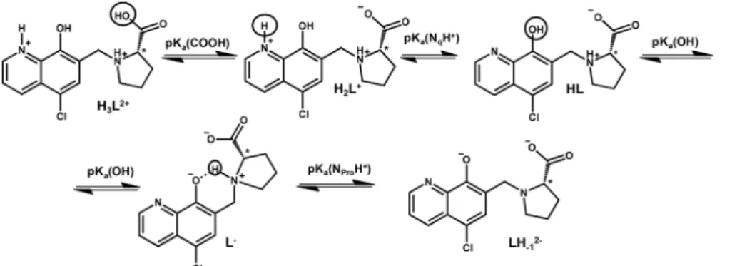
The knowledge of the pKavalue of a bioactive compound is needed not only for speciation studies, but it is also a key para- meter affecting the pharmacokinetic properties, since with the pKavalues the actual protonation state and charge of the mole- cule at a given pH can be calculated. In HQCl-Pro the incorpor- ation of the L-prolinylmethyl substituent results in two additional dissociable protons besides the quinolinium-NH+ (NqH+) and the hydroxyl group (Scheme 2) of the HQ scaffold.
N-methyl-L-proline (N-Me-Pro) is structurally similar to this substituent and it has two pKa values for the carboxylic acid and amino functions (Table 1).26
In order to characterize the proton dissociation processes of HQCl-Pro pH-potentiometric, UV-visible (UV-Vis) and 1H NMR titrations were performed. Although this compound has four dissociable protons, only two deprotonation processes could be determined adequately (with acceptable standard deviation) in the studied pH range by pH-potentiometry. The assignment of the deprotonation processes to the functional groups was done by the interpretation of the 1H NMR and UV-Vis spectral changes (Fig. 1, S15†and Table 1). Notably, HL denotes the neutral (zwitterionic) form of the ligand.
The most acidic pKa(≪2) of HQCl-Pro belongs to the car- boxylic group of the prolinyl substituent, which caused shifts of the peaks only in the aliphatic region of the 1H NMR spectra (not shown). The deprotonation of the quinolinium NqH+ (up to pH 4) and OH ( pH 5.5–9.5) groups was ascer- tained as high-field shifts of all the CH proton peaks (Fig. 1).
The pKavalues of these groups are much lower than those of HQ (see in Table 1) as a consequence of the presence of the two electron-withdrawing substituents (chlorine and the proto- nated CH2–NProH+moiety). The structurally more related HQCl suffers from very poor solubility in aqueous medium and we Open Access Article. Published on 14 May 2020. Downloaded on 9/9/2020 8:21:42 AM. This article is licensed under a Creative Commons Attribution-NonCommercial 3.0 Unported Licence.
could not determine its pKavalues accurately enough in pure water by UV-Vis titrations even at rather low concentrations (5–10 μM) (see the estimated values in Table 1). Both the
experimentally obtained and the predicted values for HQCl represent significantly lower values compared to those of HQ due to the electron withdrawing effect of the chlorine substitu- ent as it is expected. Based on the pKa values it can be observed that the prolinyl amino group deprotonates at a much higher pH than the amino group inN-Me-Pro.
This can be the result of an intramolecular H-bond between the deprotonated hydroxylate and the NProH+ moiety (Scheme 2). A similar hydrogen bond was found in the crystal structures between the hydroxylate group and a protonated morpholine or piperidinyl nitrogen in HQ derived Mannich bases, and these substituents also had a similar effect on the pKavalues.29
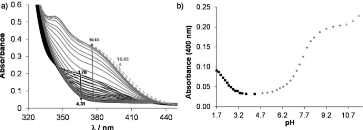
According to the 1H NMR spectra, only the singlet C6H peak seems to be sensitive to this deprotonation and a high- field shift was observed at pH > 10. The UV-Vis spectra, recorded at various pH values (Fig. 2), revealed three deproto- nation processes. However, only one pKavalue could be com- Scheme 2 Proton dissociation processes of the ligand HQCl-Pro.
Table 1 Proton dissociation constants (Ka) of HQCl-Pro and HQ, HQCl andN-methyl-L-proline (N-Me-Pro) for comparison. {T= 25.0 °C;I= 0.20 M (KNO3)}
Method
pKa (COOH)
pKa (NqH+)
pKa (OH)
pKa (NProH+)
HQ pH-potentiometry — 4.99a 9.51a —
HQCl Predicted 4.01b 8.37b —
UV-Vis 3.8c 7.6c
N-Me-Pro pH-potentiometry 1.75d — — 10.36d HQCl-Pro pH-potentiometry — 2.36 ± 0.02 7.76 ± 0.01 >11.5
UV-Vis — — 7.63 ± 0.01 —
1H NMR ≪2 2.22 ± 0.02 7.62 ± 0.01 —
aI= 0.20 M (KCl), taken from ref. 27.bCalculated with Marvin (ref. 28).
cEstimated from the data obtained by UV-Vis titrations, pH = 2.0–11.5 (I= 0.20 M (KNO3)).dI= 0.10 M (KCl), taken from ref. 26.
Fig. 1 1H NMR spectra of HQCl-Pro recorded at pH = 1.6–11.8 (low-field region). Numbering of the positions and peak assignment is shown on the central structure. {c(HQCl-Pro) = 480μM; solvent: 90% H2O/10% D2O;T= 25.0 °C;I= 0.20 M (KNO3)}.
Open Access Article. Published on 14 May 2020. Downloaded on 9/9/2020 8:21:42 AM. This article is licensed under a Creative Commons Attribution-NonCommercial 3.0 Unported Licence.
puted accurately by the deconvolution of the spectra (Table 1) as in the other two cases the whole deprotonation processes could not be seen.
On increasing the pH, the first two deprotonation processes detected in the UV-Vis spectra belong to the NqH+ and the hydroxyl groups, since changes in their protonation state have a considerable effect on the electron density of the aromatic rings. Thus, their deprotonation results in significant spectral changes, especially in the case of the hydroxyl moiety, namely the emerging strong bands in the range 330–430 nm originate from the more extended conjugated π-electron system in the deprotonated form. Surprisingly, the deprotonation of the pro- linyl amino group also affects the spectra; it is accompanied by a minor bathochromic shift (see changes at pH > 10 in Fig. 2).
Proton dissociation constants determined on the basis of the UV-Vis spectrophotometric and pH-potentiometric data are in good agreement with those obtained by the1H NMR spectro- scopic studies (Fig. S16†and Table 1).
On the basis of the obtained pKa values, species distri- bution was calculated at pH 7.4 and 63.5% of the ligand is present in its neutral form (HL+/−) that has notably a zwitter- ionic structure. In 36.5% the hydroxyl group is deprotonated
(L−) resulting in the excellent water solubility of the compound at physiological pH (vide infralipophilicity characterization).
Complex formation equilibria of HQCl-Pro with [Rh(η5-C5Me5) (H2O)3]2+and [Ru(η6-arene)(H2O)3]2+
Complex formation of the studied half-sandwich organo- metallic triaqua cations with the HQ-type ligands is simple (Scheme S1†) as generally only mono complexes are formed.
The HQ-type ligands coordinate in a (N,O−) bidentate mode based on the crystallographic and solution speciation studies.6,16–20,23Firstly, the kinetics of the complex formation was investigated spectrophotometrically. Based on the spectral changes in the [Rh(η5-C5Me5)(H2O)3]2+–HQCl-Pro system the complexation reaction was found to be fairly fast at pH 3.6 (Fig. S17†), while in the case of Ru(η6-arene) complexes it was much slower, and at least 1 h was needed to reach the equili- brium as the spectral changes show in Fig. 3.
The 1H NMR spectrum recorded for the [Rh(η5-C5Me5) (H2O)3]2+–HQCl-Pro system at pH 2 revealed peaks belonging only to a metal complex, and neither a free organometallic triaqua ion nor an unbound ligand was detected (not shown).
It suggests the formation of significantly highly stable com- Fig. 2 (a) UV-Vis absorption spectra of HQCl-Pro at pH = 1.7–11.4. (b) Absorbance values at 400 nm as a function of pH. The separated deprotona- tion steps are shown with different symbols (■,◆,▲). {c(HQCl-Pro) = 130μM;I= 0.20 M (KNO3);T= 25.0 °C;ℓ= 1 cm}.
Fig. 3 (a) Time-dependent UV-Vis absorption spectra of the Ru(η6-tol)–HQCl-Pro (1 : 1) system at pH = 3.64 (solid lines). Dotted curves show the absorption spectra of the triaqua organometallic metal ion (A) and HQCl-Pro ligand (B); the dashed curve shows their additive spectrum (A + B). (b) Absorbance values at 388 nm as a function of time. {c([Ru(η6-tol)(H2O)3]2+) =c(HQCl-Pro) = 101μM; pH = 3.65;I= 0.20 M (KNO3);T= 25.0 °C;ℓ= 1 cm}.
Open Access Article. Published on 14 May 2020. Downloaded on 9/9/2020 8:21:42 AM. This article is licensed under a Creative Commons Attribution-NonCommercial 3.0 Unported Licence.
plexes. Even though the ligand has carboxylate and amino functions that might coordinate to another metal center, there is no sign of the formation of any dinuclear species based on the recorded1H NMR spectra, and only the mono complex [Rh (η5-C5Me5)(L)(H2O)]+ is formed (where L is the coordinated form of HQCl-Pro). The Ru(η6-arene) complexes of HQCl-Pro behaved similarly. Fig. 4 shows the suggested structures for complexes [M(arene)(L)(H2O)]+in water.
However, the formation of the hydrogen bond is not indi- cated in Fig. 4, which may occur between the coordinated hydroxylate group and the protonated prolinyl nitrogen (O−⋯H+N) as it was determined for the Zn(II) complexes of various 8-hydroxyquinolines.30 A sign of this hydrogen bond may appear in methanol: the doubling found in the13C NMR spectra may belong to the isomers formed after this hydrogen bond (Fig. S13†); the biggest differences are seen in the posi- tions, which are close to this suggested hydrogen bond.
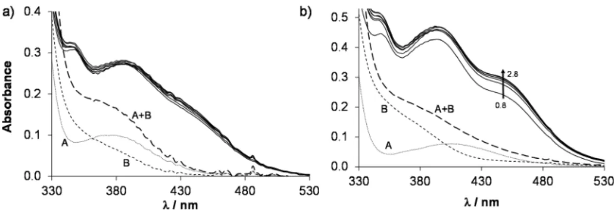
In order to determine the stability constants of the [M(arene) (L)] complexes, the UV-Vis spectra of acidic samples were recorded (pH = 0.8–2.8) to force the complex dissociation. The spectra obtained for the [Rh(η5-C5Me5)(H2O)3]2+–HQCl-Pro system were practically identical in the entire monitored pH range, however they are significantly different from the spectra of the unbound organometallic ion and ligand (Fig. 5a). It indicates that the complex formation is already complete at pH = 0.8, which hin- dered the calculation of the stability constant, and similarly no constants could be determined for the Ru(η6-arene) species (Fig. 5b). However, this approach was successfully used previously for the Rh(η5-C5Me5) and Ru(η6-p-cym) 8-hydroxyquinolinato com- plexes.23 In this work a logK [M(arene)(L)] = 16.45 ± 0.02 was obtained for the complex [Ru(η6-tol)(8-hydroxyquinolinato)(H2O)]+ based on the pH-dependent UV-Vis spectra (pH = 1.2–2.8, Fig. S18†) for comparison. Notably, this constant is slightly smaller than that of [Ru(η6-p-cym)(8-hydroxyquinolinato)(H2O)]+.23
Therefore, displacement reactions were performed to deter- mine the stability constants of these highly stable complexes.
First, ethylenediamine (en) was chosen as a competitor ligand.
It has no absorbance in the 200–800 nm wavelength range, which makes it an attractive choice for these studies. However, mixed-ligand complex formation was detected according to the
1H NMR spectra (Fig. S19†) hindering the constant determi- nation. The peaks belonging to the methyl hydrogens in the C5Me5moiety reflect the various chemical environments of the organometallic fragment.E.g.at an excess of 23 equivalents of ethylenediamine not only the peaks of binary complexes [Rh (η5-C5Me5)(en)(H2O)]2+and [Rh(η5-C5Me5)(L)(H2O)]+appear (L:
coordinated form of HQCl-Pro), but two unexpected peaks were also observed, signed with♠and♣in Fig. S19.†Addition of chloride ions decreased the ratio of these peaks (38% → 6%), which indicates that the Cl− and ethylenediamine compete for the third coordination site. Only ternary complex formation occurred in the case of the [Ru(η6-p-cym)(L)(H2O)]+ complex even at a huge excess (70 equivalents) of ethylenedia- mine, and at this highc(en)/c(HQCl-Pro) ratio ca.50% of Ru (η6-p-cym) is found in the mixed-ligand complex.
Fig. 4 The schematic representation of the mono complexes [M(arene) (L)(H2O)]+ formed with HQCl Pro and the various organometallic cations.
Fig. 5 UV-Vis absorption spectra (solid lines) of the (a) Rh(η5-C5Me5)–HQCl-Pro system at pH = 0.8–2.0 and of the (b) Ru(η6-p-cym)–HQCl–Pro system at pH = 0.8–2.8. The dotted curve shows the absorption spectrum of the free organometallic cation (A), the dashed curve (B) shows the spectrum of the free HQCl-Pro ligand and the long dashed curve (A + B) shows their additive spectrum. {c([Rh(η5-C5Me5)(H2O)3]2+) =c(HQCl-Pro, in Fig. 4a) = 60μM;c([Ru(η6-p-cym)(H2O)3]2+) =c(HQCl-Pro, in Fig. 4b) = 115μM;I= 0.20 M (KNO3);T= 25.0 °C;ℓ= 1 cm}.
Open Access Article. Published on 14 May 2020. Downloaded on 9/9/2020 8:21:42 AM. This article is licensed under a Creative Commons Attribution-NonCommercial 3.0 Unported Licence.
In the next step, 2-picolylamine ( pin) was selected and found to be an appropriate competitor in the case of the rhodium complex. Since pin has an intense ligand band in the UV region, only the use of1H NMR spectroscopy was helpful to determine the speciation. Therefore, 1H NMR spectra were recorded for the [Rh(η5-C5Me5)(H2O)3]2+–HQCl-Pro–pin ternary system at various complex-to-pin ratios (Fig. 6a). The spectra revealed that while the amount of free HQCl-Pro and [Rh(η5- C5Me5)( pin)(H2O)]2+ is increasing with the 2-pin excess, the amount of the [Rh(η5-C5Me5)(L)(H2O)]+complex is decreasing.
Based on the integrals of the peaks, fractions of the different compounds were calculated (Fig. 6b) and the stability constant was determined (Table 2) using the stability constant of [Rh (η5-C5Me5)( pin)(H2O)]2+taken from our previous work.31
However, in the case of [Ru(η6-arene)(H2O)3]2+ complexes the original yellow colour of the samples changed to pink and the peaks of the coordinated HQCl-Pro,p-cymene and toluene disappeared in the1H NMR spectra at an excess of the compe- titor 2-picolylamine ligand, which can be explained by arene loss and the probable oxidation of the Ru centre (Fig. S20†).
Thus, determination of the stability constants failed in these cases.
This side reaction aroused our interest as other competitor ligands (includinge.g.ligands from biofluids) may also cause arene loss and may affect bioactivity. Therefore, ligand displa- cement reactions were studied by UV-Vis spectroscopy. First the effect of HQCl-Pro itself was studied using different con- ditions. The addition of two equivalents of ligand to the complex [Ru(η6-p-cym)(L)(H2O)]+resulted in a too slow reaction at pH 2 to detect any changes during 1 h (while O2 passed through the solution). On the other hand, considerable changes of the spectra can be seen at physiological pH in Fig. 7a. The shape of the spectrum changes markedly, and a strong band developed at 420 nm, which is most probably related to the loss of the arene ligand and binding of the second and third HQCl-Pro. The rising broad band at higher wavelengths (λ> 500 nm) directly indicates the presence of the Ru(III) compound as it was found for [Ru(III)(8-hydroxyquino- late)3].32 Thus the change at 640 nm provides information about the rate of the Ru(III) complex formation (see Fig. 7b). In the literature the loss of arene (benzene, p-cymene) and the formation of [Ru(III)(L)3] were also found in the case of 8-hydroxyquinoline.23,32 As competitor ligands, deferiprone (1,2-dimethyl-3-hydroxy-pyridin-4(1H)-one) as an (O,O) model and 1,10-phenanthroline ( phen) as a representative of (N,N) bidentate ligands were chosen. Addition of deferiprone to the complex [Ru(η6-p-cym)(L)(H2O)]+ did not result in spectral changes, while the reaction with phen was undoubtedly fast, and there was a significant difference between the initial solu- tion (see the additive spectrum) and the spectrum recorded after 7 s for the mixed reactants (Fig. S21†). The yellow solu- tion turned red and a strong band developed with λmax = 502 nm. After the first reaction step seemed to be completed in∼90 s, another process started at∼150 s and the main band shifted to 440 nm. The sign of a tiny amount of Ru(III) was also Fig. 6 (a)1H NMR spectra of the [Rh(η5-C5Me5)(L)(H2O)]+–2-picolyla-
mine system recorded at variousc( pin)/c(HQCl-Pro) ratios (L is the co- ordinated form of HQCl-Pro). The aromatic region (with increased intensity) and C5Me5methyl protons are shown. Assignment:◆= [Rh (η5-C5Me5)(L)(H2O)]+; ♥ = [Rh(η5-C5Me5)( pin)(H2O)]+. (b) Fitted (solid line) and measured bound HQCl-Pro ratio values calculated from the integrals of HQCl-Pro (●) and C5Me5(▲). {c([Rh(η5-C5Me5)(L)(H2O)]+) = 100μM;c( pin) = 0.104–1.520 mM; solvent: 90% H2O/10% D2O; pH = 5.91 (20 mM phosphate buffer);T= 25.0 °C;I= 0.20 M (KNO3)}.
Table 2 Stability (K[M(arene)(L)]), proton dissociation (Ka[M(arene)(L)]) and water-chloride exchange constants (K’(H2O/Cl−)) of complexes of HQCl-Pro. {T= 25.0 °C;I= 0.20 M (KNO3)}
Ru(η6-p-cym) Ru(η6-tol) Rh(η5-C5Me5)
logK[M(arene)(L)] — — 13.41 ± 0.02a
pKa[M(arene)(L)]b 8.62 ± 0.04 8.45 ± 0.03 9.62 ± 0.04 logK′(H2O/Cl−)c 1.21 ± 0.01 1.09 ± 0.01 1.57 ± 0.01
aFor the [Rh(η5-C5Me5)(L)(H2O)]2++ pin⇌[Rh(η5-C5Me5)(pin)(H2O)]2+
+ L equilibrium determined at various c(pin) concentrations by 1H NMR spectroscopy. bDetermined by UV-Vis spectroscopy at pH 2.0–11.5.cpH = 5.50 (phosphate buffer). For the [M(arene)(L)(H2O)]++ Cl−⇌[M(arene)(L)Cl] + H2O equilibrium determined at various total chloride ion concentrations by UV-Vis spectrophotometry.
Open Access Article. Published on 14 May 2020. Downloaded on 9/9/2020 8:21:42 AM. This article is licensed under a Creative Commons Attribution-NonCommercial 3.0 Unported Licence.
observed at higher wavelengths (A(690 nm)∼0.05). When this experiment was repeated under argon, similar spectral changes were observed; however, the formation of Ru(III) could be successfully avoided. To explain the two main processes, the experiment was repeated with only 1 equiv. of phen under aerobic conditions (Fig. S22a†). The first step accompanied by the development of a band with 502 nm maximum was similar and was completed within 7 min (Fig. S22b†). Although the second process was different, most probably oxidation took place as the development of the strong band at 694 nm indi- cates (Fig. S22c†). Based on these findings the first step is most probably the loss ofp-cymene and the formation of the mixed-ligand complex [Ru(II)( phen)(L)(H2O)2]+(λmax= 502 nm, shown in gray rhombuses in Fig. S21b, S22b and c†). This is followed by the slow coordination of the second phen forming [Ru(II)( phen)2(L)]+when 2 equiv. of phen were provided, while the slow oxidation occurs without the second phen ligand forming [Ru(III)( phen)(L)(H2O)2]2+(shown in orange squares in Fig. S21b, S22b and c†).
Reactions of [Ru(η6-p-cym)(L)(H2O)]+ with various bio- molecules were tested as well. Addition of histidine (His),
human serum albumin (HSA) and RPMI 1640 medium com- ponents resulted in similar spectral changes (Fig. S23a, c and e†). We concluded that no Ru(III) was present in the systems, and most probably a mixed-ligand complex ([Ru(η6-p-cym)(L) (His)]+) is formed in the presence of histidine and the RPMI 1640 medium components (vide infra for NMR spectra).
Albumin is also able to bind the complex most probably through its side chains in a monodentate fashion due to the coordination of a histidine nitrogen, or cysteine thiolate or methionine thioether.33,34 The reaction with HSA was some- what slower under the same experimental setup (Fig. S23b, d and f†).
Based on these findings the following can be concluded: (i) the excess of rigid (O,O) donor bidentate ligands cannot cause the loss ofp-cymene; (ii) the excess of the rigid 8-hydroxyqui- nolate-type (N,O−) and rigid (N,N) donor bidentate ligands can cause liberation of the arene ligand followed by oxidation to Ru(III) at physiological pH in the presence of O2; (iii) the flex- ible (N,N) donor ethylenediamine and biologically available molecules (like histidine, amino acids of RPMI 1640 or human serum albumin) can readily react with the complex [Ru(η6-p- cym)(L)(H2O)]+ forming mixed-ligand complexes. A previously described class of half-sandwich complexes containing azopyr- idine ligands also showed arene-loss (even at 1 : 1 metal-to- ligand ratio), which was explained with the π-acceptor pro- perties of the bidentate ligand35 as the stronger π-acceptor bidentate ligands can compete with the arene.
Deprotonation of the coordinated water molecule and its exchange to the chloride ion in the [M(arene)(L)(H2O)]+ complexes
In the [M(arene)(L)(H2O)]+complexes (L: the coordinated form of HQCl-Pro) the coordinated aqua ligand can get deproto- nated with increasing pH and this process is reported to have an effect on the reactivity of the complex.36Namely, the hydro- xido ligand is considered as a worse leaving group than the water molecule or chloride ion, preventing the complex from interacting with biomolecules. To determine the pKavalues1H NMR spectra of the complexes were recorded in a wide pH range and representative spectra are shown for the titration of the [Ru(η6-p-cym)(L)(H2O)]+complex in Fig. 8 (and the spectra of the Ru(η6-tol) and Rh(η5-C5Me5) complexes are shown in Fig. S24 and S25†). These spectra reveal that peaks are shifted at pH > 6, and the chemical shifts draw a sigmoid curve as a function of pH (Fig. 8c).
From these sigmoid curves pKa [M(arene)(L)] values were determined (Table 2), which are much lower for the Ru con- taining complexes than for the Rh-complex. Notably, following the deprotonation the forming hydroxido complex exhibits more peaks than the aqua complex. Most probably the depro- tonation leads to the loss of the twofold symmetry of the p-cymene ligand and peaks are doubled. Due to the rigid struc- ture the rotation of thep-cymene ligand is blocked most likely.
Earlier, computational studies revealed that the rotation has a very low energy barrier in the ruthenium–arene complexes bearing 1,2-ethylenediamine (RAED),e.g.benzene completes a Fig. 7 Time-dependent UV-Vis absorption spectra of the [Ru(η6-p-
cym)(L)(H2O)]+–HQCl-Pro (1 : 2) system at pH = 7.40 in the presence of O2(L: the coordinated form of HQCl-Pro). The dashed curve shows the additive spectrum of the metal complex and 2 equivalents of the HQCl- Pro ligand. The insertedfigure shows absorbance values at 640 nm as a function of time. {c([Ru(η6-p-cym)(L)(H2O)]+) = 200μM;c(HQCl-Pro) = 400μM; pH = 7.40 (20 mM phosphate); c(KCl) = 0.10 M;T= 25.0 °C;ℓ
= 1 cm}.
Open Access Article. Published on 14 May 2020. Downloaded on 9/9/2020 8:21:42 AM. This article is licensed under a Creative Commons Attribution-NonCommercial 3.0 Unported Licence.
full rotation in 2 ps.37In that case, a steric interaction between HQCl-Pro, OH− and p-cymene can increase this rotational barrier.
Using the pKa [M(arene)(L)] values the ratio of the hydro- xido form of the complexes was calculated at physiological pH revealing the formation of [M(arene)(L)(OH)] in 6% for Ru(η6- p-cym), 8% for Ru(η6-tol) and less than 1% for the Rh(η5- C5Me5)-complex in the absence of chloride ions. pKa of this type of complexes increases with the chloride ion concentration,24,38 thus these percentages are merely maximum values. When the organometallic fragments are compared with each other, the same trend of the pKavalues was observed for complexes of (N,O) ligands such as 2-picoli- nates, HQ-5-sulfonate (HQS), HQ and 7-(1-piperidinylmethyl)- HQ (PHQ)23,38–40as Fig. 9 shows.
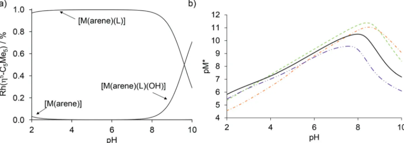
Based on the logKand pKa[M(arene)(L)] constants (Table 2) concentration distribution curves were calculated for the Rh(η5- C5Me5) complex of HQCl-Pro (Fig. 10a). A very small amount (<3%) of free organometallic ions appears at pH 2, while the for- mation of the [Rh(η5-C5Me5)(L)(H2O)]+complex is predominant at pH 7.4. The stability constants determined for the complexes of HQ derivatives cannot be compared directly due to the different basicity of ligands. For comparison, pM* (the negative logarithm of the unbound metal ion) values were calculated and plotted against the pH (Fig. 10b). (pM* is calculated by taking into con- sideration the hydrolyzed forms: pM* = −log([M(arene)] + 2×[(M(arene))2(OH)2] + 2×[(M(arene))2(OH)3]). The higher pM*
value indicates higher stability of the complex. In these calcu- lations, stability constants of the half-sandwich rhodium com- plexes with HQ, 8-hydroxyquinoline-5-sulfonate and 7-(1-piperidi- nyl-methyl)-8-hydroxyquinoline were used.23Although the HQCl- Pro complex has the highest stability among the others up to pH
∼3.5, at physiological pH the other complexes have somewhat higher stability.
Adding chloride ions to the solutions of the complexes causes changes in their1H NMR and UV-Vis spectra. This is the result of a third equilibrium process, shown in Scheme S1,†which is the exchange of the coordinated water
molecule to a chloride ion. Half-sandwich organorhodium and ruthenium complexes have a relatively high chloride ion affinity, and the chlorinated forms of the HQCl-Pro complexes are charge neutral. The chloride content of the medium has an effect on not only the charge, but also on the pKa[M(arene) (L)]24,38 and on the lipophilicity.39,41 This affinity is well described by the logK′ (H2O/Cl−) constant, which is deter- mined from the UV-Vis spectra of the complexes by varying the total concentrations of chloride ions (Fig. S27–S30† and Table 2).
The logK′(H2O/Cl−) constants show that the Rh(η5-C5Me5) complex has the highest, while [Ru(η6-tol)(L)(H2O)]+ has the lowest value (Table 2). The constants for [Ru(η6-arene)(L) (H2O)]+ complexes have tiny differences, and a similar trend was found previously in the case of 8-hydroxyquinoline, and Fig. 8 1H NMR spectra of the Ru(η6-p-cym)–HQCl-Pro 1 : 1 system recorded at pH = 5.6–10.9. (a) Aromatic protons; (b)p-cymene methyl protons.
(c) Measured (◆) andfitted (solid line) chemical shift values of one of the protons on thep-cymene ring as a function of pH. {c([Ru(η6-p-cym) (H2O)3]2+) =c(HQCl-Pro) = 300μM; solvent: 90% H2O/10% D2O;T= 25.0 °C;I= 0.20 M (KNO3)}.
Fig. 9 Comparison of pKa[M(arene)(L)] constants for the half-sandwich Ru- and Rh-complexes of (N,O) bidentate ligands (I= 0.20 M KNO3).
HL = pic (2-picolinic acid),38–40,a HQ (8-hydroxyquinoline),23,b HQS (8-hydroxyquinoline-5-sulfonate),23 PHQ (7-(1piperidinylmethyl)-8- hydroxyquinoline)23and HQCl-Pro.aI= 0.20 M KCl;bpKa([Ru(η6-tol)(8- hydroxyquinolinato)(H2O)]+) = 8.94(2) determined by UV-Vis spectro- photometric titration in this work, see Fig. S26.†
Open Access Article. Published on 14 May 2020. Downloaded on 9/9/2020 8:21:42 AM. This article is licensed under a Creative Commons Attribution-NonCommercial 3.0 Unported Licence.
Fig. S31† shows this tendency.23 The concentration of the chloride ion is around 100 mM in blood 24 mM in the cyto- plasm and 4 mM in the nucleus,42 and the actual concen- tration affects the ratio of the chloride and aqua complexes (Fig. 11). The lower thec(Cl−), the higher fraction of the aqua complex. According to the proposed activation mechanism by aquation42 the complexes are in their neutral (zwitterionic) chlorinated form in the blood serum at 100 mM chloride con- centration. The neutral chlorinated form may penetrate more easily through the cell membranes and might be trapped in the cytosol due to the lower chloride concentration and for- mation of charged aqua forms. While 79% of the rhodium complex is in the neutral form when thec(Cl−) is 100 mM, it drops to 47% and 13% at 24 mM and 4 mM chloride ion con- centrations, respectively (Fig. 11). The Ru(η6-arene) complexes show somewhat weaker chloride ion affinity.
Chloride concentration-dependence of the lipophilicity of HQCl-Pro and its complexes
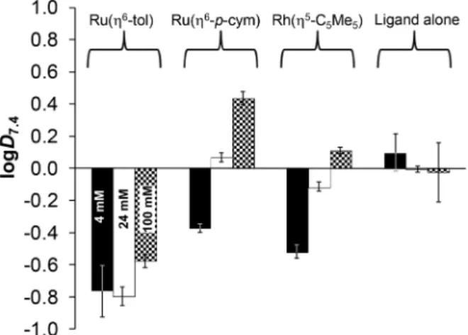
The conventional shake-flask method was used to determine the chloride-dependent lipophilicity of the ligand and its three organometallic complexes at physiological pH. Compounds were dissolved in 20 mM phosphate buffer ( pH = 7.4, saturated withn-octanol), then mixed withn-octanol (saturated with the aqueous buffer). The buffers contained chloride ions in different concentrations related to the different biofluids (4, 24, 100 mM). Distribution coefficients (logD7.4) calculated from UV-Vis quantitative analysis are shown in Fig. 12. The ligand lipophilicity slightly decreases with increasing chloride ion concentration, which is the result of the stronger ioniza- tion effect at higher ionic strength.
The actual chemical form of the studied organometallic complexes strongly depends on the chloride ion affinity and concentration, as was described previously. Fig. 11 clarifies the ratios of the neutral chlorinated and the positively charged aquated forms at pH 7.4. Based on the data in Fig. 12 it can be concluded that the most lipophilic complex is [Ru(η6-p-cym)(L) (H2O/Cl)]+/0at 100 mM of Cl−, and the most hydrophilic is [Ru (η6-tol)(L)(H2O/Cl)]+/0 at 4–24 mM of Cl−. Here the trend of lipophilicity is different from the trend of the logK′(H2O/Cl−) values: Ru(η6-tol) < Rh(η5-C5Me5) < Ru(η6-p-cym). Although the rhodium complex has the highest chloride ion affinity, other factors also affect the lipophilicity such as the higher charge of the Rh-center (+3) (vs.the +2 charge of Ru) and the negative charge of the C5Me5ligand (vs.neutral toluene/p-cymene).
In vitrocytotoxicity and antiproliferative activity
measurements of HQCl-Pro and its complexes on cancer cell lines
In order to characterize the anticancer effect of HQCl-Pro and to monitor whether the complexation with organometallic triaqua cations can affect the cytotoxic activity of the ligand Fig. 10 (a) Calculated concentration distribution curves of the [Rh(η5-C5Me5)(H2O)3]2+–HQCl-Pro system based on the stability constants from Table 2. (b) Calculated pM* curves of the Rh(η5-C5Me5)–HQ (-·-),238-hydroxyquinoline-5-sulfonate (–),237-(1-piperidinylmethyl)-8-hydroxyquino- line (-··-)23and HQCl-Pro systems (solid line), pM* =−log([M(arene)] + 2 × [(M(arene))2(OH)2] + 2 × [(M(arene))2(OH)3]). {c([Rh(η5-C5Me5)(H2O)3]2+) = c(HQCl-Pro) = 50 µM;T= 25.0 °C;I= 0.20 M (KNO3)}.
Fig. 11 The calculated ratio of aquated ([M(arene)(L)(H2O)]+) (white) and chlorinated ([M(arene)(L)Cl]) (gray) forms of the HQCl-Pro com- plexes at different chloride concentrations of modelling biofluids, based on the constants in Table 2. {c([M(arene)(H2O)3]2+) = c(HQCl-Pro) = 100 µM;c(Cl−) = 4, 24 and 100 mM;T= 25.0 °C}.
Open Access Article. Published on 14 May 2020. Downloaded on 9/9/2020 8:21:42 AM. This article is licensed under a Creative Commons Attribution-NonCommercial 3.0 Unported Licence.
itself, thiazolyl blue tetrazolium bromide (MTT) assays were performed in two human colonic adenocarcinoma cell lines under two kinds of setup. Colo 205 is a drug sensitive cell line, while Colo 320 is multidrug resistant being primarily mediated by the overexpression of P-gp. Earlier studies showed that the reference compound HQ and its organometallic Ru- and Rh- complexes are cytotoxic in MES-SA and MES-SA/Dx5 cancer cell lines (IC50= 1–13μM).23The structurally related 7-(1-piperidi- nylmethyl)-8-hydroxyquinoline and its complexes were found to be cytotoxic (IC50= 0.6–20μM), however the ligand and its Rh(η5-C5Me5) complex showed collateral sensitivity (with selectivity ratios 5.8 and 5.1, respectively).
Inhibiting the proliferation and causing cell death may occur at the same time when the drug is administered to the cells. Using a lower number of cells per well (6 × 10³) and a longer incubation time (72 h) the MTT assay provides more information about the activity of the complexes to inhibit cell
proliferation rather than growth inhibition. On the other hand, in the case of a higher number of cells per well (6 × 10⁴) and a shorter exposure time (24 h) it is possible to monitor preferably the cytotoxic effect. IC50values collected for HQCl- Pro in the absence and in the presence of [Rh(η5-C5Me5) (H2O)3]2+, [Ru(η6-p-cym)(H2O)3]2+and [Ru(η6-tol)(H2O)3]2+and for the precursor dimers [M(η5/6-arene)(μ-Cl)Cl]2are shown in Table 3. Doxorubicin and cisplatin were used as positive controls.
The organometallic precursors have no toxic effect on cancer cell lines (IC50 > 100 μM). HQCl-Pro and its Rh(η5- C5Me5) complex exhibited similar and relatively low IC50
values, while in the presence of Ru(η6-tol) and especially Ru (η6-p-cym) much higher values were obtained. Additionally, HQCl-Pro and its Rh(η5-C5Me5) complex showed higher anti- cancer activity in Colo 320 than in Colo 205. In contrast the toluene and p-cymene complexes have similar cytotoxicity in both cell lines and the antiproliferative effect is weaker in the Colo 320 cells. These data do not correlate with the lipophilicity of the complexes, suggesting that other factors seem to be more dominant in the bioactivity. Notably, the complexes have similar or weaker cytotoxicity compared to cisplatin.
Reaction with cell culture medium components
To find an answer to the decreased activity of the Ru(η6-p-cym) complex interaction of the complex [Ru(η6-p-cym)(L)(H2O)]+ with the RPMI 1640 cell culture medium was also investigated by1H NMR spectroscopy (Fig. 13). Previously we described the interaction (vide suprafor kinetics), which was followed spec- trophotometrically and no oxidation was detected; only mixed ligand complex formation was observed. This medium con- tains various amino acids and other small biomolecules which may coordinate to the metal center leading toe.g.the ( partial) release of the original ligand or the formation of mixed ligand species. According to the recorded 1H NMR spectra the free HQCl-Pro has different spectra in RPMI 1640 medium and in PBS′ (Fig. 13(3 and 4)); namely the shape of doublets are blurry, only the envelope type peak can be seen and peaks are Fig. 12 n-Octanol/water distribution coefficients at pH = 7.4 (logD7.4)
for complexes [Ru(η6-tol)(L)(H2O)]+, [Ru(η6-p-cym)(L)(H2O)]+, [Rh(η5- C5Me5)(L)(H2O)]+and the ligand HQCl-Pro at different chloride ion con- centrations: 4 mM (black), 24 mM (white) and 100 mM (checkered). {c- (compounds) = 200 µM; pH = 7.40 (20 mM phosphate buffer); T= 25.0 °C}.
Table 3 In vitroantiproliferative (72 h) and cytotoxic effects (24 h) (IC50values inμM) of HQCl-Pro and its complexes in addition to the organo- metallic precursors in sensitive (Colo 205) and multidrug resistant (Colo 320) human colonic adenocarcinoma cell lines
Antiproliferative effect Cytotoxic effect
Colo 205 Colo 320 Colo 205 Colo 320
HQCl-Pro 23.4 ± 3.3 8.5 ± 1.7 42.5 ± 7.4 17.4 ± 2.5
[Ru(η6-tol)(L)(H2O)]+ 25.3 ± 3.1 85.0± 7.5 72.6 ± 4.8 60.9 ± 8.2
[Ru(η6-p-cym)(L)(H2O)]+ 68.3 ± 10.7 >100 >100 >100
[Rh(η5-C5Me5)(L)(H2O)]+ 25.8 ± 4.8 9.7 ± 1.1 81.5 ± 3.3 24.1 ± 3.7
[Ru(η6-p-cym)(μ-Cl)Cl]2 >100 >100 >100 >100
[Ru(η6-tol)(μ-Cl)Cl]2 >100 >100 >100 >100
[Rh(η5-C5Me5)(μ-Cl)Cl]2 >100 >100 >100 >100
Doxorubicin 3.28 ± 0.22 3.12 ± 0.27 1.56 ± 0.03 6.45 ± 0.19
Cisplatin 10.1 ± 0.3 4.78 ± 0.11 83.9 ± 3.5a 18.1 ± 0.3a
a1 × 104cells were used for cisplatin.
Open Access Article. Published on 14 May 2020. Downloaded on 9/9/2020 8:21:42 AM. This article is licensed under a Creative Commons Attribution-NonCommercial 3.0 Unported Licence.
![Table 2 Stability ( K [M(arene)(L)]), proton dissociation ( K a [M(arene)(L)]) and water-chloride exchange constants ( K’ (H 2 O/Cl − )) of complexes of HQCl-Pro](https://thumb-eu.123doks.com/thumbv2/9dokorg/970598.57941/7.892.454.830.130.205/table-stability-proton-dissociation-chloride-exchange-constants-complexes.webp)
![Fig. 9 Comparison of p K a [M(arene)(L)] constants for the half-sandwich Ru- and Rh-complexes of (N,O) bidentate ligands ( I = 0.20 M KNO 3 ).](https://thumb-eu.123doks.com/thumbv2/9dokorg/970598.57941/9.892.179.719.72.291/fig-comparison-arene-constants-sandwich-complexes-bidentate-ligands.webp)
