Compactness of Protein Folds Alters Disulfide-Bond Reducibility by Three Orders of Magnitude: A
Comprehensive Kinetic Case Study on the Reduction of Differently Sized Tryptophan Cage Model Proteins
D#niel Horv#th,
[a]Njra Taricska,
[a]Erno˝ Keszei,
[b]P#l Str#ner,
[a]Viktor Farkas,
[a]G#bor K. Tjth,
[c]and Andr#s Perczel*
[a]Introduction
Structural disulfide (SS) bonds, which are stable in the harsh oxidative extracellular environment, maintain the native fold of proteins by fixing and protecting them from thermal fluctua- tion induced by elevated internal dynamics. The SS bond for- mation is perhaps the most fundamental post-translational modification that stabilizes the 3D fold of globular proteins.
The absence of regulated SS formation leads to diseases in- cluding diabetes,[1] cancer,[2] neurodegenerative conditions,[3]
and cardiovascular diseases.[4] Non-native SS bond pairing evokes backbone misfolding, which jeopardizes both function and bioactivity, although some proteins may present alterna-
tive SS states and still achieve similarly well-folded forms.[5]In protein evolution, the presence of SS bonds shows a signifi- cant correlation with the complexity of the organism.[6] Ap- proximately 50% of all cysteine residues found in proteins form SS bonds,[7]and thus, these cysteine residues become the most conserved among all amino acids, despite being added late to the genetic code during protein evolution.[8]Due to the unique pairing pattern of cysteine residues, SS bonds stabilize the 3D fold of proteins unambiguously.[9]
Contrary to structural disulfides, redox-active disulfides are highly dynamic, and their formation is reversible. The redox potential of the surrounding environment controls the regula- tion and cellular localization of these proteins.[10]Intramolecular formation of these redox-active disulfides is common for oxi- doreductases (thioredoxin[11] or glutaredoxin[12] family) and allosteric disulfides,[13–15]whereas an intermolecular SS linkage results in glutathionylated[16] or cysteinylated[17] small mole- cule–protein adducts. The redox potential and stability of the SS bond is highly dependent on several factors, such as the pKa of the thiols (the standard pKa is 8.5, but this can range from 3.5 to 12.8, depending on the local environment),[18] the strain introduced by the SS bond of the protein structure, and the entropic cost of SS bond formation.[19,20]The Cys residues of an SS bond are typically distant in the primary sequence;
49% of the SS-bond-forming cysteine residues are more than 25 residues apart from each other.[21]The SS-bond formation is thermodynamically more favorable if the cysteine residues are placed in spatial vicinity by the native fold itself before oxida- tion,[22]otherwise—in the absence of chaperones assisting fold- ing[23]—the protein precipitates. Adjacent cysteine residues oxi- A new approach to monitor disulfide-bond reduction in the vi-
cinity of aromatic cluster(s) has been derived by using the near-UV range (l=266–293 nm) of electronic circular dichro- ism (ECD) spectra. By combining the results from NMR and ECD spectroscopy, the 3D fold characteristics and associated reduction rate constants (k) of E19_SS, which is a highly ther- mostable, disulfide-bond reinforced 39-amino acid long exena- tide mimetic, and its N-terminally truncated derivatives have been determined under different experimental conditions.
Single disulfide bond reduction of the E19_SS model (with an 18-fold excess of tris(2-carboxyethyl)phosphine, pH 7, 378C) takes hours, which is 20–30 times longer than that expected, and thus, would not reach completion by applying commonly used reduction protocols. It is found that structural, steric, and electrostatic factors influence the reduction rate, resulting in orders of magnitude differences in reduction half-lives (900>
t1=2>1 min) even for structurally similar, well-folded derivatives of a small model protein.
[a]D. Horv#th, N. Taricska, Dr. P. Str#ner, Dr. V. Farkas, Prof. Dr. A. Perczel Laboratory of Structural Chemistry and Biology and
MTA-ELTE Protein Modeling Research Group at the Institute of Chemistry Eçtvçs Lor#nd University
112, P.O. Box 32, 1518 Budapest (Hungary) E-mail: perczel@chem.elte.hu
[b]Prof. Dr. E. Keszei
Chemical Kinetics Laboratory, Institute of Chemistry Eçtvçs Lor#nd University
112, P.O. Box 32, 1518 Budapest (Hungary) [c] Prof. Dr. G. K. Tjth
Department of Medical Chemistry
Faculty of General Medicine, University of Szeged Szeged Djm t8r 8, H-6720 Szeged (Hungary)
Supporting information and the ORCID identification numbers for the authors of this article can be found under https://doi.org/10.1002/
cbic.201900470.
T 2019 The Authors. Published by Wiley-VCH Verlag GmbH & Co. KGaA.
This is an open access article under the terms of the Creative Commons At- tribution License, which permits use, distribution and reproduction in any medium, provided the original work is properly cited.
dized to a SS bond are rare, although examples can be found among enzymes, receptors, and toxins.[24,25]The SS bond or SS bond pattern in prokaryotic proteins is formulated by riboso- mal mRNA translation, followed by oxidation and post-transla- tional modifications catalyzed by various enzymes located in the periplasm (DsbC, DsbG, DsbD)[26] or cytoplasm (DsbA, DsbB).[27,28] In eukaryotic species, this process is performed in specific cell organelles, such as the mitochondria (Mia40, ERV1), endoplasmic reticulum (PDI, ERO1, Erv2), and chloro- plasts (PSI, PSII, LTO1, LQY1, CYO1).[29]
The SS bonds form the core of hundreds of proteins of known 3D structures. Hydrophobic and/or aromatic residues (e.g., Trp and Tyr) may condense around the SS bond and form the network of key interactions that determine the 3D structure of a large number of different proteins.[21]In at least 50% of protein families, this type of interaction is invariant. In dozens of proteins (e.g., tick anticoagulant peptide,[30] phos- pholipase A2[31]) the SS unit(s) are reinforced by associated aro- matic–aromatic interactions[32,33] and vice versa. For instance, 92Tyr of RNase-A effectively shields the solvent-exposed nearby SS bond (40Cys–95Cys) from reducing agents (RAs), and thus, helps to maintain the native fold of the protein.[34]
If SS bonds are reduced, the thiol groups of the free cysteine residues often adopt an ensemble of local conformers that also loosen the compactness of neighboring residues. In the era of manufacturing recombinant proteins (e.g., insulin), the SS bond cyclized peptides (e.g., vasopressin, oxytocin, desmo- pressin, octreotide)[35] and human monoclonal IgG antibod- ies,[36]which are produced on a large scale by the biopharma- ceutical industry, it is vital to have reliable and fully tested methods for SS bond reduction.
In addition to b-mercaptoethanol or 1,4-dithio-d-threitol (DTT), more recently tris(2-carboxyethyl)phosphine (TCEP) has become commonly used as a RA of SS bonds because it is chemically more stable, nonvolatile, odorless, and it reduces SS bonds more effectively, even at low pH.[37,38]TCEP is claimed to selectively and completely reduce water-soluble alkyl disulfides over a wide pH range within a few minutes (<5 min).[39]Some protocols recommend using 1–100 molar equivalents of TCEP relative to protein concentration.[40,41]The reduction time and appropriate temperature greatly depend on the nature of the protein, but, generally, elevated temperature and/or TCEP[42]
concentration and longer times make the reduction more com- plete, but these conditions also initiate a multitude of side re- actions, which are poorly described, to date.
Exendin-4[43]or exenatide[44](synthetic name), which as been used in clinical practice since 2005, is an incretin mimetic[45]
glucagon-like peptide-1 (GLP-1) analogue, which is a 39-resi- due peptide with complex physiological actions[46]in multiple organs, used in the treatment of type 2 diabetes mellitus.[47]Ex- enatide acts as an agonist of the GLP-1 receptor[48](GLP-1R). Its amphipathic helix binds to the extracellular domain of the GLP-1R, the mainly unstructured N terminus activates the re- ceptor,[49] and the structure-stabilizing Trp-cage[50,51]fold is not directly involved in interactions to GLP-1R.[52,53] We have syn- thesized and studied the 3D fold of several dozens of Trp-cage folds, including analogues of exenatide, such as E19,[54,55]
which is a 39 amino acid protein of comparable bioactivity, but improved water solubility. As a “natural tool” for enhancing the compactness of the 3D fold, we introduced two solvent- exposed Cys residues into E19, making E19_SS (Figure 1), and
a loop from residues 18 to 39 in E19_A18C_S39C (E19_2SH).
E19_2SH oxidized to E19_SS spontaneously with atmospheric O2 dissolved in water at room temperature. The SS bond of E19_SS extends the hydrophobic core of the native Trp fold in the spatial proximity of 22Tyr, which is surrounded by explicit negative charges (15Glu, 16Glu, 17Glu). Although E19_SS is small in size, (MW: 4334.9 gmol@1), its reduction takes several hours to reach equilibrium with>10 molar excess of TCEP in water at room temperature. Because the SS bond reduction time turned out to be significantly longer than that expected based on literature data and common laboratory practice, we launched a comparative study, including three designed and truncated analogues of E19_SS, namely, E11_SS, E5_SS, and E2_SS. Notably, the model systems thus created (Figure 1) oxi- dize spontaneously and rapidly adopt the Trp-cage 3D fold.[56]
Moreover, the “loop size” created by the SS bond, in other words, the number of residues between the two reacting cys- teine residues, is 20 amino acids long, which is close to the average value (&17) observed in thousands of proteins.[57]
E11_SS was designed by removing the “HGEGTFTS” tail, which was the unstructured GLP-1R-activating N-terminal eight residues of E19_SS. Shortening by an additional six residues removes the outer helical part of E19_SS, namely, the
“HGEGTFTS-DLSKQM” subunit,[56] affording E5_SS. Although 14 residues shorter than that of E19_SS, E5_SS still adopts a com- pact Trp-cage fold and comprises the entire interface for bind- Figure 1.A) Structure ensemble of E19_SS, and B) amino acid sequences of GLP-1; exenatide; parent E19 and its truncated derivatives E11, E5, and E2;
their SS analogues E19_SS, E11_SS, E5_SS, and E2_SS; and their reduced 2SH analogues E19_2SH, E11_2SH, E5_2SH, and E2_2SH. The position of the SS bridge is highlighted by stick representation and underlined as C. The se- quences of E19 is divided into six major parts: 1) 2–8 unstructured N termi- nus, 2) 9–14 outer helix, 3) 15–17 kink region, 4) 18–27 inner helix, 5) 28–34 310helix, and 6) 35–39 polyproline region. This apportionment of the se- quence coincides with the truncation of the peptides.
ing to GLP-1R.[52] Finally, in E2_SS, the entire N terminus pre- ceding 18Cys of E19_SS was omitted, namely, “HGEGTFTS- DLSKQ-EEE” was cleaved, to give a folded protein with a fully exposed SS bond at its surface; this is considered to be a con- struct ready for a rapid SS bond reduction (Figure 1).
Herein, we discuss the structure and properties of both the oxidized and reduced forms of the four model proteins of dif- ferent a-helical lengths, in comparison with the parent (Cys- free) miniproteins, and the kinetics of reduction. We introduce spectroscopic approaches that make the monitoring of the re- duction progress fast and easy. The effect of the compactness of the protein fold, the accessibility, and the local explicit charges of the SS bond and the reagent type on reduction rate and the mechanism are also explained herein.
Results and Discussion
Three-dimensional fold characterized by far-UV electronic circular dichroism (FUV-ECD) spectra
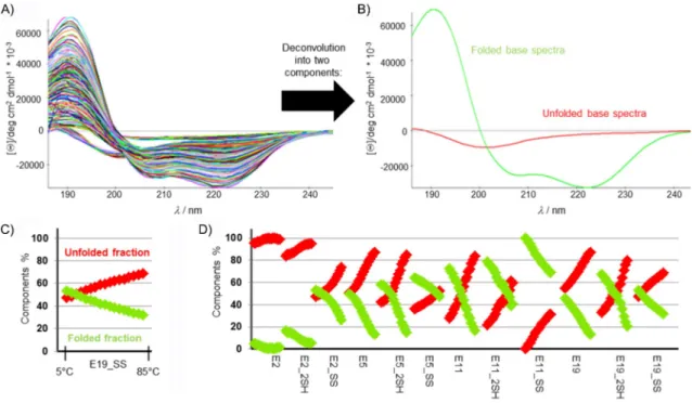
Circular dichroism (CD) spectroscopy is increasingly recognized as very sensitive indicator of protein conformation,[58,59]relying on a plethora of electronic transitions. The FUV-ECD spectra of Trp-cage proteins (e.g., Exenatide, E19, E19_SS) are typically the weighted sums of the C- (folded and highly helical) and U- type (unfolded) base curves (Figure 2A), as assigned and veri- fied by means of NMR spectroscopy.[60,61] As the temperature increases, the shape of the FUV-ECD spectra changes: those of the parent proteins—E2, E5, E11, and E19—acquire more and more U-type characteristics, as they unfold gradually. The tem-
perature-dependent FUV-ECD spectra for all four SS bond en- forced model peptides were recorded between 5 and 858C (in steps of 58C, resulting in 17 spectra for each protein; Figure S1 in the Supporting Information). Aside from E2_SS, the SS- bond-containing mutants have similar FUV-ECD spectra to that of their parent proteins at low temperatures. On the other hand, because the SS bond makes the 3D folds of SS variants more rigid, they preserve their C-type characteristics better and delay unfolding, even at higher temperatures. Once the SS bond is reduced (see below for details), the spectral properties of the SH variants revert to those of the parent proteins. Their 3D-scaffold compactness decreases as the temperature increas- es; this is less apparent in the case of E2 and E2_2SH because they both already present an ensemble of dynamic backbone structures at 58C.
Ensemble deconvolution[62, 63]of the 204 (12V17) ECD spec- tra,f(l,T), made the quantitative analysis of the relative abun- dance of secondary structural elements belonging to each peptide in each state possible because the pure ECD curves were successfully assigned.[60,64–66]The results in Figure 2B indi- cate that 1) the SS bond stabilizes the less folded protein scaf- folds more effectively, for example, whereas the difference at 48C between the E2 and E2_SS folded fraction is 48%, the same difference between E5 and E5_SS is 14%; in the case of E11 and E11_SS, it is only 28 %, and for E19 and E19_SS it is 7% (Figure 2D). 2) The ratio of the folded, helical components increases upon going from E2 to E5 and E11; however, the compact a-helical content of E19_SS, E19_2SH, and E19 is lower than those of E11_SS, E11_2SH, and E11 because the un- folded eight-residue-long N-terminal part elevates the overall
Figure 2.A) Temperature-dependent FUV-ECD spectra (204 in total) of the four primal peptides (E2, E5, E11, and E19) and their four reduced (_2SH) and four oxidized (_SS) variants. B) The two pure ECD curves were derived from the ensemble analysis of the 204 ECD spectra by using CCA+. Pure component 1 (red) represents that of the unfolded/U-type, whereas component 2 (green) represents the folded/C-type backbone structure. C) The associated relative pro- pensities [%] of the two pure components at each measured temperature are given for E19_SS as an example, D) as well for each 204 spectra starting from E2 (at 58C) up to E19_SS (at 858C).
backbone dynamics, which destabilizes the compact 3D fold.
3) All four reduced proteins (E2_2SH, E5_2SH, E11_2SH, E19_
2SH) have a higher helix content (&7–15%) than that of the parent proteins over the entire temperature range. 4) The 3D folds stabilized by SS bonds are less sensitive to temperature (Figure 2C, D).
Three-dimensional folds of proteins determined and characterized by NMR spectroscopy
The ensemble of the temperature-dependent FUV-ECD spectra confirms that SS bonds preserve the fold of the model proteins and increase thermostability. NMR spectroscopy analysis at 158C allowed further characterization of the 3D structures of each variant. Fold, chemical shift, and secondary chemical shift (SCS) information[67] were derived from the appropriate 2D homonuclear NMR spectroscopy experiments (1H,1H COSY,
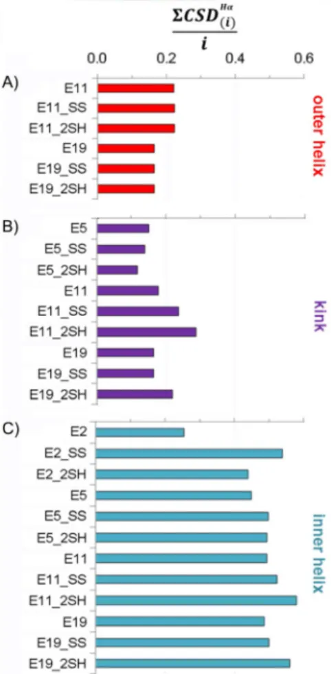
1H,1H TOCSY, and 1H,1H NOESY) at T=158C; the ensemble of the ten lowest energy structures were analyzed. This compre- hensive analysis conducted at 158C provided the following useful structural descriptors: the root-mean-square deviation (RMSD) of the 3D structures, the average chemical-shift devia- tion (CSD) of backbone Haprotons per residue ([8CSDHaðiÞ]/i) in the helical segment and the compactness of the Trp-cage core by SCS sum of selected protons: CSDcage(Table 1).
A comparison of the helices of different lengths is more straightforward if the helical segment is divided into three parts: 1) the outera-helix, 2) the kink region in the vicinity of the SS bond, and 3) the innera-helix (Figure 3).
The outer helix compactness seems to be affected by the length of thea-helix. Interestingly, this part of E11 variants is slightly more structured, in spite of being the terminal part
(usually flexible and unstructured), as opposed to the outer helices of the E19 variants, where this helical segment is flanked (Figure 3A). The above tendency is also true for the kink region, but here the presence and state (_SS or _2SH) of the SS bond are also differentiated (Figure 3B). These distant helical parts have generally lower [8CSDHaðiÞ]/ivalues than those of the inner helix. The compactness of the inner helices is simi- lar (expect for E2). Interestingly, reduced longer polypeptides show slightly increased [8CSDHaðiÞ]/i values, which may be the indicative of ring tension in the SS bond cyclized variants in these systems (Figure 3C).
1H NMR spectroscopy studies also confirm that all model proteins, except E2, have a common, compact, and folded Trp- cage core structure atT=158C (Tables 1 and S1 and Figure S2), regardless of the differently structured tails attached to them (Table 1). E2 is predominantly unfolded, even at low tempera- ture (158C), but because the SS bond joins together the N and C termini of E2_SS, the hydrophobic core folds properly. Inter- estingly, even E2_2SH forms a more compact Trp cage than that of E2. In agreement with data reported in the literature, cysteine residues promote and stabilizea-helices, if located at their N termini.[68]The cage values of the longer reduced pep- tides are close to that of their oxidized counterparts (Table 1).
Table 1.Selected measures characterizing the degree of folding of the model protein (T=158C and pH 7).
Degree of the fold
by FUV-ECD [%][a,b] Backbone
RMSD [a][c] CSDcage [8CSDHaðiÞ]/i
E19 _SS 51.6 0.7 11.7 0.5
_SH 65.8 0.7 11.1 0.6
parent 43.7 1.2 10.9 0.5
E11 _SS 96.0 0.3 11.5 0.5
_SH 76.0 0.3 11.0 0.6
parent 70.0 1.5 10.9 0.5
E5 _SS 63.4 0.2 11.4 0.5
_SH 42.8 0.1 10.4 0.5
parent 51.5 1.6 10.3 0.4
E2 SS 51.8 0.1 11.3 0.5
_SH 14.8 0.3 9.6 0.4
parent 3.7 1.5 3.8 0.3
[a]T=158C,c(protein)=20–30mmat pH&7 (typical conditions applied for CD measurements). [b] Calculated % from the joint deconvolution (CCA+) of 204 T-dependent FUV-ECD spectra (Figure 2). [c]T=158C, c(protein)=0.8–1.8 mm at pH&7 (typical conditions applied for 1H NMR spectroscopy measurements). [d] RMSD of all backbone atoms of the ten best structures. [e] Xf-cage values[51,55]were used to correlate the fold of the protein. The following “H” atoms were involved in calculations:
W25He1, L26Ha, G30Ha2, P31Hb2, R35Ha, P37Ha, P37Hb2, P38Hd1, and P38Hd2. [f] The average CSD of backbone Haprotons per residue.
Figure 3.The average CSD of backbone Haprotons per residue ([8CSDHaðiÞ]/i) in the three different helical regions: A) outer helix (i=6), B) kink region (i=3), and C) inner helix (i=10). Higher residual values imply a more struc- tureda-helix.
NMR spectroscopy data reveal that a longera-helix results in a more structured Trp cage, in all cases studied.
In general, it seems that the core of the reduced (SH@) pro- teins is almost as well folded as those that are SS bonded. The following 3D fold compactness order was established:
CSDSScage>CSD2SHcage>CSDparentcage , but the differences are small, aside from those of E2 (CSDE2cage=3.8)!E2_SS (CSDE2 SScage =11.3).
Oxidized and reduced states defined by near-UV (NUV) ECD data
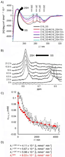
As shown above, reduction does not have a dramatic effect on the tertiary structure content of the model systems at room temperature; thus, to detect reduction, NUV-ECD spectra (in- stead of FUV) had to be used. The interpretation of the changes to the observed chiroptical properties of the Trp/Tyr/
SS!Trp/Tyr/2SH (Figure 4A) complex chromophore system is less straightforward because the assignment of “pure” NUV- ECD spectra has not yet been completed. The conformation- dependent fine structure of Tyr/Trp chromophores[60] (260, l,320 nm) comprises the 1Lb of Tyr (l&276 nm, with a shoulder at l&287 nm), 1Lb of Trp (l&281 and &293 nm), and 1La of Trp transitions appearing as superimposed broad bands. In addition, the SS bond may also contribute in form of a relatively weak but broad band with a maximum atl&260–
270 nm. For the current proteins with SS bonds, a larger nega- tive band was recorded (Figure S3). The bands of Trp and Tyr in the SS-bond-containing proteins shifted to the negative ellipticity range, which was not observed in the case of the parent proteins (E2, E5, E11, E19),[60] for which the bands of these amino acids were detected in the positive range (except the Trp band at l&293 nm). The reduction kinetics of E19_
SS!E19_2SH were monitored over time as the band intensi- ties at l&266, 281, 287, and 295 nm increased from larger negative to smaller negative and/or positive values, similar to those of the parent proteins (Figures 4A and S3). We were en- couraged to use NUV-ECD spectral changes to monitor SS to SH reduction in proteins if embedded in a suitable molecular environment such as that of the Trp cage motif.
Due to the acidic nature of TCEP, to avoid any pH shift, a phosphate buffer (50 mm, pH 7) was used to sustain near- physiological pH. The groups of Han[39] and Whitesides[37] de- scribed the chemical instability of TCEP above pH 7 in 300–
400 mmphosphate buffer. They found that the autoxidation of TCEP depended on how the reagent was stored (open air or in capped vials), whether the solution is stirred, and on the elapsed time (24,t,72 h) during storage. However, herein, we have monitored TCEP stability by means of 31P NMR spec- troscopy in 50 mmphosphate buffer, and found no significant spectral changes connected to TCEP oxidation or degradation at room temperature over 14 days.
Reduction of the E19_SS protein was followed by recording NUV-ECD spectra (&0.113 mm E19_SS, pH 7, 158C, 18-fold excess of TCEP, cell length=10 mm) at four different wave- lengths (266, 281, 287, and 293 nm). Thus, by following band intensity changes of selected (one or more)1Lb transitions of Tyr or Trp, we could monitor the redox state of the SS/SH
groups and determine the “end point” as a steady state. Thus, if a suitable aromatic residue (Tyr, Trp, Phe) is coupled to the SS bond as a chromophore, it enables its reduction/oxidation state to be monitored, even if the molecular system shows no coupled backbone conformational changes (CSDE19 SScage =11.66;
CSDE19 2SHcage =11.07). The measured absorbance was converted into concentration by using Equation (1):
Figure 4.A) NUV-ECD spectral changes measured for the reduction of E19_
SS (&0.113 mmE19_SS, pH 7, 158C, 18-fold excess of TCEP) at four different wavelengths (l=266, 281, 287, and 293 nm). No spectral changes were ob- served after about 55 h (3300 min). B)1H NMR spectra of E19_SS!E19_2SH reduction (c&0.115 mm, pH 7, 158C, 18-fold excess of TCEP) in water. The chemical shift of the indole He1 of Trp25 was used to monitor reduction:
He1 shifted upfield fromd=9.60 (SS) to 9.78 ppm (2SH during the reduc- tion). Reaching steady state, the integral ratio of E19_2SH and E19_SS was found to be 92 to 8%. C) Concentration change of E19_SS [mm] measured during reduction by different approaches plotted as a function of time.
D) The calculated rate constants (see the discussion of modeling reduction kinetics).
cðtÞ ¼ A1@A
A1@A0½SSA0 ð1Þ
Steady state was reached conclusively after about 55 h. We determined the rate constant, k1, at each wavelength by pa- rameter estimation to be kl¼266 nm1 =4.11V10@4Lmmol@1min@1, kl¼281 nm1 =5.67V10@4Lmmol@1min@1, and kl¼287 nm1 =5.98 V 10@4Lmmol@1min@1 (Figures 4C and S4). The deviations of the fitted model from the measured data at l=293 nm were re- markably large; therefore, parameter estimation was not per- formed on this dataset. NUV-ECD monitoring enables one to observe the clean and clear changes in the spectra, but it does not make it possible to extract the absolute value of the con- centration, [SS]1, at the end point of the reaction. Based only on the intensity of the molar ellipticity, it cannot be decided if reduction is fully completed or not. To ascertain the absolute values of the concentrations in the redox system, reduction was repeated under the same conditions in NMR tubes with a diameter (Ø) of 5 mm (&0.113 mmE19_SS, pH 7, 158C, 18-fold TCEP) by recording 1H NMR resonances (Figure 4B). By using both SS and SH state integrals of the signals at selected reso- nance frequencies (e.g., HeTrp), 1H NMR spectroscopy driven quantitative analysis of the reduction was performed (Fig- ure 3E) and the rate constant was determined to be kNMR1 = 8.03V10@4Lmmol@1min@1. Although 18-fold excess of TCEP was used, 1H NMR spectroscopy data showed that, at steady state,@[E19_SS]/@t&0 and@[E19_2SH]/@t&0, reduction was in- complete and about 8% of E19_SS remained oxidized. A com- parison of the calculated reaction rates of the two methods (NMR and CD spectroscopy) shows that not only are the orders of magnitudes the same, but the values are also quite similar. The reduction rate of NUV-ECD measured atl=287 nm is closest to that of kNMR1 (Figure 4C). Monitoring the intensity of the molar ellipticity by NUV is a fast and efficient method to define the end of the reaction. It also provides an approximate
value of the reduction rate if the conversion is close to com- pletion. Based on1H NMR spectroscopy integrals, it is possible to determine the rate of the conversion and obtain evidence for the reversibility of the redox system. Taking into account incomplete conversion, despite the presence of the 18-fold excess of RA, the role of dissolved oxygen and reoxidation should also be included in the kinetic mechanism.
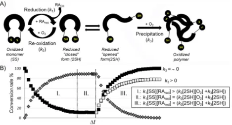
Concept of the reversible redox system
Physiological solutions contain dissolved O2 from the air, and thus, Cys-SH groups of any protein might oxidize spontaneous- ly to form the SS bond(s). The apparent rate constant depends on several microequilibrium constants, which are explicitly not elaborated herein.[69] However, it certainly depends on the width of the conformational space of the reduced molecular fold. Furthermore, the concentration of dissolved O2(and thus, Tandp), the diffusion rate of TCEP, and the protein concentra- tion are all rate-influencing factors. Because our model protein forms a coupled reaction cycle, once E19_2SH is reduced by excess TCEP, E19_SS will be instantaneously reoxidized by dis- solved O2 (Figure 5). Before exploring the mechanism of these redox-cycle-related electron-transfer processes, it should be noted that, at a macroscopic level, these coupled cycles remain hidden, as steady state (8@x/@t=0) is reached. Reduc- tion concludes in a “normal way” if all dissolved O2 is con- sumed; however, if the concentration of the RA declines faster than that of O2, then oxidation will dominate the process and spectral properties will change accordingly (Figure 5). It is hard to a priori predict the end point of the latter process because, unlike the oxidized fold of a protein, the reduced one could have a multitude of backbone conformers in exchange at vari- ous timescales (e.g., ms to ms). Among these 3D folds of the reduced state, the “closed-SH” forms (Figure S2II), in which both the C and N termini are close to each other, lead only to
Figure 5.A) A schematic illustration of the redox cycle. The oxidized state (SS) in the presence of RA (e.g., TCEP) becomes reduced (2SH), in which open and closed conformers are present in equilibrium. In the case of the closed conformer, in which the@SH groups are closely fixed to each other, intramolecular re- oxidation can occur in the presence of O2, whereas the open conformer is more likely to aggregate due to intermolecular interactions. B) Three stages of the theoretical redox setups provide the state at which reduction dominates the overall process (I), a steady state (II), and a state (III) in which excess dissolved O2
and the absence of RA lead to oxidation back to the reduced state. The black square denotes the relative concentration of the oxidized form; gray diamonds represent the reduced form. If precipitation occurs (k3>0), then at the end point of the redox cycle the soluble protein concentration has decreased relative to the initial one.
intramolecular reoxidization. However, if “open-SH” backbone forms become highly populated (e.g., as is the case of E2_2SH;
Figure S2/XIII), then intermolecular oxidation will be more prevalent, giving rise to oligo- and polymer formation (see below).
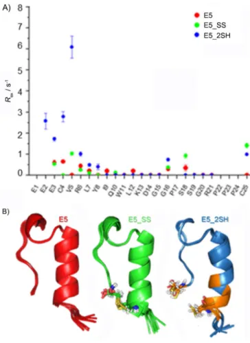
Capturing internal backbone dynamics occurring on the timescale of micro- to milliseconds was successfully attempted by means of Carr–Purcell–Meiboom–Gill (CPMG) NMR spectros- copy.[70] Herein, we present the characterization of E5, E5_SS, and E5_2SH as examples. We found that only the backbone NH groups of Glu3, Cys4, Val5, Arg6, Tyr8, and Cys25 of E5_
2SH partake in such slow motion. Considering the fact that all of these NH groups are close to both Cys residues (Figure 6), the CPMG data suggest that either E5_2SH presents alternative backbone structures, which interconvert at a slow exchange rate, or, due to incomplete reduction, the remaining oxidized form (1–8%, see a discussion of the conversion rate below) constantly interconverts with the reduced form. The minor amount of coexisting oxidized form (E5_SS) could contribute to the stabilization of the dominant backbone fold of E5_2SH.
The conformational equilibrium between the oxidized and re- duced states seems to be the most likely explanation for the above-described slow exchange; however, both scenarios of motion can occur in a concerted way.
Modeling of the SS bond reduction kinetics
The SS bond reduction by TCEP is a bimolecular nucleophilic substitution (SN2) reaction.[37] Thus, both the concentration of the oxidized form of the protein [_SS] and that of the RA con- tribute to the rate of the reduction. In an ideal case, we should consider only nucleophilic attack of the RA (k1), but, as we explained previously, in practice, we also have to take into account back oxidation (k2), which takes place simultaneously, and, in some cases, depending on the size and shape of the protein, precipitation (k3; Figure 5). The mechanism of reduc- tion, therefore, can be described by Equations (2), (3), and (4):
SSþRAredKk!2 SH1 þRAox ð2Þ
2 SHþO2K!SSk2 ð3Þ
2 SHK!SSk3 precipitated ð4Þ
By fitting this model to the concentration–time functions de- termined by means of NMR spectroscopy,k1,k2, andk3can be determined, and half-lives can be calculated. We focused on the determination of the reduction rate constantsk1; therefore, sampling was more frequent in the reduction phase (stage I;
Figure 5B). Based on parameter estimation (see the Experimen- tal Section),k2andk3are very often either negligibly small, or, due to a lack of sufficient data, cannot be confidently estimat- ed. Obtaining key kinetic parameters allowed us to describe and compare the reduction kinetics of the SS-containing mini- proteins under various experimental conditions. Some proto- cols reported in the literature apply extreme conditions, such as high temperature (e.g., 50–808C), to obtain short reduction times; this is clearly unsuitable for maintaining the integrity of the protein, or >20-fold molar excess of reagent. By perform- ing the reduction of E19_SS (0.8 mm) under such conditions (608C with 18-fold TCEP excess), the reaction seemed almost instantaneous (t1/2<5 min), but the sample became opalescent and side reactions (e.g., precipitation) were instantly detected.
Similarly to most globular proteins, the conformational ensem- ble of E19_2SH at 608C is distinctly different from that of 158C; thus presenting many more unfolded states. The folded fraction of E19_2SH is 64% at 158C, whereas it is 41 % at 608C, according to FUV-ECD analysis. Instead of intramolecular re- oxidation, undesirable intermolecular reoxidation might occur between particles. (Reducing E19_SS for 120 min, followed by centrifugation gave practically zero soluble protein concentra- tion.) In general, reduction and reoxidation at higher T (e.g., +608C) is expected to be less effective, and accompanied by multiple side reactions, such asb-elimination[71](which already occurs at a lowerT),[72, 73]racemization,[74,75]and aggregation. In principle, the reduction rate can be enhanced at lowerTby in- creasing the TCEP molar ratio (15–20-fold molar excess); how- ever, this also triggers obscure unwanted processes (Figure S5).
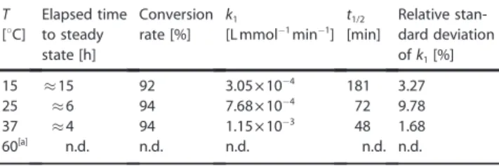
Experiments were repeated at different temperatures (15, 25, and 378C) with 0.8 mm protein and 18-fold excess of TCEP (Table 2 and Figure S6). The Arrhenius equation allows the acti- vation energy (Ea) of the redox reaction to be derived, resulting in a value of about 44.3 kJmol@1. For comparison, the activa- Figure 6.A) CPMG-determined NHRexvalues of E5 (red), E5_SS (green), and
E5_2SH (blue), and B) their backbone structures, with the key Cys residues highlighted. Slow exchange was measured for backbone NH groups of E3, C4, V5, R6, Y8, and C25 of E5_2SH only. Notably, residues that giveRexare in the proximity of the Cys residues colored orange.
tion energy of thiol–disulfide exchange between methylthio- late and oxidized DTT was calculated to be 62 kJmol@1.[76]Both FUV-ECD and NMR spectroscopy derived structural information support the high conformational similarity between E19_SS and E19_2SH; therefore, Eais likely to be used for the redox reaction, rather than for the conformational switch between the two conformational states (Table 1). Based on the NMR spectroscopy derived signal integral analysis, the reduction was almost complete (&94%) and no sign of precipitation was detected at any temperature. Additional experiments were performed to investigate the effect of the protein/RA ratio as a practical perspective (Figure S7). The above-described NMR
spectroscopy methodology provides high-resolution informa- tion about the reduction mechanism, relative to that of the more rapid NUV-ECD approach, and thus, details of the reduc- tion of all four@SS@protein models were obtained through NMR spectroscopy.
Kinetics of SS bond reduction influenced by steric factors An appropriate reduction protocol was required to unambigu- ously determine the 3D structures of the above-introduced pure reduced states. Thus, in agreement with the above dis- cussion, only mild conditions (158C and twofold molar excess of TCEP) were used for the reduction of the four different mini- proteins. Determining the structural properties and reduction rates under the same conditions allowed us to elucidate the basis of the observed differences in the reduction rates. We found that, atT=158C, thek1values of these four model pro- teins, comprising of identical core structures, but different lengths, were indeed different: theirk1andt1/2 values strongly depended on their sizes and/or molecular weights. It appears as if “cutting back” on the a-helical segment strongly affects the SS bond reducibility, even though the SS bonds of all four models are near the surfaces (Figure 7A). To our great surprise, we recorded three orders of magnitude differences between the reduction rate constants (Table 3). Whereas the reduction Table 2.Kinetic parameters of temperature-dependent E19_SS reduction
with 0.8 mmprotein and 18-fold excess of TCEP. For detailed results of parameter estimation, see Figure S6.
T Elapsed time Conversion k1 t1/2 Relative stan- [8C] to steady rate [%] [L mmol@1min@1] [min] dard deviation
state [h] ofk1[%]
15 &15 92 3.05V10@4 181 3.27
25 &6 94 7.68V10@4 72 9.78
37 &4 94 1.15V10@3 48 1.68
60[a] n.d. n.d. n.d. n.d. n.d.
[a] n.d.: not determined.
Figure 7.A) Ten superimposed structural ensembles of E19_SS, E11_SS, E5_SS, and E2_SS. Notably, all model proteins have their SS bonds at the surface, but their N termini are of different lengths and charges; thus, affecting the SS bond reducibility. Charged residues at pH 7, close to the reaction center are explicit- ly depicted: the negatively charged side chains are highlighted in red, whereas the positive ones are in blue. The C-terminal negative charge of COO@is marked by (@) and the amino group@NH3+of the N terminus by (++). Correlation between the reduction half-life versus B) helix length and C) steric factors (twofold excess of TCEP, 1.7 mmprotein, 158C) is reported. (Figure S10 shows the correlation between the reduction half-life and helix length in the case of DTT reduction.)
of E2_SS is still extremely fast, tE2 SS1=2 <&1 min, that of E5_SS occurs on the timescale of minutes: tE5 SS1=2 &14 min. E11_SS, which is elongated by six residues (1.5 turns ofa-helix) with re- spect to that of E5_SS, exhibits about a fourfold increase int1/2
(tE5 SS1=2 &14 min!tE11 SS1=2 &67 min). Finally, the unstructured short octapeptide tail HAEGTFTS- further lengthenst1/2by about 13- fold (tE11 SS1=2 &67 min!tE19 SS1=2 &909 min). The conversion rate was close to complete for the shorter peptide of E2_SS, where- as the reduction of E19_SS was only 88% complete. The kinet- ic parameters of all four model proteins were determined by using a twofold molar excess of DTT, at pH 7, and T=158C.
The mechanism of SS bond reduction by DTT is also SN2,[77]but the determined t1/2 values are significantly longer than those obtained by using the same molar excess of TCEP; however, the observed overall tendency and conclusion appear to be the same (Table 3).
Because the well-folded Trp cage motifs are identical (based on their CSD cage values; Table 1) in all four model proteins, the observedk1differences must be associated with the struc- tural properties of theira-helices and the eventually appearing unstructured tail. Although the dataset is limited (n=3 or 4), as the simplest approach, the length of thea-helix (n) and the half-lives (t1/2) of reduction could be correlated, leading to an exponential dependence for both TCEP (t1/2=2.06e0,371n, R2= 0.95) and DTT (t1/2=50.47e0,377n, R2=0.98) as the RA (Fig- ure 7B). To take into account the additional structural descrip- tors for a more complete characterization, we derived the steric factor (x) for these protein models [Eq. (5)]:
x¼
. 1
½P CSDHaðiÞA=i
-
>RMSD>n ð5Þ
in which the reciprocal of the helicity ([8CSDHaðiÞ]/i) and the bulkiness (RMSD) of the outer helical part were both calculated with respect to the length of the N terminus (n; Table 3). We observed a linear dependence of the steric factors on the re- duction half-lives as a function of the length of the N terminus
(Figure 7C). Some, but not all, of the abovek1(t1/2) differences can be explained by structural differences of the outer helix because both solvent exposure and local charges around the Table 3.Kinetic parameters of the SS bond reduction of the four model proteins. For each reduction, a protein concentration of 1.7 mmwas used with twofold excess of TCEP and DTT at 158C. For detailed results of parameter estimation, see Figures S8–S9. NMR spectroscopy derived structural properties of the outer helix are also shown.
TCEP DTT Properties of outer helix
Elapsed Conversion k1 t1/2 Relative Elapsed Conversion k1 t1/2 Relative Outer RMSD [8CSDHaðiÞ] Steric time to
steady state
rate [%] [Lmmol@1min@1] [min] standard deviation ofk1[%]
time to steady state [h]
rate [%] [Lmmol@1min@1] [min] standard deviation ofk1[%]
helix length (i)
ofouter helix
/i factor[a]
E19_SS &76 h 87 2.71V10@4 909 7.30 n.d. n.d. n.d. 30545[b] n.d. 17 1.41 0.11 213.74
E11_
SS &5–6 h 94 3.68 V10@3 67 2.61 138 &84 1.52V10@4 1659 45.152 9 0.55 0.26 19.26
E5_
SS &1 h 93 1.85 V10@2 14 3.35 9–10 95 2.18V10@3 115 1.064 3 0.39 0.14 8.56
E2_SS <5 min 100 2.59V10-@1 &1 15.37 5 100 4.04V10@3 62 5.315 0 0 0 0
[a] Steric factor comprises the following factors: the length of the outer helix, the RMSD, and the reciprocal value of [8CSDHaðiÞ]/i.[b] Half-life of E19_SS reduction by DTT was calculated according to the equation of the dependence of the half-life on outer helical length.
Scheme 1.A generalized mechanism of TCEP- and DTT-assisted mechanisms of SS bond reduction in proteins. Functional group R@stands for the N ter- minus of the protein systematically elongated here: in E19_SS the R group is equal to H+-HGEGTFTSDLSKQMEEE-, in E11: H+-DLSKQMEEE-, in E5_SS:
R=H+-EEE-, and in E2_SS it is simply H+. A brief description of the detailed reaction mechanism is provided for both TCEP and DTT in the text.
SS bonds are also different. In Scheme 1, we provide a summa- ry of the mechanistic explanation, including all of these factors and viewpoints.
Rate-determining steric and electronic factors of SS bond reduction
Apart from the steric effect of the helical part emphasized above, the SN2 mechanism of TCEP-driven reduction has to be discussed in terms of electrostatic effects.[19]In general, attack is more favorable and effective on those structures in which the C terminus is neutral. According to the average pKaof the cysteine carboxyl group (pKa=1.92) at pH 7, the proportion of COOH/COO@ is low: 1/12000. The rate-determining step is cleavage of the SS bond.[78]During the SN2 reaction, the nucle- ophilic P atom of TCEP attacks one of the SS bonds, forming a thiophosphomium salt (an S@@P+ion-pair complex; Scheme 1).
Nucleophilic attack (n!s*) is facilitated by the favorable arrow-shaped (tetrahedral: 1058) steric arrangement of the nonbonding electron pair of the P atom of TCEP. The main por- tion of the activation Gibbs free energy of reduction is con- sumed by splitting of the SS bond and not by the steric rear- rangement of the intermediate structure.[79]Better solvation of the thiol and zwitterion results in a lower activation Gibbs free energy of the reaction. Next, the positively charged @S@P+@ [(CH2)@COOH]3 complex hydrolyzes rapidly and results in the phosphine oxide and free@SH groups of the protein.
Both charged and aromatic side chains can participate, and thus, intimately influence the efficacy of TCEP-mediated reduc- tion (Figure 7A). The nucleophilic phosphine attacks the C- proximal cysteine because the intermediate cation can be sta- bilized by the proximal COO@group of the C-terminal cysteine.
A positive charge near the SS bond could enhance the reaction through electrostatic compensation of the N-proximal leaving thiolate group, whereas a negative charge might slow down the SN2 reaction.[80,81] Direct through-bond effects of any charged side chain can be ignored because they are separated by several sbonds from the negative COO@ group. Although the inductive or direct s-bond effects are negligible, both steric and spatial electrostatic effects in the vicinity ofN-proxi- mal cysteine play a major role in the reduction rate. At pH 7, the positively charged Arg near the SS bond in the inner helix may facilitate reduction; however, it is distant from the SS bond (Figure 7A), and thus, a direct charge-controlled interac- tion is less likely to occur. On the other hand, the positively charged N-terminal@NH3+can directly catalyze the instantane- ous reduction[82]of E2_SS (tE2 SS1=2 &1 min) because H@N@Ca@Cb@ S of the cysteine forms a five-membered pseudo-ring that facil- itates intramolecular NS proton transfer.[83] Thus, upon TCEP attack, these ideal local electrostatic compensations may stabi- lize the intermediate thiophosphonium salt, shifting the reac- tion equilibrium towards splitting of the SS bond. Furthermore, because the leaving thiolate anion is only positioned at the N terminus of the well-folded a-helix, the positive charge of the a-helix macrodipole also promotes progress to the re- duced state.[84,85]Moreover, due to the small protein size, the SS bond is most exposed to solvent and reagent in E2_SS.
As the N terminus is elongated on the a-helix from E2_SS toward E19_SS, the “catalyzing”@NH3+ group of the N termi- nus moves further away from the SS bond, and the effect of the macrodipole gradually vanishes; thus, the reduction rate is reduced (t1/2increases; Table 3). The role of this positive charge was directly probed by acetylating the N terminus, Ac-E2_SS, and, as expected, the half-life of reduction increased signifi- cantly: tE2 SS1=2 &1 min!tAc-E2 SS
1=2 &8 min (in both cases, a protein concentration of 1.7 mm and twofold excess of TCEP were used).
The N-terminal elongation of E2_SS by three Glu residues re- sults in E5_SS. As expected, the reduction rate is slower: tE5 SS1=2
&14 min. Although only a tripeptide is added to the dynamic N terminus, reaching the SS bond still becomes harder for both reagent and/or solvent molecules. In addition, the 3D structure (Figure 7A) shows that the three negatively charged Glu side chains (at pH 7) are flanked by the N-proximal cys- teine and the positively charged N terminus, and thus, effec- tively neutralize the catalytic effect. The structure of the en- semble determined by means of NMR spectroscopy shows a distance fluctuation from 3.7 to 10.7 a between 4Cys Cband 1Glu NH3+, whereas that of 4Cys Cband 1Glu COO@fluctuates between 3.4 and 12.4 a (Figure 7). Thus, SS bond protonation requires an active contribution from the medium; but proton transfer is perturbed by the proximity of the negatively charged glutamate side chains.
Further elongation of E5_SS by the hexapeptide -DLSKQM- leads to E11_SS. Under the same conditions, the reduction of this even larger model protein occurs more slowly (tE11 SS1=2
&67 min). The glutamate side chains are more oriented by the longer a-helix of E11_SS (Figure 7): whereas 8Glu@ turns out- ward, both 7Glu@and 9Glu@flank the SS bond from two sides.
Residues 7Glu@with 4Lys+ and 9Glu@with 12Arg+ are capable of forming salt bridges in close vicinity, and thus, could partly compensate for the slowing effect of the negatively charged side chains. E11 was found to be more helical than that of longer E19;[56] thus we find here that both E11_SS and E11_
2SH have more compact a-helices than those of E19_SS and E19_2SH, according to both [8CSDHaðiÞ]/i NMR spectroscopy measurements and FUV-ECD spectral properties. We believe that, in addition to partly compensated for negative electro- static effect(s), mainly steric effects of the elongated and stiffer a-helix cause the longer value oftE11 SS1=2 with respect to that of tE5 SS1=2 .
Finally, E11_SS elongated by the -HGEGTFTS- octapeptide re- sults in E19_SS—the largest model protein used herein—for which the longest half-life (tE19 SS1=2 =909 min) is measured. E19_
SS has the same electrostatic pattern in the vicinity of the SS bond as that of E11_SS, but its reduction rate is about 15 times slower than that of E11_SS. Although the -HGEGTFTS- segment is far from the SS bond (d7Thr-18Cys=11–14 a; Figure 7) and cannot influence reduction by electrostatic interactions, its higher internal dynamics (low S2 value),[56] as a steric effect, must slow the SS bond reduction rate further. In fact, the latter increase, in terms oft1/2, is a good estimation of the magnitude of a purely steric effect of an unstructured polypeptide chain on reduction rate.
Differences in reduction kinetics and mechanism with alternative reagents
There are a few distinct differences in terms of the general mechanism of SS reduction by TCEP and DTT (Scheme 1). 1) As an initializing step, deprotonation of the thiol group of DTT is required for successful nucleophilic attack, which depends on the pH of the medium. According to the Henderson–Hassel- bach equation,[86] taking into account the acidic dissociation constant of DTT (pKa1=9.2 and pKa2=10.1) at pH 7, deproton- ated thiolate concentration is about three to four times lower than that of the overall DTT concentration. After successful nu- cleophilic attack on the SS bond, a linear@S@S@S@transition complex has to be formed, in which the negative charge is lo- cated on the two leaving S atoms.[87] An intramolecular proto- nation, as for TCEP, also stabilizes the thiol anion leaving group if DTT is used, and thus, enhances the reaction rate. Therefore, a positive inductive/steric effect increases, whereas a negative effect decreases the reduction rate. 2) Contrary to TCEP, the active species of DTT has a negative charge. Therefore, charged amino acid side chains close to the SS bond will di- rectly affect attack by the nucleophilic RA. In line with these observations, both the negative C terminus and the SS bond flanking glutamate side chains repel DTT; thus contributing to a significant and large-scale decrease in reaction rate (Table 3).
3) Moreover, complete reduction by DTT consists of two steps:
after the first attack, the free SH group of the peptide–DTT complex has to cleave the previously formed SS bond, whereas DTT closes into a six-membered ring (Scheme 1). All of these factors jointly decrease the reduction rate if DTT is used in- stead of TCEP (Table 3). These considerations make it even more striking that, although several proteins with various num- bers of SS bonds per molecule, such as a-lactalbumin, lyso- zyme, and oxytocin, were reported to be completely reduced in 5 min by 10 mm DTT at pH 5.5 and 708C,[88] we found that the reduction of miniproteins (e.g., E11_SS) with a single and exposed SS bond might take up to 138 h (Table 3).
Spontaneous SH reoxidation accompanied by polymerization
Incomplete conversion, despite the presence of a large excess of the RA, provided evidence for the reoxidation of the re- duced SS bond of the studied model systems. To study this process in detail, the in situ reoxidation of the DTT-reduced protein samples at room temperature in sealed NMR tubes (pH 7, 158C, twofold excess of DTT) was monitored for several weeks. Spontaneous reoxidation of E2_2SH, E5_2SH, and E11_
2SH by dissolved O2 was clear after four weeks (Figure 8). The reoxidation rates (k2) have comparable orders of magnitude to that of the reduction rates (lower by one order of magnitude), but reoxidation has a pronounced role only after reaching steady state, at which the concentration of the already re- duced peptides becomes significant.
Reoxidation can take place both intra- and intermolecularly.
Whereas the former leads to a decrease of overall conversion rates, the latter results in the formation of random molecular
clusters, which may lead to precipitation. According to our semiquantitative analysis based on the recorded1H NMR spec- tra, the integral changes of the Trp He1 resonances both in the oxidized and reduced forms of the protein during reduction with DTT show a decrease in concentration over the observed period of redox time for both E2_SS and E5_SS. Precipitation can be more intense if the protein concentration is higher. Ac- cording to our present observations, increasing the length of thea-helix within the Trp cage proteins stabilizes the soluble protein fraction. This means that the elongated N terminus, namely, the outer helix in the case of E11_2SH, effectively shields the free SH@groups of the reduced protein, and thus, prevents any intermolecular reoxidation, whereas shorter var- iants, such as E2_2SH and E5_2SH, yield a significant amount of polymer formation. Due to the diversity of open 3D folds of both E5_2SH and E2_2SH, spontaneous intramolecular ring clo- sure is hindered and less likely to happen. The N-terminal Cys of E11_2SH is placed and fixed at the highly ordered inner Figure 8.The 50-membered structure ensembles of A) E11_SSQE11_2SH and B) E5_SSQE5_2SH. The fold of E11_2SH is more compact than that of E5_
2SH, which has more “open” conformers, in which the Cys residues are far from each other. This allows intermolecular, rather than intramolecular, re- oxidation. The dissolved oxidized and reduced protein concentrations of C) E11_SSQE11_2SH and D) E5_SSQE5_2SH (oxidized: blue; reduced: green) as a function of time. In the case of E5_SSQE5_2SH, the initial concentration decreased by 68%, whereas, at the end of a complete redox cycle, the con- centration of E11_SSQE11_2SH remained the same. E) Estimated parameters of the complete redox cycles. (Thek1values are slightly different from those in Table 3, for which the estimation comprises data only for phase 1.) Nota- bly, in these long-term experiments, the rate of O2diffusion characterized by the rate constantk4was also involved. Figure S11 contains all data for pa- rameter estimation of E11_SS, E5_SS, and E2_SS.
helix, with a reduced internal mobility of Cys18, and thus, mostly intramolecular ring closures take place. In the case of E5_2SH, intermolecular SS bond formation is allowed, but may be limited just by Brownian motion and concentration. A com- parison of the polymerization rates (kE11 SS3 <kE5 SS3 ) with differ- ent N-terminal lengths also supports this concept (Figure 8E).
E2_SS was N-acetylated to eliminate the reduction rate-en- hancing effect of the positively charged N terminus,@NH3+, in the vicinity of the SS bond. Upon acetylation, t1/2 has indeed increased (tE2 SS1=2 =&1 min tAc-E2 SS
1=2 =&8 min), but, in addition, the reaction reaches its steady state at a low conversion rate (50%). During reduction, almost immediately, both of the ap- propriate signal integrals of Ac-E2_SS and Ac-E2_2SH start to decrease, with a foamy precipitate gradually forming in the NMR tube. The isolated and HPLC-purified precipitant was identified as a polymer of the parent miniprotein by means of MS (Figure S12). Oligomer formation and soluble protein con- centration decrease were more advanced for Ac-E2_SS than that of E2_SS (Figure S13). Due to the absence of the shielding effect of the outera-helix, the free thiol moiety of the N termi- nus is accessible for additionally reduced peptides in which the two free SH groups can hook peptide chains together. The polymer can grow until another free N terminus and acetylated C-terminal thiol-containing peptide closes polymerization. In addition, for Ac-E2_2SH, intramolecular N!S acyl transfer could take place,[89]blocking some of the SH groups from pro- moting oligo- and polymerization through intermolecular SS bond formation.
Conclusion
The SS-bond cyclized exenatide derivate and its variants were synthesized. Both the oxidized (E19_SS) and reduced (E19_
2SH) forms, along with the parent molecule, E19, and all three of their truncated variants (E11_SS, E11_2SH, E11, E5_SS, E5_
2SH, E5, E2_SS, E2_2SH, and E2) comprised the same Trp cage/
SS/SH bond motif as that of their core structures. The SS bond stabilized model proteins showed improved thermostability and 3D fold compactness, with respect to their reduced and parent forms. Key residues for receptor binding remained in position in all of these models; therefore, E19_SS might be promising agonists for GLP-1R and as a lead compound for type 2 diabetes mellitus.
The reduction rate of E19_SS was found to be unexpectedly slow compared with that reported in the literature. The reac- tion takes hours (t1/2=48 min), even at 378C, although the pro- tein is small, and its single SS bond is exposed at the surface, and thus, accessible for reducing reagents. All four Trp cage variants studied herein have an almost equally compact core structure, witha-helical segments of different length and inter- nal mobility. By performing a complete NMR spectroscopy based structure elucidation, we found that the progress of re- duction could be monitored by means of1H NMR by using se- lected resonance frequencies. We have established that these four model proteins of differenta-helical lengths have signifi- cantly different reduction rate constants. Although it is gener- ally complicated to discriminate each factor that affects the SS
bond reduction rate, the present set of miniproteins enabled them to be deciphered separately. We have focused special at- tention on the importance of the intramolecular protonation of the SS bond; this step greatly enhances the reaction rate.
From CPMG measurements, we found that, at steady state, se- lected residues in the vicinity of the SS bond presented a slow exchange on the micro- to millisecond timescale of motion.
This redox cycle lasts as long as active RA can be found in so- lution. We found that structural, steric, and electrostatic factors influenced the reduction rate greatly, resulting in almost three orders of magnitude differences in reduction half-lives (t1/2) for otherwise structurally similar and globularly folded model pro- teins.
Notably, in addition to intramolecular reoxidation within the redox cycle, intermolecular oxidation could also occur. The rate of these two concerted reactions depended on 1) the internal dynamics of the backbone conformers in the proximity of the SS bond, and 2) the shielding effect of the a-helix on the SS bond. Intramolecular N!S acyl transfer in Ac-E2_SS inhibits intramolecular reoxidation, but increases intermolecular reoxi- dation, which leads to oligo- and polymerization.
We found that easy-to-collect NUV-ECD spectral properties were indeed useful for monitoring the SS!SH reaction, even quantitatively, without the time-consuming assignment of the high-resolution NMR spectroscopy data. If the SS bond were situated in the vicinity of an aromatic cluster, NUV-ECD spectral changes could be used to monitor the transformation, which was proportional to the extent of reduction and clearly sig- naled when steady state had been reached. Thus, we encour- age the use of CD spectroscopy for monitoring protein reduc- tion rate in the manufacture of recombinant proteins (e.g., in- sulin, human monoclonal IgG antibodies) on a large scale, to control and provide information on the state of SS–SH bonds.
Experimental Section
ECD: FUV-ECD spectra were recorded on a Jasco J810 spectropho- tometer by using a 1.0 mm path length cuvette with protein con- centrations of 20–30mm. Data accumulation was performed over a range of 185–260 nm, with 0.2 nm step resolution at a scan rate of 50 nmmin@1 with a 1 nm bandwidth. The spectral accumulations were resolved between 5 and 858C in steps of 58C. The tempera- ture was controlled by using a Peltier-type heating system. Each spectrum baseline was processed by subtracting the solvent spec- trum from that of the protein and the raw ellipticity data were converted into mean residue molar ellipticity units, [V]MR.
Reduction monitoring by NUV-ECD: The spectra were recorded on a Jasco J810 spectrophotometer by using a 10 mm path length cuvette with protein concentrations of 120–150mm. Data accumu- lation was performed over a range of 240–325 nm, with 0.2 nm step resolution at a scan rate 50 nmmin@1with a 1 nm bandwidth.
The sample was tempered by using a Peltier-type heating system.
Each spectrum baseline was processed by subtracting the solvent spectrum from the peptide spectrum and the raw ellipticity data were normalized by the concentration [V]. Reduction was followed for 75 h. Each intensity [V] at 266, 281, 287, and 293 nm was con- verted into concentration by using Equation (6).