Infrared neuromodulation:
a neuroengineering perspective
Z. Fekete1, Á. C. Horváth1, A. Zátonyi1
1 Research Group for Implantable Microsystems, Pázmány Péter Catholic University
Keywords: infrared neuromodulation, infrared neural stimulation, infrared inhibition, photobiomodulation, thermal stimulation, near-infrared laser stimulation, neural interfaces
Abstract
Infrared neuromodulation (INM) is a branch of photobiomodulation techniques, which offers direct or indirect control of cellular activity through elevation of temperature in a spatially confined region of the target tissue. Research on INM, started around one and a half decade ago, is gradually gaining attention of the neuroscience community, as numerous experimental studies give evidence of the safe and reproducible excitation and inhibition of neuronal firing in both in vitro and in vivo conditions. However, its biophysical mechanism is not fully understood, several engineered interfaces have been created to perform infrared stimulation in both the peripheral and central nervous system. In this review, we first summarize recent applications and present knowledge on the effects of INM on cellular activity, and provide an overview on technical approaches to deliver infrared light to cells and to interrogate the optically-evoked response. Micro- and nanoengineered interfaces to investigate the influence of infrared neuromodulation will be also described in details.
Content
Abstract ... 1
1. Introduction ... 3
2. Applications ... 5
2.1 INM restore auditory perception ... 5
2.2 INM to investigate brain diseases ... 6
2.3 INM to control peripheral nerves ... 7
2.4 INM for cardiac pacing ... 9
3. Mechanisms of action ... 9
3.1. Infrared neural stimulation ... 11
3.1.1 Heat-sensitive ion channels ... 11
3.1.2 Electrostatic mechanism ... 11
3.1.3 Intracellular Ca2+ waves ... 13
3.1.4 Network effects ... 13
3.2 Infrared neural inhibition ... 14
4. Neural interfaces for INM ... 16
4.1 Direct photomodulation using infrared light ... 16
4.1.1 Fiber-based approaches ... 16
4.1.2 MEMS based devices ... 17
4.2 Indirect photomodulation using infrared light ... 20
4.2.1 Polymer-mediated approaches ... 20
4.2.2 Upconversion nanoparticle mediated applications ... 23
4.2.3 Thermoplasmonic neurostimulation ... 24
5. Future perspectives ... 25
Acknowledgement ... 25
References ... 26
Figures ... 41
1. Introduction
The number of disabled individuals of the ageing population, draws attention to neurological diseases, which may cause socioeconomic burden in the long term [Olesen, 2012]. The most commonly known disorders, like Alzheimer, depression, Parkinson’s disease, show a variety of symptoms, which makes patients unable to take care of themselves in the everyday life. Improvement of sensory deficits, like the partial or total loss of hear or vision is now feasible using dedicated implantable devices.
Neuromodulation is a technology that directly intervenes the physiological processes of neural ensembles by physical, chemical or electrical means. One success story of neuromodulation is deep brain stimulation (DBS), which acts directly on the target area in the central nervous system by programmed electrical pulses [Benabid, 2009]. The feasibility of the technique has been proven in clinical practice in PD, but it is also gaining attention in the treatment of other diseases. The operation of DBS relies on a stimulator probe implanted permanently into the brain tissue and on an external device, which delivers current pulses in a pre-defined sequence to terminate tremor. The implanted electrodes of DBS devices cause a potential gradient across a neuron, which leads to intracellular ionic current flow and localized depolarization and hyperpolarization of the cell membrane. This way, a region of cell membrane hyperpolarization is created, which is sufficient to block action potential propagation, thereby achieving neural inhibition. Briefly, stimulation appears to block the signals that cause disabling motor symptoms and so helps provide greater control over movement. Due to the increasing number of patients, an intensive research has been started to seek alternative technological options in neuromodulation strategies [Luan, 2014].
Transcranial magnetic stimulation (TMS). Like in the case of electrical DBS, potential gradients are induced in the tissue by a rapidly changing strong magnetic field (>1 T). Recently, Bonmassar et al.
[Bonmassar, 2012] reported on the design of a micro-TMS system, which reduces the spatial resolution by an order of magnitude. Nevertheless, the large power consumption is still an obstacle for building a fully implantable stimulator.
Optogenetic stimulation. Optogenetics is a new neuromodulation technology in which light-sensitive proteins (“opsins”) are genetically expressed in the cell membrane of neurons [Boyden, 2005]. These proteins act as light-activated ion pumps, channels or enzymes to provide precise optical manipulation of electrical and biochemical processes in the living cells [Yizhar, 2011]. Due to high safety and ethical standards in the clinical practice, the wide use of this genetic technology is not expected in the near future.
Other concerns include photoelectrical artefacts and uncertainty in the physiological responses to a stimulus because of variations in gene expressions.
Transcranial ultrasound stimulation. Experiments have indicated that temporally modulated ultrasound waves can elicit action potentials in brain cells [Menz, 2013]. Up to now, very little is understood about mechanobiology of affected cells, although proposed mechanisms have included cavitation and thermal effects.
Thermal stimulation. The mechanism of thermal modulation of neural activity likely relies on the combination of the temperature induced changes in the transmembrane capacitance and non-uniform changes in the conductance dynamics of specific ionic channels [Peterson and Tyler, 2014]. Two strategies have been demonstrated. In case of Microwave/RF heating, magnetic nanoparticles are applied to absorb RF radiation and therefore heat surrounding tissue [Dutz, 2013]. Combining these particles with proteins known to bind to specific protein targets on neural cell membranes has been shown to enable focused heating of target cells. Although this approach could potentially give very good spatial resolution, the temporal resolution and power consumption is still to be improved. In the case of optically induced thermal modulation, a rapid heating is achieved through the targeting of near infra-red laser light, therefore the technique is often referred to as infrared neuromodulation (INM).
Infrared neuromodulation (INM) uses pulsed or continuous-wave infrared light (between the wavelength of 1400-2100 nm) to generate highly controlled temperature change in neurons, leading them to either elicit or suppress action potentials (see Figure 1) [Wells, 2007; Cayce, 2011; Duke, 2013; Lothet, 2017;
Baumhoff, 2019; Horváth, 2020]. The induction of a temperature gradient (dT/dz or dT/dt) or elevation of background temperature of the tissue are required for pulsed or continuous-wave infrared light to generate an action potential. The advantages of the modulation technique are the following:
(1) it can be applied to neural tissue without prior genetic incorporation of light sensitive channel rhodopsin unlike optogenetic stimulations,
(2) it produces a focal stimulation spot size matching the size of an optical fiber (e.g. a few tenth of microns) and does not suffer from current spread (unlike electrical stimulation),
(3) due to the controlled heat transients, within certain parameters, it can be applied repeatedly to tissue without damage.
(4) it is insensitive to external magnetic fields, therefore this technique could be compatible with magnetic resonance imaging typically used to check implants in humans and can be also concurrently used with electrophysiology methods.
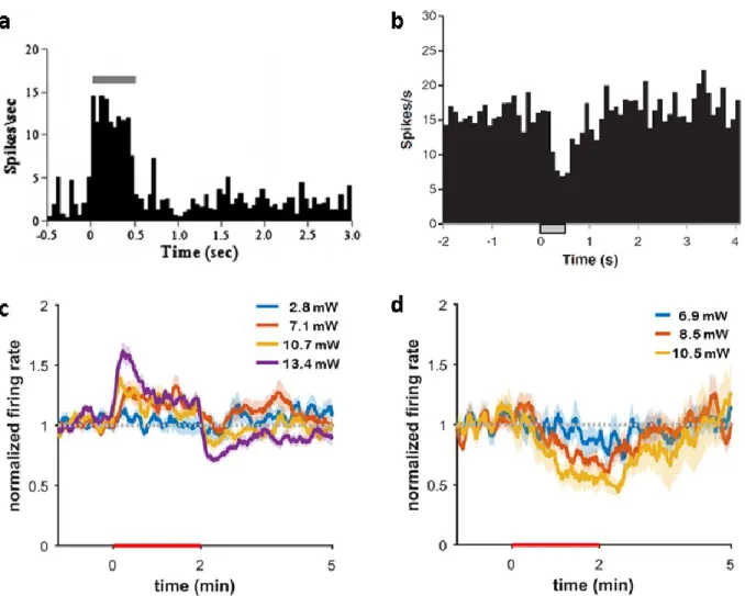
Two specific neural response to INM is identified and are shown on Figure 1. The effect of infrared neural stimulation (INS) (Fig.1.a,c) and infrared neural inhibition (INI) (Fig.1b,d) can be elicited by either pulsed (Fig. 1.a,b) or continuous-wave (Fig.1.c,d) IR governed by different cellular processes to be introduced in later chapters.
The aim of the current review is to guide the community through the recent technological advances introduced into the field of infrared neuromodulation. Description of the latest in vivo and in vitro demonstration will be given to cover applications in the both the central and peripheral nervous system.
Hypothesized mechanisms of action will be also discussed. Recently developed neural interfaces engineered to exploit the above advantages of infrared stimulation will be presented in details.
2. Applications
2.1 INM to restore auditory perception
Cochlear implants are complex bionic devices that are able to restore lost hearing of deaf people. The core components of today’s systems rely on an intracochlear electrode array. These interfaces transmit electrical signals used to stimulate the auditory nerve directly. The conventional stimulation method has low spatial selectivity due to the inherent current spread, activating several spiral ganglion neuron (SGN) at a time. To circumvent the disadvantage of this phenomenon several groups has investigated alternative stimulation protocols applying pulsed infrared light (Tian et al., 2017; Guan et al., 2016; Tan et al., 2015;
Richter et al., 2011a). Early demonstration of Izzo and co-workers suggested that compound action potential in gerbil’s auditory neurons can be induced with infrared pulses (λ=1994 nm, pulse duration = 1-100 µs, repetition rate = 2 Hz, radiant exposure = 50 mJ/cm2). Later on, several paper has challenged the idea that SGN could be directly activated by heat, but rather stimulated through remaining hair cells, which are sensitive to infrared radiation (Richardson et al., 2017; Thompson et al., 2015; Rettenmaier et al., 2014; Schultz et al., 2014; Verma et al., 2014; Teudt et al., 2011). Thompson et al performed experiments to reveal the mechanism of INS in the cochleae of both normal hearing and profoundly deaf guinea pigs, and found that optically induced response could only be evoked in subjects with normal hearing. Their statement that such response from INS is hair-cell mediated is still debated. By the development in transgenic animal models, Tan et al demonstrated mice INS evoked auditory responses in
Vglut3−/− despite the inability of hair cells in these mice to release glutamate transmitter (Tan, 2018), which indicates the direct stimulation of SGN cells is indeed possible. Besides the possible indirect effect of INS on primary auditory structures, it may also be utilized in combination with electrical stimulation implants to carry acoustic envelope information (Baumhoff, 2019). However, INS has been only exploited to activate signaling in the auditory pathway, Jiang et al showed that 980-nm laser pulses could reversibly and repeatedly inhibit acoustically induced neural activities in the cochlear nucleus, which opens up new opportunities to influence the acoustic input through neuromodulation in the brainstem (Jiang, 2019).
2.2 INM to investigate brain diseases
Neurodegenerative diseases refer to pathological conditions accompanied by death of neurons.
Several common disorders like Alzheimer's and Parkinson's disease, epilepsy, traumatic brain disorder produces severe symptoms of affected patients, which ultimately changes their quality of life. Studies in this field use both invasive and non-invasive methods, where transmission of NIR radiation is performed intracranially or transcranially, respectively. In the clinical settings, such neuromodulation techniques are often referred as Transcranial photobiomodulation (tPBM), Transcranial Near-infrared laser therapy (TNILT) or Low-level laser Therapy (LLLT) (Lapchak, 2012).
Motor symptoms of Parkinson’s disease are mainly due to a loss of neurons that produce dopamine, an important chemical messenger in the brain. Peoples et al have shown in MPTP induced mouse model of Parkinson’s disease that exposure to NIR (λ = 670 nm, radiant exposure = 5 J/cm2) either at the same time or well after chronic MPTP insult saved dopaminergic cells from degeneration in SNc (Peoples, 2012).
In a similar animal model, Moro et al demonstrated the neuroprotective effect of NIr on these cells using an intracranially implanted surgical device composing of an optical fiber coupled to an LED of the same wavelength is possible (Moro, 2014). Yoo et al. stimulated the globus pallidus (GP) and STN for 2 minutes with 808 nm wavelength at various optical power (10-40 mW) has found that for GP and STN excitatory and inhibitory effect was dominant, respectively. The responsiveness to NIS was linearly dependent on the power of radiation exposure. Reinhart et al has proved in hemi-parkinsonian rat model injected with 6-OHDA that NIR exposure of STN led to improved behavior and neuroprotection (Reinhart, 2016).
Traumatic brain injury is caused by severe traumatic impact to the head inducing impairment in the cerebral blood flow. Transcranial NIR stimulation to patients with traumatic brain disorders proved to be effective in mitigating cognitive impairment (Naeser, 2011). Among the diverse mechanisms of NIR to TBI,
stimulation of ATP production, regional cerebral blood flow, acupoint, neurogenesis and synaptogenesis, as well as a reduction in anti-inflammatory effect are hypothesized (Yao, 2018).
Alzheimer-disease is a type of dementia that is featured by a continuous decline in thinking, behavioral and social skills, and is believed to be caused through a variety of molecular mechanism (e.g. extracellular aggregation of amyloid beta plaque) (Crews, 2010). Purushothuman et al presented in transgenic APP/PS1 mouse model that Alzheimer-related pathological cues in the cerebral cortex can be mitigated using transcranially delivered NIR light (λ = 670 nm, radiant exposure = 4 J/cm2) (Purushothuman, 2014;
Purushothuman, 2015).
More recently, the effect of photomodulation in the NIR range (between 808-1064 nm) on the oscillatory rythms of the brain is explored in more details (Wang, 2019; Zomorrodi, 2019) and several groups hypothesize the possibility to intervene the pathological mechanism of brain disorders indirectly through the modulation of brain waves. Of course, the application of longer wavelength is accompanied by some increase in penetration depth and increase in spatial selectivity, which may be also beneficial like in case of suppression of focally induced epileptic seizures. Epilepsy is a central nervous system disorder caused by abnormal excitement of neural activity. Blocking the convulsive effects of seizures can be potentially performed by NIR light. First, Xia et al have shown that induced epileptiform neural activities in cortical slices can be reduced by 80 % under direct continuous-wave IR exposure (λ = 1550 nm, radiant exposure = 14.5 mW/mm2) (Xia, 2019). Furuyama and co-worker also investigated BMI induced epileptic activity in CA1 region of Mongolian gerbils (Furuyama, 2019). They found that firing rate during seizure- like activity can be efficiently suppressed with near-radiation through an optical fiber placed in the seizure focus (λ = 830 mn, radiant exposure = 17.5 mW/mm2). These studies have given further evidence on the feasibility of INM in the central nervous system, which may be gradually recognized as an alternative tool to investigate or even treat neurodegenerative diseases.
2.3 INM to control peripheral nerves
First demonstration of the infrared stimulation of peripheral nerves dates back to the mid-2000s.
Researchers from Vanderbilt University explored the sensitivity of rat sciatic nerve to pulsed infrared irradiation. Using a tunable free electron laser source (λ = 2 to 10 micron) and a solid state Holmium:YAG laser (λ = 2120 nm). They determined both upper (damage) and lower (activation) threshold to generate compound action potentials, (Wells, 2005). These parameters were found to be wavelength dependent, and safety ratio (ratio of damage and activation threshold) was the highest (about 6) at 2100 nm. It was
also determined experimentally that radiant exposures required for reliable stimulation of nerve fibers was between 0.34–0.48 J/cm2. The upper limit for safe laser stimulation repetition rate was identified near 5 Hz, however, low repetition rate like 2 Hz for long-term stimulation is adequate. Results of the pioneering work of Wells et al still serves as important reference for pulsed infrared stimulation.
Nerve recruitment was also evaluated using electrical co-stimulation with the concurrent application of a cuff electrode in the rat sciatic nerve, which showed that preconditioning the nerve fibers with IR pulse train (λ = 1875 nm, tpulse = 2 ms, repetition rate = 15-20 Hz ) may increase the force response to both electrical and hybrid (electrical/optical) stimulation (Duke, 2012). This yield was believed to be induced by the elevated baseline temperature of the tissue, which further enhanced cell excitability in the target region. The highly localized stimulation was repeated in rabbit sciatic nerve, and experiments revealed that 81% of the nerves were recruited (Peterson, 2014). Differences in sensitivity between rat and rabbit nerve fibers were believed to occur due to the differences in nerve size. This hypothesis regarding the importance of the axonal diameter was confirmed by Lothet et al, who selectively inhibited small and large diameter axons in Aplysia californica (Lothet, 2017). In contrast to IR-induced activation, which is believed to result from the spatiotemporal temperature gradient, they aimed to create an increase of baseline temperature (ΔT = 2.9-9.7 K) using a different set of laser parameters (λ = 1860 nm, tpulse = 0.2 ms, repetition rate = 200 Hz, radiant exposure = 0.05-0.11 J/cm2/pulse). In the mouse sciatic nerve, motor fibers were recruited at substantially lower wavelength (λ = 1470 nm), but at similar exposure rate (0.05- 0.23 J/cm²) and pulse duration (100-500 us), which may also open up new possibilities towards miniaturization of potential stimulation devices not relying on a tunable Capella laser (Dautrebande, 2018). Such efforts may further increase when carbon nanoparticles are used as mediators for IR light absorption, and therefore the threshold energy can be reduced to evoke compound muscle action potentials (Wang, 2017). Regarding the inhibition of nerve fiber activity, more recent results relying on the stimulation of crayfish unmyelinated motor axons indicated that IR pulses have influence on axonal excitability and locally initiated action potentials can be reversibly blocked. Authors of this study used a fiber-coupled laser diode (7.1 mW) with a wavelength centered at 1994 nm and a pulse duration between 20-500 ms, and an estimated radiant exposure of 0.44 J/cm2 – 11.38 J/cm2 on the axonal surface (Zhu, 2019).
2.4 INM for cardiac pacing
Besides the modulation of activity of cells in the CNS and PNS, irradiation of cardiac cells to control heart rate and cell development may be also exploited. Jenkins et al have proposed first pulsed IR light can be utilized for optical pacing of the intact hearts of quail embryos (Jenkins, 2010). Using a threshold (50% probability point) of 0.81 J/cm2, they managed to synchronize the frequency of the heart beats to that of the laser pulses without detectable damage to the tissue. Based on ECG measurements, they found that each pulse was initiating and not modulating heart beats, while the conduction was propagated through the AV node. The same phenomenon was later demonstrated in the adult rabbit heart ex vivo as well (Jenkins, 2013). The principle was further exploited in a screening instrument for development of human cardiac cells derived from pluripotent stem cells (McPheeters, 2017). The origin of the underlying mechanism was discussed by Dittami et al, who reported that intracellular Ca2+ transients induced by the incident IR light were responsible for the elicited contractile response in neonatal rat ventricular cardiomyocytes (Dittami, 2011). Later on, as the inhibitory mechanism of action of IR light had been revealed, IR irradiation was also demonstrated to stop the beating of heart in early embryonic avian (Wang, 2016). Based on measurement with a thermocamera, the tissue was exposed to an increase in temperature between 10-15°C, however, histological staining did not revealed thermally induced cell necrosis. A more recent study reported optical stimulation and pacing of the embryonic chicken heart using a pulsed infrared thulium laser with a wavelength of 1927 nm (Chung, 2019). Using a radiant exposure of 1.01 J/cm2 and targeting the atrium, they achieved full pacing.
3. Mechanisms of action
In principle, electromagnetic waves can be exploited to modulate neuronal activity through photoelectrical, photochemical, photomechanical and photothermal processes. Photoelectrical interaction has been demonstrated in particular cases, when photoactive intermediate layers were used to inject electrons into the neighboring tissue, and altered the growth and differentiation characteristics of cells (Akhavan, 2013; Abdullaeva, 2016).
Photochemical reactions happen, when the absorbed energy of the incident light induces chemical reaction governed by endogenous or exogenous molecular chromophores. To evoke photochemical
reaction, the energy of individual photons should exceed that of typical ion bond energies ranging from 100-1000 kJ/mol. Among the very first demonstrations of INS, Wells et al have showed that using free electron laser, no enhancement of laser stimulation was observed at any particular wavelength between 2 and 10 µm (Wells, 2007). Based on their experiment, the role of photochemical reaction in the behind mechanism of INS was excluded. The most often used system of Aculight Corporation (λ = 1875 nm) is featured by photon energies of ∼0.66 eV, which corresponds to ∼64 kJ/mol bond energy (Cayce, 2011).
In some recent work, the application of short-wave near infrared regime (800-1200 nm) is getting into the focus of INS experiment. If one aims to describe the mechanism of elicited tissue response to photon energies in this regime, careful considerations are required.
Photomechanical effects may occur when stress confinement happens in the tissue in response to pulsed irradiation. Stress waves generated by thermoelastic expansion needs time to propagate out of the irradiated volume. If pulse duration is shorter than this time period, optical energy is accumulated. To produce such laser-induced stress waves inside the tissue sub-microsecond pulse duration is needed.
Typical values of pulse duration are significantly higher, therefore photomechanical component of infrared neuromodulation is also excluded (Richter, 2014).
Photothermal mechanism is considered to govern INM, however, various physiological event may have influence on the outcome of cell activation or blocking through IR irradiation. In fact, the energy of light is converted to heat in the exposed tissue, which is enhanced by water molecules acting as chromophores in INM. The major impact of water was already proven in the study of Wells et al, who determined that cellular response to IR irradiation depends on the emission wavelength of the laser source (Wells, 2005). Later, Shapiro et al confirmed that water is an important component in INM, when they replaced with heavy water exhibiting much lower absorption coefficient (Shapiro, 2012). As a result, the evoked cellular response reduced by 80%. In view of these findings, tuning the wavelength of light sources to specific values where absorption coefficient of water is high, may significantly increase the sensitivity of INM. Since INM studies utilize a variety of laser source and wavelength, these values should be carefully considered, when comparing their effectivity.
In the following sections, cellular mechanisms behind the photothermal effect revealed so far will be described.
3.1. Infrared neural stimulation
3.1.1 Heat-sensitive ion channels
Discovery of heat-sensitive ion channels dates back to the late 90’s, when Caterina et al described the first TRPV (transient receptor potential vanilloid) channel (Caterina, 1997). TRPV1 was also termed as capsaicin receptor due to its well-known response to hot chili pepper. Besides its sensitivity to various physical and chemical stimuli, TRPV1 channel is activated above 43 °C, which is also important in peripheral nociperception. It is important to note that channels of TRPV family can be found in both the peripheral and central nervous system. They are basically nonselective cation channels that are permeable to Na+, K+ and Ca2+ (Kauer, 2009). Rhee et al showed first that TRPV1 channels mediate the response to infrared irradiation as well (Rhee, 2008). Using TRPV1 antagonists, they demonstrated that pulsed IR light failed to evoke membrane depolarization in primary sensory neurons if TRPV1 channels are blocked. They also showed that TRPV1 channels can be rapidly and reversibly activated with brief trains of 2-ms long IR pulses. This initial result was confirmed by the Richter group, who tested cochlear response of TRPV1 knock-out animals to infrared stimulation (λ = 1850 nm, tpulse = 30-1000 µs, repetition rate = 10 Hz), and found that action potential on the auditory nerve cannot be evoked in mice lacking of the gene for TRPV1 (Suh, 2009). Sensitivity of TRPV2 and TRPV3 was also proved using HEK293 cells and patch-clamp recording (Yao, 2009). Later, Albert et al demonstrated that TRPV4 (activated above 34°C) also mediates the response of sensory neurons to external heat stimuli (Albert, 2012). Using TRPV4 channel blocker, they proved that the operation of voltage gated calcium and sodium channels is indirectly influenced by TRPV4, and this extra calcium influx contributes to laser-evoked neuronal response. Another work of the same pointed out that activation is rather governed by the absolute temperature and not by the rate of heat (Bec, 2012). Such temperature thresholds of TRPV members for the HEK 293 cells and Xenopus oocytes were determined as follows: TRPV1 >43 °C, TRPV2 >52°C, TRPV3 >32°C and TRPV4 >27°C.
3.1.2 Electrostatic mechanism
INS has been demonstrated to act in a similar manner in the case of various cell types including peripheral nerves, sensory ganglion and cardiac cells (Wells, 2005; Izzo, 2008; Jenkins, 2010). This implies that a general mechanism behind these responses may be present besides the effect exerted by heat- sensitive ion channels. It was first hypothesized by Shapiro et al, that the temperature dependent change
in the membrane capacitance may have key role during the induction of action potential as cells are exposed to pulsed IR light (Shapiro, 2012). Their voltage and current clamp measurement in three species (X. laevis oocytes, embryonic kidney (HEK) cells and artificial bilayer) revealed that current response to the electrostatic change of the plasma membrane depends on the temporal gradient of heating and not on the absolute temperature. The increase in membrane capacitance was found to be dose dependent, and this transient behavior lasted up to 100-200 ms after individual stimuli. The experimental results were explained using the Gouy–Chapman–Stern (GCS) theory, modeling the capacitance of the phospholipid bilayer and the in-series capacitance of ionic double layers on each side of the plasma membrane as a resultant capacitor (Grahame, 1947). Shapiro et al formulated a coupled double layer mathematical model based on the extension of the original GCS equations completed with an extra Stern layer. Another study on the neuromuscular junction of Caenorhabditis elegans confirmed that very fast heat-shocks (like 500°C/s heat shock for 500 μs) induced a charge redistribution on the membrane surface, and the resultant asymmetric excitatory currents led to depolarization of the cell membrane (Liu, 2013).
They also constituted a mathematical description to quantify the capacitive membrane currents evoked by this photothermal phenomenon. Carvalho-de-Souza also showed using gold nanoparticles (AuNPs) conjugated to cell ligands that AuNP-mediated infrared stimulation of neurons also elicits action potentials due to changes in membrane capacitance (Carvalho-de-Souza, 2015). A few years later Shapiro’s theoretical model was revisited by Plaksin (Plaksin, 2017, Shapiro, 2017), who highlighted a mathematical error in the original paper, which actually results a prediction of decrease in membrane capacitance instead of increase. To accurately explain the experimental outcomes related to the electrostatic mechanism of INS, Plaksin formed the MechanoElectrical Thermal Activation (META) theory (Plaksin, 2018). This predicts an approximately 0.3% change in membrane capacitance per Kelvin, originated from a deformation of the phospholipid bilayer causing extra ionic displacement currents.
Extending the existent model of Shapiro, Ebtehaj et al proposed an improved model, which takes the temperature dependence of the surface charge density and the hydrophobic core dielectric constant of the lipid bilayer into account (Ebtehaj, 2018).
Besides the temperature dependent change in membrane thickness, formation of small diameter nanopores through the plasma membrane are also hypothesized to participate in the cellular response to INS (Beier, 2014). Beier and coworkers hypothesized that temporary nanopores allows the influx of extracellular ions into the cell cytoplasm due to the destabilization of phospholipids within the membrane elicited by the temperature gradient. Using radiant exposure between 1.7–6.5 J/cm2 for a few millisecond long pulse duration, increase in intracellular Ca concentration was observed. The tracked Ca2+
influx happened in synchrony with IR stimulation. Their group has also given evidence on the role of nanoporation in a follow-up work, where calcium was replaced with thallium to mimic the dimensional and electrostatic property of calcium without its biological functionality (Roth, 2016).
3.1.3 Intracellular Ca2+ waves
One of the earliest explanation of the mechanism behind INS relied on the generation of transient intracellular Ca2+ waves, originating from the endoplasmic reticulum (ER) (Iwanaga, 2006). Infrared stimulation has been proved to increase intracellular [Ca2] in cardiomyocytes (Dittami et al. 2011), in cortical neurons and glia (Cayce et al. 2014a), in hippocampal neurons of mice and human glioblastoma cell lines (Moreau et al. 2018) as well as in spiral ganglion neurons (Barrett, 2018).
The study of Cayce et al., utilizing 2-photon calcium imaging, also proved that subcellular processes of different time scale may be concurrently present, and demonstrated that pulsed INM elicit transient calcium events in astrocytes and in apical dendrites of neurons in the rat’s living brain (Cayce, 2014). They hypothesized that these two events may also interact somehow. Barrett et al, investigating this Ca release in cultured spiral ganglion neurons, also pointed out that no Ca influx through the plasma membrane is needed to evoke action potentials, which confirms the major role of intracellular stores (Barrett, 2018).
They described that Ca release from the ER is mediated by TRPV4 channels colocalizing with ER (Barrett, 2018). The involvement of heat-sensitive ion channels in the regulatory mechanism of intracellular Ca may lead to revisiting the already established fundamental contribution of TRPV family to INS. The work of Moreau et al has provided additional experimental evidence on the role of phospholipase C and IP3 (inositol-3-phosphate receptor) in the Ca2+ release process from the ER (Moreau, 2018). A more recent study on the modulation of astroglial cells via pulsed infrared light has confirmed the participation of TRPV4 and IP3 in the shaping of calcium dynamics, and enlightened the contribution of further channels, TRPA1 and Aquaporin-4 (AQP4) (Borrachero-Conejo, 2020). The importance of AQP4, a water transport channel, was validated in AQP4 knockout mice.
3.1.4 Network effects
Most of the approaches used for INM propose technical solutions that aim to produce a spatially confined spot of IR irradiation. Besides the evoked processes in small cellular ensembles, the influence of INM on the network activity in the CNS should be also understood, so even secondary effects of local
stimuli can be taken into account in later studies. Uozumi et al. showed that 15-45 minutes long irradiation of 808 nm NIR light at 1.6 W/cm2 can increase the intensity of cerebral blood flow by 30% compared to the control group in mouse subjects (Uozumi, 2010). A study of INS concurrently used with intrinsic optical signal imaging found that heat-induced vasodilation is not likely to be the primary effect of change in intrinsic signals, since the peak intensity was reached well-after the stimulus and decay of that did not follow an exponential pattern (Cayce, 2011). Tian et al using functional near-infrared spectroscopy revealed that irradiance of 1064 nm laser at a power density of 0.25 W/cm2 is enough to improve cerebral oxygenation in healthy humans (Tian, 2016). A recent model exploiting hemodynamic data from human experiments also claimed that increasing the concentration oxygenated hemoglobin indeed happens, however, there is a large variation between individual responses (Wu, 2019).
Related results using magnetic resonance imaging also draw the attention towards consequences on functional patterns likely due to the change in the blood flow characteristics. Khoury et al showed that transcranial near-infrared (λ = 808 nm) stimulation has no effect on resting state functional connectivity (Khoury, 2019). Nevertheless, they pointed out that functionally active brain regions are definitely influenced. A study has shown improvement of functional connectivity in chronic TBI patients was achieved (Naeser, 2019). Similar conclusion was drawn after treatment of dementia patients, where the increased functional connectivity between the posterior cingulate and lateral parietal cortex was found (Chao, 2019).
Besides, fMRI, other non-invasive approaches like EEG has been utilized to investigate the effect of NIR stimulation on network activity. A recent report suggests that INM (λ = 1064 nm) increases the power of electrophysiological oscillations as measured by scalp electroencephalogram (EEG) (Wang et al., 2019). Similar results of Zomorrodi et al confirmed the significant increase of higher oscillatory frequencies in the resting state at pulsed NIR (λ = 810 nm at 40 Hz), and the change in the clustering properties of various brain regions (Zomorrodi, 2019).
However, there are some preliminary recent results from human experiments, regarding the causal relationship between NIR stimulation and brain oscillations, further studies are necessary not only in human subjects, but in established animal models as well.
3.2 Infrared neural inhibition
Infrared neural stimulation as detailed in the previous chapter activates neural activity as a result of spatiotemporal gradient (dT/dt and dT/dx) in temperature induced by local irradiation of infrared light.
Laser-induced inhibition in the intact rat somatosensory cortex was first described by Cayce et al (Cayce, 2011). They observed that pulsed INS led to a reduction in firing rate in vivo lasting about 1.5–2.0 s, followed by a return to the baseline level (λ =1875 nm, radiant exposure = 0.019 J/cm2, tpulse = 250 μs, pulse train length = 500 ms). They hypothesized that INS may have an excitatory effect on inhibitory neurons in superficial layers of the cortex. The underlying mechanism was first discussed in the work of Duke et al., who used preparations of motor axon of Aplysia californica and rat sciatic nerve, and transiently suppressed the electrically initiated action potential generation and propagation (Duke, 2013).
They achieved inhibitory modulation within a spot as small as 100 µm in diameter in as little as a few hundred µs. They believed that baseline of tissue temperature plays a crucial role in the inhibitory response, which was controlled in species below a maximum temperature change of 9 °C. The inhibition of AP generation and block of AP propagation was carried out using different sets of laser parameters.
Inhibition of generation required 500 ms long episodes of 5 or 0.5 ms long pulses with a radiant exposure between 4-8 J/cm2 depending on the wavelength of laser source (1450 and 1860 nm, respectively). In contrast, nerve conduction was blocked using 3 seconds long protocol consisting of 0.2 ms long pulses with a radiant exposure of 0.5 J/cm2 delivered at 200 Hz. Later, the inhibitory cardiac activity in embryonic heart of quails was demonstrated by Wang et al (Wang, 2016). With optical mapping, they showed that INI stopped contractions in cardiac cells by blocking action potentials, which eventually led to the abolition of intracellular calcium transients and contraction.
To investigate the mechanism of INI, Mou et al proposed a COMSOL and NEURON based physical model (Mou, 2012). Their work relied on the implementation active nerve fiber model incorporating the temperature dependent Hodgkin-Huxley gating mechanism. This theory of heat block, experimentally validated in early works (Hodgkin, 1949; Huxley, 1959), implies that the activation of the potassium ion channels of a cell alongside the inactivation of sodium channels reduces the generation and propagation of action potential. Using potassium and sodium channel blockers independently, Ganguly et al proved that compound action potential propagation in Aplysia californica nerves was dominantly blocked by potassium channel activation during IR exposure, and sodium channels play no significant role in the thermal response (Ganguly, 2019a). They have also modified the original Hodgkin-Huxley model to fit the experimental data, and showed that an increase in temperature leads to a net increase in hyperpolarizing currents actually activated depolarizing current (Ganguly, 2019b). This increase in hyperpolarizing current eventually antagonizes depolarizing currents. Their findings were confirmed by Zhu et al., who additionally observed that AP amplitude and
duration recovered at different rates in the post-stimulus period (Zhu, 2019). As most of the experimental work of INI was limited to the use of high-power expensive laser sources in pulsed operation so far, the paper of Xia et al meant a novel approach towards the commercialization of the method (Xia, 2019). They showed the reversible suppression of neural activity in rat cortical slices using a 1550 nm IR laser in continuous-wave mode at a laser power between 2-111 mW. Spike-rate decrease up to 80% was safely demonstrated at a change in temperature between 5-9 °C. A more recent study applying integrated optrodes has also shown that in CW IR inhibition in the rat cortex in vivo is possible (Horvath, 2020).
Horvath et al gave evidence of successful suppression of spike rate by at least 50% in the infragranular layer utilizing even smaller temperature changes (below 5 °C) induced at substantially lower laser power levels (below 13.5 mW). Interestingly, they also proved that not only suppression, but increase in the spike rate can be also elicited in both the cortex and the hippocampus even by exceeding 1K change in temperature, which may fuel further studies to reveal the cell-specific features of IR responses in vivo.
4. Neural interfaces for INM
Infrared light can be directly or indirectly used to modulate neuronal activity depending on the interaction of the incident electromagnetic wave with the surrounding medium. In the case of direct exposure, the cellular activity is either thermally stimulated or suppressed due the absorption of IR light in the tissue. In contrast, an indirect scenario utilizes mediatory components that are used to convert incident IR light to heat or high energy visible photons, which will eventually induce neuromodulatory effects.
4.1 Direct photomodulation using infrared light
4.1.1 Fiber-based approaches
Similarly to the delivery of visible light in optogenetic methods, IR light can be also guided through positioned optical fibers incorporated into cannulas for in vivo use. Several groups have demonstrated INM by the irradiation of nerve preparations, brain slices or the superficial layer of the intact brain using this simple, spatially well-confined approach (Wells, 2007; Xia, 2019; Cayce, 2014). However, INM utilizing optical fibers positioned above the intact tissue is robust and can be efficiently used up to a depth of a
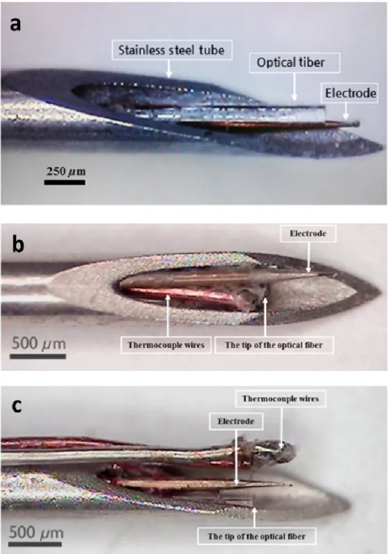
few hundred micron , the often used high-power laser sources limits the core diameter of fibers, which are too bulky for intracortical use. Additional disadvantage of these fragile components that does not provide any information on the evoked cellular response, therefore they make the use of further recording implants necessary (Furuyama, 2019). An early attempt of Duke et al showed that combination of optical fibers with nerve cuff electrodes is a feasible solution to extend functionality of fiber-based optical stimulation (Duke, 2012). They demonstrated that the IR excitation of the rat sciatic nerve with concurrent electric stimulation is possible through the transparent silicone based cuff electrode using 2.03±0.43 J/cm2 at 2 Hz. The 0.6 mm thick nerve cuff was suitable to transmit 93% of IR light (λ = 1875 nm). The very first deep-brain application of a fiber-based IR stimulation was presented by Yoo et al (Yoo, 2013), who modulated the activity of thalamus through a stainless steel guide electrode hosting a 62.5 µm core diameter optical fiber, a recording wire and a thermocouple (see Figure 2). A more recent work of Dautrebande et al showed the operation of a hybrid cuff electrode consisting of a standard, flexible cuff electrode combined with and a 200 µm core diameter multimode optical fiber (Dautrebande, 2018). The cuff also contained three stainless steel wires to either stimulate or record electrical activity of the targeted nerve.
4.1.2 MEMS based devices
Microtechnology, developed for several decades by the semiconductor industry, also plays an important role in today’s implantable microdevices. Due to the advancement in micromachining processes, especially in photolithography, truly integrated optrodes can be manufactured. These devices may hold multiple functionalities on top of the delivery of IR light. Since the mass-production scheme of these microscale optrodes provides low variability in operational parameters between individual devices, reproducibility and spatial precision of the stimulation in experiments are outstanding (Fekete, 2015;
Goncalves, 2017). Further advantage of the applied micro- and nanoengineering techniques that location of additional integrated sensors (like electrophysiological recording sites, temperature or neurochemical sensors) can be tailored at micronscale precision with respect to the optical interface (Fekete, 2017).
Regarding the operation of the integrated optical actuator in such optrodes, passive or active neural probes can be distinguished.
There are passive and active MEMS based optrodes. Passive optrodes are able to deliver electromagnetic radiation from external light sources into the deep tissue, usually laser diodes or LEDs, through integrated waveguides. Regarding the hardware configuration of passive optrodes, Utah microelectrode arrays (UMEA) and Michigan-type neural probes are commonly used. Since there is inherently a significant distance between the light source and the stimulation target, several loss mechanism should be considered along the beam path (Goncalves, 2017). Due to Fresnel reflection, large number of photons are lost in the boundary of waveguide material air interface and at the end facet of the waveguide. Due to imperfections in total internal reflection through the waveguide (e.g. surface roughness, change in cross-section, surface impurities), there is propagation loss. In contrast, active optrodes hold light source located in the close vicinity of the target tissue, and emitted photons are directly interacting with cells (Iseri, 2017). Nevertheless, current supply of these integrated active optical sources (e.g. µLEDs, µSELD, VCSEL) is to be implemented on the implantable part of such optrodes, which may contribute to parasitic tissue heating and cross-talk between stimulation current and electrophysiological functions (Dong, 2018).
Utah arrays
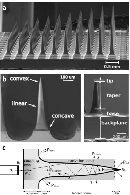
Utah microelectrode arrays (UMEA) are implantable microdevices developed to carry out intracortical interrogation of neurons (Maynard, 1997). A typical device is made of silicon and it consists of a microneedle matrix each needle holding a recording site at its tip. The characteristic length of these out-of-plane microstructures are in the range of 0.5-2 mm, which is inherently limited by the thickness of silicon wafers used as substrate materials for microfabrication. The safety use of UMEA even in human subjects has been proven in several works in the past (Normann, 2016). For this reason of approved biocompatibility in the clinical practice makes it a worthwhile platform to be also used for stimulation purposes. The first concept of a UMEA capable of infrared stimulation was proposed by the Solzbacher group (Abaya, 2012; Abaya, 2014) (see Figure 3). They demonstrated that microneedles of UMEA can be utilized as individual optical waveguides, however, the tapering profile is accompanied by a large quantity of scattered light, which deteriorates spatial precision of the infrared stimulation. Nevertheless, they demonstrated that such a device coupled to a 50 micron thick optical fiber can transmit the incoming IR light (λ = 1550 nm) with an efficiency of 34.7%, which may be well sufficient for a reasonable use for INS.
Based on their geometrical study, the output power was depending mainly on length/taper profile and masking the coupled light from large diameter fibers were found critical. An important aspect of the work was that the created microsystem was connected to high-power Capella lasers, and is therefore
compatible with experiment using high energy lasers at pulsed operation without device failure. However, there is a great potential in using an optical UMEA in preclinical studies, further efforts in device development was not continued and in vivo validation has never been presented.
Michigan probes
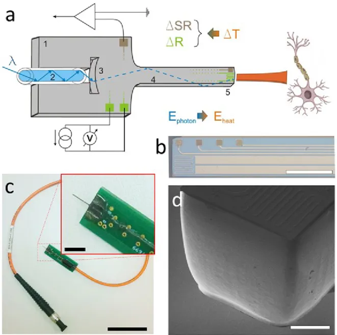
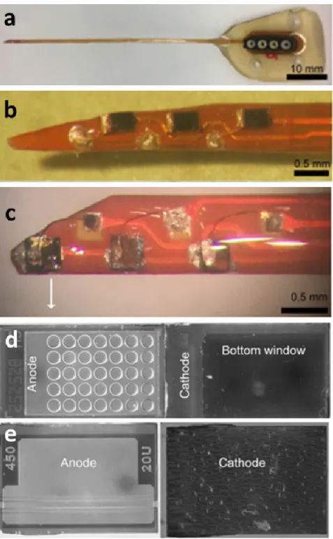
Michigan-type optrodes are relying on in-plane needle like structures fabricated using planar microtechnology. Essential feature of such devices is that on top of integrated light sources or light delivery microstructures, they are able to hold multiple recording sites offering high-density neural recording around the stimulation spot. Early devices developed for optogenetics have been demonstrated (Royer, 2010; Zorzos, 2012). The first Michigan-type optrode microdevice, entirely addressed for IR delivery and stimulation was proposed by Kiss et al (Kiss, 2016). They developed a technical solution, relying wet chemical polishing, to transform the mechanical carrier of silicon neural probes into optical waveguides. Later on, Horvath et al. proved that these devices, also holding integrated electrophysiological sensors, are able to deliver the infrared light in a spatially controlled fashion [Horváth, 2018a; Horváth, 2018b] (see Figure 4). Boros et al provided a coupled multiphysical model to predict the thermally affected region around the device (Boros, 2018), and also determined guidelines to optimize the optical system to increase efficiency, while reducing the risk of mechanical failure (Boros, 2019). Based on the in vivo demonstration of the device safe operation was confirmed in the rat somatosensory cortex and hippocampus [Horváth, 2020]. Evaluating the artefact-free electrophysiological recordings, they found that both excitation and inhibition of neuronal activity can be achieved in a reversible manner using their integrated optrode.
Active optoelectronic probes
Optrodes holding active light sources provide freedom to tailor the stimulation pattern and location inside the tissue. Furthermore, the use of active light emitting components on the spot eliminates the inherent optical loss exhibited by waveguide structures, and therefore promotes high power density stimulation. Inorganic GaN based microLEDs integrated on neural probes have been demonstrated to control individual neurons in the mouse hippocampus in optogenetic experiments (Wu, 2015; Scharf, 2016, Klein, 2018). However, these microengineered solutions mainly aim the activation of opsins sensitive to visible light, configuration of these optical platforms may be adapted in later development of INS-oriented brain implants. Due to the evolution of red-shifted opsins, further technological approaches have been utilized, which are getting closer to the NIR regime regarding stimulation wavelength. Lee et al
introduced a flexible AlGaInP vertical light-emitting diodes (VLEDs) for activation of specific functional regions of the mouse cortex (Lee, 2018). The size of these components is still substantially greater than that of microLEDs, and therefore are not particularly suitable for deep tissue applications, their approach to combined VLEDs with ECoG opened up new opportunities towards infrared stimulation as well. The first active optrode device developed for INS has been presented recently, by proposing vertical-cavity surface emitting laser (VCSEL) and superluminescent (SLED) diode arrays integrated in a flexible optical delivery system implanted in the scala tympani of cats and guinea pigs (Xu, 2018) (see Figure 5). Their device held 15 light sources along a 24 mm long array. These devices with a center wavelength around 1850 nm, and high energy (up to 20 µJ/pulse) makes these chip-scale components good candidates in future devices, especially in the field of hear implants where the insertion of passive fiber bundles may potentially cause mechanical damage inside the cochlea (Balster, 2014). Besides the beneficial features of integrated light sources like VCSELs for infrared neural stimulation (Dummer, 2011), such specific device for INS research in the brain is still awaited.
4.2 Indirect photomodulation using infrared light
There are several examples in the literature of neuromodulation utilizing infrared light indirectly to control cellular activity. In these cases, micro- and nanoscale transducers are addressed by IR light, which eventually exploits the beneficial propagation properties of NIR radiation in the tissue. These emerging approaches are presented, because they hold a high potential in future studies in neuromodulation, and may provide additional engineered features to experimental setups aiming to reveal the biophysical mechanism of INM.
4.2.1 Polymer-mediated approaches
Among other polymer materials conjugated polymers (CPs) with extended - conjugation along molecular backbone, are extensively used in various research field related to organic optoelectronic applications, such as fluorescence imaging, photodynamic therapy (PDT) (Celli et al., 2010; Ethirajan, Chen, Joshi, & Pandey, 2011; Lucky, Soo, & Zhang, 2015; Z. Zhou, Song, Nie, & Chen, 2016; Huang et al., 2016), photothermal therapy (PTT) (Shengliang Li et al., 2016; Cheng, Wang, Feng, Yang, & Liu, 2014), two – photon live cell imaging (Rahim et al., 2009; Shuang Li et al., 2014; Lv et al., 2015; Geng et al., 2014) and in neuromodulation to tune neural firing (Feyen et al., 2016). As a result of the delocalized - electron
cloud, CPs are own exceptional optoelectronic characteristic, however CPs are inherently hydrophobic organic macromolecules. For their application in biological environment, chemical modification on the backbone with charged side chains should be implemented. Few CPs exhibit remarkable absorption in the near infrared (NIR) wavelength range (Liu, 2018). Feyen et al. investigated the feasibility of poly[2,6-(4,4- bis-(2-ethylhexyl)-4H-cyclopenta[2,1-b;3,4-b′]dithiophene)-alt-4,7(2,1,3-benzothiadiazole)] (PCPDTBT) as a potential tool for polymer mediated indirect photomodulation of neural activity, as PCPDTBT is photosensitive in the NIR range. The group managed to modulate firing activity at the interface between CP and mouse hippocampal slices by prolonged NIR illumination. They observed a sustained reduction of spiking activity during the light pulse and surprisingly the inhibitory effect extended to the post – stimulus phase. In PDT, CPs either act as photosensitizer (PS), where emission energy is directly transferred from CPs to tissue oxygen in order to produce toxic molecular species, such as reactive radicals and singlet oxygen (1O2) or CPs act as energy transfer structure to PS (Feng et al., 2017; Yu et al., 2019). Wang et al.
demonstrated a NIR – responsive conjugated polymer nanoparticles (CPNs) by coprecipitating conjugated polymer, poly[2,6′-4,8-di(5-ethylhexylthienyl)benzo[1,2-b;3,4-b]dithiophenealt-5-dibutyloctyl-3,6-bis(5- bromothiophen-2-yl)pyrrolo[3,4-c]pyrrole-1,4-dione] (PDPP-DBT) with matrix polymer 1,2-distearoyl-sn- glycero-3-phosphoethanolamine-N-[maleimide(polyethylene glycol)-2000] (DSPE-PEG2000-MAL) from tetrahydrofuran (THF) solution into water (PDPP-DBT/MAL CPNs). Synthetized CPNs can be further modified with positively charged Tat peptide, that assists in the interaction between CPNs and cells. The resultant CPNs – Tat was observed to have rapid and superior NIR laser induced photothermal conversion efficiency. As a proof – of – concept, due to NIR laser illumination (808 nm), heat – induced gene expression was activated by CPNs – Tat in living cells. Heat inducible heat shock protein – 70 promoter (HSP – 70) was used to mediate gene expression and EGFP gene was selected as target gene. In their experiment HeLa cells were transfected with pCDNA3.1-HSP-70-EGFP, and CPNs – Tat produced sufficient heat ( 42 C) to trigger the activation of pCDNA3.1-HSP-70-EGFP plasmid expression. The HSP – 70 promoter started the transcription of the downstream EGFP gene, thus the so produced green fluorescent protein (GFP) was detected with confocal laser scanning fluorescence microscopy (Wang et al., 2018). M.
Leccardi et al. presented a photovoltaic organic interface tailored for retinal stimulation in the NIR spectral range, and to induce artificial vision in blind patients. They provided a new blend of polymers to improve the stability of nanocomposites in aqueous environment and to modulate photoelectric stimulation efficiency, in order to be responsive in the NIR spectral range. Poly[2,6-(4,4-bis-(2-ethylhexyl)-4H- cyclopenta [2,1-b;3,4-b′]dithiophene)-alt-4,7(2,1,3-benzothiadiazole)] with [6,6]-Phenyl-C61-butyric acid methyl ester (PCPDTBT: PC60BM) was blended to form bulk heterojunction (BHJ). Ex vivo electrophysiology
experiment on explanted retinas from 4 – months old retinal degeneration, a mice model for retinitis pigmentosa was used to validate the functionality of the NIR – responsive photovoltaic prosthesis. They concluded that due to the presence of BHJ and consequently due to photovoltaic stimulation, firing activity in retinal ganglion cells were induced and recorded under NIR light pulses at a safe irradiance level (Leccardi et al., 2020).
Lyu et al. designed and synthetized a semiconducting polymer nanobioconjugates (SPNbc) for targeted photothermal nanomodulation of thermosensitive ion channel is neurons and provided a platform for specific and rapid activation of the intracellular Ca2+ influx. SPNbc comprises of poly (cyclopentadithiophene-alt-diketopyrrolopyrrole), designed to efficiently absorb the NIR light at 808 nm and coprecipitated with polystyrene-b-poly (acrylic acid) (PS – PAA). PS – PAA is an amphiphilic polymer that ensures good water – solubility, besides it has another role in the nanosystem to provide reactive functional groups (carboxyl groups) on the surface for postconjugation. They confirmed the conjugation with anti – TRPV1 (transient receptor potential vanilloid 1) antibody via carbodiimide coupling reaction was successful. They also analyzed the targeting capability of SPNsbc with fluorescence live cell imaging.
Mouse neuroblastoma / rat dorsal root ganglion (DRG) neuron hybrid ND7/23 cells that intrinsically express TRPV1 on their plasma membrane were chosen as positive control cells, while human cervical carcinoma HeLa cells were used as the TRPV1 negative control. They found that the main advantages of SPNsbc over eg. gold nanorods that can be used in similar applications, are its superior photothermal conversion efficiency ( 20 %), rapid heating capability, excellent photothermal stability and its also completely organic and biologically inert in contrast with heavy metal ion – induced toxicity. Maximum photothermal temperature of 70 °C was reached and plateaued at t = 360 sec. To sum it up, SPNbc with its rapid heating capability was able to quickly increase the local temperature above the threshold of 43 C, therefore mediated photothermal activation of TRPV1 ion channels, inducing intracellular Ca2+ influx. Due to the efficient photothermal conversion, the laser irradiation time could be minimized in living organisms, accordingly the probability of noxious heat could be reduced (Lyu, Xie, Chechetka, Miyako, & Pu, 2016).
Later on Lyu et al. developed a dendronized semiconducting polymer (DSP) as a gene delivery system that has NIR conversion efficiency around 44 %, which means the nanocarrier can be used to mediate remote photothermal activation of gene expression upon NIR irradiation (808 nm) at a designated time and location. It is useful in applications, where a heat inducible promoter is being involved in the gene regulatory process (eg. HSP70). DSP has a hydrophobic backbone acts as a NIR photothermal transducer, a cationic polyamidoamine (PAMAM3) side chains as gene vectors and poly(ethylene glycol) (PEG) blocks to ensure water-solubility. In this case, the maximum temperature of 58.8 °C was reached and plateaued
at t = 480 sec. Their work is a proof-of-concept to optically regulate transgene systems and may pave the way for therapeutic approaches to treat inherited genetic diseases and cancer with minimizing the chance of off – target gene expression (Lyu et al., 2017).
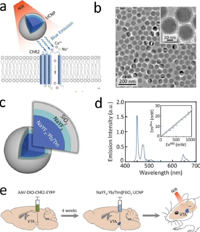
4.2.2 Upconversion nanoparticle mediated applications
Nanoscale particles that exhibit photon upconversion are called upconverting nanoparticles (UCNPs). During photon upconversion, the incident low energy photons are absorbed and a photon with higher energy is emitted [Wang, 2009]. The synthetic nanoparticles are composed of a host lattice, an activator ion and a sensitizer ion. Each of them has an important role to tailor conversion efficiency and spectral characteristics. In the case of host material, cations like alkali, alkaline-earth, rare-earth and transition metal ions and anions like fluorides are good candidate, because they provide high chemical stability to hold the activator and the sensitizer ion and have low phonon energy which helps the energy transfer efficiently. Activator and sensitizer ions are co-doped into the host lattice to produce higher energy anti-Stokes luminescence. Selection of the proper dopants determines results in wavelength (color) selective upconversion, such as NIR to shorter NIR, visible (blue, green, red), or UV [Wen, 2013].
Lanthanide doped nanocrystals are one of the most popular systems that convert near infrared into visible emission, and has been exploited in applications like molecular sensing [Gu, 2018], bioimaging [Da Costa, 2014] and therapy [Chen, 2014].
NIR excitation of optogenetic targets is new popular line of research, which offers the outstanding penetration depth of NIR in the deep tissue, and preserves the anatomic precision of the molecular sensitization (see Figure 6). First, Wu and co-workers demonstrated that upconversion nanoparticles can be efficiently utilized as optical transducers to excite neural firing using near-infrared light [Wu, 2016].
Later on, Lin et al came up with an alternative UCNP construct with an emission wavelength matching excitation wavelength of inhibitory opsins and that way they suppressed the electrical activity of nearby cells [Lin, 2018]. Stimulation following this strategy has been demonstrated with a transcranial method (Chen, 2019). These efforts rely on primitive means of UCNP administration into the living body, and would definitely benefit from the micro-engineered environment, which guarantees the spatially controlled in vivo use of these particles, also complemented with direct feedback of their operation through the integrated features of the implants. The very first attempt has been presented by Wang et al, presenting polypropylene based spinal cord stimulator, which provided wireless excitation of upconversion nanoparticles dispersed in the device substrate (Wang, 2020). Further solutions to exploit the capability of these optical nanotransducers may be expected in the future.
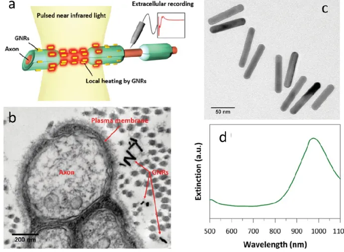
4.2.3 Thermoplasmonic neurostimulation
The phenomenon of surface plasmon resonance is commonly used in biosensors to monitor the interaction between a mobile molecule in an analyte and its biospecific complement immobilized on the surface (Nguyen, 2015). Oscillations of free electrons of a smooth metal surface also called as surface plasmons are excited and detected using polarized light coming at a certain incident angle. In the presence of metal nanoparticles of a size comparable to the wavelength of the incident light waves, surface plasmons are confined or in other words localized. When this localized surface plasmons are excited by the incident light of appropriate wavelength, hot charge carriers are created. Redistribution of these hot carriers inside the nanoparticles eventually transfers the absorbed photon energy into the metallic lattice through electron-photon collisions, and dissipates heat to the surrounding medium. Besides the potential use in bioimaging, drug delivery and tumor therapy, plasmonically triggered localized heat was proposed for controlled modulation of neural activity by Eom et al (Eom, 2014) (see Figure 7). They activated the rat sciatic nerve using pulsed infrared light (λ = 980 nm). Eliciting compound nerve action potentials, the stimulation thresholds were found to be 0.159 J/cm2 with gold nanorods and 0.480 J/cm2 without gold nanorods. They also observed in histological tests that there was no apparent damage in the irradiated tissue below 2.23 J/cm2 radiant exposure. The Stoddart group also presented the NIR stimulation (λ = 780 nm) of primary auditory neurons with gold nanorods using a constant 90 mW peak laser power with manually specified pulse length (Yong, 2014). Suppression of the hyperactivity of the cardiac sympathetic nerves was also demonstrated using gold nanorods injected into the left stellate ganglion of dogs (Ye, 2019). As the nanoparticle assisted stimulation of neurons has been explored in more details, complex microengineered solutions have been proposed to modulate cultured neurons through NPs embedded in microelectrode arrays (MEA). Neuronal firing evoked by electrical stimulation and signal propagation along neurites were inhibited with NIR via an integrated platform (Yoo, 2016). The gold nanorod coated MEA of Jung et al was combined with a digital micromirror device to achieve patterned NIR modulation of E18 rat hippocampal neurons (Jung, 2017). Inhibition of the firing of hippocampal neurons through micronscale resolution of the thermoplasmonic heating was first demonstrated by Kang et al using inkjet- printed method to create gold nanorod patterns (Kang, 2018). These efforts of combining micro- and nanotechnology may potentially lead to novel results through high spatial precision of NIR excitation and inhibition in both in vitro and in vivo experiments.
5. Future perspectives
Our study pointed out that due to the increasing number of experimental works in this field, infrared neuromodulation can be used for safe stimulation of neuronal ensembles either in vitro or in vivo.
Numerous studies involving various biological species confirmed the controlled manipulation of action potential generation and propagation. Behind the mechanism of INM various biophysical, molecular and physiological events are hypothesized, however, the recent advancements evoked by both in-depth theoretical explanations and models as well as novel experimental results provide a much clear view on the role of each component. The concurrent use of electrophysiology, fluorescence neuroimaging, pharmacology and genetic methods is gradually help us to understand the complex nature of INM, and will likely lead to new discoveries also regarding the biophysical mechanism. In spite of the thorough work on the fundamentals of INM, there are only a few attempts in the literature aiming the use of INM in a preclinical or clinical setting. Further efforts towards the translation of current results should be urged.
Researchers and clinicians paving the way of INM towards human-oriented applications should unite the existing knowledge of neighboring disciplines like transcranial low level laser therapy, transcranial photobiomodulation and infrared neural stimulation, which seems to fail to exploit the experimental findings of one another. Especially the therapy of brain disorders and management of pain in humans using INM holds a great potential, which may be promoted by the state-of-the-art implantable micro- and nanoengineered solutions, like integrated optrodes and microelectrode arrays or nanoparticle-mediated approaches.
Acknowledgement
The author is thankful to the National Brain Research Program (grant: 2017_1.2.1-NKP-2017-00002), New National Excellence Program of the Ministry for Innovation and Technology (UNKP-19-4-PPKE-9) and Bolyai János Scholarship of the Hungarian Academy of Sciences. The support of the European Union through the grant EFOP-3.6.3-VEKOP-16-2017-00002 co-financed by the European Social Fund is also acknowledged.
References
Abaya, T. V. F., Diwekar, M., Blair, S., Tathireddy, P., Rieth, L., Clark, G. A., & Solzbacher, F. (2012).
Characterization of a 3D optrode array for infrared neural stimulation. Biomedical optics express, 3(9), 2200-2219.
Abaya, T. V. F., Diwekar, M., Blair, S., Tathireddy, P., Rieth, L., & Solzbacher, F. (2014). Deep-tissue light delivery via optrode arrays. Journal of biomedical optics, 19(1), 015006.
Abdullaeva, O. S., Schulz, M., Balzer, F., Parisi, J., Lützen, A., Dedek, K., & Schiek, M. (2016).
Photoelectrical stimulation of neuronal cells by an organic semiconductor–electrolyte interface.
Langmuir, 32(33), 8533-8542.
Akhavan, O., & Ghaderi, E. (2013). Flash photo stimulation of human neural stem cells on graphene/TiO 2 heterojunction for differentiation into neurons. Nanoscale, 5(21), 10316-10326.
Balster, S., Wenzel, G. I., Warnecke, A., Steffens, M., Rettenmaier, A., Zhang, K., ... & Reuter, G. (2014).
Optical cochlear implant: evaluation of insertion forces of optical fibres in a cochlear model and of traumata in human temporal bones. Biomedizinische Technik/Biomedical Engineering, 59(1), 19-28.
Barrett, D. W., & Gonzalez-Lima, F. (2013). Transcranial infrared laser stimulation produces beneficial cognitive and emotional effects in humans. Neuroscience, 230, 13-23.
Barrett, J. N., Rincon, S., Singh, J., Matthewman, C., Pasos, J., Barrett, E. F., & Rajguru, S. M. (2018).
Pulsed infrared releases ca2+ from the endoplasmic reticulum of cultured spiral ganglion neurons.
Journal of neurophysiology, 120(2), 509-524.
Bec, J. M., Albert, E. S., Marc, I., Desmadryl, G., Travo, C., Muller, A., ... & Dumas, M. (2012).
Characteristics of laser stimulation by near infrared pulses of retinal and vestibular primary neurons.
Lasers in surgery and medicine, 44(9), 736-745.
Beier, H. T., Tolstykh, G. P., Musick, J. D., Thomas, R. J., & Ibey, B. L. (2014). Plasma membrane
nanoporation as a possible mechanism behind infrared excitation of cells. Journal of neural engineering, 11(6), 066006.
Benabid, A.L., Chabardes, S., Mitrofanis, J. and Pollak, P., 2009. Deep brain stimulation of the subthalamic nucleus for the treatment of Parkinson's disease. The Lancet Neurology, 8(1), pp.67-81.
Bonmassar, G., Lee, S.W., Freeman, D.K., Polasek, M., Fried, S.I. and Gale, J.T., 2012. Microscopic magnetic stimulation of neural tissue. Nature Communications, 3, p.921.
Boros, Ö. C., Horváth, Á. C., Beleznai, S., Sepsi, Ö., Csősz, D., Fekete, Z., & Koppa, P. (2019). Optimization of an optrode microdevice for infrared neural stimulation. Applied optics, 58(14), 3870-3876.
Boros, Ö. C., Horváth, Á. C., Beleznai, S., Sepsi, Ö., Lenk, S., Fekete, Z., & Koppa, P. (2018). Optical and thermal modeling of an optrode microdevice for infrared neural stimulation. Applied optics, 57(24), 6952-6957.
Borrachero‐Conejo, A. I., Adams, W. R., Saracino, E., Mola, M. G., Wang, M., Posati, T., ... & Hutchinson, M. R. (2020). Stimulation of water and calcium dynamics in astrocytes with pulsed infrared light. The FASEB Journal.
Boyden, E.S., Zhang, F., Bamberg, E., Nagel, G. and Deisseroth, K., 2005. Millisecond-timescale, genetically targeted optical control of neural activity. Nature Neuroscience, 8(9), p.1263.
Carvalho-de-Souza, J. L., Treger, J. S., Dang, B., Kent, S. B., Pepperberg, D. R., & Bezanilla, F. (2015).
Photosensitivity of neurons enabled by cell-targeted gold nanoparticles. Neuron, 86(1), 207-217.
Caterina MJ, Schumacher MA, Tominaga M, Rosen TA, Levine JD, Julius D (1997) The capsaicin receptor:
a heat-activated ion channel in the pain pathway. Nature 389:816–824
Cayce, J. M., Friedman, R. M., Jansen, E. D., Mahavaden-Jansen, A., & Roe, A. W. (2011). Pulsed infrared light alters neural activity in rat somatosensory cortex in vivo. Neuroimage, 57(1), 155-166.