Glial architecture and extracellular matrix of circumventricular organs
Ph.D. theses
Dr. Károly Pócsai Semmelweis University
János Szentágothai Doctoral School of Neurosciences
Supervisor: Dr. Mihály Kálmán D.Sc.
Reviewers: Dr. Klára Matesz D.Sc.
Dr. Anna L. Kiss Ph.D.
Chair of the Ph.D. examination committee: Dr. Pál Röhlich D.Sc.
Members of the Ph.D. examination committee: Dr. Attila Magyar Ph.D.
Dr. Zsuzsanna Környei Ph.D.
Budapest 2015.
1
Introduction
The circumventricular organs are areas with special structure in the wall of the cerebral ventricles. They are characterized by specific ependyma and other glia and take part in the regulation of energy- and salt-water homeostatis, apetite, circulation, fever and immune responses. Their main function is neurosecretion (see e.g. median eminence: releasing and release inhibiting hormones to the adenohypophysis; neurohypophysis: oxitocin and vasopressin; pineal body: melatonin) and chemoperception of several humoral factors (e.g. angiotensin II, cholecystokinin, ghrelin, leptin – see McKinley et al. Adv Anat Embryol Cell Biol 172:1-127).
Because of their function the circumventricular organs require free access to cerebral blood, so the vascular structure of them (except for the subcommissural organ’s) is different from that of other brain areas, and lack the blood-brain barrier.
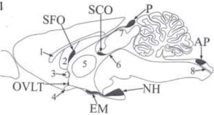
Figure 1. shows the location of the circumventricular organs of the mammalian brain. The circumventricular organs investigated by us: area postrema (AP), median eminence (EM), neurohypophysis (NH), vascular organ of the lamina terminalis (OVLT), subfornical organ (SFO) and pineal body (P). The anatomical structures for better orientation: 1 – corpus callosum, 2 – fornix, 3 – anterior commissure, 4 – optic chiasm, 5 - interthalamic adhesion, 6 –cerebral aquaeduct, 7 - superior collicle, 8 – central canal. The subcommissural organ (SCO - having a blood-brain barrier) and the choroid plexuses (not unanimously recognized as a circumventricular organ) were not investigated.
In the circumventricular organs investigated there is a perivascular space between the glial (parenchymal) and vascular basal laminae (Krisch et al.
Cell Tissue Res 195:485-497). Otherwise, in mature, intact brain this
2
phenomenon is restricted to the initial segments of brain vessels intruding from the pial surface into the brain parenchyma, while along the deeper segments the two basal laminae are fused into a common basal lamina.
Laminin is one of the main components of the basal laminae, even so the brain vessels in general are characterized by the absence of laminin- immunoreactivity. The reason of this pehomenon, according to Krum et al.
(Exp Neurol 111:152-165) is the ’hiding’ of the laminin-epitopes by the fusion of the glial and vascular basal laminae. That is why only those vessel segments are delineated by laminin-immunoreactivity along which this fusion is not complete.
The dystroglycan is a ubiquitous cell surface receptor anchoring the cells to the basal lamina. It consists of two subunits (α,β) and forms the dystroglycan complex together with other proteins, e.g. utrofin and dystrobrevin. In the brain the meningeal- and perivascular glia end-feet contain the dystroglycan complex.
Tanycytes are held to be the characteristic ependymal cells covering the circumventricular organs. These cells have kept their radial glia-like features: their long process link them to brain vessels or to the meningeal surface.
The role played by the glia in the induction of the blood-brain barrier and in the production of laminin is known. GFAP (glial fibrillary acidic protein), vimentin and nestin are components of intermediate filaments of glial cells. GFAP is known as an astroglia marker. Vimentin and nestin as characteristic proteins of immature neuroepithelial cells are capable for marking, among others the radial glia and the glia precursor cells. Further astroglia markers are: the S100 calcium-binding protein and the glutamine synthetase, which enzyme takes part in the elimination of unwanted and toxic glutamate leftover after the synaptic transmission.
The central nervous system has an extracellular matrix as well found dispersed in the neuropil, or forming perineuronal nets. It has an important role in brain development, cell migration, axon growth, synaptogenesis, brain vascularization, cell adhesion, tissue hydration, the binding of the soluble factors, and the control of their diffusion.
The main extracellular matrix components are: i) the hyaluronic acid, a glycosaminoglycan molecule with great water binding capability; ii) proteoglycans, mainly the chondroitin sulphate containing and hyaluronic acid binding, so-called ‘hyalectans’; iii) glycoproteins, in the brain mainly laminin, fibronectin and tenascin (in the mature brain mainly the –R isoform of the latter one). The most abundant brain ‘hyalectans’, as the versican, aggrecan, neurocan and brevican are also called 'lecticans' because of their
3
capability for binding certain lectins, e.g. the Wisteria floribunda agglutinin (WFA).
Aims
In circumventricular organs lacking the blood-brain barrier (median eminence, OVLT, SFO, pineal body, neurohypophysis, area postrema) we investigated what kind of modifications can be found regarding the glial architecture and the extracellular matrix with special respect to the: - basal lamina;
- gliovascular connections;
- glial architecture bordering the organs.
By these regards what kind of similarities and differences can be found:
i) comapring the circumventricular organs with the rest of the brain, ii) between the circumventricular organs, and
iii) between the subdivisions of a circumventricular organ.
In circumventricular organs of adult rats we investigated:
a) with immunohistochemical method:
- a main component of the basal lamina (laminin);
- the dystroglycan-complex (β-dystroglycan, α1-dystrobrevin, utrofin) important in the gliovascular connection;
- the water channel protein (aquaporin-4) ’anchored’ by the dystroglycan- complex;
- specific proteins (GFAP, glutamine synthetase, nestin, S100, vimentin) of mature and immature glial cells;
- the distribution of some components of the extracellular matrix (aggrecan, brevican, neurocan, versican, tenascin-R), and
b) with lectinhistochemical method the distribution of the WFA-binding loci.
We applied several combinations of double immunohistochemical labelings, which were examined with a confocal laser scanning microscope.
Methods
The animals used were adult male and female rats (Wistar, 250-300g).
The animal experiments were performed according to the directive 86/609/EEC of The Council of the Europen Communities (license number:
22.1/3453/003/2009). Following ketamine-xylasine anaesthesia (i.m., 20 and 80 mg/kg, respectively) the animals were perfused through the aorta (physiological saline solution followed by 4% paraformaldehyde).
4
The sections (50μm) were cut in the coronal, sagittal or horizontal planes and most of them underwent immunohistochemical processing using fluorescent secondary antibodies or the avidin-biotinylated peroxidase method. The WFA-binding was also visualised with the latter method. In most cases the fluorescent immunolabelings were investigated with a Radiance-2100 (BioRad, USA) confocal laser scanning microscope.
Colocalization of antibodies was confirmed on the basis of records made in two perpendicular phocus planes.
Electron microscopic observations were made on specimens a) underwent pre-embedding immunohistochemical reactions against laminin or β-dystroglycan; or b) without immunohistochemical processing.
Results
Vessel types distinguished on the basis of laminin-β-dystroglycan double labeling in the circumventricular organs
a) ’double-walled’, i.e. surrounded by two, laminin-immunopositive layers. Along the outer ’wall’ the β-dystroglycan-immunoreactivity was faint and uncertain, mainly it was visible only on the corresponding monochromatic photo;
b) ’double-walled’, the inner ’wall’ was only laminin-immunoreactive, while the outer one showed laminin-β-dystroglycan double immunoreactivity;
c) ’single-walled’, a perivascular space was not recognisable under the light microscope, the colocalization of laminin and β-dystroglycan was seen in yellow;
d) ’single-walled’, only β-dystroglycan-immunoreactive, like the brain vessels in general.
These types of vessels usually represent vessel segments which continue in each other gradually and show zonal distribution, although not every type was found in all circumventricular organs.
5
SFO
Figures 2AB help to illustrate our results on the SFO (sagittal and horizontal planes, respectively). The three surfaces of the SFO: i) pial (dashed line); ii) ventricular (continual line), which faces to the 3rd ventricle (3V); iii) ’parenchymal’, which faces to the ventral hippocampal commissure (VHC). The pail surface forms invaginations (’holes’ on the figure of the horizontal plane) for the organ’s great vessels from which rise (arrowheads) the smaller vessels supplying the organ. On the border of the ventricular and pial surfaces attaches (arrows) the lamina epithelialis choroidea to the SFO. A shell-like (dotted) and a core-like (plain) area can be distinguished in the organ. The ependymal cover is cuboidal on the former and flattened on the latter part (thick and thin lines, respectively).
The immunoreactivity of GFAP, S100 and aquaporin-4 marked the ’shell’, the glutamine synthetase-immunoreactive cells were also found mainly in this area but too scarcely to mark a territory. The vessels were type ’d’ in the ’shell’, i.e. β-dystroglycan-immunoreactive but laminin- immunonegative, and their glial coverage and the aquaporin-4- immunoreactivity around them was continuous.
The ’core’ is rich in nestin-immunoreactive elements, but poor in GFAP-immunoreactive ones. The vessels of this area were types ’a’ and ’b’.
The type ’a’ vessels were located in the zone closest to the ventricle (like the ’core of the core’), this zone was more or less surrounded by the zone of the type ’b’ vessels. The immunoreactivity of GFAP, vimentin and aquaporin-4 was not continuous around the vessels of the ’core’. At the border of the ’core’ and ’shell’ there were transitional forms (type ’c’).
The vimentin-immunoreactivity distributed evenly throughout the organ, so in this case there was no ’core-shell’ difference, but it almost disappeared
6
at the parenchymal surface of the organ. Vimentin’s colocalization was scarce with GFAP, but it was frequent with nestin. The flattened ependymal cells covering the ’core’ were aquaporin-4-immunonegative, in contrast to the ependyma in general, including the ependymal coverage of the ’shell’.
The cell bodies to which the perivascular processes belonged were seen only occasionally. In the case of vimentin these cell bodies were found either in the parenchyma or the ependymal coverage of the SFO, while the GFAP-immunoreactive processes rised almost exclusively from parenchymal (not ependymal) cell bodies.
Area postrema
The location of the area postrema (Figures 3AB, sagittal and frontal planes, respectively) is similar to that of the SFO. The parenchymal surface of this organ faces to the nucleus tractus solitarii (NTS), while the ventricular one faces to the 4th ventricle (4V). The organ’s ependymal coverage consists of flattened cells (thin, continual line) in contrast to the cuboidal cells (thick, continual line) covering the nucleus tractus solitarii and the lamina epithelialis choroidea, the latter attaches (arrow) to the area postrema at the border of the pial (dashed line) and vintricular surfaces.
From the intrusions of the pial surface smaller vessels penetrate (arrowheads) the organ.
At the parenchymal surface of the organ there is a ’glial-fence’ (dotted area) which showed more intense GFAP-, vimentin- and nestin- immunoreactivity than the organ’s inner part (plain area). The same trend was seen in the case of aquaporin-4, in the inner part of the organ it was found mainly along the vessels, but it did not delineate them as continuously as the brain vessels in general. The ependymal cells covering the organ were aquaporin-4-immunonegative. Vimentin was frequently seen in colocalization with either GFAP or nestin. While only GFAP- immunoreactivity marked a glial process-system running perpendicularly to the ’glial-fence’ as a connection between the nucleus tractus solitarii and the
7
area postrema (and its vessels). The immunoreactivity of S100 was intense throughout the organ, while that of glutamine synthetase marked only few cells.
The vessel types distinguished on the basis of laminin-β-dystroglycan double labeling showed zonal distribution also in the area postrema. The types ’a’ and ’b’ were found mainly in the anterodorsal part of the organ, the type ’c’ occurred quite scarcely, mainly near to the parenchymal surface of the organ. The laminin-immunoreactivity of the vessels disappeared where they passed the ’glial-fence’ toward the nucleus tractus solitarii, where only type ’d’ vessels were seen, like in the brain tissue in general.
OVLT
Figures 4AB of the OVLT (mediansagittal and frontal planes, respectively) show that at the rostromedial part of the organ the pial surface (dashed line) forms a pit-like intrusion (in the frontal plane it is a ’hole’), which - altogether with the vessels penetrating into it - is called the ’vascular subdivision’ (RV). From the ’pit’ vessels penetrate (arrowheads) the organ’s so-called ’posterior-’ (PPv) and ’lateral periventricular’ (LPv) subdivisions, where they form an ’inner capillary plexus’. The mentioned subdivisions are ’covered’ by the so-called ’dorsal cap’ (DC) subdivision. The ependymal cells covering the 3rd ventricle (3V) are flattened (thin, continual line) at the PPv and LPv subdivisions, but cuboidal (thick, continual line) at the optic chiasm (OC) and at the more dorsal parts of the lamina terminalis.
8
Two glial process-systems can be distinguished in the glial architecture of the organ. There are shorter glial processes running from the ependymal cell bodies in radial orientation, these occupied the plain area and showed vimentin-nestin double immunoreactivity. GFAP-immunoreactivity was found only scarcely if at all in this area. The other glial process-system occupied the dotted area and rised from the ependymal- and parenchymal-, astrocyte-like cell bodies residing farer, dorsocaudally to the OVLT, in the wall of the 3rd ventricle. The ependymal cells showed vimentin- and nestin- immunoreactivity, while the parenchymal, astrocyte-like ones were GFAP- immunoreactive. Both glial process-systems ran to the pial surface of the OVLT - including also the ’wall’ of the ’pit’ – and to vessels.
The immunoreactivity of aquaporin-4 in accordance with that of GFAP was seen in the dotted area and their colocalization was frequent. While in the GFAP-poor area there was no aquaporin-4-immunoraectivity too, even along the pial ’pit’ and the vessels or in the flattened ependymal cells. The S100-immunoreactivity was intense in the organ, mainly in the plain area, while the immunohistochemical reaction against glutamine synthetase labeled only few cells. In the case of laminin-β-dystroglycan double immunoreaction there were ’a-c’ type vessels in the plain area and mainly ’d’ type vessels in the dotted area.
Median eminence
In the glial structure of the median eminence three layers can be found constitued by two separated cell populations. The cell bodies of the tanycytes lining the the floor of the 3rd ventricle and their basal processes barnched like a broom showed vimentin-nestin double immunoreactivity.
These tufty processes cover the glove finger-like pial basal lamina of the vessels intruding at the basal surface of the organ. The other cell population is composed of astrocytes situated in a ’middle lane’ under the cell bodies of the tanycytes. Only few of these latter cells had perivascular process.
The immunoreactivity of nestin was almost limited to the tanycytes, while that of S100 occurred in the astrocytes too. The immunoreactivity of glutamine synthetase was found in the ’astrocyte-layer’ in colocalization with that of GFAP, like the immunoreactivity of aquaporin-4. So the latter one did not follow the course of the vessels.
Applying laminin-β-dystroglycan double labeling the vessels of the organ were types ’a’ and ’b’, while types ’c’ and ’d’ were found scarcely, in the subependymal area.
9
Neurohypophysis
Applying immunohistocheical reactions against glial markers most of the cells in the neurohypohysis proved to be immunopositive to glutamine synthetase and S100. These two astroglial markers did not show complete colocalization. GFAP-immunoreactive cells were relatively scarce and occurred mainly at the periphery of the organ.
The type ’b’ vessels were characteristic to this organ as well as to the pineal body. The aquaporin-4-immunoreactivity did not follow the vasculature, it occurred at the periphery, most abundantly where the neurohypohysis is attached to the intermediate lobe.
Pineal body
While S100 and glutamine synthetase were more or less evenly distributed, the immunoreactions against aquaporin-4 or GFAP divided the organ to an immunoreactive, proximal part and another bigger, immunonegative, distal part. GFAP-immunoreactive cells were found only in the aquaporin-4-immunoreactive part, with partial colocalization. The neurofilament-immunoreactive cells occurred in the distal part, quasi as a complement for the part containing the GFAP-immunoreactive astrocytes.
The immunoreactivity of utrofin and α1-dystrobrevin
The immunoreactivity of utrofin delineated the vasculature of the circumventricular organs investigated, in contrast with the findings on brain vessels in general. Laminin-utrofin double immunoreaction showed that the immunoreactivity of utrofin occurs at the inner-, vascular side of the laminin-immunoreactive layer.
The immunoreactivity of α1-dystrobrevin, which in general delineates the brain vessels (Lien et al. Cell Tiss Res 327:67-82), occurred also along the vessels of the circumventricular organs, with the exception of the neurohypophysis and the pineal body.
Extracellular matrix
In the SFO the immunoreactions against versican, tenascin-R, neurocan and brevican showed similar, net-like labeling patterns and marked the territory of the organ, but in general the ’shell-core’ difference was visible
10
also in these cases because of the less intense immunoreactivity in the ’core’.
The WFA-binding was limited to a ’shell’, leaving unlabeled the inner part.
The area postrema was marked by the immunoreactions against brevican, neurocan, versican and mostly tenascin-R. Abundant labeling was found at the ventrolateral border of the organ, corresponding to the ’glial-fence’.
WFA-binding also marked the territory of the organ with a more intense labeling at the ventrolateral border than in the inner zone.
In the OVLT the immunoreactivity of versican was the most intense and it was limited to that area which might correspond to the ’posterior periventricular’ subdivision.
In the median eminence two similar patterns were found. The immunoreactivity of brevican and neurocan labeled a middle zone, while the subpial and periventricular zones were immunonegative. In the cases of tenascin-R and versican the immunoreactivity extended from the ependyma to the pial surface with interruptions corresponding to the basal intrusions of the vessels. In all of these cases the intense immunoreactivity decreased at the lateral border of the organ. The WFA-binding marked the whole territory of the median eminence as well as the neighbouring ventromedial part of the arcuate nucleus where it outlined also neurons, while otherwise it was amorphous and continuous.
In the neurohypophysis and also in the pineal body the immunoreactivity of brevican, versican, neurocan and tenascin-R, similarly to that of laminin showed a vascular system-like distribution pattern.
Conclusions
The importance of the vascular dystroglycan- and laminin- immunoreactivity
The brain vessels and the meningeal glia limitans are β-dystroglycan- immunoreactive (Tian et al. Eur J Neurosci 8:2739–2747; and from our laboratory e.g. Szabó and Kálmán Neuropathol Appl Neurobiol 30:169-177).
On the other hand laminin-immunoreactivity is missing from the brain vessels in general. According to Krum et al. (see also in the Introduction) this is a masking effect, the glial- and the vascular basal laminae fuse, which process hides the laminin-epitopes, these become inaccessible for the antibodies.
Therefore the laminin-immunoreactivity marks those loci where the two basal laminae are not completely fused. According to our results this is characteristic for only a part of the circumventricular organs’ vessels, where the outer ’wall’ is actually the continuation of the pial surface while the
11
inner ’wall’ corresponds to the vascular basal lamina. Since β-dystroglycan occurs in the glial-endfeet (Tian et al. Eur J Neurosci 8:2739–2747), its immunoreactivity is found only in the outer ’wall’. The perivascular space between the two ’walls’ forms a complex channel-system which is in continuation with the subarachnoid space, actually this is a modification of the Virchow-Robin spaces known at the brain’s meningeal surface in general.
The alteration of the laminin-immunoreactivity and the progressively tightening colocalization of the laminin- and β-dystroglycan- immunoreactivity may correspond to the gradual reduction of the perivascular space and finally to its disappearance.
Gliovascular connection, perivascular space and the blood-brain barrier
It is to be emphasized that where there is perivascular space the glial end-feet do not have even an indirect (i.e. through the common basal lamina) connection with the vessels. Several authors suggested that the larger is the distance between the glial- and endothelial cells, i.e. the wider is the perivascular space, the weaker is the astroglial effect to induce characteristics of the blood-brain barrier in the endothelium. The loose and discontinuous glial cover may not be enough for the induction and maintenance of the blood-brain barrier.
The vessel types (’a-d’) distinguished by us on the basis of the laminin- and β-dystroglycan-immunoreactivity seem to correspond to the vessel types (I-III) described by Gross (Can J Physiol Pharmacol 69:1010-1025) on the basis of electron microscopic observations.
The type III vessels of Gross were frequently fenestrated and had wide perivascular spaces. From such vessels silver-grain extravasation was detected. In our studies these vessels may correspond to the ’a’ and ’b’
types, which are ’double-walled’ applying immunoreaction against laminin, and along their outer ’wall’ weak (type ’a’) or intense (type ’b’) β- dystroglycan-immunopositivity is seen, the former may be considered as a sign of the gliovascular connection’s weakness. It is worth noting that around these vessels there was no continuous aquaporin-4- immunoreactivity.
According to Gross the brain vessels in general belong to type II in which the presence of endothelial fenestrations and perivascular space is not characteristic, he found also such vessels in the periphery of the SFO and in the nucleus tracrus solitarii. Gross also distinguished type I vessels which are transitional forms between the types III and II in the SFO. So here is also a comparison with the vessel types described on the basis of
12
immunohistochemical staining, the type II corresponds to our type ’d’, while the type I to our type ’c’.
Utrofin and α1-dystrobrevin
The immunoreactivity of utrofin and laminin was found to be parallel in the circumventricular organs and in the Virchow-Robin spaces. According to our former studies the immunoreactivity of utrofin similarly to that of laminin appeared in the vessels of the lesioned brain areas, and it was found in the vessels of immature brain.
It seems that utrofin’s immunohistochemical detectability, like that of laminin is also related to the presence of the perivascular space and indirectly to the lack of the blood-brain barrier. Nevertheless utrofin occurs inside the endothelial cells therefore its immunoreactivity can not be influenced (e.g. hided, like that of laminin) directly by the fused or detached state of the basal laminae. So a more complex, indirect molecular mechanism must be presumed.
In turn the α1-dystrobrevin-immunopositivity, which outlines the brain vessels in general is not a sign for the presence of the blood-brain barrier, since it can be found also along the vessels of the circumventricular organs, except for the neurohypohysis and the pineal body.
The distribution of aquaporin-4
In the circumventricular organs (in contrast to e.g. the cerebral gray matter) there is an uneven distribution of aquaporin-4, preferring the areas rich in GFAP. However, it did not correlated with vimentin, except in the cuboidal ependymal cells. The flattened ependymal cells, which are thought to be specific in the circumventricular organs are vimentin- and S100- immunoreactive, similarly to other brain regions’ ependmya, but do not contain aquaporin-4.
Another curiosity is that the distribution of aquaporin-4 does not always follow the course of the vessles in the circumventricular organs, while along the vessels of other brain regions there is continuous aquaporin-4- immunoreactivity. It is to be mentioned that according to Nico et al. (J Cell Sci 114:1297-1307) the appearance of the aquaporin-4-immunopositivity is a sign of the maturing of the blood-brain barrier.
Aquaporin-4 probably takes part in the osmosensitive function: it helps in the diffusion of water molecules, so they can easily move toward the higher osmolarity and this can change the cell’s shape, what may be the main trigger for the osmoreceptors. To this may be related that the immunoreactivity of aquaporin-4 marks a ’shell’ in the SFO as well as the
13
lateral part and the’dorsal cap’ subdivision of the OVLT, since these areas take part in the osmoperception and the regulation of the salt-water balance.
Nestin
Nestin was found in every case to be colocalized with vimentin (but never with GFAP). It is known that vimentin can form filaments in copolimerization with nestin, moreover this is how nestin mainly occurs.
Several authors suggested that the circumventricular organs are also stem cell niches in the brain. The presence of long glial processes containing also vimentin suggests similar function to what is known in the case of the radial glia of the immature brain, i.e. the guidance of the cell migration. The environment rich in extracellular matrix is specific for the immature brain and the subventricular zone as well as for the circumventricular organs (see later).
The localization of vimentin and GFAP is often different
However, vimentin and GFAP was colocalized in certain places (e.g. in the area postrema) but rather mainly the separation of the two populations was specific (OVLT, median eminence, perivascular glia of the SFO). In other brain areas where vimentin possibly occurs in the mature astrocytes (corpus callosum, Bergmann-glia, reactive glia) it is colocalized with GFAP, moreover they form copolimers.
The characteristics of the glial processes obviously depend on the presence of GFAP or vimentin in them and on their GFAP/vimentin proportion, the former is maybe responsible for the ’stability’ while the latter is for the ’motility’. The presence of a Na(x) channel was described in the vimentin-immunopositive cells, which is however, missing from the GFAP-immunopositive ones, and it was supposed that the presence of GFAP influences the composition of the extracellular matrix and cell adhesion factors synthesized by the astrocytes.
In the circumventricular organs aquaporin-4 colocalized with GFAP, while nestin with vimentin. The OVLT, SFO and median eminence actually can be quasi divided into a GFAP-aquaporin-4 containing- and another vimentin-nestin containing part, in the case of the area postrema these parts overlap.
Tanycytes do not dominate absolutely
Our studies modify some details of the general opinion about the circumventricular organs. Athough the tanycytes are known to be important
14
components of the area postrema, SFO and OVLT, they can be considered as dominant glial cells at most in the median eminence.
In the OVLT many of the processes reaching the pial surface belong to astrocyte-like subependymal cells. However, there are also such long processes which originate from non-ependymal cells, and do not terminate on the pial surface, but rather on more distant vessels (see SFO, area postrema). The long processed subependymal astrocytes as well as the tanycytes may take part in the transport of certain substances, such function was already suggested by Hajós et al. (1982) in the embryonal radial glia.
Glutamine synthetase is scarce while S100 is abundant compared to GFAP
There are only few data yet about the distribution of the glutamine synthetase in the circumventricular organs. Relatively few cells were found to be glutamine synthetase-immunoreactive, in accordance with our results.
Nevertheless it is possible that the ’detoxicating’ function of glutamine synthetase has less importance in the specific milieu (wide perivascular spaces, excellent blood supply) of the circumventricular organs, therefore this protein does not occur in every astrocyte. The same antibody against glutamine synthetase labeled many astrocytes in the neighbouring brain parenchyma.
However, in several areas (primarily inside the area postrema) many cells proved to be S100-immunopositive, but GFAP-immunonegative. It is understandable since not all of the astrocytes show GFAP-immunoreactivity.
Extracellular matrix
The extracellular matrix is mainly produced by glial cells in the brain, thus its distribution may be similar to that of GFAP. It binds certain molecules, e.g. growth factors, which therefore may have slighter but protracted effect. The extracellular matrix may cooperate with aquaporin-4 in the regulation of tissue hydration in the circumventricular organs. It is known that retarding the cation mobility, the extracellular matrix increases the osmotic pressure of the tissues, controls their hydration and contributes to the maintenance of the distance between cells. The large extracellular space may be required for the diffusion and other processes important in the chemosensory and neurosecretory function of these organs. (The extracellular matrix does not fill out the intercellular space but rather creates it!) The extracellular matrix may form channels for the diffusion so it may be facilitated in certain directions. On the other hand the extracellular matrix may also form barriers to the diffusion. The decrease of extracellular
15
matrix beyond the border of the circumventricular organs may contribute to the control of the diffusion of molecules from the organs into the surrounding brain substance.
It is to be emphasized that typical perineuronal nets, a prevalent form of the arrangement of the extracellular matrix were not recognized in the circumventricular organs.
’Shell’ and ’core’ in the SFO
The SFO can be divided into a ’core’ and a ’shell’ considering the distribution of the vessel types distinguished on the basis of laminin-β- dystroglycan double labeling and most of the glial- and extracellular matrix markers investigated. The glial architecture of the two parts was different as well. The long processes resembling tanycytes occurred rather in the ’core’.
Most of them however, were not of ependymal orirgin, but rather belonged to cell bodies residing in the ’shell’.
Reviewing i) the vascular ultrastructure, ii) mappings about the distribution of neurotransmitters and neuron types and iii) functional studies McKinley et al. (see Introduction) also considered ‘core’ and ‘shell’ as the most applicable terms for the SFO’s subdivisions.
The comparison of the area postrema and the SFO
The comparison of the area postrema with the SFO is interesting because of their similar localization (see Results).
Similarly to SFO a zonal arrangement was found here also, the vessel types ’a-d’ were arranged as concentric zones in the caudoventral direction, however, the type ’d’ was found outside the ’glial fence’, in the nucleus tractus solitarii.
The ’glial fence’ found at the ventrolateral border of the area postrema may be compared to the ’shell’ of the SFO. Several authors described structures similar to the ’glial fence’, it may correspond to the funiculus separans (according to e.g. McKinley et al., see Introduction) separating the area postrema from the ala cinerea. However, the funiculus separans was described actually as an ependymal thickening, see Gray’s Anatomy (1995).
Others although showed the ’fence’ on their figures, but did not distinguish it from the area subpostrema. Pecchi et al. (J Comp Neurol 501:353-368) described that the ’glial fence’ consists of long, parallel glial processes of ependymal origin, so in this respect it differs from the ’shell’ of the SFO.
The GFAP-immunoreactive process-system crossing the ’fence’ may establish connection between the cells of the nucleus tractus solitarii and the
16
vessels of the area postrema. This arrangement resembles the processes running from the ’shell’ to the ’core’ in the SFO.
Several authors suggested that the ’glial fence’ is a barrier zone to the diffusion of blood-borne molecules from the area postrema into the surrounding brain parenchyma. This may be related to the intense labeling of several extracellular matrix markers at the ventrolateral border of the area postrema, where the ’glial fence’ is situated, as well as in the ’shell’ of the SFO.
In the area postrema, including the ’glial fence’ the GFAP- and vimentin-immunopositive glial elements are not separated, contrary to what was found in the ’shell’ and ’core’ of the SFO.
OVLT
A vimentin-nestin containing glial population, around the ’vascular’
subdivision, and another GFAP-aquaporin-4 containing glial population surrounding the former one can be distinguished also in the OVLT.
Laminin-immunoreactive vessels occurred mainly in the territory of the former cell popualtion. This distribution reminds to the ’shell’ and ’core’ of the SFO.
Several authors described the occurrence of tanycytes in the OVLT. To our knowledge however, it was not former described that some of the glial processes seen in the OVLT originate from a more distant part of the 3rd ventricle’s lateral wall, and converge to the OVLT.
Median eminence
The capillaries inside the glove finger-like pial intrusions at the basal surface of the median eminence were described as type III capillaries by Gross according to their fenestrations. In our studies these capillaries were surrounded by β-dystroglycan- and double laminin-immunopositive layers.
The laminin-immunoreactivity found in several subependymal capillaries refers to that these are also rather ’circumventricular type’ vessels than ’brain type’ ones. However, Gross described here mainly ’brain type’-, type II vessels i.e. those without perivascular space and fenestrations. It is possible that in this case the two different vessel types refer to different morpho-functional states of the same vessels.
The three zones distinguished on the basis of the glial architecture can be corresponded to the former described ependymal-, fibro-reticular- and palisade zones of the organ. These zones consist of the combination of the two distinct glial populations: tanycytes and astrocytes which have been
17
already described, the new data are the colocalizations, nestin with vimentin, while aquaporin-4 with GFAP.
The distribution of certain extracellular matrix proteins (brevican, neurocan) seemed to be in accordance mainly with that of the astrocyte population in the median eminence. Others (versican, tenascin-R) were distributed in the whole organ, but respected the territories of the vessel- containing glove finger-like pial intrusions.
Neurohypophysis and pineal body
The vessels of both organs are of type ’b’, and show a mixture of brain vessel- (β-dystroglycan-immunopositivity) and meningeal vessel (absence of the α1-dystrobrevin-immunoreactivity) characteristics.
The aquaporin-4-immunoreactivity did not delineate vessels in the neurohypophysis, rather labeled astrocyte-like cells, mainly at the periphery of the organ, where the neurohypophysis is attached to the intermediate lobe – we still do not have any idea on the functional relevance of this phenomenon.
Our data on the pineal body further strengthen the existence of proximal-distal differences in the organ. The results about neurofilament protein and the extracellular matrix components should be considered as new ones. The uneven distribution of GFAP has been already known, as well as that of aquaporin-4, the latter was also described at first by our group (Goren et al. Anat Embryol 211:155-172).
The main new results of the dissertation
Based on laminin-β-dystroglycan double labeling 4 different vessel types can be distinguished, which may correspond to the gradual tightening of the perivascular space, and usually showed zonal distribution.
The laminin-immunopositive vessels in the same time, contrary to brain vessels in general showed also utrofin-immunopositivity. The immunoreactivity of α1-dystrobrevin also delineated them, as well as the brain vessels in general, except for those of the pineal body and the neurohypophysis. Such vessels usually had loose perivascular glia and discontinuous aquaporin-4-imunoreactivity.
The immunoreactivity of aquaporin-4 was mainly localized apart from the vessels, contrary to what is seen in other brain regions.
There is a well separated GFAP-aquaporin-4 containing territory and another vimentin-nestin containing one in the SFO (’shell’ and ’core’), OVLT and median eminence, but in the area postrema these territories
18
usually overlap.
Relatively few glutamine synthetase-immunopositive-, but many S100- immunopositive (but GFAP-negative) cells were found. In the SFO the distribution of S100 also delineated a ’shell’.
The tanycyte-like long glial processes belonged not just to ependymal- but also to parenchymal cell bodies.
The extracellular matrix components investigated, except for aggrecan, usually marked the circumventricular organs or their subdivisions. Classical perineuronal nets were not found even in those organs which contain neuronal cell bodies.
Publications of the author
Bagyura Z, Pócsai K, Kálmán M (2010). Distribution of components of basal lamina and dystrophin-dystroglycan complex in the rat pineal gland: differences from the brain tissue and between the subdivisions of the gland. Histol Histopathol 25:1-14.
Kálmán M, Mahalek J, Adorján A, Adorján I, Pócsai K, Bagyura Z, Sadeghian S (2011). Alterations of the perivascular dystrophin-dystroglycan complex following brain lesions. An immunohistochemical study in rats.
Histol Histopathol 26:1435-1452.
Pócsai K, Bagyura Z, Kálmán M (2010). Components of the basal lamina and dystrophin-dystroglycan complex in the neurointermediate lobe of rat pituitary gland: different localizations of beta-dystroglycan, dystrobrevins, alpha1-syntrophin, and aquaporin-4. J Histochem Cytochem 58:463-479.
Pócsai K, Kálmán M (2014). Extracellular matrix components mark the territories of circumventricular organs. Neurosci Lett 566:36-41.
Pócsai K, Kálmán M (2014). Immunohistochemical detectability of cerebrovascular utrophin depends on the condition of basal lamina.
Neurosci Lett 583:182–187.
Pócsai K, Kálmán M (2015) Glial and perivascular structures in the subfornical organ: distinguishing the shell and core. J Histochem Cytochem 63:367-383.