The evolving beta cell phenotype
Doctoral thesis
Ágnes Jermendy MD
Semmelweis University Doctoral School of Clinical Medicine
Supervisor: László Madácsy M.D., Ph.D., D.Sc.
Head of the Comprehensive Exam Committee:
András Szabó M.D., Ph.D., D.Sc.
Members of the Comprehensive Exam Committee:
László Gerő M.D., Ph.D., D.Sc.
Tamás Halmos M.D., Ph.D., D.Sc.
Beáta Dérfalvi M.D., Ph.D.
Budapest 2014
Introduction
The very essence of diabetes is the failure of the insulin-producing pancreatic beta cells, either due to autoimmune destruction in type 1 diabetes or as a consequence of permanent glucotoxicity combined with peripheral insulin resistance in type 2 diabetes. Diabetes mellitus in all its forms is a serious and costly disease and it has become a major health problem worldwide during the past few decades. Patients with diabetes require a life-long therapy, and complications can decrease quality of life substantially. Both type 1 and type 2 diabetes is complicated by severe end-organ damage in vital organs of the body including the retina, the kidney glomerulus and peripheral nerves, because of the long-term effects of chronic hyperglycaemia on the microvasculature. People with diabetes are also at greater risk for developing artherosclerosis, affecting coronary, cerebrovascular and peripheral arterial circulation. Data strongly imply that normalization of blood glucose levels is essential early in the time course of diabetes in order to prevent or delay micro- and macrovascular complications. There has been an impressive improvement in the treatment of diabetes with higher standards of care, advances in insulin therapy and new medications;
however, the quest for cure still remains. Consequently, much research effort has been made to understand beta cell development, beta cell biology with detailed mechanisms of insulin secretion and the pathophysiological changes that occur to beta cells in diabetes.
Characterizing the embryogenic processes of pancreas and islet development and understanding the transcription factor cascade that drive beta cell specialization may hold the key for cell-based therapies for diabetes. Since 1970’s beta cell replacement has been attempted with whole pancreas transplantation, or isolated islet transplantation. However, the need for life-long immunosuppression, the limited availability of donor tissue, the potential surgical complications and graft function failure rendered these therapies suboptimal and not widely used. Therefore, research efforts have
turned to alternative means of generating beta-cells. Theoretically, beta cells could be derived in vitro from stem or precursor cells thorough gene transfer of transcription factors, under experimental conditions. In the future, these in vitro generated beta cells could potentially be transplanted into patients.
Beta cell differentiation process has been studied extensively in mice, and today we know that there are four distinct phases of beta cell generation. The first phase is the primary endocrine cell phase (before embryonic day 13 in mice) during which a few endocrine cells are generated in the dorsal and ventral pancreatic bud, which are mostly glucagon positive, with few insulin positive cells. These cells contain low levels of insulin, co-express glucagon, and lack mature beta cell markers. The next phase is called secondary transition (embryonic days 13-18 in mice), during which beta cell neogenesis from ductal epithelium (tubular progentiors) accelerates dramatically, and the newly formed cells are more similar to mature beta cells. The process of differentiation of progenitor cells down the endocrine linage is initiated by the transient expression of transcription factor Ngn3, and beta cells are also characterized by expressing Pdx1 (pancreatic and duodenal homeobox 1) and NeuroD1 (neurogenic differentiation 1). Simultaneously, exocrine acinar cells start to differentiate. The third phase of beta cell formation starts shortly before birth and lasts through the first few weeks of postnatal life and is characterized by a significant expansion in beta cells due to proliferation. Similar beta cell expansion is also described in human neonates. Beta cell maturation also occurs in this period, leading to a mature beta cell phenotype with regulated, glucose-stimulated insulin secretory response. Transcription factor MafA (musculoaponeurotic fibrosarcoma oncogene homolog A) appears in beta cells from this period. The final phase of beta cell generation occurs in adulthood, when replacement or expansion of beta cell mass happens in response to metabolic needs. This period is not well characterized, and latest research suggests that in mice, adult beta cell regeneration mostly depends on proliferation of preexisting beta cells rather than
neogenesis; whereas in humans, neogenesis from pancreatic ductal epithelium and beta cell proliferation probably contributes equally to the regeneration process.
Transcription factors seem to have a dual role in beta cell function:
determining early cellular development, and later maintaining the phenotype of terminally differentiated cells. Regulated expression of transcription factors is critical, because disruption of these genes has resulted in phenotypes of impaired development of the pancreas and consequent diabetes. Furthermore, lessons learned from the gene knock-outs in mice have been used to successfully identify mutations in several of the corresponding orthologous genes in individuals with familial monogenic type 2 diabetes (MODY). In pathological conditions such as in chronic glucotoxity, beta cell dysfunction develops with impaired glucose-stimulated insulin secretion, down- regulation of several important genes of the beta cell metabolism and also lower levels of MafA. Taken together, detailed characterization of beta cell specific transcription factors are highly important to understand the molecular mechanisms of diabetes, and it seems realistic that these molecules may become targets of drug development in the near future.
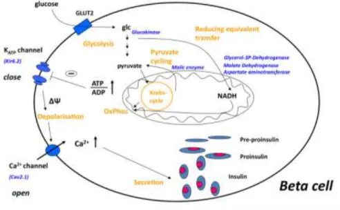
Adult, functional beta cells are characterized by rapid and precise changes in insulin secretion in response to nutrient secretagogues, particularly glucose, that maintain blood glucose levels in the physiologically relevant range. Specialised metabolic pathways ensure the coupling of external nutrient stimuli to insulin secretion in adult beta cells (Figure 1), and insulin gene transcription is regulated by Pdx1, NeuroD1 and MafA together. Glucose entering the beta cell is quickly metabolized to pyruvate and the glucose-derived carbons are then oxidized in the mitochondria. The very low expression of lactate dehydrogenase and monocarboxylate transporters results in negligible lactate production. Simultaneously, glycolysis-derived cytosolic NADH is reoxidized in mitochondrial membrane shuttles, and the strong pyruvate carboxylase activity of beta cells further
facilitates the flux of pyruvate to the shuttles. Glucose oxidation promotes the production of ATP, the key metabolic coupling factor for glucose-stimulated insulin secretion (GSIS). The closing of ATP- dependent K+ channels results in depolarisation, opening of voltage- activated Ca2+ channels and subsequent exocytosis of the preformed insulin granules. The fine tuning of GSIS is ensured by the distinct expression pattern of metabolic pathways in beta cells compared to other cell types.
Figure 1. The current concept of glucose-stimulated insulin secretion in pancreatic beta cells, described in details in the text. Abbreviations: glc, glucose;
OxPhos, oxidative phosphorylation; ΔΨ, membrane depolarisation.
Although the metabolism of adult beta cells has been characterised extensively, that of immature fetal/neonatal beta cells, which are glucose unresponsive, is less well understood. Similarly, molecular mechanisms in diabetic, dysfunctional beta cells are poorly characterized. Neonatal, immature and diabetic, dysfunctional beta cells may seem quite different, but they share a common phenotype:
lack normal GSIS.
My research work was focused on the metabolic gene and transcritption factor expression patterns that drive the change of beta cell phenotype.
Aims
The general objective of my PhD thesis is to provide a molecular description of the evolving beta cell phenotype; from the immature neonatal period, through the mature, glucose-responsive stage, to the development of glucotoxicity and a dysfunctional beta cell phenotype.
AIM 1. To characterize the underlying mechanisms responsible for the immature, glucose-unresponsive phenotype of neonatal beta cells.
Postulating that the mechanisms responsible for neonatal beta cell immaturity are complex, we used microarray analysis to compare the gene expression profile of neonatal (postnatal day P1) and adult beta cells excised by laser-capture microdissection. Sets of genes with differential expression were confirmed with quantitative realtime PCR (qPCR) and immunostaining. Moreover, expression pattern of these genes were followed during the first weeks of postnatal life, as the insulin secretion of the islets matures.
AIM 2. To assess whether maturation of neonatal beta cells into glucose-responsive, mature insulin secreting cells is regulated by transcription factor MafA and/or Pdx1.
We characterized the expression of MafA and Pdx1 and that of other key beta cell genes during the first 4 weeks of postnatal life. We hypothesized that enhancing the expression levels of these factors in neonatal beta cells could drive the acquisition of glucose-responsive insulin secretion.
AIM 3. To characterize the importance of MafA in regulating beta cell function in adult rat islets.
We hypothesized that there is a causal relationship between MafA expression and functional integrity of adult beta cell; and we aimed at studying MafA targets besides insulin with the inhibition of MafA function in adult rat beta cells.
AIM 4. To test the role of MafA in dysfunctional beta cells.
It is known that MafA expression level is lower and GSIS is impaired in beta cells under glucotoxicity. We investigate the ability of MafA overexpression in a diabetic model to restore glucose-stimulated insulin secretion.
Methods
1. Animal studies
Adult male and female Sprague-Dawley rats were used for animal studies, and 1-day-old pups were used for neonatal beta-cell experiments. As a diabetic model, Goto-Kakizaki (GK) rats were investigated with age-, gender-matched Wistar-Kyoto rats as controls. Animals were purchased from Taconic Farms (Germantown, NY). All procedures were approved by the Joslin Institutional Animal Care and Use Committee. Rats were anaesthetized with Nembutal. For immunostaining, excised pancreases were fixed by immersion in 4% paraformaldehyde, and were then embedded in paraffin. For laser capture microdissection (LCM), pancreases were immersed in TissueTek OCT medium and then were rapidly frozen. Islet isolation were done by collagenase digestion and gradient separation according to the protocol published by Gotoh et al. in 1987.
2. Laser-capture microdissection
Beta cell-enriched cores of islets were microdissected using a PixCell II LCM system (Arcturus, Mountain View, CA) from frozen
pancreatic sections. For each sample, two to five islets per section were excised from 10–20 sections, then total RNA from was extracted and amplified for the microarray.
3. Microarray hybridization
Biotinylated cDNA of four adult and four neonatal beta cell-enriched samples were made of total RNA and were subsequently run on Affymetrix GeneChip Rat Genome U34A (Affymetrix, Santa Clara, CA). Analysis was performed using a DNA-Chip Analyzer (dChip;
Harvard School of Public Health, Boston). Lower confidence boundary (LCB) and p values (<0.050) were used to assess differentially expressed genes between neonate and adult rats. Genes were then classified into functionally related clusters using DAVID software.
4. Adenovirus infection
Recombinant adenoviruses were generated to direct expression of transcription factors in beta-cells using the AdEasy system (Stratagene, La Jolla, CA). The following adenoviruses were used:
AdMafA, expressing the full human MafA coding sequence; AdDN- MafA, containing a dominant negative mutant of MafA lacking the N-terminal transcriptional activation domain; AdNeuroD1, expressing the hamster NeuroD1 coding sequence; and an adenovirus encoding green fluorescent protein alone (AdGFP) served as control.
AdPdx1 was a kind gift from Dr. D. Melton. All adenoviruses were amplified in 293Ad cells and purified with the Vivapure AdenoPACK 100 kit (Sartorius, Gottingen, Germany). Virus titers were established based on quantification of plaque formation. For beta-cell infections, islets were dispersed to single cells using trypsin to ensure uniform adenoviral infections during an overnight incubation, and were then reaggregated.
5. RNA extraction and reverse transcription
Total RNA from isolated islets or dispersed and reaggregated islet cells was extracted using RNeasy Plus Mini Kit (Qiagen, Germantown, MD). Next, cDNA was produced using Superscript II reverse transcriptase (Invitrogen, Carlsbad, CA).
6. Quantitative real-time PCR (qPCR)
The qPCR with SYBR Green detection was performed using the ABI7300 Real-time PCR System (Applied Biosystems, Foster City, CA) with primers designed using Primer Express. The comparative CT (threshold cycle) method was used to calculate relative gene expression levels, after normalization of the gene of interest to a control ribosomal gene (L32 or S25).
7. Insulin secretion in vitro
Insulin secretion of the islet cell aggregates was measured in the supernatant with sequential static incubations at low (2.8mM) and high (16.7mM) glucose Krebs-Ringer bicarbonate buffer. The Insulin Rat EIA kit (ALPCO Diagnostics, Salem, NH) was used for analysis.
8. Insulin secretion in vivo: intra-peritoneal glucose tolerance test For intraperitoneal glucose tolerance tests (IPGTT), rats were fasted with free access to water only, for 8–12 h. Rats were then injected intraperitoneally with 10% glucose solution at a dose of 2 g/kg body weight. Blood glucose readings were performed from the tail vein every half hour for two hours in total with OneTouch Ultra blood glucose meter (LifeScan, Milpitas, CA).
9. Western Blot
Protein quantities were established by Western Blot analysis. After AdMafA and AdDN-MafA infections, islet cell aggregates were sonicated, and then boiled in the presence of β-mercaptoethanol.
Next, protein extract was resolved on SDS-PAGE electrophoresis, transferred to PVFD membranes and probed with either MafA antibody (developed by Nishimura et al.) or HSV antibody (Abcam,
Cambridge, MA), which detects the HSV-tagged DN-MafA protein.
Secondary antibodies were conjugated to horseradish peroxidase, and blots were visualized based on chemiluminescence (SuperSignal West Dura reagent, Pierce, Thermo Fisher Scientific, Waltham, MA).
10. Immunohistochemistry
Paraffin embedded pancreatic sections were stained for pyruvate- kinase and glycerol-3P-dehydrogenase following general immunostaining protocols, antibodies were developed by US Biological, Swampscott, MA and Dr. M. MacDonald. As a second step, biotin-streptavidin amplification was used. Finally protein quantities were detected based on peroxidase reaction for pyruvate- kinase and immunofluorescence for glycerol-3P-dehydrogenase. In another set of experiments, GK and WKY rat pancreases were studied for beta cell composition, and sections were double stained with anti-insulin and a cocktail of anti-non-beta cell hormones.
Histological sections were imaged by Olympus BH2 and Zeiss 710 LSM microscopes.
11. Statistical analysis
For statistical analysis, unpaired Student’s t test was used. To see differences among groups, ANOVA was used followed by post hoc analysis (Tukey’s). A p value <0.050 was considered statistically significant.
Results
Neonatal and mature beta-cells have distinct gene expression patterns, several metabolic enzymes important for insulin secretion are expressed lower in the neonatal period.
In the first phase of my research, I studied the gene expression profile of immature, neonatal beta cells. Previous studies have described single genes being expressed lower in neonatal cells; however,
according to our hypothesis, generalized low expression of key metabolic enzymes may account for the poor glucose-responsiveness of neonatal beta cells. Gene expression profile of neonatal, immature beta cells, excised by laser capture microdissection from rat pancreatic islets was compared to adult, mature cells. Several mRNA-s of beta cell important enzymes were found to be lower in the neonatal period (pyruvate-carboxylase, GLP1-receptor, enzymes of the reducing equivalent transport through mitochondrial membrane, such as malate-dehydrogenase, glycerol-3-phosphate- dehydrogenase, malic enzyme), which could explain the insufficient glucose-stimutaled insulin secretion at this age. Microarray results were confirmed by real-time PCR, and by immunohistochemical staining of selected metabolic enzyme proteins (pyruvate-kinase, glycerol-3-phosphate-dehydrogenase). While studying the development of the characteristic metabolic profile of adult beta cells, we confirmed that the expression level of these genes gradually increase over the first postnatal month, in parallel with the appearance of the glucose-stimulated insulin secretion.
Expression level of transcription factors change over the postnatal period. MafA plays an important role in the induction of glucose-stimulated insulin secretion.
The reduced expression of many key beta cell genes in neonatal beta cells suggests that regulated expression of transcription factors may drive beta cell maturation. Key beta cell specific, glucose-regulated transcription factors Pdx1, NeuroD1 and MafA were studied, and compared with the expression profile of metabolic enyzmes and insulin during the first postnatal month in rats. All these transcription factors are glucose regulated; Pdx1 plays a role in early endocrine differentiation, NeuroD1 is a late differentiation factor, and whereas MafA is a beta cell maturation factor that aso regulates insulin synthesis and secretion, according to our hypothesis. Of these studied transcription factors, expressional profile of MafA gradually increased over the first postnatal months, similarly to metabolic enzymes. Protein levels of MafA were quantitated by Wester blot
analysis, and found to be extremely low in day 1, markedly increased by day 10, but still lower than in adults. In the second phase of my studies, expression level of MafA was exogenously enhanced in neonatal rat islet cells by adenoviral infection and overexpression the MafA protein, which resulted in induction of important metabolic enzymes. Importantly, by enchancing the expression level of MafA, glucose-stimulated insulin secretion also significantly increased when compared to control neonatal cells. In contrast, overexpression of Pdx1 did not influence the insulin release. Taken together, we established that beta cell maturation could be accelerated with overexpression of MafA.
Transcription factor MafA as a regulator molecule in adult, mature beta-cells.
Next, I aimed to characterize the importance of MafA in regulating beta cell function in adult rat islets, and tested the effect of MafA inhibition in adult, mature beta cells. MafA function was selectively inhibited by a “dominant-negative” MafA construct (DN-MafA), which codes a mutant DN-MafA protein, lacking the N-terminal transcriptional activation domain. The DN-MafA construct was transfected into freshly isolated rat islet cells by adenovirus mediated infection. The expressed DN-MafA protein would bind to the endogenous MafA and inhibit its transcriptional activity. Endogenous MafA mRNA and protein levels decreased to 10% as compared to control untreated beta cells, suggesting MafA autoregulation. Low levels of MafA resulted in decreased expression of several metabolic enzymes, beta cell specific genes and other transcription factors.
Gene expressional changes were followed for 72 hours after DN- MafA infections, and mRNA levels of glucokinase, GLUT1, insulin, PCSK enzyme, GLP1-receptor, Kir6.2 potassium channel, and multiple enzymes of the reducing equivalent transport significantly reduced. In contrast, mRNA level of lactate-dehydrogenase, which is virtually not expressed in adult beta cells, increased significantly.
Functional studies revealed that glucose-stimulated insulin secretion becomes impaired at 36 hours after DN-MafA infection, which
coincides with the time when expression of beta cell important metabolic genes decrease. The results suggest that beta cells become dysfunctional, dedifferentiated with the loss of functional MafA, and they acquire a phenotype similar to neonatal, immature beta cells.
MafA overexpression in dysfunctional, diabetic beta-cells can increase glucose-stimulated insulin secretion.
Previous studies have shown that chronic hyperglycaemia, typical of diabetes, induces similar phenotypic changes as described above, as beta cells become dedifferentiated. It is also known that glucose- stimulated insulin secretion of diabetic beta cells is impaired.
According to a recent animal study, hyperglycaemia causes a decrease in MafA expression. Based on these results, in the last phase of my studies I aimed to restore MafA function in impaired beta cells of diabetic rats by overexpressing MafA, and sought to improve glucose-stimulated insulin secretion. I studied Goto-Kakizaki rats, the prototype animal model of spontaneous type 2 diabetes. As expected, adenovirus mediated overexpression of MafA increased glucose-stimulated insulin secretory response in 48-72 hours.
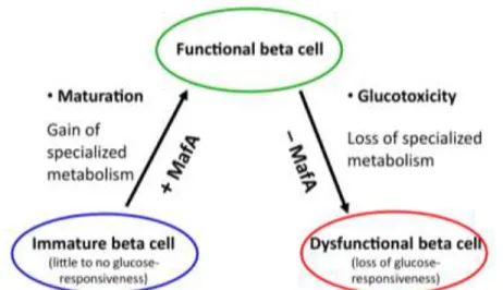
Figure 2: Paradigm of the beta cell, with MafA as a critical regulator of beta cell phenotype.
Conclusions
Low levels of transcription factor MafA causes low expression of several beta-cell important genes and blunted glucose-stimulated insulin secretion, a phenotype typical of neonatal, immature beta- cells as well as diabetic, dedifferentiated beta-cells (Figure 2).
Restoring MafA function can the increase insulin secretory response.
Transcription factor MafA regulates the expression of several beta cell important genes directly or indirectly, besides regulating insulin gene expression as well. Thus, the presence of MafA is indispensable for mature beta cell function and physiologic glucose-stimulated insulin secretion.
Based on our results, transcription factor MafA may serve as a potential candidate for drug development for treating patients with type 2 diabetes, since it could prevent beta cell dedifferentiation and may enhance insulin secretion.
Bibliography of the candidate’s publications
Publications related to the thesis:
Jermendy Á, Toschi E, Aye T, Koh A, Aguayo-Mazzucato C, Sharma A, Weir GC, Sgroi D, Bonner-Weir. Neonatal beta cells lack the specialized metabolic phenotype of mature beta cells.
Diabetologia 2011; 54:594-604.
IF: 6.551
Aguayo-Mazzucato C, Koh A, El Khattabi I, Li WC, Toschi E, Jermendy Á, Juhl K, Mao K, Weir GC, Sharma A, Bonner-Weir S.
Mafa expression enhances glucose-responsive insulin secretion in neonatal rat beta cells. Diabetologia. 2011; 54:583-93.
IF: 6.551
Publications not related to the thesis:
Molvarec A, Jermendy Á, Kovács M, Prohászka Z, Rigó J Jr. Toll- like receptor 4 gene polymorphisms and preeclampsia: lack of association in a Caucasian population. Hypertens Res 2008; 31: 859- 64.
IF: 3.146
Molvarec A, Jermendy Á, Nagy B, Kovács M, Várkonyi T, Hupuczi P, Prohászka Z, Rigó J Jr. Association between tumor necrosis factor (TNF)-alpha G-308A gene polymorphism and preeclampsia complicated by severe fetal growth restriction. Clin Chim Acta 2008;
392: 52-7.
IF: 2.960
Jermendy Á, Körner A, Kovács M, Kaszás E, Balázsovics J, Szőcs A, Madácsy L, Cseh K. A Toll-like rceptor polimorfizmusainak hatása a tumornekrózis faktor- és szolúbilis receptorainak szintjére elhízott gyermekekben és serdülőkben. Diabetologia Hungarica 2009; 3: 241-248.
Lukács K, Szatmári I, Jermendy Á, Krikovszky D, Körner A, Pánczél P, Madacsy L, Hermann R: A PTPN22 gén C1858T és az inzulin génrégió -23HphI polimorfizmusának összefüggése az 1-es típusú diabetesszel magyar populációban. Gyermekgyógyászat 2009;
60: 42-47.
Jermendy Á, Szatmári I, Laine AP, Lukács K, Horváth KH, Körner A, Madácsy L, Veijola R, Simell O, Knip M, Ilonen J, Hermann R.
The interferon-induced helicase IFIH1 Ala946Thr polymorphism is associated with type 1 diabetes in both the high-incidence Finnish and the medium-incidence Hungarian populations. Diabetologia 2010; 53: 98-102.
IF: 6.973
Jermendy Á, Körner, Kovács M, Kaszás E, Balázsovics J, Szőcs A, Madácsy L, Cseh K. Association between toll-like receptor polymorphisms and serum levels of tumor necrosis factor-α and its soluble receptors in obese children. Med Sci Monit 2010; 16: 180- 185.
IF: 1.699
Jermendy Á, Körner A, Kovács M, Madácsy L, Cseh K. PPAR-γ2 Pro12Ala polymorphism is associated with post-challenge abnormalities of glucose homeostasis in children and adolescents with obesity. J Pediatr Endocr Met 2011; 24: 55–59.
IF: 0.875
Jermendy Á, Körner A, Kovács M, Cseh K, Madácsy L: A PPAR-ƴ Pro12Ala polimorfizmus összefüggése a glükóz-homeosztázissal elhízott gyermekek és serdülők körében. Gyermekgyógyászat 2012;
4: 147-151.
Cavelti-Weder C, Shtessel M, Reuss JE, Jermendy Á, Yamada T, Caballero F, Bonner-Weir S, Weir GC. Pancreatic duct ligation after almost complete β-cell loss: exocrine regeneration but no evidence of β-cell regeneration. Endocrinology 2013; 154: 4493-4502.
IF: 4.717