MOLECULAR DIVERSITY AND ANTIBIOTIC RESISTANCE GENE PROFILE OF SALMONELLA ENTERICA SEROVARS ISOLATED FROM HUMANS
AND FOOD ANIMALS IN LAGOS, NIGERIA
ABRAHAM AJAYI1*, STELLA IFEANYISMITH2, JULIEN COULIBALY KALPY3, IBIDUNNIOREOLUWABODE-SOJOBI4, YAO KOUAMÉ RENÉ3 and
ADEYEMIISAAC ADELEYE1
1Department of Microbiology, University of Lagos, Akoka, Nigeria
2Molecular Biology and Biotechnology Department, Nigerian Institute of Medical Research, Lagos, Nigeria
3Laboratoire de Bactériologie et Virologie Institut Pasteur de Côte d’Ivoire, Centre Nationale de Référence de Salmonella, Paris, France
4Department of Medical Microbiology, Lagos University Teaching Hospital, Idi-Araba, Lagos, Nigeria
(Received: 17 April 2019; accepted: 23 May 2019)
Outbreaks of Salmonellosis remain a major public health problem globally.
This study determined the diversity and antibiotic resistance gene profile ofSalmo- nella enterica serovars isolated from humans and food animals. Using standard methods,Salmonellaspp. were isolated from fecal samples, profiled for antimicrobial susceptibility and resistance genes. Seventy-oneSalmonellaisolates were recovered from both humans and food animals comprising cattle, sheep, and chicken. Forty-four serovars were identified, with dominantSalmonellaBudapest (31.8%). Rare serovars were present in chicken (S. Alfort,S. Wichita,S. Linton,S. Ealing, andS. Ebrie) and humans (S. Mowanjum, S. Huettwillen, S. Limete, and S. Chagoua). Sixty-eight percent of isolates were sensitive to all test antibiotics, while the highest rate of resistance was to nalidixic acid (16.9%;n=12), followed by ciprofloxacin (11.3%;
n=8) and tetracycline (9.9%;n=8). Five isolates (7%) were multidrug-resistant and antimicrobial resistance genes coding resistance to tetracycline (tetA), beta-lactam (blaTEM), and quinolone/fluoroquinolone (qnrBandqnrS) were detected. Evolutionary analysis of gyrA gene sequences of human and food animal Salmonella isolates revealed variations but are evolutionarily interconnected. Isolates were grouped into four clades withS. Budapest isolate from cattle clustering withS. Budapest isolated from chicken, whereasS. Essen isolated from sheep and chicken was grouped into a clade. Diverse S. enterica serovars with high antibiotic resistance profile are
*Corresponding author; E-mail:ajayiabraham2013@gmail.com
First published online October 29, 2019
ubiquitous in food animals; hence, there is a need for surveillance and prudent use of antibiotics in human and veterinary medicine.
Keywords:antibiotic resistance genes, serotype, quinolone, diversity Introduction
Salmonellosiscontinue to pose substantial health challenges in developed and developing countries [1]. Over 2,500Salmonella entericaserovars have been identified and some of which are the etiology of salmonellosis that is presented mainly as enteric fever in cases caused by Salmonella Typhi or Paratyphi and gastroenteritis caused by otherS. entericaserovars. While the former is most of the time severe and invasive, the latter tends to be self-limiting; however, it could be severe and systemic in infants, older people, and immunocompromised indivi- duals [2–4]. Epidemiological surveillance overtime has revealed that outbreaks of salmonellosis in human are linked to ingestion of foods mainly of animal origin that are contaminated withSalmonella; thus, mostSalmonellainfections could be considered as zoonotic. However, host-specific serovars likeS. Typhi transmission is often through the fecal–oral route and not considered to be zoonotic. Establish- ing the route of transmission is more or less difficult, since the transmission chain is complex [2,5,6]. Food animals (chicken, pork, cattle, sheep, and evenfish) are a major reservoir of diverse serovars ofS. enterica as has been documented in several reports [7–11]. This persistence ofS. entericain the intestinal tract of food animals creates a chronic or non-symptomatic carrier state that provides for continuous shedding of bacteria in feces, thereby serving as a reservoir for subsequent spread by contaminated meat, milk, eggs, and agricultural products cultivated onSalmonella-containing manure lands [12]. Not only food animals are sources ofSalmonellainfections, they also serve as a channel for the dissemination of multidrug-resistant (MDR) serovars, which in recent times has emerged as a threat to the effective treatment of infections. In a 2014 World Health Organization (WHO) report on resistance of selected bacteria of international concern to antibacterial drugs, non-typhoidal Salmonella (NTS) resistance to fluoroquino- lones made the list with a total number of reports with data sets based on ≥30 tested isolates to be as high as 75% [13]. Resistance of Salmonellaspp. to most widely used classes of antibiotics spans from aminoglycosides, chloramphenicol, tetracycline, beta-lactams (including cephalosporin), quinolones, sulfonamide and trimethoprim to polymyxins [14–16]. In Nigeria, there has been reports of the isolation and antibiotic resistance profiling of Salmonella serovars isolated from humans, chicken, and pigs [17–19] but there is drought of information on studies that evaluates human and a broader range of food animals at once.
Hence, this study explores the diversity and antibiotic resistance genes profile of Salmonellaserovars isolated from humans and three major food animals in Lagos, Nigeria, a cosmopolitan city in West Africa.
Materials and Methods Study design
The study is composed of humans and food animals (cattle, sheep, and chicken).
Group 1: This group comprised human subjects who were apparently healthy food handlers recruited based on the following criteria: (1) adults with
≥18 years of age (both male and female) and (2) not on any form of antibiotic treatment. Sample size for human subject was defined as:
n=Z2pq d2 , where:
Z→Standard normal deviate=1.96
p→Prevalence≅0.304 according to Smith et al. [20]
q→1−p=1−0.304
d→Tolerable margin of error=0.05 n=n=ð1.96Þ2ð×0.5ð0.304ÞÞ20.0025×ð0.696Þ =0.8128211 n=325.1.
With a gazette 10% non-response rate n=325+33=358.
Group 2: This comprised apparently healthy food animals (cattle, sheep, and chicken) that were being processed for slaughter. One hundred and two fecal samples were collected from each animal type making the total sample size from animals to be 306. Therefore, the total number of samples collected for both group was 664.
Sample collection
Fecal samples were collected from humans, cattle, sheep, and chicken between June 2016 and March, 2017. Samples were collected from animals randomly selected each day of sample collection. Fecal samples were obtained after slaughter from viscera (intestine) in sterile specimen bottles appropriately
labeled and transported in a thermobox at 4 °C to the laboratory immediately.
Fecal samples were also collected from humans in sterile sample bottles and transported immediately to the laboratory under the same conditions earlier stated.
Isolation and identification of isolates
Five grams of each fecal sample were enriched in 25 ml of selenite F broth (Oxoid, Basingstoke, UK) and incubated at 37 °C for 18–24 h. This was followed by plating ontoSalmonella–Shigellaagar (Oxoid) and incubated at 37 °C for 24 h.
Presumptive colonies were further purified by subculturing on nutrient agar (Oxoid). Pure colonies were identified using biochemical tests including motility, indole, urease, mannitol, lysine decarboxylase, citrate, ortho-nitrophenyl- β-galactoside, Kliger Iron Agar to determine H2S and gas production, lactose and glucose fermentation, oxidase and catalase.
Serotyping
Serotyping of all biochemically confirmedSalmonellaisolates was carried out at Centre Nationale de Référence de Salmonella, Laboratoire de Bactériologie et Virologie Institut Pasteur de Côte d’Ivoire according to White Kauffmann–Le Minor scheme [21]. Serotyping was carried out by slide agglutination test to characterize O and H antigens using commercially available antisera (Bio-Rad, F-92430 Marnes-La-Coquette, France).
Antimicrobial susceptibility testing
Antimicrobial susceptibility testing was performed using the disk diffusion method according to the European committee on antimicrobial susceptibility testing [22] guidelines.Salmonellaisolates were inoculated into brain–heart infusion broth (Oxoid) and incubated at 37 °C for 18 h after which they were subcultured on Mueller–Hinton agar (Hi-Media Laboratories Pvt. Ltd., India) at 37 °C for 24 h.
Two to three distinct colonies were emulsified in 5 ml of sterile physiological saline and adjusted to 0.5 McFarland standard and then a sterile swab stick was in applying bacteria suspension to the surface of Mueller–Hinton agar. Antibiotic disks were then applied comprising ampicillin (10 μg), ciprofloxacin (5 μg), tetracycline (30 μg), minocycline (30 μg), colistin (50 μg), tobramycin (10 μg), nalidixic acid (30 μg), norfloxacin (10 μg), imipenem (10 μg), cephalotin (30 μg), ceftazidime (10μg), gentamycin (10μg), aztreonam (30μg), ceftriaxone (30μg),
cefuroxime (30μg), amikacin (30μg), chloramphenicol (30μg), cefotaxime (5μg), trimethoprim–sulfamethoxazole (25μg), amoxicillin+clavulanic acid (30μg), and cefepime (30 μg) (Bio-Rad). The diameter of inhibition zone was measured using ADAGIO (Bio-Rad) and was interpreted as resistance (R), intermidiate (I), and sensitive (S). Escherichia coliATCC 25922 was used as quality control organism.
DNA extraction
Phenol–chloroform–isoamyl alcohol method of DNA extraction according to Adi et al. [23] was adapted with modifications. Three to five colonies of bacterial isolates grown on Colombia Blood Agar overnight was emulsified in nuclease-free water to form a suspension. Cell suspension was refrigerated at −20 °C for 15 min and then transferred to thermomixer held at 95 °C and incubated for 15 min at 500 rpm and then centrifuged at 14,000 rpm for 10 min, after which the supernatant containing the DNA was separated into a new sterile Eppendorf tube. To the supernatant, 500 μl of phenol+chloroform+isoamyl (25:24:1) was added, vortexed, and then centrifuged at 13,000 rpm for 10 min at 4 °C. Eight hundred microliter of supernatant was obtained to which 80μl of sodium acetate+500μl of absolute (100%) ethanol was added, vortexed gently, and stored at −20 °C overnight. After centrifugation at 13,000 rpm for 20 min at 4 °C, supernatant was discarded while 1 ml of ethanol (70%) was added to the residue, vortexed gently, and then centrifuged at 13,000 rpm for 10 min at 4 °C;
then, supernatant was discarded; later, the residue was maintained at 70 °C to allow the leftover ethanol to evaporate for 25–30 min. Then, it was reconstituted with 60 μl of Elu Buf (NucliSENS, BioMerieux, France). Purity of DNA was determined using a spectrophotometer and DNA was stored for further use at −20 °C.
Detection of antibiotic resistance genes by polymerase chain reaction (PCR)
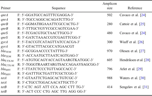
Nine antibiotic resistance genes (tetA,tetB,blaTEM,blaSHV,blaCTX-M,qnrA, qnrB, qnrS, and gyrA) were assayed by PCR using primers listed in Table I.
A 50-μl PCR reaction was used that contained 28.8 μl of nuclease-free water, 3.5μl MgCl2(25 mM), 10μl of 5×PCR buffer, 1μl of dNTPs (10 mM), 0.75μl of each forward and reverse primers (10μM), 0.2μl of one Taq DNA polymerase (5,000 U/ml) (New England Biolabs, Hertfordshire, UK), and 5μl DNA template.
PCR was performed in a GeneAmp PCR system 9700 thermal cycler (AB Applied Biosystems, Singapore) with programming conditions determined empirically.
For qnrA, qnrB, qnrS, gyrA, blaSHV, blaCTX-M, and tetB, 35 cycles of initial denaturation at 95 °C for 5 min, denaturation at 95 °C for 30 s, annealing at 60 °C for 30 s, elongation at 72 °C for 1 min, andfinal elongation at 72 °C for 10 min were used; whereas forblaTEMandtetA, PCR conditions were similar only with a different annealing temperature of 56 °C. The negative control was water, whereas positive controls includeS.Typhimurium P5002212DT104 forgyrA,S.Virchou 58.67 Holland for blaCTX, S. Bredeney TEM-104 (Gisele) for blaTEM, S.Keurmassar DAK2 forblaSHV,Enterobacter cloacae03-577 forqnrA,Klebsi- ella pneumoniae KP15 for qnrB, Escherichia coli pHC19 for qnrS, E. coli NCTC50078 fortetA, and E. coli CSH50:TN10 fortetB. PCR products were separated on a 1.5% agarose gel at 120 V and a 100-bp DNA ladder (New England Biolabs) was used as molecular weight maker.
DNA sequencing
PCR amplification products were sent to a commercial facility (Eurofins Genomics, France) for sequencing. Sequence results were analyzed and compared with sequences in the GenBank using BLAST (https://blast.ncbi.nlm.nih.gov/
Blast.cgi) and CARD (https://card.mcmaster.ca/analyze/blast). Phylogenetic analysis was performed using the MEGA 7.0 software [32].
Table I.Primers used in the detection of antibiotic resistance genes
Primer Sequence
Amplicon
size Reference qnrA F: 5′-GGATGCCAGTTTCGAGGA-3′ 502 Cavaco et al. [24]
qnrA R: 5′-TGCCAGGCACAGATCTTG-3′
qnrB F: 5′-GGMATHGAAATTCGCCACTG-3′ 280 Cattoir et al. [25]
qnrB R: 5′-TTTGCYGYYCGCCAGTCGAA-3′
qnrS F: 5′-TCGACGTGCTAACTTGCG-3′ 480 Cavaco et al. [24]
qnrS R: 5′-GATCTAAACCGTCGAGTTCGG-3′
gyrA F: 5′-TACCGTCATAGTTATCCACGA-3′ 300 Wiuff et al. [26]
gyrA R: 5′-GTACTTTACGCCATGAACGT
blaTEM F: 5′-GCGGAACCCCTATTTG-3′ 970 Olesen et al. [27]
blaTEM R: 5′-ACCAATGCTTAATCAGTGAG-3′
blaCTX-M F: 5′-ATGTGCAGYACCAGTAARGTKATGGC-3′ 605 Hendriksen et al. [28]
blaCTX-M R: 5′-TGGGTRAARTARGTSACCAGAAYSAGCGG-3′
blaSHV F: 5′-TTATCTCCCTGTTAGCCACC-3′ 796 Arlet et al. [29]
blaSHV R: 5′-GATTTGCTGATTTCGCTCGG-3′
tetA F: 5′-GTAATTCTGAGCACTGTCGC-3′ 988 Waters et al. [30]
tetA R: 5′-CTGCCTGGACAACATTGCTT-3′
tetB F: 5′-CTC AGT ATT CCA AGC CTT TG-3′ 414 Sengeløv et al. [31]
tetB R: 5′-ACT CCC CTG AGC TTG AGG GG-3′
Statistical analysis
Statistical analysis and graphics were performed using GraphPad Prism software 5.01 (La Jolla, CA, USA) and Microsoft Excel (Microsoft Cooperation, 2013 USA). Statistical significance of proportions was assessed using theχ2test consideringp<0.05 as significant.
Results Isolation and identification
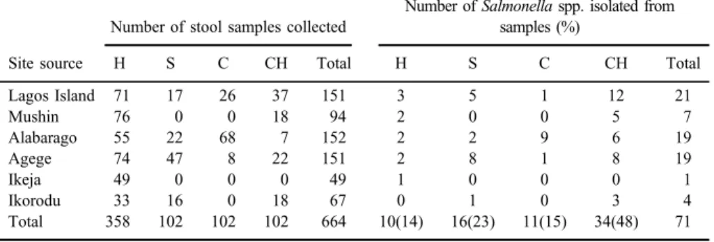
A total of 71 Salmonellaisolates were recovered from 664 stool samples collected from humans, cattle, sheep, and chicken with a recovery rate of 10.7%.
Forty-eight percent of the isolates were obtained from chicken, whereas 22.5%
was from sheep (Table II). Forty-four different serovars were identified with S.Budapest having the highest occurrence 31.8% (14/44) followed by S.Essen 15.9% (7/44) withS.Paratyphi C having the least with 4.5% (2/44) (TableIII).
Antimicrobial susceptibility testing
Altogether 68% of allSalmonellaisolates (n=48) were susceptible to all 21 test antibiotics. The highest rates of resistance was found to nalidixic acid, (12/71;
16.9%) followed by ciprofloxacin 11.3%, while resistance to tetracycline and minocycline was 9.9%. Thirty-one percent (n=22) of the entire isolates exhibited resistance to one or more of the test antibiotics, which was significant (p<0.001) compared to the susceptible isolates. Seven percent (n=5) of the antibiotic resistant isolates were MDR. Antibiotic resistance within population revealed
Table II.Distribution of the isolates from various sources and location
Site source
Number of stool samples collected
Number ofSalmonellaspp. isolated from samples (%)
H S C CH Total H S C CH Total
Lagos Island 71 17 26 37 151 3 5 1 12 21
Mushin 76 0 0 18 94 2 0 0 5 7
Alabarago 55 22 68 7 152 2 2 9 6 19
Agege 74 47 8 22 151 2 8 1 8 19
Ikeja 49 0 0 0 49 1 0 0 0 1
Ikorodu 33 16 0 18 67 0 1 0 3 4
Total 358 102 102 102 664 10(14) 16(23) 11(15) 34(48) 71
Note:Sample source–H: human; S: sheep; C: cattle; CH: chicken.
Table III.The occurrence ofSalmonellaserovars identified
CODE Antigenic formula Serotype Frequency of isolation
Isolates from chicken
A115/190 (1,3,19: z: I,w) S.Carno 1
CF10/191 (1,4,12,[27]: g,t: _) S.Budapest 13
F2/194 A152/198 E14/200 CF13/201 A6/202 CF3/206 F4/207 Fe/213 CF6/215 CF9/216 F5/219 AF13/220
B11y/196 (35: gst: _) S.Anecho 3
AF5/199 D10/204
E7/203 (3{10}{15}15,34:eh:1,5) S.Muenster 1
D8/209 (4,12:eh:1,7) S.Kaapstad 1
D6/205 (35: gt: _) S.Agodi 2
E15/211
A2/212 (8,20:Z4Z23:l,w) S.Dabou 1
A11/214 (4,[5],12:g,z51:e,n,z15) S.Tennyson 1
F12/217 (6,8:r:l,w) S.Goldcoast 1
AF1/218 (35:g,m,t:_) S.Ebrie 1
AF6/221 (4,12:g,m:_) S.Essen 3
E8/224 CF14/226
E6/222 (4,[5],12: l,v:e,n,z15) S.Brandenburg 1
E4/225 (3,10:f,g:e,n,x) S.Alfort 1
F1/227 (1,6,14,25:c:l,w) S.Minna 1
D3/193 (13,23:r:1,6) S.Linton 1
D25/210 (1,13,23:d:1,6) S.Wichita 1
D14/223 (35:g,m,s:_) S.Ealing 1
Total 34
Isolates from sheep
S59/230 (6,7,14:d:l,w) S.Livingstone 1
S63B/231 (6,7:d:1,6) S.Kivu 1
S43/232 (17:c:1,5) S.Berlin 1
S57/233 (4,12:g,m:_) S.Essen 4
S38B/235 S56/236 S81/247
S36C/237 (17:b:1,5) S.Dahra 2
S36/241
S37/240 (4,12:I,w:1,5) S.Mono 1
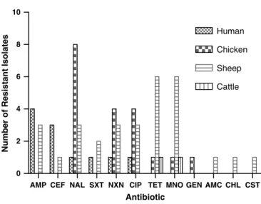
that 43.8% (n=7/16) of isolates from sheep showed resistance to one or more antibiotics closely followed by isolates from humans with 40% (4/10) resistance.
Percentage of antibiotic resistance ofSalmonellaisolates from chicken was 29.4%
(10/34; Figure 1).
Detection of resistance genes by PCR
From PCR analysis, Salmonella isolates that displayed phenotypic resistance to antibiotics were positive for some antibiotic resistance genes assayed
Table III.The occurrence of Salmonella serovars identified(Continued)
CODE Antigenic formula Serotype Frequency of isolation
S61/239 (1,4,12:z10:I,w) S.Mura 1
S28/242 (8,20:z10:e,n,z15) S.Chomedey 1
S38/243 (8,20:d:1,5) S.Yovokome 1
S43/244 (16:d:1,5) S.Sculcoates 1
S51/246 (1,4,12[27]:b:l,w) S.Wien 1
S76/234 (3{10}{15}{15,34}:y:1,5) S.Orion 1
Total 16
Isolates from cattle
CO18/249 (3,10:d:1,7) S.Onireke 1
CO67/248 (6,7:z4z24:_) S.Somone 1
CO20/251 (13,23:z:1,6) S.Farmsen 4
CO15/252 CO26/257 CO38/255
CO101/250 (28: z4z24:_) S.Ketheabarny 1
CO19/253 (1, 4,12[27]:gt:_) S.Budapest 1
CO37/254 (43,:g,z62:enx) II 1
CO50/256 (1,4,12,27:lz13z28:e,n,z15) S.Vom 1
CO12/258 (1,4,[5],12:eh:enz15) S.Sandiego 1
Total 11
Isolates from human
H132/266 (1,4,12,[27]:b:1,5) S.Limete 1
H44/267 (9,12:z10:1,5) S.Portland 1
H45/268 (1,4,12:a:I,w) S.Huettwillen 1
H63/269 (6,8:z:1,5) S.Mowanjum 1
HL8/263 (6,7[vi]:c:1,5) S.Paratyphi C 2
H418/274
H363/271 (1,4,12,27:i:1,2) S.Tyhpimurium 1
H209/272 (6,8:i:1,5) S.Takoradi 1
H183/273 (1,4[5],12:b:1,2) S.Paratyphi B 1
H117/275 (1,13,23:a:1,5) S.Chagoua 1
Total 10
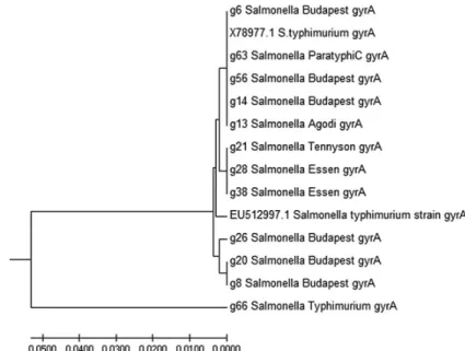
(TableIV).qnrB gene was detected in 10 (45.4%) of the isolates andqnrSwas detected in 1 isolate (Figures2and3).blaCTX,blaSHV, andqnrAwere not detected in any of the isolates; however, 22.7% (5/22) was positive forblaTEMtetAwas also detected in 18.1% (4/22) of the isolates. The gyrA sequences of 12 isolates (GenBank accession numbers: MG593259–MG593270) were grouped into four clades withS.Typhimurium largely diverged.S.Budapest isolated from cattle and S. Budapest isolated from chicken were close, whereasS. Essen from sheep and chicken was grouped together in a clade, indicating evolutionary relatedness.
Some isolates were grouped alongside S. Typhimurium (accession numbers:
X78977 and EU512997) from GenBank (Figure4).
Discussion
Diversity and gene variation among antibiotic-resistantS. entericaserovars of food animal sources continues to expand. Of the 71Salmonellaisolates from human, cattle, sheep, and chicken, 44 serovars were obtained with isolates from chicken accounting for 17 of these serovars. This is not surprising as poultry has been known to be a major reservoir and vehicle of transmission ofS.serovars to humans [33]. S. Budapest had the highest rate of occurrence in poultry from different locations sampled. Although this serovar has not been reported in any
AMP CEF NAL SXT NXN CIP TET MNO GEN AMC CHL CST 0
2 4 6 8 10
Human Chicken Sheep Cattle
Antibiotic
Number of Resistant Isolates
Figure 1.Distribution of resistance to antibiotics amongSalmonellaisolates from human, chicken, sheep and cattle. AMP: ampicillin; CEF: cephalotin; NAL: nalidixic acid; SXT: trimethoprim/
sufamethoxazole; NXN: norfloxacine; CIP: ciprofloxacin; TET: tetracycline; MNO: minocycline;
GEN: gentamycin
Table IV.Antimicrobial resistance phenotype, and genes ofSalmonellaserovars isolated from humans, chicken, sheep, and cattle
Code Origin Phenotype Genes Serotype
F5/219 Chicken NAL, NXN, and CIP gyrAandqnrB S. Budapest
CF3/206 Chicken NAL gyrAandqnrB S. Budapest
Fe/213 Chicken NAL, NXN, and CIP gyrAandqnrB S. Budapest
A152/198 Chicken NAL qnrB S. Budapest
A115/190 Chicken NAL gyrA S. Carno
D6/205 Chicken NAL, NXN, and CIP gyrAandqnrB S. Agodi
A11/214 Chicken NAL, gyrAandqnrB S. Tennyson
AF6/221 Chicken NAL, NXN, and CIP gyrAandqnrB S. Essen A2/212 Chicken TET and MNO qnrB, qnrS,andTetA S. Dabou
E14/200 Chicken GMI – S. Budapest
CO19/253 Cattle TET and MNO TetA S. Budapest
H117/275 Human AM, NAL, SXT, NXN, and CIP gyrAandTetA S. Chagoua
H63/269 Human AM and CEF – S. Mowanjum
H44/267 Human AM and CEF – S. Portland
H363/271 Human AM and CEF – S. Typhimurium
S57/233 Sheep AM and TET blaTEMandTetA S. Essen
S38B/235 Sheep AM, TET, and SXT blaTEM S. Essen
S56/236 Sheep NAL, NXN, and CIP gyrAandqnrB S. Essen
S81/247 Sheep MNO and TET blaTEM S. Essen
S61/239 Sheep NAL, NXN, and CIP gyrA S. Mura
S28/242 Sheep NAL, TET, SXT, NXN, and CIP qnrBandblaTEM S. Chomedey
S51/246 Sheep MNO and TET blaTEM S. Wien
Figure 2.Agarose gel image of PCR products showing positive bands forqnrB. Lane 1–M: 100-bp DNA marker, lane 2–PC: positive controlKlebsiella pneumoniaKP15, lane 19–NC: negative control, 1-S. Carno, 6, 8, 14, 20, 26-S. Budapest, 13-S. Agodi, 19-S. Dabou, 21-S. Tennyson, and
28-S. Essen
human outbreak in Nigeria, a 2016Salmonella annual report of the Centers for Disease Control and Prevention USA reportedS.Budapest isolated from at least one patient with enteric fever in the USA between 2003 and 2013 [34].
NTS serovars accounted for majority ofS.serovars from all three food animals
Figure 3.Agarose gel image of PCR products showing positive bands forqnrS. Lane 1–M: 100-bp DNA marker, lane 2–PC: positive controlEscherichia colipHC19, lane 16–NC: negative control,
19-S. Dabou
Figure 4.gyrAphylogenetic tree constructed based on an alignment ofgyrAsequences of 12 isolates from human, chicken, cattle, and sheep withgyrAgene ofSalmonellaTyphimurium (EU512997 and
X78977) from GenBank
(cattle, sheep, and chicken). NTS serovars have a vast host range including ruminants and birds with their products (eggs, meat, and milk) acting as vehicle for the transmission ofSalmonellainfections to humans and have been implicated in bacterial bloodstream infections in children and adults in sub-Saharan Africa [35–37]. Rare serovars were present in chicken, which includedS. Alfort, S. Wichita,S. Linton,S. Ealing, andS. Ebrie. In a similar study, Smith et al. [6]
reported the occurrence of rare serovars including S. Amoutive, S. Ealing, S. Urbana,S. Bargny,S. Drac, and S. Nyborg in beef, chicken, goat, and pork sold in different markets in Lagos Nigeria. Food animals remain a major source of diverse serovars of Salmonella. This study reports the first isolation of rare serovarsS. Mowanjum,S. Huettwillen,S. Limete, andS. Chagoua from apparently healthy humans in Nigeria. Although these serovars have been isolated from animals, animal products, and animal feeds in Denmark and Thailand, respectively [38,39]. Thisfinding is of public health relevance because these individuals who are food handlers are likely to disseminate the bacteria, sinceSalmonellahas the tendency of persisting in macrophages and other immune cells coupled with chronic carrier status, which entails excreting the bacteria for some time that may be the case in this study [40]. No same serovar was recovered from human and animal sources, which indicates the difficulty in establishing the route of trans- mission of Salmonella serovars within the food chain. However, one isolate of S. Budapest was isolated from cattle and isolates ofS. Essen were recovered from both chicken and sheep.Salmonella serovars displayed varied antibiotic suscep- tibility pattern with 16.9% resistance to nalidixic acid and 11.3% resistance to ciprofloxacin and norfloxacin. Most of these resistances to tested quinolone and fluoroquinolones were observed in isolates from chicken and sheep, which corroborates thefindings of Raufu et al. [41] who reported a high-level resistance ofSalmonellaisolates of poultry origin to ciprofloxacin and nalidixic acid in the northern–eastern region of Nigeria. Zhu et al. [42] also reported a very high rate of resistance to nalidixic acid (99.5%) and ciprofloxacin (43%) inSalmonellaisolates of poultry origin in China. This indicates a high use of quinolones in both human and veterinary medicine. Some isolates phenotypically exhibited resistance to some of the test antibiotics with no concomitant detection of resistance genes responsible for such resistance. This was observed in both human and animal isolates, for example S. Mowanjum, S. Portland, and S. Typhimurium were resistant to ampicillin and cephalotin yet no beta-lactamase (bla) genes were detected. In addition,S. Wien isolated from sheep was resistant to tetracycline but notetAnortetBgenes were detected. Conversely, S. Chagoua,S. Essen, andS.
Wien possessed the resistance genes but resistance phenotype was not observed.
Similar observation was made by Afzal et al. [43] who reportedS.Typhi isolates that were resistant to ampicillin and tetracycline with no detected corresponding
resistance genes. In the opinion of previous study of Adesiji et al. [44], such phenomenon may be due to non-expression of the genes responsible referred to as silent gene.qnr genes that indicates plasmid-mediated resistance of quinolones and fluoroquinolones were detected. Among antibiotic-resistant serovars, 43%
were positive forqnrBgene with only oneS. Dabou havingqnrSin addition to qnrB; however, this isolate did not exhibit any phenotypic resistance to nalidixic acid nor ciprofloxacin. In a previous study, Fashae and Hendriksen [45] reported the detection ofqnrB19inS. Corvallis andqnrS1in S. Derby isolated from pig farms in Ibadan, Nigeria. In this study, presumed mutations ingyrAgene would have resulted in ciprofloxacin resistance exhibited by serovars in which qnr genes were not detected, since mutation ingyrAgene resulting in ciprofloxacin resistance is widespread in Africa [46]. According to Baker et al. [47], selective and sustained pressure is the driving force for the evolution of antimicrobial resistance.
Evolutionary analysis of gyrA genes in this study showed that gyrA gene in S. enterica from both human and food animals exhibits variations but are evolutionarily interconnected. Three major clades and a single largely diverged lineage were observed from the phylogenetic relationship analysis of isolates from both human and food animals using sequences ofgyrAgenes. The major clade had fiveSalmonellaserovars from human (S. Paratyphi C) and chicken (S. Budapest and S. Agodi) clustering with X789771.1 S. Typhimurium serovar from the GenBank (NCBI), which was isolated from infected human subjects in the United Kingdom with a presumed zoonotic source: including chicken, turkey, eggs litter and imported exotic birds [48]. The second clade comprisedS. Essen (g28) from chicken,S. Essen (g38) from sheep, andS. Tennyson (g21) from chicken, which was close to reference strain EU512997.1 S. Typhimurium from the GenBank, which was isolated from patients in Korea. Hence, a clonal expansion has been indicated by Chattaway et al. [49] who reported the difficulty in discerning clonal origin and distinguishing clonal expansion from evolutionary convergence of clonal lineages of fluoroquinolone-resistant S. Typhimurium DT104 and S. Kentucky. In this study, allblaTEMgenes detected were from isolates of sheep origin and belong to the TEM-144, TEM-135, and TEM-4 variants. This points to the fact that contaminated mutton could also be a potential source of beta- lactamase genes. Little or no attention has been paid to this food animal in terms of being a reservoir of antibiotic-resistant Salmonella serovars even when largely consumed in Nigeria. In this study, there was no resistance to third and fourth generation cephalosporins; however, utmost caution should be observed since dependency on this group of antibiotics in treatment most especially of children is high [37]. Thus, the need for prudent use of these drugs in both veterinary and human medicine is advocated to prevent the development of resistance.
Food-borneSalmonellaserovars remains a public health risk; hence, there is a need for continuous antimicrobial resistance surveillance and monitoring in food animals and humans. Antibiotic surveillance systems is generally lacking in Africa. Thus, the results from this study can serve as a template for broader and further studies to provide a comprehensive data that can be used for formulating policies for antibiotics regulation in human and animal medicine to prevent and control outbreaks of multidrug resistance in food animals and humans.
Acknowledgements
To the authors would like to express their profound gratitude to the International Center for Genetic Engineering and Biotechnology (ICGEB) for supporting this study through ICGEB-SMART Fellowship (Fellowship number: S/NGA 16-02), National Food Institute (DTU Food) Denmark for providing all the reference strains used as positive controls, Prof. M. Dosso of Institute Pasteur Cote d’Ivoire for providing a research space for this work, and Mr. Perri Ehue and Mr. A. Sylla for their contributions to the success of this work.
Ethical approval
Ethical approval for this study was obtained from the Human Research and Ethical Committee (HREC) of the Lagos University Teaching Hospital with code number ADM/DCST/HREC/APP/1118 and Nigerian Institute of Medical Research Institutional Review Board, with project number IRB/12/180.
Conflict of Interest The authors declare no conflict interest.
References
1. Crump, J. A., Sjolund-Karlsson, M., Gordon, M. A., Parry, C. M.: Epidemiology, clinical presentation, laboratory diagnosis, antimicrobial resistance and antimicrobial management of invasive Salmonella infections. Clin Microbiol Rev28, 901–937 (2015).
2. Freitas, N. O. C., Penha, F. R. A. C., Barrow, P., Berchier, J. A.: Source of human non- typhoidal salmonellosis: A review. Braz J Poultry Sci12, 1–11 (2010).
3. Mellon, G., Delanoe, C., Roux, A. L., Heym, B., Dubourg, O., Hardy, P., Chevallier, B., Perronne, C., Rouveix, E., Salomon, J.: Non-typhi Salmonella enterica urinary tract infection. Med Mal Infect47, 389–393.
4. Marks, F., von Kalckrevth, V., Aaby, P., Adu-Sarkodie, Y., El-Tayeb, M. A., Ali, M., Aseffa, A., Baker, S., Biggs, H. M., Bjerregaard-Andersen, M., Breiman, R. F., Campbell, J. I., Cosmas, L., Crump, J. A., Espinoza, L. M., Deerin, J. F., Dekker, D. M., Fields, B. S., Gasmelseed, N., Hertz, J. T., Van Minh Hoang, N., Im, J., Jaeger, A., Jeon, H. J., Kabore, L. P., Keddy, K. H., Konings, F., Krumkamp, R., Ley, B., Løfberg, S. V., May, J., Meyer, C. G., Mintz, E. D., Montgomery, J. M., Niang, A. A., Nichols, C., Olack, B., Pak, G. D., Panzner, U., Park, J. K., Park, S. E., Rabezanahary, H., Rakotozandrindrainy, R., Raminosoa, T. M., Razafindrabe, T. J., Sampo, E., Schütt-Gerowitt, H., Sow, A. G., Sarpong, N., Seo, H. J., Sooka, A., Soura, A. B., Tall, A., Teferi, M., Thriemer, K., Warren, M. R., Yeshitela, B., Clemens, J. D., Wierzba, T. F.: Incidence of invasive Salmonella disease in Sub-Saharan Africa: A multicenter population based surveillance study. Lancet Glob Health5, e310–323 (2017).
5. Deekshit, V. K., Kumar, B. K., Rai, P., Rohit, A., Karunasagar, I.: Simultaneous detection of Salmonella pathogenicity island 2 and its antibiotic resistance genes from seafood. J Microbiol Methods93, 233–238 (2013).
6. Smith, S., Branu, S., Akintimehin, F., Fesobi, T., Bamidele, M., Coker, A., Monecke, S., Ehricht, R.: Serogenotyping and antimicrobial susceptibility testing of Salmonella spp. isolated from retail meat samples in Lagos, Nigeria. Mol Cell Probe30, 189–194 (2016).
7. Kjeldsen, M. K., Torpdah, I. M., Campos, J., Pedersen, K., Nielsen, E. M.: Multiple locus variable number tandem repeat analysis of Salmonella entericasubsp. entericaserovar Dublin. J Appl Microbiol116, 1044–1054 (2014).
8. Raufu, I. A., Lawan, F. A., Bello, H. S., Musa, A. S., Ameh, J. A., Ambali, A. G.:
Occurrence and antimicrobial susceptibility profiles ofSalmonella serovars fromfish in Maiduguri Sub-Saharan Nigeria. Egypt J Aquat Res 40, 59–63 (2014).
9. Niemann, J., Tietze, E., Ruddat, I., Fruth, A., Prager, R., Rabsch, W., Blaha, T., Munchhausen, C., Merle, R., Kreienbrock, L.: Epidemiological analysis of the dynamic and diversity ofSalmonellaspp. infive German pig production clusters using phenol and genotyping methods: An exploratory study. Vet Microbiol176, 190–195 (2015).
10. Aboud, O. A. A., Adaska, J. M., Williams, D. R., Rossitto, P. V., Champagne, J. D., Lehenbauer, T. W., Atwill, R., Li, X., Aly, S. S.: Epidemiology of Salmonella sp. in California cull dairy cattle: Prevalence of faecal shedding and diagnostic accuracy of pooled enriched broth culture of faecal samples. Peer J4, e2386 (2016).
11. Fagbamila, I. O., Barco, L., Mancin, M., Kwaga, J., Ngulukun, S. S., Zavagnin, P.:
Salmonella serovars and their distribution in Nigeria commercial chicken layer farms. PLoS One12, e0173097 (2017).
12. Yue, M., Schifferli, D. M.: Allelic variation in Salmonella: An underappreciated driver of adaptation and virulence. Front Microbiol4, 419 (2014).
13. World Health Organization. Antimicrobial Resistance. Global Report on Surveillance.
Geneva, Switzerland: WHO Press, World Health Organization, 2014. Available at www.who.int
14. Abatcha, M. G., Zakaria, Z., Kaur, D. G., Thong, K. L.: Review article: A trend of Salmonellaand antibiotic resistance. Adv Life Sci Technol17, 9–21 (2014).
15. Yi, L., Wang, J., Gao, Y., Liu, Y., Doi, Y., Wu, R., Zeng, Z., Liang, Z., Liu, J.-H.: MCr-1- HarboringSalmonella enterica serovar Typhimurium sequence type 34 in pigs, China.
Emerg Infect Dis23, 291–295 (2017).
16. Djeffal, S., Bakour, S., Mamache, B., Elgroud, R., Agabou, A., Chabou, S., Hireche, S., Bouaziz, O., Rahal, K., Rolain, J. M.: Prevalence and clonal relationship of ESBL- producing Salmonella strains from humans and poultry in Northeastern Algeria. BMC Vet Res13, 132 (2017).
17. Fashae, K., Ogunsola, F., Aarestrup, F. M., Hendriksen, R. S.: Antimicrobial susceptibility and serovars ofSalmonellafrom chicken and humans in Ibadan, Nigeria. J Infect Dev Ctries 4, 484–494 (2010).
18. Smith, S. I., Fowora, M. A., Goodluck, H. A., Nwaokorie, F. O., Aboaba, O. O., Opere, B.:
Molecular typing ofSalmonellaspp. isolated from food handlers and animals in Nigeria. Int J Mol Epidemiol Genet2, 73–77 (2011).
19. Agada, G. O. A., Abdullahi, I. O., Aminu, M., Odugbo, M., Chollom, S. C., Kumbish, P. R., Okwori, A. E. J.: Prevalence and antibiotic resistance profile ofSalmonellaisolates from commercial poultry and poultry farm-handlers in Jos, Plateau state Nigeria. Br Microbiol Res J4, 462–479 (2014).
20. Smith, S. I., Fowora, M. A., Tiba, A., Anejo-Okopi, J., Fingesi, T., Adamu, M. E., Omonigbehin, E. A., Ugo-IJeh, M. I., Bamidele, M., Odeigah, P.: Molecular detection of some virulence genes inSalmonella spp. isolated from food samples in Lagos, Nigeria.
Anim Vet Sci3, 22–27 (2015).
21. Grimont, P. A. D., Weill, F. X.: Antigenic Formulae of the Salmonella Serovas, 9thEdition.
World Health Organization Collaborating Center for Reference and Research on Salmonella Institute Pasteur, Paris, France, 2007.
22. The European Committee on Antimicrobial Susceptibility Testing (EUCAST): Break Point Tables for Interpretation of MICs and Zone Diameters Version 1.0, 2016. Available at http://www.eucast.org(Accessed 16 April 2017).
23. Adi, P. J., Naidu, J. R., Matcha, B.: Multiplex quantification of Escherichia coli, Salmonella typhi and Vibrio cholera with three DNA targets in single reaction assay.
Microb Pathog110, 50–55 (2017).
24. Cavaco, L. M., Frimodt-Moller, N., Hasman, H., Guardabassi, L., Nielsen, L., Aarestrup, F. M.: Prevalence of quinolone resistance mechanisms and associations to minimum inhibitory concentrations in quinolone-resistant Escherichia coli isolated from humans and swine in Denmark. Microb Drug Resist14, 163–169 (2008).
25. Cattoir, V., Weill, F. X., Poirel, L., Fabre, L., Soussy, C. J., Nordmann, P.: Prevalence of qnrgenes inSalmonellain France. J Antimicrob Chemother59, 751–754 (2007).
26. Wiuff, C., Madsen, M., Baggesen, D. L., Aarestrup, F. M.: Quinolone resistance among Salmonella entericafrom cattle broilers, and swine in Denmark. Microb Drug Resist6, 11–17 (2000).
27. Olesen, I., Hasman, H., Aarestrup, F. M.: Prevalence of beta-lactamases among ampicillin- resistantEscherichia coliandSalmonellaisolated from food animals in Denmark. Microb Drug Resist10, 334–340 (2004).
28. Hendriksen, R. S., Mikoleit, M., Kornschober, C., Rickert, R. L., Duyne, S. V., Kjelso, C., Hasman, H., Cormican, M., Mevius, D., Threlfall, J., Angulo, F. J., Aarestrup, F. M.:
Emergence of multidrug resistantSalmonellaConcord infection in Europe and the United States in children adopted from Ethiopia 2003–2007. Pediatr Infect Dis J28, 814–818 (2009).
29. Arlet, G., Rouveau, M., Philippon, A.: Substitution of alanine for aspartate at position 197 in the SHV-6 extended spectrum beta-lactamase. FEMS Microbiol Lett 152, 163–167 (2007).
30. Waters, S. H., Rogowsky, P., Grinsted, J., Altenbuchner, J., Schmitt, R.: The tetracycline resistance determinants of RP1 and Tn1721: nucleotide sequence analysis. Nucleic Acids Res11, 6089–6105 (1983).
31. Sengelov, G., Agerso, Y., Halling-Sorensen, B., Baloda, S. B., Andersen, J. S., Jensen, L. B.: Bacterial antibiotic resistance levels in Danish farmland as a result of treatment with pig manure slurry. Environ Int 28, 587–595 (2003).
32. Kumar, S., Stecher, G., Tamura, K.: MEGA 7: Molecular Evolutionary Genetic Analysis version 7.0 for bigger datasets. Mol Biol Evol33, 1870–1874 (2016).
33. Raufu, I., Hendriksen, R. S., Ameh, J. A., Aarestrup, F. M.: Occurrence and characteriza- tion ofSalmonellaHiduddify from chicken and poultry meat in Nigeria. Foodborne Pathog Dis6, 425–430 (2009).
34. Centers for Disease Control and Prevention (CDC): National Salmonella Surveillance Annual Report, 2013. Atlanta, Georgia: US Department of Health and Human Services, CDC, 2016. Available at www.cdc.gov/nationalsurveillance/Salmonella-surveillance.
html.Accessed on: September 20, 2017).
35. Morpeth, S. C., Ramadhani, H. O., Crump, J. A.: Invasive non-TyphiSalmonelladisease in Africa. Clin Infect Dis49, 606–611 (2009).
36. Mezal, E. H., Stefanova, R., Khan, A. A.: Isolation and molecular characterization of Salmonella enterica serovar Javiana from food, environment and clinical samples. Int J Food Microbiol164, 113–118 (2013).
37. Medalla, F., Gu, W., Mahon, B. E., Judd, M., Folster, J., Griffin, P. M., Hoekstra, R. M.:
Estimated incidence of antimicrobial drug-resistant nontyphoidal Salmonella infections, United States, 2004–2012. Emerg Infect Dis23, 29–37 (2017).
38. Boqvist, B. S., Hansson, I., Bjerselius, U. N., Hamilton, C., Wahlstrom, H., Noll, B., Tysen, E., Engvau, A.:Salmonellaisolated from animals and feed production in Sweden between 1993–1997. Acta Vet Scand44, 181–197 (2003).
39. Trongjit, S., Angkititrakul, S., Tuttle, R. E., Poungseree, J., Padungtod, P., Chuanchuen, R.:
Prevalence and antimicrobial resistance in Salmonella enterica isolated from broiler chicken, pigs and meat products in Thailand-Cambodia border provinces. Microbiol Immunol61, 23–33 (2017).
40. Wen, S. C. H., Best, E., Nourse, C.: Non-typhoidalSalmonella infections in children:
Review of literature and recommendations for management. J Paediatr Child Health53, 936–941 (2017).
41. Raufu, I., Bortolaia, V., Svendsen, C. A., Ameh, J. A., Ambali, A. G., Aarestrup, F. M., Hendriksen, R. S.: Thefirst attempt of an active integrated laboratory basedSalmonella surveillance programme in the north-eastern region of Nigeria. J Appl Microbiol 115, 1059–1067 (2013).
42. Zhu, Y., Lai, H., Zou, L., Yin, S., Wang, C., Han, X., Xia, X., Hu, K., He, L., Zhou, K., Chen, S., Ao, X., Liu, S.: Antimicrobial resistance and resistance genes in Salmonella strains isolated from broiler chickens along the slaughtering process in China. Int J Food Microbiol259, 43–51 (2017).
43. Afzal, A., Sarwar, Y., Ali, A., Maqbool, A., Salman, M., Habeeb, M. A., Haque, A.:
Molecular evaluation of drug resistance in clinical isolates ofSalmonella entericaserovar Typhi from Pakistan. J Infect Dev Ctries7, 929–940 (2013).
44. Adesiji, Y. O., Deekshit, V. K., Karunasagar, I.: Antimicrobial-resistant genes associated withSalmonella spp. isolated from human, poultry and seafood sources. Food Nutr2, 436–442 (2014).
45. Fashae, K., Hendriksen, R. S.: Diversity and antimicrobial susceptibility ofSalmonella entericaserovars isolated from pig farms in Ibadan, Nigeria. Folia Microbiol59, 69–77 (2013).
46. Tadesse, G., Tessema, T. S., Beyene, G., Aseffa, A.: Molecular epidemiology of fluoroquinolone resistant Salmonella in Africa: A systematic review and meta-analysis.
PLoS ONE13, e0192575 (2018).
47. Baker, S., Duy, P. T., Nga, T. V. T., Dung, T. T. N., Phat, V. V., Chau, T. T., Turner, A. K., Farrar, J., Boni, M. F.: Fitness benefits influoroquinolone resistantSalmonellaTyphi in the absence of antimicrobial pressure. eLife2, e01229 (2013).
48. Griggs, D. J., Gensberg, K., Piddock, L. J. V.: Mutations ingyrAgene of quinolone resistant Salmonellaserotypes isolated from humans and animals. Antimicrob Agents Chemother 40, 1009–1013 (1996).
49. Chattaway, M. A., Aboderin, A. O., Fashae, K., Okoro, C. K., Opintan, J. A., Okeke, I. N.:
Fluoroquinolone-resistant enteric bacteria in sub-Saharan Africa: Clones, implications and research needs. Front Microbiol7, 558 (2016).