Activation of HER3 Interferes with Antitumor Effects of Axl Receptor Tyrosine Kinase Inhibitors:
Suggestion of Combination Therapy
1Robert Torka*, 2, Kinga Pénzes*, §, 2,
Simone Gusenbauer*, Christine Baumann*, István Szabadkai†, Lászlȯ Őrfi†,‡,
György Kéri†, § and Axel Ullrich*
*Department of Molecular Biology, Max Planck Institute of Biochemistry, Martinsried, Germany;†Vichem Chemie Research Ltd, Budapest, Hungary;‡Department of Pharmaceutical Chemistry, Semmelweis University, Budapest, Hungary;§MTA-SE (Magyar Tudományos Akadémia -Semmelweis Egyetem) Pathobiochemistry Research Group, Department of Medical Chemistry, Semmelweis University, Budapest, Hungary
Abstract
The Axl receptor tyrosine kinase (RTK) has been established as a strong candidate for targeted therapy of cancer.
However, the benefits of targeted therapies are limited due to acquired resistance and activation of alternative RTKs. Therefore, we asked if cancer cells are able to overcome targeted Axl therapies. Here, we demonstrate that inhibition of Axl by short interfering RNA or the tyrosine kinase inhibitor (TKI) BMS777607 induces the expression of human epidermal growth factor receptor 3 (HER3) and the neuregulin 1(NRG1)–dependent phosphorylation of HER3 in MDA-MB231 and Ovcar8 cells. Moreover, analysis of 20 Axl-expressing cancer cell lines of different tissue origin indicates a low basal phosphorylation of RAC-α serine/threonine-protein kinase (AKT) as a general requirement for HER3 activation on Axl inhibition. Consequently, phosphorylation of AKT arises as an independent biomarker for Axl treatment. Additionally, we introduce phosphorylation of HER3 as an independent pharmacodynamic biomarker for monitoring of anti-Axl therapy response. Inhibition of cell viability by BMS777607 could be rescued by NRG1-dependent activation of HER3, suggesting an escape mechanism by tumor microenvironment. The Axl-TKI MPCD84111 simultaneously blocked Axl and HER2/3 signaling and thereby prohibited HER3 feedback activation. Furthermore, dual inhibition of Axl and HER2/3 using BMS777607 and lapatinib led to a significant inhibition of cell viability in Axl-expressing MDA-MB231 and Ovcar8 cells. Therefore, we conclude that, in patient cohorts with expression of Axl and low basal activity of AKT, a combined inhibition of Axl and HER2/3 kinase would be beneficial to overcome acquired resistance to Axl-targeted therapies.
Neoplasia (2014) 16, 301–318
Introduction
Axl is a member of the unique Tyro3, Axl, MerTK family of receptor tyrosine kinases (RTKs).Axlwas first identified as an oncogene in patients with chronic myelogenous leukemia[1]and was shown to have transforming activity when transfected into NIH/3T3 cells[2].
Axl activation occurs by binding ofgrowth arrest–specific gene 6, its only validated ligand [3,4]. Axl signaling stimulates phosphatidyli- nositide 3-kinase/RAC-α serine/threonine-protein kinase (PI3K/
AKT), extracellular signal-regulated kinase (ERK) and p38 mito- gen-activated protein kinase cascades, the nuclear factor-kappa B (NF-κB) pathway, and signal transducer and activator of transcription signaling [5]. Consequently, Axl signaling modulates biologic
processes, including invasion, angiogenesis, resistance to chemother- apeutics and targeted drugs, survival, and proliferation, which represent characteristics associated with malignancies[6].
www.neoplasia.com
Address all correspondence to: Robert Torka, PhD, Department of Molecular Biology, Max Planck Institute of Biochemistry, Am Klopferspitz 18, D-85152, Martinsried, Germany. E-mail:torka@biochem.mpg.de
1This work was supported by Max Planck Society. Conflict of interest: The authors declare no conflict of interest.
2These authors contributed equally to this work.
Received 14 February 2014; Revised 10 March 2014; Accepted 11 March 2014 Copyright © 2014 Neoplasia Press, Inc. All rights reserved 1476-5586/14 http://dx.doi.org/10.1016/j.neo.2014.03.009
Subsequent to its original discovery in patients with chronic myelogenous leukemia, Axl expression was observed in more than 20 human tumor entities and is associated in most cases with tumor progression [7]. High expression of Axl in primary lung and pancreatic adenocarcinomas correlates with lymph node status and disseminated disease [8–10]. In glioblastoma multiforme, Axl expression is associated with an actively migrating cell population that predicts aggressive behavior [11]. Importantly, primary ovary carcinoma[12], lung carcinoma[13], breast carcinoma[14], renal cell carcinoma [15], glioblastoma multiforme [16], or acute myeloid leukemia [17]with high Axl expression levels correlate with shorter progression-free and overall survival. These studies suggest that expression of Axl confers aggressive tumor behavior, leading to tumor dissemination and mortality from metastasis.
Axl up-regulation has also been described in cisplatin-resistant ovarian cancer [18], doxorubicin-resistant acute myeloid leukemia [19], lapatinib-resistant breast cancer [20], imatinib-resistant gastrointestinal stromal tumors [21], and imatinib-resistant chronic myeloid leukemia[22]. Lately, Axl activation has been reported as a cause of resistance to epidermal growth factor receptor (EGFR)- targeted therapy in non–small cell lung cancers [23,13]. These studies support the well-known fact that the benefits of single- targeted therapies are limited due to resistance formation by activation of alternative RTKs. Hence, we asked if targeted Axl therapies would result in the activation of other RTKs to circumvent Axl inhibition. To answer this question, we blocked Axl phosphorylation in MDA-MB231 cells overexpressing Axl by short interfering RNA-mediated knockdown or by treatment with three selective Axl tyrosine kinase inhibitors (TKIs), namely, BMS777607, R428, and MPCD84111. BMS777607 was recently published as a Met inhibitor but also targets Axl RTK very potently at low nanomolar concentrations [24]. R428 is a selective TKI published by Holland et al. entering clinical phase I studies in the near future[25]. MPCD84111 was recently patented as a new Axl TKI by our group.
In contrast to MPCD84111, BMS777607 and R428 induce a v-erb-b2 avian erythroblastic leukemia viral oncogene homolog 3/
human epidermal growth factor receptor 3 (ERBB3/HER3)- activating feedback loop mechanism depending on inhibition of PI3K/AKT signaling. Inhibition of cell viability by BMS777607 was rescued with the HER2/3 ligand neuregulin 1 (NRG1), suggesting a possible mechanism of tumor cell escape. MPCD84111 simulta- neously blocked Axl and HER2/3 signaling, thereby prohibiting this HER3 feedback loop. Importantly, blocking of Axl by BMS777607 and HER2/3 by lapatinib led to synergistic or at least additive inhibition of cell viability of MDA-MB231 and Ovcar8 cells. This provides a strong rationale for combined inhibition of Axl and HER2/3 kinase signaling in patients with Axl-amplified tumors.
Materials and Methods
Cell Culture Techniques
Cell lines were grown in a humidified, 5% CO2incubator at 37°C.
The human cancer cell lines MDA-MB231, A549, C4-I, A172, U373, Panc-1, SF126, U118, and NIH/3T3-Axl fibroblasts were maintained in Dulbecco's modified Eagle medium supplemented with 1% sodium pyruvate and 10% FBS. Hs578T, NCI-H1975, NCI-H1299, NCI-H292, Ovcar8, BxPC3, BT549, MDA-MB436, and MDA-MB468 were routinely passaged in Roswell Park
Memorial Institute 1640 medium supplemented with 1% gluta- mine and 10% FBS. MDA-MB231-D3H2LN and Capan-2 were cultivated in minimum essential media supplemented with 1%
sodium pyruvate, 1% glutamine, 1% nonessential amino acids, and 10% FBS. McCoy 5A medium (modified) supplemented with 10%
FBS was used for SKOV-3 cells. Cell culture media were purchased from Gibco (Invitrogen, Bleiswijk, The Netherlands), and supplements were ordered from GE Healthcare (Wien, Austria).
Three dimensional (3D) Spheroid Culture
BD Matrigel Basement Membrane Matrix (BD Biosciences, Bedford, MA, No. 354234) was diluted at a concentration of 3 mg/ml in cell line–corresponding serum-free medium. Eighty microliters of precooled Matrigel was pipetted into the wells of precooled 96-well plates. The Matrigel polymerized at least 16 hours at 37°C. Cells (7000 per well) in 120μl of medium were seeded on top of the solid Matrigel and formed spheroidlike structures within 72 hours. Inhibitor treatments were initiated at the moment of cell seeding into Matrigel.
RNA Interference
Transfection of 21-nucleotide siRNA duplexes (Axl No. s1845, Met No. s8701, HER2 No. 540, HER3 No. s4780, and Ambion Control No. 1; Ambion/Invitrogen, Darmstadt, Germany and EGFR, No. J-003114-13; Thermo Scientific Dharmacon, Schwerte, Germany) was carried out using Lipofectamine RNAiMAX (Invitro- gen, Darmstadt, Germany) and OPTI-MEM media (Gibco/Invitro- gen, Bleiswijk, The Netherlands). After 6 hours, medium was changed to normal medium containing 10% FBS. Cells were used for subsequent experiments after additional 48 hours of incubation.
Cell Viability
We measured cell viability using a luciferase-coupled ATP quantitation assay (CellTiter-Glo; Promega, Mannheim, Germany).
Cells were incubated for 72 hours, and the CellTiter-Glo reagent was added according to the manufacturer's instructions. The lumines- cence signal was recorded using a microplate luminometer, LB 96V (Berthold Technologies, Bad Wildbad, Germany). The experiments were performed at least in triplicate.
Human Phospho-RTK Array
Human Phospho-RTK array (R&D Systems GmbH, Wiesbaden- Nordenstadt, Germany, No. ARY001) was used according to the manufacturer's instructions. Briefly, cells were lysed on ice in lysis buffer [50 mM Hepes (pH 7.5), 150 mM NaCl, 1 mM EGTA, 10% glycerol, 1% Triton X-100, 100 mM NaF, 10 mM Na4P2O7*10 H2O, 1 mM Na3VO4, 1 mM PMSF, and 10 mg/ml aprotinin, all purchased from Sigma-Aldrich, Steinheim, Germany] for 15 minutes. Five hundred micrograms of lysates was incubated with blocked array membranes overnight. Detection was performed using enhanced chemilumines- cence (Western Lightning; PerkinElmer, Rodgau, Germany).
Western Blot Analysis
Cells were lysed in lysis buffer for 15 minutes. Equal amounts of protein were resolved by sodium dodecyl sulfate–polyacrylamide gel electrophoresis. Proteins were transferred to nitrocellulose mem- branes (Schleicher and Schuell BioScience GmbH, Dassel, Germany), blocked for 1 hour, and incubated at 4°C overnight with the corresponding primary antibody. pHER3 Y1289 (No. 4791), pHER2 Y1248 (No. 2247), EGFR (No. 2232), and pAKT S473
(No. 9271) antibodies were purchased from Cell Signaling Technology (Cell Signaling Technology/New England Biolabs GmbH, Frankfurt am Main, Germany). The anti-HER2 (No. 06- 562) and anti-HER3 (No. 05-390; Millipore, Schwalbach, Germa- ny), anti-Met (No. sc-161; Santa Cruz Biotechnology, Heidelberg, Germany), and the anti-Tubulin (No. T9026) antibody was purchased from Sigma-Aldrich. For phospho-tyrosine (p-Tyr) detection, the homemade anti–p-Tyr clone 4G10 was used.
Membranes were blocked and incubated with an HRP-conjugated anti-rabbit or anti-mouse secondary antibody (Jackson Immuno Research Europe Ltd., Suffolk, UK) for 1 hour at room temperature.
Detection was performed using enhanced chemiluminescence (Western Lightning; PerkinElmer, Rodgau, Germany). Densitomet- ric analysis was conducted using ImageJ software from the National Institutes of Health (Bethesda, MD). The changes of intensity reported in the figures were obtained as a ratio between the DMSO control or siRNA control band intensity and the band intensity of interest after indicated treatment. All bands were normalized to Tubulin as loading control.
Immunoprecipitation
Respective antibodies were precoupled to 40 μl of A-Sepharose beads (GE Healthcare, Munich, Germany, No. 17-5280-04) in lysis buffer for 1 hour and washed three times with lysis buffer. Lysates and precoupled antibody-beads were incubated at 4°C for 16 hours, and precipitates were washed three times with 1 ml of lysis buffer, suspended in Laemmli buffer, boiled for 10 minutes, and analyzed by Western blot. Antibodies used for immunoprecipitation (IP) were HER3 (No. 05-390) and HER2 (No. 06-562) purchased from Millipore or HER2 homemade antibody clone 13D1B1.
Enzyme-Linked Immunosorbent Assay (ELISA)
Phospho-Axl ELISA. Axl phosphorylation was determined using phospho-tyrosine Axl ELISA (p-Tyr–Axl ELISA). Onto six-well plates, 75,000 NIH/3T3-Axl cells were seeded, starved for 24 hours, and then treated with serially diluted inhibitor concentrations for 1 hour. Cells were lysed on ice in 400μl of lysis buffer for 15 minutes.
Ninety-six–well Nunc MicroWell plates (Fisher Scientific GmbH, Schwerte, Germany) were coated overnight with 2μg/ml homemade anti-Axl capture antibody (homemade clone 259/2, IgG1 isotype) in phosphate-buffered saline (PBS) (100μl per well). Subsequently, 96- well plates were blocked with PBS/0.05% Tween 20 (Sigma-Aldrich, Steinheim, Germany) + 10% FBS for 4 hours at 37°C. Plates were washed five times with PBS/0.05% Tween 20, and 95μl of lysate was transferred per well for incubation overnight at 4°C. Plates were washed five times with PBS/0.05% Tween 20. For detection of phosphorylated tyrosine, we used the biotinylated homemade anti–p- Tyr clone 4G10 antibody (0.5μg/ml) in PBS/0.05% Tween 20 + 10% FBS (100 μl per well) and incubated the 96-well plate for 2 hours at room temperature. The 4G10 antibody was biotinylated with Sulfo-NHS-Biotin according to the supplier’s protocol (Pierce, Rockford, IL) and purified by Micro Bio-Spin 6 chromatography columns (Bio-Rad Laboratories, Inc, Hercules, CA) using PBS as diluent. Plates were washed five times with PBS/0.05% Tween 20.
Alkaline phosphatase–conjugated streptavidin SA110 (Millipore, Schwalbach, Germany) (1:4000) was used in PBS/0.05% Tween 20 + 10% FBS (100μl per well) and incubated for 30 minutes at room temperature. Plates were washed five times with PBS/0.05%
Tween 20. For fluorometric detection of alkaline phosphatase,
AttoPhos substrate set (Roche Diagnostics GmbH, Mannheim, Germany) was used (100 μl per well). The fluorometric signal was quantified after 90 minutes at a wavelength of 430/560 nm using a TECAN Ultra Evolution plate reader (Tecan Deutschland GmbH, Crailsheim, Germany). The Patent No. WO2011045084A owned by the Max Planck Society contains a detailed description of the p-Tyr–
Axl ELISA and the generation of Axl-expressing NIH/3T3-Axl cells used for this ELISA.
Neuregulin 1–enzyme-linked immunosorbent assay. We used a specific human NRG1-ELISA (R&D Systems GmbH, Wiesbaden- Nordenstadt, Germany, No. DY377) to quantify protein amounts according to the manufacturer's protocol with the following modifications: After the incubation with a biotinylated detection antibody specific for NRG1 (1:180), we used an alkaline phosphatase–conjugated streptavidin SA110 (Millipore, Schwal- bach, Germany) (1:4000) at room temperature. For fluorometric detection of alkaline phosphatase, AttoPhos substrate set (Roche Diagnostics GmbH, Mannheim, Germany) was used (100 μl per well). The fluorometric signal was quantified after 90 minutes at a wavelength of 430/560 nm using a TECAN Ultra Evolution plate reader (Tecan Deutschland GmbH, Crailsheim, Germany). All antibodies were diluted in PBS/0.05% Tween 20 + 1% BSA (100μl per well). The plates were washed five times with PBS/0.05%
Tween 20.
Phospho-AKT S473 ELISA. The Pan AKT-specific ELISA kit (R&D Systems GmbH, Wiesbaden-Nordenstadt, Germany, No.
DYC887B-5) was used for quantification of phospho-AKT S473 according to the manufacturer's protocol with the following modifications: After the incubation with a biotinylated phospho- AKT (S473) panspecific detection antibody (1:180), we used an alkaline phosphatase–conjugated streptavidin SA110 (Millipore, Schwalbach, Germany) (1:4000) at room temperature. For fluoro- metric detection of alkaline phosphatase, AttoPhos substrate set (Roche Diagnostics GmbH, Mannheim, Germany) was used (100μl per well). The fluorometric signal was quantified after 90 minutes at a wavelength of 430/560 nm using a TECAN Ultra Evolution plate reader (Tecan Deutschland GmbH, Crailsheim, Germany).
All antibodies were diluted in PBS/0.05% Tween 20 + 1% BSA (100μl per well). The plates were washed five times with PBS/0.05%
Tween 20.
Quantitative Polymerase Chain Reaction
MDA-MB231 cells were treated with 10 μM BMS777607 or DMSO for the indicated periods of time. RNA preparation [RNeasy Mini Kit (50); QIAshredder; Qiagen, Hilden, Germany] and first- strand cDNA synthesis (First Strand cDNA Synthesis Kit; Fermentas/
Fisher Scientific GmbH) were performed according to the manu- facturer's protocol. For cDNA synthesis, 1 μg of total RNA and 200 ng of random hexamer primer were used. The Gene database search on National Center for Biotechnology Information platform (Bethesda, MD) revealed 17 different NRG1 isoforms inHomo sapiens (Gene ID: 3084): No. 1, NM_001159995.1; No. 2, NM_001159996.1; No. 3, NM_001159999.1; No. 4, NM_001160001.1; No. 5, NM_001160002.1; No. 6, NM_001160004.1; No. 7, NM_001160005.1; No. 8, NM_001160007.1; No. 9, NM_001160008.1; No. 10, NM_004495.3; No. 11, NM_013956.3; No. 12, NM_013957.3;
No. 13, NM_013958.3; No. 14, NM_013959.3; No. 15, NM_013960.3; No. 16, NM_013962.2; and No. 17,
NM_013964.3. For HER3 (ERBB3 v-erb-b2 avian erythroblastic leukemia viral oncogene homolog 3; Gene ID: 2065), two different isoforms are described: No. 1, NM_001982.3 and No. 2, NM_001005915.1. Primers were designed to cover most of the 17 isoforms of NRG1. For cDNA quantitation, one sixtieth of the reverse transcriptase reaction was mixed with 0.5μM gene-specific primers.
Primers were given as follows: NRG1 primer set 1 (binding all isoforms
except Isoforms No. 2 and No. 14)—5′- TTCGCATTAACAAAG- CATCACTGG-3′ (forward) and 5′- ATCTCGAGGGGT TTGAAAGGTCTT-3′ (reverse); NRG1 primer set 2 (binding to isoform No. 2, No. 15 and No. 17)—5′- ACCTTTCAAACCCCTC- GAGATAC-3′(forward) and 5′- TCATGGGCACATTCTCAGTA- CAT-3′ (reverse); and HER3 (binding only isoform No. 1)—5′- TGTGTAGCCAGCTGTCCCCATAAC-3′ (forward) and 5′-
0 10 20 30 40 50 60 70 80 90 100
Axl Met Src
Abl Kit
Ret PDGFRbeta
Tie2 AurA VEGFR-2 BRAF FGFR3 FLT3 DDR1 CSK JAK3 Syk HER2 PAK4 IRAK4 PIM1 InsR ZIPK CHK1 PKCa PAK1 RockII ERK1 CDK2 TrkA IKKbeta
AKT1 JNK1
PLK3 mTORCDK4
HER3 Tubulin
0% FCS DMSO BMS 84111 BMS 84111 BMS 84111 BMS 84111 BMS 84111
0 h 1 h 6 h 16 h 24 h 48 h
MDA-MB231
B
55kD- 250kD- 250kD-
DMSO
BMS 84111R428
pHER3 Y1289 HER3 Tubulin
C
250kD- 250kD-
55kD-
D
10 µM MPCD84111% Inhibition
A
NIH/3T3-Axlfold change fold change
pHER3 Y1289 HER3
pHER3 Y1289
GAGGCCGGTGATCAGAAAGTCC-3′ (reverse); reference gene HPRT1–5′- GCTATAAATTCTTTGCTGACCTGCTG-3′ (for- ward) and 5′ AATTACTTTTATGTCCCCTGTTGACTGG-3′
(reverse) and Fast SYBR Green Master Mix (Applied Biosystems) to a total volume of 12μl. The polymerase chain reaction (PCR) was carried out on a StepOnePlus instrument (Applied Biosystems, Darmstadt, Germany), according to the manufacturer's instruction.
Kinase Selectivity Profiling
The kinase selectivity profiling was performed by Proteros biostructures GmbH (Martinsried, Germany) according to the company's standard operation procedure. The calculations of percent- age of inhibition have been performed using three different types of assays, namely, IMAP (Molecular Devices, Sunnyvale, CA), binding, and HTRF assays (Molecular Devices, Sunnyvale, CA). The radar blot inFigure 1Dcontains the data for the selectivity screening of compound MPCD84111 against 36 protein kinases normalized to a maximal inhibition of 100%. The experiments have been performed in triplicate.
IMAP assay. The IMAP assay (Molecular Devices, Sunnyvale, CA) detects kinase activity in solution. A fluorescently labeled substrate peptide is phosphorylated in the kinase reaction. After the reaction, a binding solution containing large trivalent metal-based nanoparticles is added, and the phosphorylated substrate binds to these beads. This reduces the rotational speed of the substrate, which can be detected using fluorescence polarization. The following kinases were used as substrates: Abl, AKT1, AurA, Axl, Cyclin-dependent kinase 2 (CDK2), CDK4, Serine/threonine-protein kinase Chk1/Checkpoint kinase-1 (CHK1), Kit, Met, Tyrosine-protein kinase CSK/C-Src kinase (CSK), Fibroblast growth factor receptor 3 (FGFR3), Receptor-type tyrosine- protein kinase FLT3/Fms-like tyrosine kinase 3 (FLT3), Inhibitor of nuclear factor kappa-B kinase subunit beta (IKKβ), InsR, Interleukin-1 receptor-associated kinase 4 (IRAK4), Tyrosine-protein kinase JAK3/
Janus kinase 3 (JAK3), Mitogen-activated protein kinase 8/c-Jun N-terminal kinase 1 (JNK1). Mitogen-activated protein kinase 3/
Extracellular signal-regulated kinase 1 (ERK1), Serine/threonine- protein kinase PAK 1/p21-activated kinase 1 (PAK1), PAK4, Platelet-derived growth factor receptor beta (PDGFRβ), Serine/
threonine-protein kinase pim-1 (PIM1), Protein kinase C alpha type (PKCα), Serine/threonine-protein kinase PLK3/Polo-like kinase 3 (PLK3), Ret, RockII, Src, Syc, Tie2, TrkA, Vascular endothelial growth factor receptor 2 (VEGFR-2), and Death-associated protein kinase 3/Zipper-interacting protein kinase (ZIPK).
Binding assay. The binding assay is based on reporter probes that are designed to bind to the site of interest of the target protein. The binding of the reporter probe to the protein results in the emission of an
optical signal. Compounds that bind to the same site as the reporter probe displace the probe, causing signal diminution. Probe displace- ment is calculated in percentage. Signal reflecting 100% probe displacement is determined in the absence of enzyme, whereas 0%
probe displacement is measured in the absence of compound. The reporter probe is used at a concentration reflecting its own Kd is the equilibrium dissociation constant (probe) value. The following kinases were used as substrates: Serine/threonine-protein kinase B-raf/v-Raf murine sarcoma viral oncogene homolog B1 (BRAF), Epithelial discoidin domain-containing receptor 1 (DDR1), and Serine/threo- nine-protein kinase mTOR/Mammalian target of rapamycin (mTOR).
HTRF assay. This assay detects kinase activity with time-resolved fluorescence transfer (FRET). A biotinylated, kinase-specific peptide is phosphorylated in the kinase reaction. After the reaction, two detection reagents are added: first, an antibody recognizing the phosphorylated amino acid residue that is labeled with europium cryptate as a FRET donor and second, streptavidin that binds to the peptide through its biotin group and carries XL665 as a FRET acceptor. If the substrate becomes phosphorylated, the close proximity between the FRET donor and acceptor allows for the measurement of the time-resolved FRET signal. The HER2 kinase was analyzed by HTRF assay.
TKIs and Therapeutic Monoclonal Antibodies
Lapatinib and MPCD84111 were obtained from Vichem Chemie Research Ltd (Budapest, Hungary). MPCD84111 is patented under application example 12 from WO2011045084. BMS777607 and R428 were kind gifts from Lead Discovery Center GmbH (LDC, Dortmund, Germany). All inhibitors were dissolved in DMSO and stored at room temperature in 10 mM stock solution. The therapeutic monoclonal antibodies Herceptin and Erbitux were purchased from the Max Planck Pharmacy (Martinsried, Germany).
Ligands and Batimastat
The recombinant human NRG1 (No. 396-HB-050) and batima- stat (BB94, No. 2961) were purchased from R&D Systems GmbH.
Microscopy
Phase-contrast images were captured on a Axiovert 300 microscope (Carl Zeiss, Jena, Germany) using MetaMorph (Molecular Devices, Sunnyvale, CA).
Statistical Data Analysis
All assays were performed at least in triplicate. Mean values and SEM are shown. Half maximal inhibitory concentration IC50values
Figure 1.HER3 activation is a common feedback mechanism of Axl inhibitors, and MPCD84111 blocks phosphorylation of HER3 in contrast to BMS777607. (A) Inhibition of Axl phosphorylation was determined 1 hour posttreatment with BMS777607, MPCD84111, and R428 by p-Tyr–Axl ELISA in NIH/3T3-Axl cells. IC50values were calculated by four-parameter log curve fit. Axl kinase activity was inhibited in a dose-dependent manner, with an IC50value of 0.006μM for BMS777607, 0.027μM for MPCD84111, and 0.043μM for R428. (B) Axl Inhibitors induce HER3 expression. Western blot analysis of MDA-MB231 cells treated with 10μM BMS777607, MPCD84111, and R428 for 24 hours. BMS777607 and R428 caused an increase in pHER3 Y1289 after 24 hours of treatment in contrast to MPCD84111.
Treatment with all three Axl inhibitors leads to a six- to 7.5-fold up-regulation of HER3 protein levels. The diagrams show the densitometric analysis of Western blots for pHER3 Y1289 and HER3. Mean values and SEM are shown (n= 3). (C) MPCD84111 blocks phosphorylation of HER3 in contrast to BMS777607. Western blot analysis of MDA-MB231 cells treated with 1μM BMS777607 or MPCD84111 up to 48 hours is shown. BMS777607 caused an increase in pHER3 Y1289 after 6 hours of treatment in contrast to MPCD84111. Both inhibitors induced a significant increase in protein expression of HER3 after 16 hours of treatment. (D) The kinase selectivity profile against a panel of 36 human kinases proves HER2 as a direct target of MPCD84111. The selectivity profiling was performed in triplicate at a compound concentration of 10μM. The plots indicate the percentages of inhibition for each individual kinase.
were determined from dose-response curve generated by four- parameter curve fitting. For statistical analysis, we performed a Mann-Whitney test or a Kruskal-Wallis test in combination with
Dunn multiple comparison posttest using GraphPad Prism 5 (GraphPad Software, Inc, La Jolla, CA). Differences with *Pb .05,
**Pb.01, and ***Pb.001 were considered as statistically significant.
BMS 1 µM BMS 10 µM
Axl siRNA ctrl siRNAAxl siRNA ctrl siRNA DMSO ctrl
pHER3 Y1289 Axl 250kD-
p-Tyr
250kD- pHER3
Y1289
Axl siRNA ctrl siRNA
IP: Her3
WB: total lysate
ctrl Axl siRNA BMS MDA-MB231
A
D C
B
250kD-
116kD-
HER3 InR Axl
+ - + - - - -
10% FBS
Tubulin 55kD-
pHER3 Y1289 Met 250kD-
116kD-
Tubulin 55kD-
ctrl siRNA
Met siRNA
- BMS
ctrl siRNA
Met siRNA
+
E
- +
EGFR Met
HER4
NIH/3T3-Axl
HER2 IGF-1R
Tubulin 55kD-
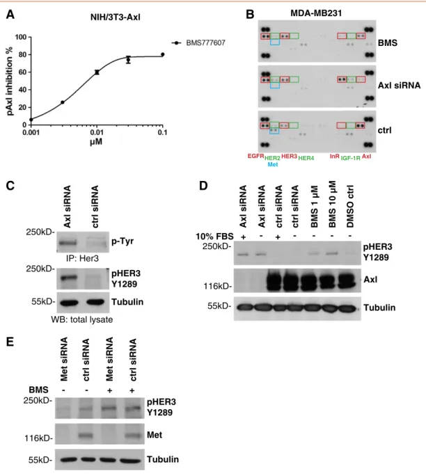
Figure 2.Inhibition of the Axl RTK leads to up-regulation of HER3 phosphorylation. (A) Inhibition of Axl phosphorylation was determined 1 hour posttreatment with BMS777607 by p-Tyr–Axl ELISA in NIH/3T3-Axl cells. IC50values were calculated by four-parameter log curve fit.
(B) Axl inhibition led to an up-regulation of HER3 phosphorylation as determined by the Human Phospho-RTK Array Kit. MDA-MB231 cells were incubated with 1μM BMS777607 for 24 hours or Axl-specific siRNA for 48 hours. The capture antibodies have been spotted in duplicates. The coordinates in the membrane for EGFR, HER2, Met, HER3, HER4, InR, IGF1R, and Axl are highlighted by rectangles. The corresponding antibody names are labeled at the bottom of the three displayed membranes from left to the right. (C) Validation of HER3 phosphorylation by HER3 immunoprecipitation with a HER3-specific antibody (Millipore, No. 05-390) and the Western blot for p-Tyr. The site-specific pHER3 Y1289 antibody displayed the same up-regulation of HER3 phosphorylation in Western blot experiments after treatment of MDA-MB231 cells with Axl-specific siRNA for 48 hours. Tubulin served as loading control. (D) Validation of HER3 phosphorylation by Western blot analysis of MDA-MB231 cells treated with Axl-specific siRNA for 48 hours or with 1 or 10 μM BMS777607 for 24 hours. The depletion of Axl kinase by siRNA was confirmed by anti-Axl Western blot analysis. Independent of the conditions, an increase of pHER3 Y1289 levels was evident in contrast to control treatments. Tubulin served as loading control. (E) Met- specific knockdown displayed no effect on pHER3 Y1289 levels compared to control siRNA treatment. The phosphorylation of HER3 Y1289 was induced by 10μM BMS777607 treatment for 24 hours independent of Met expression. The depletion of Met kinase from the cells was confirmed by Western blot analysis performed on cell lysates harvested 48 hours after Met-specific siRNA transfection. Tubulin served as loading control.
Results
Inhibition of the Axl RTK Leads to Up-Regulation of HER3 Phosphorylation
The Axl RTK activates prosurvival and metastatic pathways, and Axl overexpression was shown to correlate with aggressive tumor behavior. Therefore, we evaluated multiple TKI as potential Axl inhibitors and finally selected BMS777607 as the most potent Axl inhibitor published up to now. BMS777607 is currently undergoing phase I clinical trials and was originally described as a Met TKI.
However, BMS777607 displays a higher affinity to Axl RTK (IC50of
1.1 nM) than to Met (IC50of 3.9 nM) as published by Schreoder et al., 2009 [24]. In the first step, we validated the efficiency of BMS777607 to inhibit Axl phosphorylation by treating Axl- overexpressing NIH/3T3-Axl cells for 1 hour with serially diluted compound concentrations. Subsequently, we performed a phospho- Axl–specific ELISA and calculated the IC50 values by applying a four-parameter logistic curve fit. BMS777607 inhibits Axl autopho- sphorylation at low nanomolar concentrations, displaying an IC50
value of 0.006 μM as shown inFigure 2A. This NIH/3T3-Axl cell line was generated by Axl cDNA transfection because it represents a useful tool to screen the efficacy of Axl inhibitors.
Ovcar8 MDA-MB231
Hs578T
SKOV-3 250kD-
250kD-
250kD-
250kD-
BMS DMSO ctrl
Ovcar8 MDA-MB231
Hs578T
SKOV-3 250kD-
250kD-
250kD-
250kD-
Axl siRNA ctrl siRNA
A B
C
pAKT S473 pHER3 induction no pHER3 induction
55kD- Tubulin 55kD- Tubulin
55kD- Tubulin
55kD- Tubulin
55kD- Tubulin
55kD- Tubulin
55kD- Tubulin
55kD- Tubulin
pHER3 Y1289
pHER3 Y1289
pHER3 Y1289
pHER3 Y1289
pHER3 Y1289
pHER3 Y1289
pHER3 Y1289
pHER3 Y1289
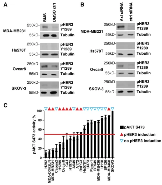
Figure 3.Inhibition of Axl leads to phosphorylation of HER3 in multiple cell lines and correlates with low basal AKT phosphorylation. (A) Western blot analysis of MDA-MB231, Hs578T, Ovcar8, and SKOV-3 cells treated with 10μM BMS777607 for 24 hours. A significant increase of pHER3 Y1289 levels was evident in MDA-MB231 and Ovcar8 cells in contrast to Hs578T and SKOV-3 cells. Tubulin served as loading control. (B) Western blot analysis of MDA-MB231, Hs578T, Ovcar8, and SKOV-3 cells treated with Axl-specific siRNA for 48 hours.
A significant increase of pHER3 Y1289 levels was evident in MDA-MB231 and Ovcar8 cells in contrast to Hs578T and SKOV-3 cells.
Tubulin served as loading control. (C) Low basal levels of AKT S473 phoshorylation correlate with induction of pHER3 Y1289 after Axl inhibition by 10μM BMS777607 and Axl knockdown with siRNA. The levels of basal AKT S473 phosphorylation for the indicated 20 cell lines are shown in ascending order on the abscissas axis. In parallel, we determined the up-regulation of HER3 phosphorylation of those 20 cell lines by Western blot analysis for pHER3 Y1289. The cell lines displaying activation of HER3 after Axl depletion were marked with red triangles on the top. The cell lines without HER3 response to Axl inactivation were marked with blue triangles. A positive correlation between a low basal phosphorylation of AKT S473 and the induction of HER3 phosphorylation was evident as 7 of 10 cell lines with low pAKT S473 levels, but only 2 of 10 cell lines with high pAKT S473 levels responded with up-regulation of pHER3 Y1289 to the inhibition of Axl by BMS777607 or Axl-specific siRNA. AKT phosphorylation was normalized to the highest values represented by SKOV3 cells (100%).
Mean values and SEM are shown (n= 3).
For the subsequent experiments, we used MDA-MB231 cells as a model system to study the effect of Axl inhibitors on human cancer cells. We selected the triple-negative breast cancer cell line MDA- MB231, first, because it is characterized by overexpression of the Axl RTK, and second, because multiple studies proved in MDA-MB231
cells that aggressive cell behavior depends on the expression of Axl RTK [14,26,27]. To confirm the activity of BMS777607 on MDA-MB231 cells and to analyze the activation pattern of other RTKs after Axl inhibition, we performed Human Phospho-RTK arrays. Therefore, we depleted Axl protein by Axl-specific siRNA knockdown or blocked Axl
A B C
HER3
Tubulin
1 h 3 h 6 h 16 h 24 h 48 h
BMS - + - + - + - + - + - +
55kD- 250kD-
250kD-
D
E
HER3 NRG1 NRG1
Supernatant
70kD-
pAKT S473 12 h 24 h 36 h 48 h
NRG1 [% of ctrl]
0 100 80 60 40 20 0
2 4 6 8
fold change
1 3 6 16 24 48 hours
0 1 2 3
fold change
1 3 6 16 24 48 hours
0 1 2 3
fold change
1 3 6 16 24 48 hours
1 3 6 16 24 48 hours 15
10 5 0
1 3 6 16 24 48 hours 15
10 5
fold change fold change 0
pHER3 Y1289 HER3
pHER3 Y1289
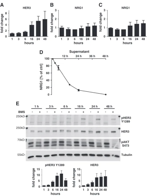
Figure 4.Inhibition of the Axl RTK activates HER3 transcription and phosphorylation. HER3 correlates with consumption of NRG1. (A) Quantification of HER3 mRNA induction in MDA-MB231 cells. Cells were treated with 10μM BMS77607 for 1 hour up to 48 hours. A three- to six-fold increase of HER3 mRNA was evident between 16 to 48 hours posttreatment. Mean values and SEM are shown (n= 3).
(B) Quantification of NRG1 mRNA induction with primer set 1 detecting all isoforms of NRG1 except isoforms No. 2 and No. 14. Cells were treated with 10μM BMS777607 for 1 hour up to 48 hours. No significant increase of NRG1 mRNA levels was detected. Mean values and SEM are shown (n= 3). (C) Quantification of NRG1 mRNA induction with primer set 2 detecting isoforms No. 2, No. 15, and No. 17 of NRG1. Cells were treated with 10μM BMS777607 for 1 hour up to 48 hours. No significant increase of NRG1 mRNA levels was detected.
Mean values and SEM are shown (n= 3). (D) Quantification of NRG1 protein levels in conditioned medium was assayed using NRG1- ELISA. Cells were treated with 10μM BMS777607 for 1 to 48 hours. A time-dependent consumption of NRG1 was measured. Mean values and SEM are shown (n= 3). (E) Western blot analysis of MDA-MB231 cells. Cells were treated with 10μM BMS777607 for 1 to 48 hours. A significant increase of the pHER3 Y1289 levels was evident in MDA-MB231 after 6 hours of treatment. The up-regulation of HER3 protein expression was evident 16 hours posttreatment. BMS777607 treatment continuously suppressed the phosphorylation of AKT S473. Tubulin served as loading control. The diagrams show the densitometric analysis of Western blots for pHER3 Y1289 and HER3.
Mean values and SEM are shown (n= 3).
tyrosine kinase activity by BMS777607 treatment. Surprisingly, 24 hours after exposure of MDA-MB231 cells to 1 μM BMS777607, phosphorylation of Axl and Met decreased significantly, whereas phosphorylation of HER3 was markedly enhanced. The phosphory- lation of the other three HER family members (EGFR, HER2, and HER4) remained unchanged (Figure 2B). In contrast to the treatment with 1 μM BMS777607, the Axl-specific siRNA knockdown additionally induced the phosphorylation of the insulin receptor (InR) and a slight up-regulation of the insulin-like growth factor 1 receptor (IGF1R) and of HER2 levels (Figure 2B). Unfortunately, we were not able to verify the phosphorylation of InR, IGF1R, and HER2 in Western blot experiments (data not shown).
To validate this Axl-specific siRNA-induced HER3 phosphoryla- tion, we performed an immunoprecipitation for HER3 from MDA- MB231 cell lysates under the same conditions as described for the RTK array. Tyrosine phosphorylation of HER3 was quantified by Western blot analysis using a homemade anti–p-Tyr antibody. The same induction of HER3 phosphorylation was evident with the site- specific pHER3 Y1289 antibody (Figure 2C). pHER3 Y1289 is a PI3K-binding site, and this phosphorylation was shown to be essential for PI3K/AKT signaling cascade activation[28]. Therefore, we used this site-specific antibody for all subsequent experiments.
To further validate the RTK array results and to elucidate the impact of serum on HER3 phosphorylation, we treated MDA- MB231 cells with Axl-specific siRNA to induce depletion of Axl protein either under full-serum conditions (10% FBS) or under starving conditions (0% FBS). The depletion of Axl kinase from the cells was confirmed by Western blot analysis performed on cell lysates harvested 48 hours after Axl-specific siRNA transfection. Indepen- dent of the serum conditions, a significant increase of pHER3 Y1289 levels was evident in contrast to control siRNA treatment (Figure 2D). The treatment with 1 and 10μM BMS777607 for 24 hours led also to an enhancement of HER3 phosphorylation in a concentration-dependent manner (Figure 2D).
As BMS777607 was originally published as Met inhibitor but also targets Axl RTK very potently at low nanomolar concentrations, it was necessary to elucidate a possible role of Met in the up-regulation process of phospho-HER3 [24]. Therefore, we used Met-specific siRNA to deplete the protein from MDA-MB231 cells. The Met knockdown was confirmed by Western blot analysis 48 hours after Met-specific siRNA transfection. Met knockdown displayed no effect on pHER3 Y1289 levels compared to control siRNA treatment (Figure 2E). Additionally, independent from the Met knockdown, the treatment with 10μM BMS777607 for 24 hours increased the phosphorylation level of HER3. On the basis of this result, we conclude that Met inhibition is not related to the up-regulation of HER3 phosphorylation.
To sum up, HER3 remained the only validated and consistently up-regulated RTK after treatment with BMS777607 or knockdown with Axl-specific siRNA in MDA-MB231 cells.
Inhibition of Axl RTK Leads to Phosphorylation of HER3 in Multiple Cell Lines and Correlates With Low Basal AKT Phosphorylation
Because we had shown that pHER3 is an activating feedback mechanism on Axl inhibition in MDA-MB231 cells, we asked if this is a general phenomenon in Axl-overexpressing tumor cells. Therefore, we analyzed the activation of HER3 in different breast and ovary cancer cell lines expressing Axl. First, we blocked Axl activity pharmacologically
with 10μM BMS777607 (Figure 3A), and second, we depleted Axl protein by Axl-specific siRNA (Figure 3B). Phospho-HER3 was inducible after Axl protein depletion as well as after inhibition of phosphorylation by treatment with 10μM BMS777607 in MDA- MB231 and Ovcar8 cells but not in Hs578T and SKOV-3 cells (Figure 3, A and B). From this finding, we concluded that Axl expression itself is not a biomarker for the induction of a HER3 feedback loop.
To further characterize the HER3 activation loop, we analyzed 20 different Axl-expressing cell lines originating from brain, breast, ovary, cervix, lung, and pancreatic tumors. Knowing that the AKT signaling pathway is influenced by Axl RTK, we performed phospho- AKT S473 ELISAs to determine the basal AKT phosphorylation levels. The levels of pAKT S473 are shown in ascending order on the axis of abscissas in Figure 3C. In parallel, we determined the activation of HER3 in those 20 cell lines after treatment with 10μM BMS777607 and Axl-specific siRNA by Western blot analysis with the pHER3 Y1289 antibody. The cell lines displaying activation of HER3 after Axl depletion were marked with red triangles on the top of Figure 3C. The cell lines without HER3 response to Axl inactivation were marked with blue triangles. Thereby, we discovered a positive correlation between a low basal phosphorylation of AKT S473 and the induction of HER3 activation on Axl inhibition by BMS777607 and Axl-specific siRNA treatment. Seven of 10 cell lines with low pAKT S473 levels but only 2 of 10 cell lines with high pAKT S473 levels responded with up-regulation of pHER3 Y1289 after inhibition of Axl by BMS777607 or Axl-specific siRNA treatment (Figure 3C).
From these results, we conclude that inhibition of Axl induces an activation feedback loop of HER3, in various tumor types, characterized by low basal AKT S473 phosphorylation and by a high dependency on Axl/PI3K/AKT signaling.
Inhibition of the Axl RTK Activates HER3 Transcription but Not of Its Ligand NRG1
Next, we performed quantitative PCR to determine the levels of HER3 mRNA. Therefore, we treated MDA-MB231 cells with 10 μM BMS777607 and harvested them at different time points. A significant induction of HER3 mRNA production became evident after 16 hours of treatment reaching a 3.9-fold level, whereas the maximum increase of HER3 mRNA was achieved after 48 hours reaching 5.9-fold values (Figure 4A). As breast cancer cells often express high levels of HER RTK activating ligands [29], we performed quantitative PCR and ELISA analysis of the HER3 ligand NRG1 to compare the relative levels after treatment with 10 μM BMS777607. The Gene database search on National Center for Biotechnology Information platform revealed 17 different NRG1 isoforms inH. sapiens(Gene ID: 3084). Due to the fact that NRG1 appears in different isoforms deriving from the NRG1 gene by alternative splicing, we used two different primer sets for quantitative PCR analysis. One primer set binds to the mRNA of all isoforms except of isoforms No. 2 and No. 14. The second primer set binds to the mRNA of isoforms No. 2, No. 15, and No.
17. In this way, we were able to cover all isoforms with the exception of isoform No. 14. NRG1 mRNA expression was detectable in MDA-MB231 cells, but it was induced only reaching values between 1.2- or 1.5-fold within 48 hours (Figure 4,BandC).
Accordingly, we excluded NRG1 mRNA up-regulation as being responsible for HER3 activation.
Induction of HER3 Phosphorylation Correlates with Consumption of NRG1
Although we did not expect an up-regulation of NRG1 ligand on the basis of the quantitative PCR results, we analyzed NRG1 protein levels in the supernatant of BMS777607-treated MDA-MB231 cells by NRG1-ELISA. Strikingly, the levels of NRG1 protein dropped in the supernatant of 10μM BMS777607-treated MDA-MB231 cells in a time-dependent manner compared to DMSO-treated control levels from 97% after 1 hour to less than 1% after 48 hours (Figure 4D). To determine if there is any connection between the decrease of NRG1 ligand in the supernatant and HER3 activation, we treated the MDA-MB231 cells in a time course experiment up to 48 hours with 10 μM BMS777607 and analyzed in parallel the supernatant by Western blot. Consistent with the previous results,
BMS777607 caused a 4.2-fold activation of pHER3 Y1289 after 6 hours of treatment (Figure 4E). The activation of pHER3 Y1289 correlated with the decrease of NRG1 in the supernatant (Figure 4D).
The climax of 11.1-fold HER3 activation was reached after 24 hours of compound treatment and decreased at the 48-hour time point.
Parallel to the pHER3 Y1289 decrease, NRG1 levels of the supernatant approximated the background level of the NRG1 ELISA assay after 48 hours. At the same time, we proved that the 7.8- to 8.8-fold increased expression of HER3 protein, after 16 to 48 hours of treatment, results from the elevated level of HER3 mRNA as shown inFigure 4A. Simultaneously, BMS777607 was able to inhibit phosphorylation of AKT S473 constantly for up to 48 hours.
However, the phosphorylation of HER3 does not result in a downstream activation of pAKT S473. These results reveal that Axl
inhibition by BMS777607 leads to transcription of HER3 protein and to the NRG1 ligand–dependent phosphorylation of HER3.
Additionally, these results are in line with the mechanism of AKT inhibition–dependent transcription of HER3 described recently by Chandarlapty et al., 2011 and Chakrabarty et al., 2012[30,31].
HER3 Induction is a Common Feedback Mechanism of Axl Inhibitors
Because we could show that induction of pHER3 is part of an activation feedback mechanism induced by BMS777607, we asked whether this is a general phenomenon of Axl inhibitors. Therefore, we compared BMS777607 to R428, a recently described Axl inhibitor [25], and an Axl inhibitor recently developed by our group, namely, MPCD84111. First, we determined the activity of these Axl TKIs in NIH/3T3 Axl-overexpressing cells. For this reason, we treated the cells for 1 hour with serially diluted compound concentrations of BMS777607, MPCD84111, and R428 and subsequently performed a phospho-Axl–specific ELISA to calculate the IC50 values by applying a four-parameter logistic curve fit. As shown inFigure 1A, the compounds inhibited Axl phosphorylation at nanomolar concentrations, displaying IC50 values for BMS777607 of 0.006 μM, MPCD84111 of 0.027μM, and R428 of 0.043μM.
Next, we analyzed if MPCD84111 and R428 have the same impact on the HER3 feedback loop as BMS777607 in MDA-MB231 cells. Interestingly, treatment with 10μM BMS777607 and 10μM R428 induced the phosphorylation of HER3 (6.7- and 2.8-fold), whereas 10 μM MPCD84111 prohibited the phosphorylation of HER3 by 68% in comparison to DMSO control within 24 hours (Figure 1B). We could also demonstrate that 10μM BMS777607 and 10μM R428 as well as 10 μM MDCD84111 induce HER3 expression 6.1-, 7.5-, and 7.4-fold in comparison to DMSO treatment. Our data reveal that the transcriptional feedback loop of HER3 was initiated similarly by BMS777607, R248, and MPCD84111, even though MPCD84111 was able to block phosphorylation of HER3 Y1289 efficiently.
To elucidate these results in more detail, we treated MDA-MB231 cells in a time course experiment for up to 48 hours with 1 μM BMS777607 or MPCD84111 and analyzed the cell lysates by Western blot. In contrast to BMS777607, MPCD84111 inhibited the phosphorylation of HER3 Y1289 during the entire time course (Figure 1C). Interestingly, both inhibitors increased the total protein levels of HER3 after 16 hours of treatment. It was published earlier by Myatt and Lam, 2007 that inhibition of AKT has an impact on the expression and activity of the Forkhead box protein O (FoxO) family members [32]. Therefore, the induction of HER3 RTK expression might result from FoxO-dependent transcription. Our data point out that the phosphorylation of HER3 is blocked by MPCD8411 independently from transcription cascade of HER3 and might result from a direct inhibition by the compound.
MPCD84111 Inhibits the Dimerization Partner for HER3
Subsequently, we addressed the question why MPCD84111, but not BMS777607, inhibits phosphorylation of HER3. It is widely accepted that HER3 has an impaired kinase activity. Because of this reason, activation of the HER3 RTK occurs only after the dimerization with other RTKs, such as HER2 or EGFR [33]. We asked if MPCD84111-dependent inhibition of HER3 phosphoryla- tion (Figure 1,BandC) might be a result of blocking HER2 as a heterodimerization partner for HER3. Therefore, we determined the kinase selectivity profile of MPCD84111 using three different types of assays, namely, IMAP, binding, and HTRF assays. The kinase profile of BMS777607 was published earlier by Schroeder et al., 2009 [24]. The kinase profile of MPCD84111 displayed an inhibition of Axl as well as Met, Src, Abl, Kit, Ret, PDGFRβ, Tie2, VEGFR-2, and DDR1 with efficacy of 100% when used at 10 μM concentration (Figure 1D). The comparison of both kinase selectivity profiles identified common targets of BMS777607 and MDCD84111 such as Axl, Met, Kit, PDGFRβ, Tie2, AuroraA/B, and VEGFR-2. In contrast to BMS777607, MPCD84111 uniquely targets HER2, a
Figure 5.Inhibition of HER2 blocks HER3 phosphorylation in triple-negative breast cancer cells. (A) Validation of HER2 being a target of MPCD84111. MPCD84111 inhibits phosphorylation of HER2 in contrast to BMS777607 as demonstrated on HER2-expressing MCF7 cells. Phosphorylation of HER2 Y1248 was analyzed 1 hour after addition of BMS777607 and MPCD84111 by Western blot. Both compounds were used in three-fold serial dilutions starting with 10μM. Tubulin served as loading control. (B) Herceptin and lapatinib block HER3 phosphorylation in MDA-MB231 cells. Representative Western blots of MDA-MB231 cells treated with 10μM BMS777607 for 24 hours compared to DMSO-treated control cells are shown. Cells were additionally incubated with 5μM BB94 (batimastat), 10μg/ml Erbitux, 10μg/ml Herceptin, or 5μM lapatinib for 2 hours. One hundred micrograms of lysate was subjected to sodium dodecyl sulfate– polyacrylamide gel electrophoresis, blotted, and probed with corresponding antibodies for pHER3 Y1289, HER3, pAKT S473, pERK1/2, and Tubulin as loading control. BB94, Herceptin, and lapatinib were able to suppress the BMS777607-induced HER3 Y1289 phosphorylation in contrast to Erbitux. (C) EGFR in combination with HER2-specific knockdown exhibits a significant effect on pHER3 Y1289 levels compared to control siRNA treatment. MDA-MB231 cells were treated with EGFR, HER2, HER3, and a combination for EGFR and HER2-specific siRNA for 48 hours. Subsequently, the phosphorylation of HER3 Y1289 was induced by 10μM BMS777607 treatment for 24 hours. Only HER3 and the combination of EGFR and HER2 siRNA blocked HER3 phosphorylation completely. HER2-specific knockdown leads to a less pronounced reduction of HER3 phosphorylation, and EGFR-specific knockdown even induces pHER3 y1289 activity by 1.4-fold. The depletion of EGFR, HER2, and HER3 from the cells was confirmed by Western blot analysis. Tubulin served as loading control. The diagram shows the densitometric analysis of Western blots for pHER3 Y1289. Mean values and SEM are shown (n= 3). (D) MDA-MB231 cells were treated with 10μM BMS777607 for 24 hours to further validate the involvement of HER2 in the phosphorylation of HER3. The HER2 RTK was immunoprecipitated with a commercial HER2-specific antibody (Millipore, No. 06-562) from 2 mg of total lysate. Western blots for p-Tyr and total HER2 are shown. BMS777607 treatment increased the phosphorylation of HER2 compared to DMSO-treated control, and 50 ng/ml NRG1 further enhanced the phosphorylation level. (E) Validation of HER2 activity by immunoprecipitation of HER2. MDA-MB231 cells were treated with 10 μM BMS777607 for 24 hours. The HER2 RTK was immunoprecipitated with a homemade HER2-specific antibody (clone 13D1B1) from 2 mg of total lysate. Western blots for p-Tyr, HER2, as well as for Tubulin are shown. Twenty micrograms of the total lysate was used for the Tubulin Western blot analysis as loading control for the immunoprecipitation. BMS777607 treatment increased the phosphorylation of HER2 compared to DMSO-treated control, and 50 ng/ml NRG1 further enhanced the phosphorylation level. Therefore, HER2 remains a hardly detectable, but active dimerization partner for HER3 in MDA-MB231 cells.
dimerization partner of HER3 (Figure 1D). This finding implicates that blocking of HER3 phosphorylation by MPCD84111 might be achieved by inhibition of HER2.
Inhibition of HER2 Blocks HER3 Phosphorylation in Triple-Negative Breast Cancer Cells
In breast cancer cells withHER2gene amplification, HER2 is the main kinase that phosphorylates HER3 [34]. As BMS777607 in contrast to MPCD84111 does not affect the catalytic activity of HER2 as demonstrated in MCF7 cells expressing HER2 (Figure 5A), we assumed that in HER2 low-expressing MDA-MB231 cells, HER2 might also act as the kinase maintaining phosphorylation of HER3 on inhibition of Axl as well.
Therefore, we examined the effect of BMS777607 in combination with either 10 μg/ml HER2-blocking antibody Herceptin, 5 μM
EGFR/HER2 TKI lapatinib, or 5μM the broad spectrum matrix metalloprotease inhibitor BB94 (batimastat). In MDA-MB231 cells, all of these combinations effectively inhibited phosphorylation of HER3, in contrast to the treatment with 10 μg/ml Erbitux (Figure 5B). Particularly, the specific blocking of HER2 by the monoclonal antibody Herceptin as well as lapatinib suggested that HER2 plays a significant role in maintaining phosphorylation of HER3 in MDA-MB231 cells. It is well known that active ligands of the HER family are proteolytically cleaved by metalloproteases and afterwards released from the cell surface[35]. BB94 (batimastat) is a broadband inhibitor of the Disintegrin and metalloproteinase domain-containing protein ADAM family of metalloproteases and blocks shedding of EGFR ligands [36,37]. Inhibition of HER3 phosphorylation by 5μM BB94 underlined our findings that HER3 is activated by an extracellular mechanism. BMS777607 consistently
blocked phosphorylation of AKT S473 but had only a minor effect on the Mitogen-activated protein-kinase pathways as proven by pERK1/2 Western blot analysis (Figure 5B).
Knowing that lapatinib was most efficient in blocking HER3 phosphorylation, we evaluated the impact of EGFR and HER2 on HER3 phosphorylation by siRNA experiments. Therefore, we depleted EGFR, HER2, HER3, and EGFR in combination with HER2 from MDA-MB231 cells by siRNA knockdown and subsequently induced HER3 phosphorylation by 10 μM BMS treatment for 24 hours (Figure 5C). Only HER3 siRNA and the combination of EGFR and HER2 siRNA treatment blocked HER3 phosphorylation, completely exhibiting less than 5% of phosphor- ylation compared with control siRNA treatment. Comparable to Herceptin, HER2-specific siRNA treatment remained a residual HER3 phosphorylation of 22% (Figure 5C).
Although MDA-MB231 cells represent a prototype of triple- negative breast cancer cell lines characterized by the loss of HER2 expression, we investigated HER2 as one of the probable dimerization partners of HER3. To elucidate in more detail the phosphorylation status of HER2 in triple-negative breast cancer cells, we performed immunoprecipitation experiments with a vast protein amount of 2 mg. We treated MDA-MB231 cells with 10μM BMS777607 for 24 hours, and we further enhanced the phosphorylation of HER2 by stimulation with 50 ng/ml NRG1. The HER2 RTK was immunoprecipitated with a homemade anti-HER2–specific antibody (clone 13D1B1) or a commercial anti-HER2 antibody (Millipore, No. 06-562). The subsequent Western blot analysis for p-Tyr resulted in a slight, but reproducible, induction of tyrosine phosphorylation after BMS777607 treatment and NRG1 stimulation (Figure 5,DandE).
Hence, we assume that this minimal amount of HER2 phosphorylation might contribute to the pronounced HER3 activation and thereby maintains the feedback loop counteracting Axl inhibitor treatment. Additionally, lapatinib emerged as the most efficient treatment strategy for inhibition of HER3 phosphorylation.
Exogenous Application of NRG1 Rescues AKT Phosphorylation and Cell Viability
Even though complete recovery of pAKT S473 was not achieved with a pharmacological dose of 1μM BMS777607, the feedback up- regulation of HER3 expression and HER3 phosphorylation was clearly evident, further suggesting that inhibition of the Axl/PI3K/
AKT pathway leads to the reactivation of HER3 (Figure 4E). These data imply that HER3 phosphorylation partially restores the prosurvival and proliferation signaling that, in turn, may limit the effect of Axl inhibitors. To support this hypothesis, we performed rescue experiments in combination with cell viability assays. MDA- MB231 cells were treated with pharmacological concentrations of BMS777607 under starving conditions for 72 hours. Addition of the HER3 ligand (50 ng/ml NRG1) completely compensated the inhibitory effect of 0.1 and 1μM BMS777607 on cell viability. In contrast, the cell viability of the untreated cells was not affected by NRG1 stimulation (Figure 6A). The compensatory up-regulation of HER3 expression and partial maintenance of HER3 phosphorylation on inhibition of Axl suggested that combined inhibition of Axl and HER2/HER3 could synergistically inhibit tumor cell viability.
Monolayer cell cultures represent oversimplified models for tumor studies, due to the loss of extracellular matrix rigidity on artificial plastic surfaces and high serum concentrations. These conditions poorly mimic the tumor cell biology in vivo. A more robust and reproducible test system is the three dimensional (3D) spheroid culture. To test the combined inhibition of Axl and HER2/HER3 on tumor cell viability, we used these 3D spheroid cultures.
We treated two Axl-expressing cell lines, namely MDA-MB231 and Ovcar8, with MPCD84111 and with a combination of BMS and lapatinib, to elucidate the hypothesis that an inhibition of Axl and HER2/HER3 might be essential for treatment of Axl overexpressing cells. First, we proved the efficacy of 1μM MPCD84111 in contrast to 1μM BMS777607 treatment on MDA-MB231 and Ovcar8 cells.
One micromolar MPCD84111 significantly inhibited cell viability to 37% in MDA-MB231 spheroids and to 42% in Ovcar8 spheroids in
Figure 6.Exogenous application of NRG1 rescues AKT phosphorylation and cell viability. (A) Exogenous application of NRG1 rescues the inhibition of cell viability by BMS777607. Cell viability of MDA-MB231 cells was measured by CellTiter-Glo assay after 72 hours of treatment under starving conditions (0% FBS). Cells were incubated with increasing concentrations of BMS777607 (0.1, 1, 10μM) with and without 50 ng/ml NRG1. Addition of NRG1 completely compensates the proliferation inhibitory function of 0.1 and 1μM BMS777607.
The DMSO control does not react to NRG1 stimulation. Mean values and SEM of three independent experiments are shown. Differences with *Pb.05, **Pb.01, and ***Pb.001 were considered statistically significant (Mann-Whitney test; ns, nonsignificant). (B) Cell viability of MDA-MB231 and Ovcar8 cells is inhibited by MPCD84111 in contrast to BMS777607. Cell viability of MDA-MB231 and Ovcar8 cells was measured by CellTiter-Glo assay after 72 hours of treatment. 3D spheroids were incubated with 1 µM BMS777607 or 1 µM MPCD84111 using starving conditions (0% FBS). Cell viability was reduced to 37% by MPCD84111 in MDA-MB231 cells and to 43% in Ovcar8 cells. Mean values and SEM of three independent experiments are shown. Differences with *Pb.05, **Pb.01, and ***Pb.001 were considered statistically significant (Mann-Whitney test). (C) BMS777607 and lapatinib exhibit a synergistic inhibitory effect on HER3 phosphorylation. Western blot analysis of MDA-MB231 cells is shown. Cells were treated with 1μM BMS777607 or a combination of 1 μM BMS777607 and 5μM lapatinib relative to DMSO control for 24 hours with or without 50 ng/ml NRG1 stimulation for 15 minutes. A significant increase of the pHER3 Y1289 levels was evident after BMS777607 treatment. BMS777607 treatment suppressed the phosphorylation of AKT S473 to 24%. Additional NRG1 stimulation enhances HER3 phosphorylation and restores pAKTS473 phosphorylation. The combination of BMS777607 and lapatinib blocks completely the HER3 and AKT phosphorylation. Tubulin served as loading control. The diagrams show the densitometric analysis of Western blots for pHER3 Y1289 and pAKT S473. Mean values and SEM are shown (n= 3). (D) BMS777607 and lapatinib exhibit a synergistic inhibitory effect on HER3 phosphorylation. Western blot analysis of Ovcar8 cells is shown. Cells were treated with 1μM BMS777607 or a combination of 1μM BMS777607 and 5μM lapatinib relative to DMSO control for 24 hours with or without 50 ng/ml NRG1 stimulation for 15 minutes. A 2.4-fold increase of the pHER3 Y1289 levels was evident after BMS777607 treatment. BMS777607-induced HER phosphorylation treatment completely rescued AKT phosphorylation.
Additional NRG1 stimulation enhances HER3 and pAKTS473 phosphorylation. The combination of BMS777607 and lapatinib blocks completely the HER3 and AKT phosphorylation. Tubulin served as loading control. The diagrams show the densitometric analysis of Western blots for pHER3 Y1289 and pAKT S473. Mean values and SEM are shown (n= 3).
contrast to 1μM BMS777607 treatment exhibiting cell viability rates of 97% in MDA-MB231 and 92% in Ovcar8 in comparison to DMSO-treated controls (Figure 6B).
Second, we proved the complete inhibition of HER3 phosphory- lation by combined treatment of 1 μM BMS777607 and 5 μM lapatinib in MDA-MB231 and Ovcar8 cells. Additionally, we analyzed
if BMS treatment or the stimulation with HER3 ligand NRG1 might lead to the reconstitution of the AKT signaling pathway.
One micromolar BMS777607 leads to up-regulation of total HER3 protein levels and to a six-fold induction of HER3 phosphorylation after 24 hours of treatment in MDA-MB231 cells (Figure 6C). The phospho-AKTS473 levels are reduced to 24% compared with DMSO- treated cells, and additional stimulation with 50 ng/ml NRG1 restores pAKTS473 signal to DMSO control levels. The combination of 1μM BMS777607 and 5 μM lapatinib inhibited HER3 and AKT phosphorylation to 0.9%, respectively, 6.1% in comparison to BMS777607 single treatment. Especially, NRG1 ligand stimulation was not able to induce HER3 phosphorylation any more than in contrast to DMSO (two-fold induction) and 1 μM BMS777607 treatment (13-fold induction). HER2 phosphorylation was below detection threshold in MDA-MB231 cells due to the low expression level of HER2 in this cell line (Figure 6C).
The Ovcar8 cells react to 1μM BMS777607 treatment, analogous to MDA-MB231, by up-regulation of total HER3 protein levels and a 2.4- fold HER3 phosphorylation compared to DMSO-treated cells. This HER3 phosphorylation results in a 1.4-fold induction of AKT phosphorylation. Additional 50 ng/ml NRG1 ligand stimulation leads in a 10- or 17.8-fold HER3 phosphorylation in DMSO control and 1μM BMS777607-treated cells. The combined treatment with 1 μM BMS777607 and 5μM lapatinib completely prevents HER3 and AKT phosphorylation and additionally prohibits the activation by NRG1 ligand. In Ovcar8 cells, HER2 Y1248 exhibits an identical phosphorylation pattern to pHER3 Y1289, emphasizing the impor- tance of the HER2/HER3 heterodimer in maintaining the Her3 phosphorylation in cell lines with significant HER2 levels (Figure 6D).
We conclude that BMS777607-induced HER3 phosphorylation is able to rescue or at least stabilizes the AKT signaling in Ovcar8 and MDA- MB231 cells, andvice versa,a combination of 1μM BMS777607 and 5 μM lapatinib completely inhibits HER3 and AKT phosphorylation.
Pharmacological Inhibition of Axl Sensitizes for Lapatinib Treatment
We further tested the combined inhibition of Axl and HER2/
HER3 on tumor cell viability in 3D spheroid cultures of MDA- MB231 and Ovcar8 cells. Therefore, we treated MDA-MB231 3D spheroids with 1μM BMS777607 and 5μM lapatinib and assessed their viability under starving conditions (0% FBS). Cell viability was significantly reduced up to 50% by a combination of 1 μM BMS777607 and 5 μM lapatinib compared to the effect of either inhibitor administered alone (Figure 7A). The cell viability of Ovcar8 cell spheroids was significantly inhibited by 5μM labatinib and the combination of 1μM BMS777607 and 5μM lapatibnib. The cell viability was inhibited to 56% by the combination of 1 μM BMS777607 and 5μM lapatinib (Figure 7B).
The images ofFigure 7,CandD, visualize the spheroids formed after 72 hours in Matrigel. Ovcar8 cell spheroids react more sensitively to 5 μM lapatinib treatment alone due to the fact of higher HER2 expression levels and resulting dependency on HER2 signaling.
Analogous but less pronounced results were achieved with MDA- MB231 3D spheroids cultured under full-serum conditions (10%
FBS). The cell viability of MDA-MB231 spheroids decreased significantly under full-serum conditions by combination treatment with 1μM BMS777607 and 5μM lapatinib. To better present the differences in cell viability, we displayed the inhibition of cell viability in Figure 7, E and F. The cell viability of MDA-MB231 3D spheroids was not significantly affected by 1 μM BMS777607 or 5 μM lapatinib alone, whereas MDA-MB231 3D spheroids treated with the TKI combination displayed a statistically significant reduction in viability of 24% (Figure 7E). To underline the efficacy of combined targeted therapies toward Axl and HER3, we performed siRNA experiments to knockdown Axl and HER3 (Figure 7F).
Analogous to the pharmacological inhibition, the cell viability of MDA-MB231 3D spheroids was not significantly affected by Axl
Figure 7.Pharmacological inhibition of Axl sensitizes for lapatinib. (A) BMS777607 and lapatinib exhibit a synergistic effect on inhibition of cell viability. Cell viability of MDA-MB231 cells was measured by CellTiter-Glo assay after 72 hours of treatment. MDA-MB231 3D spheroids were incubated with 1μM BMS777607, 5μM lapatinib, or a combination of both compounds relative to DMSO control under starving conditions (0% FBS). Cell viability was reduced to 49.5% by the combination of 5μM lapatinib and 1μM BMS777607 compared to either compound administered separately. Mean values and SEM of three independent experiments are shown. Differences with *Pb .05, **Pb.01, and ***Pb.001 were considered statistically significant (Kruskal-Wallis test; ns, nonsignificant). (B) BMS777607 and lapatinib exhibit an additive cell viability inhibition effect. Cell viability of Ovcar8 cells was measured by CellTiter-Glo assay after 72 hours of treatment. Ovcar8 3D spheroids were incubated with 1μM BMS777607, 5μM lapatinib, or a combination of both compounds relative to DMSO control under starving conditions (0% FBS). Cell viability was reduced to 56% by the combination of 5μM lapatinib and 1μM BMS777607 compared to either compound administered separately. Mean values and SEM of three independent experiments are shown. Differences with *P b .05, **P b .01, and ***P b .001 were considered statistically significant (Kruskal-Wallis test; ns, nonsignificant). (C) Representative phase-contrast images of MDA-MB231 spheroids. MDA-MB231 3D spheroids were incubated with 1 μM BMS777607, 5μM lapatinib, or a combination of both compounds relative to DMSO control under starving conditions (0% FBS) for 72 hours. Scale bars indicate 100μm. (D) Representative phase-contrast images of Ovcar8 spheroids. Ovcar8 spheroids were incubated with 1μM BMS777607, 5μM lapatinib, or a combination of both compounds relative to DMSO control under starving conditions (0%
FBS) for 72 hours. Scale bars indicate 100μm. (E) Validation of the synergistic inhibitory effect of BMS777607 and lapatinib treatment on cell viability. Cell viability of MDA-MB231 cells was measured by CellTiter-Glo assay after 72 hours of treatment. MDA-MB231 3D spheroids were incubated with 1μM BMS777607, 5μM lapatinib, or a combination of both compounds relative to DMSO control under full-serum conditions (10% FBS). Inhibition of cell viability was significantly increased to 24% by a combination of 5μM lapatinib and 1μM BMS777607 compared to either compound administered separately. Mean values and SEM of six independent experiments are shown.
Differences with *Pb.05, **Pb.01, and ***Pb.001 were considered statistically significant (Kruskal-Wallis test; ns, nonsignificant). (F) Validation of synergistic inhibitory effect by knockdown of Axl and HER3 on cell viability. MDA-MB231 cell were transfected with Axl- specific siRNA, HER3-specific siRNA, or a combination of both siRNAs for 48 hours. Subsequently, MDA-MB231 3D spheroids were grown for 72 hours under full-serum conditions (10% FBS), and cell viability was measured by CellTiter-Glo assay. Inhibition of cell viability was significantly increased to 22% by a combination of Axl-specific siRNA and HER3-specific siRNA compared to either siRNA transfected separately. Mean values and SEM of five independent experiments are shown. Differences with *Pb.05, **Pb.01, and ***P b.001 were considered statistically significant (Kruskal-Wallis test; ns, nonsignificant).