STRUCTURE, GATING, AND REGULATION OF THE CFTR ANION CHANNEL
László Csanády, XPaola Vergani, and David C. Gadsby
Department of Medical Biochemistry, Semmelweis University, Budapest, Hungary; MTA-SE Ion Channel Research Group, Budapest, Hungary; Department of Neuroscience, Physiology and Pharmacology, University College London, London, United Kingdom; and Laboratory of Cardiac/Membrane Physiology, The Rockefeller University, New York, New York
L
Csanády L, Vergani P, Gadsby DC. Structure, Gating, and Regulation of the CFTR Anion Channel. Physiol Rev 99: 707–738, 2019. Published December 5, 2018;doi:10.1152/physrev.00007.2018.—The cystic fibrosis transmembrane conduc- tance regulator (CFTR) belongs to the ATP binding cassette (ABC) transporter super- family but functions as an anion channel crucial for salt and water transport across epithelial cells. CFTR dysfunction, because of mutations, causes cystic fibrosis (CF). The anion- selective pore of the CFTR protein is formed by its two transmembrane domains (TMDs) and regulated by its cytosolic domains: two nucleotide binding domains (NBDs) and a regulatory (R) domain. Channel activation requires phosphorylation of the R domain by cAMP– dependent protein kinase (PKA), and pore opening and closing (gating) of phosphorylated channels is driven by ATP binding and hydrolysis at the NBDs. This review summarizes available information on structure and mechanism of the CFTR protein, with a particular focus on atomic-level insight gained from recent cryo-electron microscopic structures and on the molecular mechanisms of channel gating and its regulation. The pharmacological mechanisms of small molecules targeting CFTR’s ion channel function, aimed at treating patients suffering from CF and other diseases, are briefly discussed.
I. INTRODUCTION 707
II. CFTR DOMAIN TOPOLOGY AND... 709 III. THE CFTR ANION PERMEATION... 711 IV. REGULATION OF CFTR GATING... 715 V. REGULATION OF CFTR GATING... 725 VI. TARGETING CFTR FUNCTION TO... 728
VII. CONCLUDING REMARKS 730
I. INTRODUCTION
A. CFTR and Cystic Fibrosis
Cystic fibrosis (CF) is the most common life-threatening, inherited monogenic disorder among Caucasian popula- tions; it affects 1 in ~2500 newborns in Europe and 1 in
~3500 newborns in the United States. CF is a multiorgan disorder, with symptoms including airway blockage by thickened mucus, leading to chronic lung infections, inflam- mation and bronchiectasis, blockage of pancreatic ducts and consequent pancreatic insufficiency, bowel obstruction in newborns, male infertility because of obstruction of the vas deferens, and a characteristic high-salt sweat diagnostic of the disease. Although at present efficient causative treat- ment is still restricted to a small subset of CF patients, in the past decades, improvements in patient care and symptom- atic treatment have greatly prolonged the life expectancy of people born with CF, from ~1 yr in 1950 to ~40 yr at present (171).
The primary defect in CF patients is a reduction in chloride (190, 259) and bicarbonate (208) transport capacity across the apical membrane of epithelial cells. In 1989, the gene mutated in CF patients was identified on chromosome 7 by positional cloning (201), and the protein product was named the cystic fibrosis transmembrane conductance reg- ulator (CFTR) to reflect its presumed involvement in the regulation of other anion transport proteins. Purification of the CFTR protein and its functional reconstitution in lipid bilayers soon provided proof that CFTR is, itself, the anion channel responsible for cAMP– dependent anion transport across epithelial surfaces (16).
B. The ABC Protein Superfamily
CFTR is a member of the large superfamily of tens of thou- sands of ATP binding cassette (ABC) proteins that are found in all kingdoms of life (147) and that serve to trans- port a large variety of substrates into and out of cells at the expense of ATP hydrolysis. ABC proteins share a conserved general architecture based on the modular assembly of four canonical domains: two transmembrane domains (TMDs) and two cytosolic nucleotide binding domains (NBDs). The number of polypeptide chains that this “core” functional unit comprises is variable. In prokaryotes, the four domains are often expressed as four individual polypeptides or as two TMD/NBD “half transporters” that coassemble post- translationally. The human genome encodes 48 ABC pro-
teins, which have been grouped into seven subfamilies (ABCA–G) (69). Except for the E and F subfamilies, which contain no TMDs, the human ABC proteins consist either of half transporters that homo- or heterodimerize or of full transporters in which the four canonical domains are linked in a single polypeptide chain. The ABCC subfamily, to which CFTR (ABCC7) belongs, falls into the latter class. In CFTR, each TMD contains six transmembrane (TM) heli- ces, and the two homologous TMD-NBD halves (TMD1- NBD1 and TMD2-NBD2) are linked by a contiguous, unique, cytosolic regulatory (R) domain (201) (FIGURE 1A).
At the sequence level, CFTR’s two homologous halves dis- play a marked asymmetry, a general feature of ABCC sub- family proteins (187). Within the entire ABC superfamily, CFTR is the only protein shown to form a transmembrane ion channel pore, in contrast to the vast majority of its homologs that serve as active transporters. The only other exceptions among human ABC proteins are the soluble E and F subfamily members, that are not involved in trans- membrane transport, and CFTR’s close relatives SUR1 (ABCC8) and SUR2 (ABCC9) that serve as regulatory sub- units of ATP-sensitive potassium (KATP) channels (69).
C. Basic Functional Properties of the CFTR Anion Channel
Opening and closing (gating) of the CFTR anion pore is largely regulated by two processes. First, for a CFTR chan- nel to become activated, its cytosolic R domain must be phosphorylated by cAMP– dependent protein kinase (PKA) (20, 48, 183, 225). Second, gating of a phosphorylated CFTR channel is driven by binding of ATP to its cytosolic NBDs (9). In single-channel recordings, CFTR channel ac- tivity displays typical bursting behavior, with brief (in- traburst) closures interrupting longer periods of channel opening, yielding open bursts that are separated by long (interburst) closures (32, 95, 261). Gating kinetics are rela- tively slow; the duration of a burst and interburst cycle is on a second timescale (0.1–2 s, depending on species, phos- phorylation level, and temperature) rather than the milli- second timescale of voltage-gated channels. Moreover, ex- cept for the kinetics of intraburst closures, which little affect channel open probability (30), CFTR gating is largely volt- age independent (20). Anion permeation through the open pore follows simple ohmic behavior: in symmetrical 140 mM of chloride, the unitary current/voltage relationship is relatively linear (20, cf. Ref. 30) with a slope conductance of
~10 pS at 35–37°C (20) or ~7– 8 pS at 20 –25°C (227).
These basic biophysical properties serve as a fingerprint that allows reliable identification of CFTR currents both in na- tive tissues and in heterologous expression systems.
D. Root Cause of CF Disease Symptoms To date, more than 2,000 CFTR mutations have been iden- tified in CF patients (http://www.genet.sickkids.on.ca), a
remarkable number for a 1480-residue protein, although a subset of these variations is likely to have no functional consequence. The roughly 200 mutations that are known to cause CF are traditionally classified (68, 282) based on how they affect the encoded protein, that is, whether they abol- ish or reduce the production of the full-length CFTR poly- peptide (truncation mutations, Class I; alternative splicing, Class V), impair protein trafficking/maturation (Class II), impair regulation of channel gating (Class III) or anion permeation through the open channel pore (Class IV), or affect the lifetime of the channel protein in the apical mem- brane (Class VI). Despite the large number of identified CFTR mutations, a single mutation, deletion of phenylala- nine 508, is responsible for the majority of CF cases world- wide. The⌬F508 allele represents ~70% of all CF-associ- ated alleles; thus, given the recessive inheritance of the dis- ease,⬎90% of CF patients carry at least one⌬F508 allele.
The⌬F508 mutation belongs to several classes. Because of a severe folding defect resulting in degradation of most of the protein translated at the endoplasmic reticulum (Class II) (42, 148) coupled with thermal instability and an in- creased rate of degradation once at the plasma membrane (Class VI) (173, 251), the amount of mature, fully glycosy- lated⌬F508 CFTR protein in the plasma membrane is es- timated to be only ~2% of that of wild type (WT) (239). In addition, the small amount of⌬F508 CFTR present in the plasma membrane is phosphorylated by PKA at a dimin- ished rate (246), and even fully phosphorylated ⌬F508 channels display a severe gating defect (Class III) character- ized by a⬎40-fold reduction in channel open probability because of a lower rate of pore opening (66, 125, 161).
A reduction of CFTR anion permeability is undoubtedly one of the root causes of abnormal lung secretions that lead to the ultimately lethal CF lung symptoms. However, CF airway epithelia also show enhanced amiloride-sensitive transepithelial potentials and short-circuit currents (28, 121, 122). Loss of an inhibitory effect of WT CFTR on the amiloride-sensitive epithelial Na⫹ channel (ENaC), and consequent ENaC overactivation, was thought to underlie this observation, and increased Na⫹ absorption together with the loss of Cl⫺secretion was suggested to cause dehy- dration of the airway surface liquid, impairing mucociliary clearance and increasing susceptibility to infection (27). Al- though initial electrophysiological studies of coexpressed CFTR and ENaC in Xenopusoocytes did not detect any inhibitory effect of CFTR on ENaC (165), studies in more native systems do support this hypothesis. In primary nasal epithelia, a Na⫹-permeant channel had a higher open prob- ability in cells obtained from CF patients than in those from normal individuals (44). Biochemical studies demonstrated that WT CFTR could protect ENaC from protease-depen- dent activation in airway epithelial cells, but⌬F508 CFTR failed to do so (89, 128). This consistent picture was ques- tioned, however, when CF pigs (both⫺/⫺and homozygous
⌬F508) were developed by the Welsh laboratory. These,
like humans, developed lung disease and showed increased susceptibility to bacterial infection when newborn. A re- duced pH of the airway surface liquid was found to be crucial in developing the disease (181) (see Section IIIC), but increased Na⫹absorption was not detected (39, 110), sug- gesting that amiloride-sensitive changes in epithelial prop- erties might be secondary to reduced apical Cl⫺permeabil- ity. However, using a biophysical model of transepithelial ion fluxes, alterations in bioelectric properties of CF epithe- lia were found to be too large to be accounted for by elec- trical coupling alone (170). Thus, a more complex interpre- tation of CF pathogenesis might be required before decades of controversy can be finally laid to rest.
II. CFTR DOMAIN TOPOLOGY AND STRUCTURE
A. Domain Boundaries
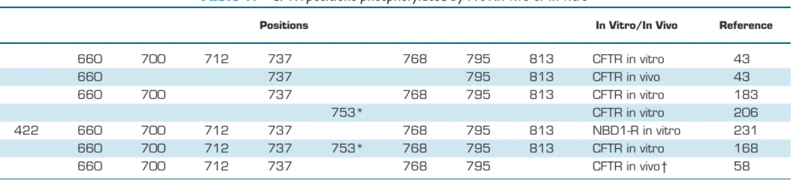
Cloning of the CFTR sequence revealed its domain organiza- tion(FIGURE 1A)and allowed a rough prediction of transmem- brane topology and domain boundaries (201). The suggested topology has stood the test of time, except for some small adjustments of helical boundaries [e.g., (86, 255)]. However, the originally predicted NH2- and COOH-termini of NBD1 and 2 turned out to be quite inaccurate. Exploiting ABC trans- porter modular architecture, coexpression of complementary CFTR segments was used to provide a functional definition of NBD1 boundaries (33): this approach extended the NBD1 COOH-terminus from amino acid position (a.a.) 586 to 633, but left its NH2-terminus uncorrected. The crystal structure of mouse CFTR NBD1 (130) finally assigned correct NBD1 NH2- and COOH-terminal boundaries to ~a.a. 390 and ~670, respectively, although residues distal from ~a.a. 645 form a helix that is not conserved among ABC proteins and contains two consensus serines (S660 and S670) phosphorylated by PKA, suggesting that it might be considered part of the R domain. For NBD2, the crystal structure of a fusion protein of human CFTR NBD2 with the regulatory domain ofEsche- richia coli MalK (PDBID: 3GD7) allowed adjustment of NBD2 NH2- and COOH-terminal boundaries to a.a. 1208 and ~1427, respectively, largely confirmed (1207–1436) by the first atomic structure of full-length human CFTR (145).
Given that much of the R domain of CFTR is unstructured (145, 177), its exact NH2- and COOH-terminal boundaries are still uncertain and might be assigned to ~a.a. 645 (670) and
~845, respectively, largely based on the boundaries of its bracketing domains (NBD1 and TMD2; see FIGURE 1A) as well as on the locations of consensus sites for PKA phosphor- ylation.
B. The ATP Binding Cassettes
NBD1 and NBD2 are also known as CFTR’s ATP binding cassettes, the highly conserved (both at a sequence and
three-dimensional structure level) ATPase subunits charac- teristic of all ABC proteins(FIGURE 1B). ABC NBD struc- tures consist of two subdomains. The nucleotide binding core subdomain (the “head”) comprises an F1-like parallel
-sheet (FIGURE 1B,light green), which is stabilized by␣-he- lices (FIGURE 1B, dark green) and contains the conserved Walker A (consensus GXXXXGKS/T;FIGURE 1B,red) and B (consensus⌽⌽⌽⌽DE,⌽hydrophobic;FIGURE 1B,ma- rine) motifs important for Mg-ATP binding (245) and is completed by an ABC-specific, three-stranded antiparallel
-sheet (FIGURE 1B, cyan). The two -sheets surround a central␣-helix preceded by the P-loop, which is formed by residues of the Walker A motif (FIGURE 1B,dark green helix andred loop). The NBD␣-helical subdomain (the “tail”;FIG- URE 1B,orange) contains the highly conserved, ABC-specific
“signature sequence” (consensus LSGGQ; FIGURE 1B, ma- genta). In nucleotide-bound, high-resolution NBD structures, the P-loop is seen to coordinate the phosphate chain, with the conserved Walker A lysine (K464 and K1250 in CFTR;FIG- URE 1B,red sticks) playing a dominant role by coordinating all three phosphates of ATP. The antiparallel-sheet provides a conserved aromatic residue that stacks against the adenine base of the bound nucleotide (W401 and Y1219 in CFTR;
FIGURE 1B,blue sticks). The Walker B motif ends in a con- served aspartate (D572 and D1370 in CFTR;FIGURE 1B,ma- rine sticks) important for Mg2⫹coordination and is followed by a conserved glutamate (E1371 in NBD2 of CFTR;FIGURE 1B,salmon sticks), which acts as the general base that polar- izes the attacking water molecule during the ATP hydrolysis reaction (162, 174, 175). A conserved glutamine (Q493 and Q1291 in CFTR;FIGURE 1B;orange sticks) in the loop that links the head and tail subdomains (the “Q-loop”) acts as the
␥-phosphate sensor and plays a key role in an induced fit conformational change elicited by ATP binding; an ~15° rota- tion of the tail subdomain toward the core subdomain in ATP compared with ADP-bound, or apo, structures (115, 268).
These key catalytic residues are held together by the conserved
“switch histidine” (H1402 in NBD2 of CFTR; FIGURE 1B, light magenta sticks), also called the “linchpin” (174, 269). In CFTR, there is substantial asymmetry between NBD1 and NBD2 regarding the key consensus motifs. In NBD1, the post–
Walker B glutamate and the switch histidine are replaced by serines (S573 and S605, respectively), whereas in NBD2, the signature sequence is atypical (LSHGH). In addition, CFTR’s NBD1 contains two unique sequence segments (130): an ~30- residue unstructured segment (a.a. 406 – 436) inserted into the antiparallel -sheet [regulatory insertion (RI)], and an ~30- residue helical extension [a.a. 641– 670; regulatory extension (RE)]. Both segments contain consensus serines phosphory- lated by PKA (S422 and S660 and S670, respectively), and both are unstructured in full-length CFTR (FIGURE 1B;light magenta dotted lines).
In the presence of ATP, but under conditions that preclude ATP hydrolysis, isolated ABC NBDs form head-to-tail dimers that occlude two molecules of ATP at the dimer
interfaces. Such NBD dimerization can be observed both for isolated soluble NBD domains [e.g., (38, 215, 269)] or in the context of full-length ABC proteins [e.g., (46, 67, 256)], including CFTR [(99, 159, 207, 274);FIGURE 1C]. In both composite nucleotide binding sites, an ATP is sandwiched between the Walker motifs of one NBD (FIGURE 1C, Walker A,red) and the signature sequence of the other NBD (FIG- URE 1C,magenta). This arrangement explains the crucial role the signature sequence plays in catalysis despite its distance from the bound nucleotide within an NBD mono- mer (FIGURE 1B). Furthermore, in CFTR, the head-to-tail arrangement of the NBD dimer collects all noncanonical substitutions into a single-composite binding site formed by the head of NBD1 and the tail of NBD2 (site 1;FIGURE 1D, upper site); in this degenerate site, the catalytic glutamate and the switch histidine are ablated by mutation, and the signature sequence is aberrant. In contrast, in the composite binding site formed by the head of NBD2 and the tail of NBD1 (site 2;FIGURE 1D,lower site), all key residues are canonical. A similar asymmetrical distribution of consensus versus atypical residues is found throughout the entire ABCC subfamily as well as in many other prokaryotic and eukaryotic heterodimeric ABC proteins (101, 187).
C. Structural Organization of Full-Length CFTR
The overall three-dimensional arrangement of full-length CFTR was resolved in a series of recent atomic resolution structures obtained by cryo-electron microscopy (cryo- EM), two from zebrafish CFTR (273, 274) and one from the human protein (145). Although at present no functional information is available on zebrafish CFTR, which is only
~55% identical in sequence to the human protein, the struc- tures of the two orthologs in their dephosphorylated apo- states are virtually identical (root mean square deviation
~1.9Å across the entire protein), suggesting that structural information obtained from the zebrafish ortholog is largely relevant to the human protein.
The global arrangement of the CFTR protein(FIGURE 1E–F)
resembles that of other ABC exporters (46, 67, 212, 256), as had been predicted by extensive cross-linking studies (99, 159, 207). The membrane-spanning components are formed by the twelve transmembrane helices, six from TMD1 (FIGURE 1E–F,light gray) and six from TMD2 (FIG- URE 1E–F,dark gray), which also extend deep into the cy- tosol. TMD/NBD interactions occur via four short coupling helices (CH1– 4;FIGURE 1E–F,magenta) formed by intracel- lular loops 1 (CH1; aa. 168 –174) and 2 (CH2; aa. 269 – 275) of TMD1 and the analogous intracellular loops 3 (CH3; aa. 961–966) and 4 (CH4; aa. 1062–1068) of TMD2. As for other ABC exporters, TMD1 and TMD2 do not form distinct, separate bundles of transmembrane heli- ces but are closely intertwined with each other as well as with the NBDs. In particular, a unit formed by TM4/CH2/
TM5 reaches out from TMD1 and contacts NBD2 and in a similar fashion, TM10/CH4/TM11 extends from TMD2 toward NBD1 [domain swap (67)]. Thus, the full-length CFTR molecule can be seen to be formed by two structural halves [TM helices 1, 2, 3, 6 plus 10, and 11 with NBD1 and TM helices 7, 8, 9, 12 plus 4, and 5 with NBD2 (274),
FIGURE 1E]. The coupling helices run roughly parallel with the plane of the membrane and fit into corresponding clefts on the NBD surfaces, forming ball-and-socket–like joints that are the transmission interfaces for communications be- tween the NBDs and the TMDs. Because of deletion of a short helix from NBD1, the “socket” on the NBD1 surface that accepts CH4 is shallower than the NBD2 socket that accepts CH2 (compare sockets inFIGURE 1B), rendering the NBD1/CH4 interface more sensitive to deleterious effects of mutations. This explains the severe structural destabiliza- tion caused by deletion (or mutations) of phenylalanine 508 (FIGURE 1B, NBD1,purple sticks), which contributes a hy- drophobic side chain to formation of the shallow NBD1 socket (273).
Experimentally observed conformations of ABC proteins fall into two major classes. First, in most structures solved in the absence of nucleotide (apo-structures), the TMDs adopt an inward-facing conformation in which the extra- cellular ends of the TM helices are tightly bundled, whereas their cytosolic extensions, including the coupling helices, are spread apart, and the NBDs are separated (8, 101, 212, 256). Among the many solved inward-facing struc- tures of ABC proteins, the observed degree of separation between NBD interfaces is highly variable. In the struc- ture of dephosphorylated apo-CFTR [both zebrafish (273) and human (145)], the NBD interface separation is relatively large (⬎17Å; FIGURE 1E). Second, in nucle- otide-bound ABC-exporter structures solved under con- ditions that preclude ATP hydrolysis, the TMDs typically adopt an outward-facing orientation in which the cyto- solic ends of the TM helices are tightly bundled, the coupling helices approach each other, and the NBDs are tightly dimerized. In such structures, a variable degree of separation is observed between the extracellular ends of the TM helices, ranging from widely splayed extracellu- lar loops (67, 256) to more compact bundling (46). The structure of phosphorylated, ATP-bound zebrafish CFTR is in an outward-facing conformation (274) resembling the latter, tighter extracellular bundling arrangement, with the TM helices largely parallel to each other [FIGURE 1F; see also (52)].
In addition to these mostly expected general ABC protein characteristics, the recent CFTR structures revealed several unpredicted features. The NH2-terminal ~60 residues, con- served throughout the ABCC subfamily and unique to it, form a “lasso motif” (FIGURE 1E–F,red). The lasso contains two ␣-helices, the first of which is partly inserted in the membrane, packed against TMD2. The second, amphi-
pathic helix, which runs parallel to the membrane and ex- poses a highly charged surface to the aqueous environment, has been implicated in both channel trafficking and gating regulation (166, 167). Further unique features of CFTR’s TMDs are likely essential for its ion channel function. The pseudosymmetry of the TM helices is disrupted by a discon- tinuity of TM helix 8 [TM8 (51)], which makes two sharp breaks within the membrane (FIGURE 1F,right; TM8 exter- nal segment and helical breaks are highlighted in cyan), thereby displacing TM7 from its ABC-typical location (FIG- URE 1F,right,pale green). As a consequence, the central ion pore is mostly lined by TM1 and 6 of TMD1, and by TM8 and 12 of TMD2, consistent with earlier accessibility stud- ies (12, 85, 88, 188, 254, 255, 271). In ABC transporters, access to the substrate translocation pathway is gated at both ends: the external gate is open in the outward-facing TMD conformation, whereas the internal gate opens in the inward-facing state. In contrast, in the CFTR channel, the open pore provides a continuous aqueous transmembrane pathway permeable to anions. Given that the open CFTR pore corresponds to an outward-facing TMD conforma- tion, an aqueous pathway must exist that bypasses the closed internal ABC transporter gate. Consistent with re- sults of functional (76, 77) and modeling (52, 163) studies, in the corresponding CFTR structure (274), that pathway is formed by a lateral opening (FIGURE 1F,red arrow) between TM4 (FIGURE 1F, yellow) and 6 (FIGURE 1F,orange) that connects the cytosolic environment with the internal vesti- bule of the pore.
D. Structural Information on the R Domain The entirely unique amino acid sequence of the R domain is a consequence of its evolutionary origin from an intronic DNA sequence (205). Although early circular dichroism (CD) spectra of an R domain peptide, based on the origi- nally suggested domain boundaries (a.a. 595– 831), re- ported some ␣-helical content to a degree influenced by phosphorylation and identified an NH2-terminal sub- domain with high sequence conservation among CFTR or- thologs (residues 587– 672) (72), the latter segment, in fact, largely belongs to NBD1. In contrast, CD spectra of an R domain peptide encompassing a.a. 708 – 831 predicted this domain to be largely unstructured (177), consistent with its origin from noncoding sequence. Nevertheless, biochemical pull-down assays and NMR studies with isolated peptides suggested that the R domain interacts with other parts of the channel in a phosphorylation-dependent manner (29, 36, 249). In the dephosphorylated closed apo-structures of both zebrafish (273) and human (145) CFTR, a large amor- phous density corresponding to the R domain is seen wedged between the two CFTR halves, interacting with NBD1 and the cytosolic ends of the TM helices; in the dephosphorylated human structure, a part of the density that interacts with the TM helices can be modeled as an
␣-helix (FIGURE 1E, yellow surface plot) and likely corre-
sponds to the COOH-terminal end of the R domain (a.a.
825– 843). Such an intercalated arrangement of the dephos- phorylated R domain is sterically incompatible with NBD dimerization or with an outward-facing conformation of the TMDs. Indeed, in the phosphorylated, ATP-bound, out- ward-facing zebrafish CFTR structure (274) (FIGURE 1F), no density corresponding to the R domain is observed, in- dicating that this region does not adopt a common confor- mation in most of the analyzed particles but, instead, be- comes disordered.
III. THE CFTR ANION PERMEATION PATHWAY
A. Structural Segments Lining the Pore Long before the availability of high-resolution structural information, several of CFTR’s helices had been probed for their contributions to the ion permeation pathway by use of the substituted cysteine accessibility method. Such studies identified TM helices 1 (85, 88, 254), 5 (271), 6 (11, 75, 88), 11 (78, 255), and 12 (17, 78, 188) as pore lining. The accessibility patterns were generally consistent with helical structures, with each third-to-fourth-substituted residue be- ing accessible from the ion permeation pathway, and the patterns even predicted some symmetry break by suggesting that TM7 does not participate in forming the pore (255, 271). A lateral portal serving as the cytosolic entrance of the pore vestibule between the cytosolic extensions of TM4 and 6 was hypothesized (52, 163) and later experimentally con- firmed. It is lined by a number of positively charged amino acid side chains that play a role in attracting cytoplasmic chloride ions to the inner mouth of the pore [(76, 77); some of the corresponding residues in zebrafish CFTR are high- lighted asblue spheresinFIGURE 1F,right]. Except for the unanticipated helical break observed in TM8 (FIGURE 1F, right, cyan), the recent cryo-EM structures largely con- firmed the predictions of these functional studies and re- vealed that the entire inner surface of the pore is lined by positively charged residues, as expected for an anion chan- nel (273).
B. Location of the Channel Gate
A deep and wide intracellular (12, 141, 211, 221, 279), and a shallower extracellular (169, 218), vestibule predicted from the voltage dependence of pore block by various large organic anions suggested an asymmetric hourglass shape for the CFTR pore. This shape positioned the gate, corre- sponding to a narrow constriction in the permeation path- way [with a functional diameter of ~5.3A (106, 144) in the open conformation], close to the extracellular membrane surface, at the level of TM6 residues 338 –341 (63, 75, 87).
Consistent with a gate located at the extracellular end of the pore, binding of large organic anion pore blockers (59) or
ATP (219) in the intracellular vestibule does not prevent gate closure. In the inward-facing structures of zebrafish (273) and human [(145); FIGURE 1E], CFTR, the funnel- shaped intracellular vestibule, indeed tapers down to a nar- row tunnel at the predicted location of the gate, consistent with those structures representing closed CFTR channels.
Interestingly, however, in the outward-facing structure of phosphorylated ATP-bound zebrafish CFTR (274), al- though this gate constriction between TM6, TM1, and TM11 widens as expected, access to the pore from the ex- tracellular space is nevertheless prevented by the extracel- lular segments of TM helices 8 and 12 (FIGURE 1F,right, cyananddark cyan); this results from a local reorientation of these two secondary structure elements with respect to the rest of the half molecule. A structure of the CFTR pore in its fully conductive conformation remains to be captured.
C. Mechanism of Anion Selectivity
The CFTR pore shows high anion versus cation selectivity [the relative permeability of Na⫹compared with Cl⫺(pNa/ pCl) is ~0.03 (226)] but poorly selects among anions. Ex- perimentally obtained anion permeability sequences (which report relative ease of ion entry into the pore) are consistent with a lyotropic selection mechanism; relative permeability of a given anion is inversely proportional to its energy of dehydration (140, 157, 216). For instance, compared with chloride, large anions like SCN⫺ and nitrate, which more easily shed their hydration shell, display higher permeabil- ity through CFTR [e.g., pSCN/pCl⬎2.4 (140, 157)]. On the other hand, these high-permeability, large anions also typ- ically bind very tightly within the pore, resulting in a low throughput rate operationally defined by measuring relative
conductance [e.g., the relative conductance for SCN⫺com- pared with Cl⫺(gSCN/gCl) is⬍0.2 (135, 157)]. Thus, evolu- tion seems to have optimized the CFTR pore to provide maximal conductance for the physiologically most relevant anion, chloride (135). Besides chloride, the other physiolog- ically relevant ion that permeates through CFTR is bicar- bonate. CFTR’s relative permeability (pHCO3/pCl) and rela- tive conductance (gHCO3/gCl) for bicarbonate are both
~0.25 (144, 184). Low permeability ratios were initially also found in isolated pancreatic ducts (91), and CFTR’s role in bicarbonate secretion was thought to be indirect, mediated by regulation of Cl⫺/HCO3⫺ exchangers of the SLC26A family [reviewed in (129)]. However, several stud- ies have raised the possibility that direct HCO3⫺ perme- ation through CFTR might become important, especially in some physiological conditions (108), and that CFTR anion permeability might be dynamically regulated (178, 198). Re- gardless of the exact mechanism, CFTR-dependent bicarbon- ate secretion clearly plays an important physiological role in controlling the pH of the fluid layers that line various epithelial surfaces (189, 202), including the lung (181, 210), as demon- strated by the correlations seen between levels of CFTR, bicar- bonate fluxes, and strength of lung host defense defects (208).
The region responsible for CFTR’s lyotropic selectivity (i.e., the region that provides sites of interaction for permeating anions) corresponds to the narrow region of the pore, as evidenced by changes in anion selectivity sequences upon mutation of residues F337, T338, S341, S1118, or T1134 (90, 135, 140, 157, 158, 275). In addition, the large number of positively charged residues that line the entire internal surface of the pore [in particular, residue K95 in the internal vestibule (137, 139)] or flank the cytoplasmic lateral open- ing [(76, 77); cf.,FIGURE 1F,right,blue spheres] contribute to enhancing chloride conductance by attracting chloride ions to the pore (139). Thus, it has been suggested that the anion selectivity characteristics of CFTR might result from at least two distinct selectivity filters operating in series, interactions between permeant anions and the pore con-
striction around F337 being largely responsible for deter- mining the selectivity of permeability, whereas anion/pore interactions in the inner vestibule and around the entrance of the lateral portal determine anion over cation selectivity and boost anion conductance (139). Some evidence sug- gests that overall high conductance and tight pore binding of CFTR might result from simultaneous, multiple interac- tions of permeating anions along the permeation pathway (138). Channel pores with multiple ion binding sites often show a nonlinear dependence of unitary conductance on the mole fractions of two types of permeant ion that are simul- taneously present. In the case of CFTR, such anomalous mole fraction behavior has been reported for the anion pairs Cl⫺/SCN⫺(136, 139) and Cl⫺/SO42⫺(60).
By stabilizing the open-pore structure, anion/pore interac- tions contribute to the energetic stability, and therefore the life time, of the open-channel state. Thus, anion replace- ment affects not only permeation properties, but also the kinetics of gating transitions: nitrate and bromide, which bind more tightly than chloride delay, whereas formate, which binds less tightly than chloride, accelerates pore clo- sure (219, 266). These effects of tight-binding permeating ions to retard the closing of the CFTR gate might be anal- ogous to the influence exerted by a substrate bound to an outward-facing exporter; extracellular release of the sub- strate favors closing of the extracellular gate, and thus res- toration of the inward-facing conformation [see Section IVG; see also (266)].
D. Pore Blockers
Many large organic anions block the CFTR pore when ap- plied from the intracellular side. The resulting brief closed events (flickery block) reflect the brief residence time of the blocker at its binding site in the pore. Pore block is more pronounced at hyperpolarized (more negative) membrane potentials that drive the negatively charged blocker into the
FIGURE 1. CFTR domain topology and structure.A: CFTR domain topology. TMD1 (light gray), TMD2 (dark gray), intracellular loops (light purple), NBD1 (blue), NBD2 (green), regulatory (R) domain (rose), and membrane (yellow).B: Ribbon representation of NBD1 (left) and NBD2 (right) from the cryo-electron microscopy (cryo-EM) structure of the phosphorylated, ATP-bound form of zebrafish CFTR (PDBID: 5W81). F1-like parallel-sheet plus␣-helices (green), ABC-specific antiparallel-sheet (cyan),␣-helical subdomain (orange), Walker A motif (red), Walker B motif (marine), signature motif (magenta), ATP (yellow sticks), and Mg2⫹ion (slate sphere). The numbering of the conserved residues shown in stick representation is based on the human CFTR sequence. An E–Q mutation of the catalytic glutamate in NBD2 was used to trap the protein in an ATP-bound form. In NBD1,light magenta dotted linesmark the locations in the primary sequence of the unresolved regulatory insertion (RI) and regulatory extension (RE).C: Organization of the ATP-bound, head-to-tail NBD1/NBD2 heterodimer (from PDBID: 5W81). NBD1 (blue), NBD2 (green), ATP molecules (yellow sticks), Walker A motifs (red), and signature sequences (magenta). The conserved Walker A lysines are shown asred spheres.D: Cartoon representation of residue asymmery in the CFTR NBD dimer; color coding of conserved residues as inB. The upper site (site 1, degenerate site) harbours all noncanonical substitutions, whereas in the lower site (site 2, canonical site), all catalytically important side chains are intact.E: Ribbon representation of the dephosphorylated human CFTR apo-structure (PDBID: 5UAK); domain color coding as inA. Lasso motif (red), R domain helix modeled into observed density (yellow surface), coupling helices (magenta), ATP (yellow sticks), and membrane (horizontal gray lines).F: Ribbon representations of the structure of phosphorylated, ATP-bound CFTR (PDBID: 5W81) viewed from two different orientations; left view and domain color coding as inE. The view to the right shows the cytoplasmic opening of the ion permeation pathway (red arrow, lateral opening) flanked by transmembrane (TM) helices 4 (yellow) and 6 (orange); positively charged residues lining the opening are shown asblue spheres. Outer segments of TM8 and TM12 are colored (cyananddeep cyan); TM7 ispale green; the lasso motif has been removed for clarity.
intracellular pore vestibule but is alleviated at depolarized (more positive) membrane potentials. The steepness of this voltage dependence allows estimation of how deep the blocker binding site is [i.e., what fraction of the membrane electrical field the blocker traverses before reaching its bind- ing site ()]. Similar voltage dependences suggest a common binding site for a structurally diverse group of blockers including diphenylamine-2-carboxylate, flufenamic acid [⫽~0.41 for both (156)], glibenclamide [⫽~0.45– 0.48 (211, 279)], 5-nitro-2-(3-phenylpropylamino)benzoate (NPPB) [⫽~0.5 (59)], 3-(N-morpholino)propanesulfonic acid (MOPS) [ ⫽ ~0.5 (59, 109)], and anthracene-9- carboxylic acid [⫽~0.5 (1)]. In line with the notion of a common binding site, glibenclamide and isethionate (279), or NPPB and MOPS (60), were shown to compete for pore block. Mutagenesis studies highlighted how the positively charged side chain of the pore-lining residue K95 plays an important role in interacting not only with permeant an- ions, but also with blockers (137). In the atomic structure of human CFTR, K95 is located at a position where the intra- cellular vestibule tapers down to the narrow tunnel believed to comprise the gate (145). Of note, an intrahelical salt bridge between the side chains of K95 and E92 was recently noted in the cryo-EM structure of human CFTR and pro- posed to play a role in anion/pore and blocker/pore inter- actions (104). Consistent with the binding site for diverse blockers being located intracellular to the channel gate, the presence of NPPB or MOPS in the pore does not delay gate closure (59). More modest voltage dependences reported for block by the disulfonic stilbenes 4,4=diisothiocyanostil- bene-2,2=-disulfonic acid and 4,4=-dinitrostilbene-2,2=- disulfonic acid [ ⫽ ~0.16 and ⫽ ~0.34, respectively (141)] suggest that a more superficial blocker binding site might also exist.
Whereas the above compounds block from the cytosolic side and show low affinity for the CFTR pore (Kd in the hundreds of micromolar to millimolar range at 0 mV mem- brane potential), high-throughput screening has led to the discovery of a higher-affinity pore blocker that acts from the extracellular side; N-(2-naphthalenyl)-[(3,5-dibromo-2,4- dihydroxyphenyl)methylene]glycine hydrazide (GlyH-101) blocks CFTR currents with aKdof ~4M at 0 mV mem- brane potential and shows the inverse voltage dependence expected for an anionic blocker that binds in the extracel- lular vestibule (164). The electrical distance of the GlyH- 101 binding site (⫽0.35) and strong reductions in appar- ent affinity by mutation R334C (62) or upon covalent mod- ification of a cysteine engineered into position 338 (169) suggest that GlyH-101 binds at the bottom of the shallow extracellular vestibule, just above the constriction that forms the channel gate (169).
E. Intraburst (Flickery) Closures
Under all experimental conditions, gating of single CFTR channels shows clear bursting behavior; groups of open
events interrupted by brief (~1–3 ms at 37°C and ~10 ms at 25°C) flickery closed events form bursts that are sep- arated from each other by long (~0.1– 0.2 s at 37°C and
~0.4 –2 s at 25°C) interburst closed events. Some of the brief intraburst closures represent block by large cytoso- lic anions (278) or by anionic buffer molecules present in the recording solution that bathes the cytosolic mem- brane surface (109); indeed, even cytosolic ATP causes low-affinity pore block (219). But not all intraburst clo- sures may be accounted for by such a mechanism, be- cause flickery closures can be observed for locked/open channels long after ATP has been washed away, even in a cytosolic solution buffered with a cationic buffer (279), and also when channel currents are studied at positive voltages (243), which deter cytosolic anion entry. Thus, at least a fraction of the observed flickery closures must represent a gating mechanism intrinsic to the channel protein.
Several studies have addressed the dependence of in- traburst gating kinetics on a variety of factors, including voltage, pH, or the concentration of ATP used for chan- nel activation. Both the frequency and the duration of flickery closures increases at positive membrane poten- tials, reporting a weak voltage dependence of intraburst gating (30). Acidification of the bath solution to pH⫽6.3 prolongs the average duration of flickers by approximately twofold (40). In contrast, neither the fre- quency nor the duration of intraburst closures is sensitive to the concentration of applied ATP (243, 261). Finally, mean flickery closed time is prolonged by catalytic site mutations that disrupt ATP hydrolysis (243) because of the appearance of a second population of intraburst closed events with an average life time of ~50 –100 ms (25, 61). As a result, intraburst closures lasting up to several hundred milliseconds may be observed during long locked/open bursts and have been dubbed “gating”
in some reports (172). Although temperature dependence of intraburst kinetics has not yet been addressed system- atically, comparison of studies conducted at room tem- perature (20 –25°C) and those obtained at 37°C suggests approximately three- to fivefold briefer flickers at the higher temperature [e.g., (243) versus (30)].
Two alternative kinetic mechanisms, Cslow↔Cfast↔O and Cslow↔O↔Cfast, with Cslowand Cfastdenoting the inter- burst and intraburst closed states, respectively, have been used to model CFTR bursting behavior. Because these two schemes cannot be distinguished by steady-state recordings, all data available to date may be explained equally well by either scheme. In such situations, it is customary to prefer the scheme which requires adjustment of the smaller num- ber of parameters to describe two data sets obtained under two different experimental conditions. However, even that
“parsimony argument” has been of no help so far as for either model, only one rate was found to be sensitive to ATP
concentration (243, 261), but several to both voltage (30) and intracellular pH (40).
The physical mechanism underlying flickery closures is still elusive. The slow and fast gates that cause inter- and in- traburst closures, respectively, may or may not be formed by two physically distinct protein regions. But slow and fast gating certainly reflect two distinct types of TMD confor- mational change; slow gating (i.e., entering and exiting a burst) likely represents flipping between TMD conforma- tions that are analogous to inward- and outward-facing conformations of ABC exporters, respectively (146, 242), whereas fast gating (i.e., intraburst flickering) is likely caused by a smaller-scale, more localized conformational change. One possibility is that the outward-facing occluded structure seen for phosphorylated zebrafish CFTR (274), in which the pathway is blocked by a local distortion of the outer segments of TM helices 8 and 12, represents the flick- ery closed state. As described above, in that conformation, the gate constriction is widened, but the external extremity of the ion conduction pathway is blocked by a localized conformational change of the outer-leaflet segments of TM helices 8 and 12 (FIGURE 1F, right, cyan and dark cyan).
However, given that the (human) CFTR channel dwells in the flickery closed state only for a small fraction of the total duration of a burst, capturing this conformation in a cryo-EM structure is unlikely, unless it is stabilized by some factor specific to the cryo-EM conditions (e.g., spe- cies difference or low temperature). Thus, an alternative interpretation of the outward-facing occluded zebrafish CFTR structure is that it represents an intraburst closed state with a high occupancy probability but a lifetime too short (~1 s) to be resolved in limited-bandwidth elec- trophysiological recordings; in that case, the experimen- tally measured unitary conductance of 7–10 pS would reflect the full conductance multiplied by the fraction of time the pore is truly open within a burst (274). Molec- ular dynamics simulations, encompassing 1.5 s, high- light the relative stability of the outward-occluded con- formation seen in the ATP-bound zebrafish structure (51), disfavoring the latter scenario. Thus it remains pos- sible that for both interburst and conventional flickery closures, the ion conduction pathway is interrupted by a similar conformation of the narrow constriction between TM helices 1, 6, 8, and 12 observed at the height of TM6 residues 338 –341 [the gate (273)].
IV. REGULATION OF CFTR GATING
THROUGH NUCLEOTIDE INTERACTIONS AT THE NBDs
Because phosphorylation and ATP regulate slow gating, in the following sections, channel “opening” and “closing”
will be used synonymously with entering and exiting a burst.
A. ATP Hydrolysis at One of Two
Nonequivalent Composite ATP Binding Sites
The catalytic turnover rate of CFTR ATPase activity (0.5–1 s⫺1), estimated for phosphorylated human CFTR protein purified to homogeneity (131, 145), falls into the range of channel gating (bursting) rates. An early hint that ATPase activity might be coupled to pore gating was provided by the observation that lowering free Mg2⫹to 4 nM, or adding Na-azide, inhibited both processes (131). The catalytic ac- tivity of the CFTR protein must originate from composite site 2 of the NBDs, because mutation of the Walker A lysine in site 2, but not that in site 1, abolishes ATPase activity (192). Indeed, photocrosslinking experiments revealed that site 1 retains ATP bound and unhydrolyzed for up to tens of minutes (7, 14). The presence of canonical consensus motifs in site 2, but noncanonical residues in site 1(FIGURE 1D), readily explains such functional asymmetry between CFTR’s two composite ATP binding sites and is likely a shared feature of asymmetric ABC proteins (102, 186, 235) [including the entire human ABCC subfamily as well as many heterodimeric prokaryotic homologs (187)].
B. Coupling of Pore Opening/Closure to Formation/Disruption of a Head-to-Tail NBD Heterodimer
Decades of experimental work gathering information on both CFTR and related ABC proteins have clarified the basic mechanism by which ATP binding and hydrolysis at the NBDs drives pore gating in CFTR. Early studies dem- onstrated that preventing (or attenuating) ATP hydrolysis in site 2 by mutations of the Walker A lysine (K1250A/G/
M/T) or the catalytic glutamate (E1371Q/S) in NBD2 (FIG- URE 1B,right;FIGURE 1D)locks channels in the open burst- ing state (32, 96, 242, 243) for time intervals at least two orders of magnitude longer than the mean burst duration of WT CFTR. These results clearly demonstrated that site 2 ATP hydrolysis is required for normal (fast) termination of a burst. Mixtures of ATP either with nonhydrolyzable ATP analogs, such as 5=-adenylyl-imidodiphosphate (AMPPNP) or adenosine 5=-(gamma-thiotriphosphate) (ATP␥S), or with pyrophosphate (PPi) also lock channels open (95, 103), suggesting that these analogs prevent closure by bind- ing at site 2. A similar lock-open effect of mixtures of ATP with the inorganic phosphate (Pi) analog orthovanadate (Vi) is believed to reflect formation of a stable ADP:Vicom- plex that resembles the pentacovalent transition state of the ATP hydrolysis reaction in site 2 (15, 95). Formation of such complexes by the hydrolysis product ADP and a Pi analog that binds tightly in place of the released Pihas been observed in atomic structures of ABC proteins (174).
Contrary to early conclusions obtained mostly on single CFTR channels (9, 103, 203), nonhydrolyzable ATP ana-
logs, such as AMPPNP, ATP␥S, ,␥-methyleneadenosine 5=-triphosphate (AMPPCP) (4, 243), or PPi(234), alone are capable of opening CFTR channels, although the nucleotide analogs are poor substitutes for ATP, supporting a maximal opening rate only ~5% of that observed in saturating ATP (243). Although the hydrolysis-abolishing K1250A muta- tion was found to reduce not only closing, but also channel opening rate (32, 192), the latter effect was later shown to be largely due to a reduced ATP binding affinity in site 2 (243). Indeed, another site 2 mutation expected to disrupt hydrolysis, E1371S, does not impair channel opening (243).
Thus, pore opening requires nucleotide binding, but, in con- trast to channel closure, not hydrolysis.
By what mechanism is ATP binding at the NBDs translated into pore opening? The hyperbolic dependence of channel opening rate (i.e., the rate of entering a burst) on ATP concentration [K1/2⫽ ~50M (55, 241, 243, 270)] indi- cates a rate-limiting step for pore opening other than ATP binding. That step is Mg2⫹dependent (71, 131) and must follow ATP binding because its rate is sensitive to nucleo- tide structure; maximal opening rates supported by 8-azido-ATP (14) or AMPPNP (243) are much lower than that observed in ATP. Furthermore, ATP binding must have happened at least at the NBD2 head before the pore opens, because mutations of the Walker A lysine (K1250A,red), the Walker B aspartate (D1370N,marine), or the stacking aromatic residue (Y1219G, blue) in NBD2 (FIGURE 1B, right), all of which destabilize ATP binding there, dramati- cally reduce the apparent affinity for ATP to open the pore (243, 281). In contrast, prior ATP binding to the NBD1 head (FIGURE 1B,left) seems less essential for channel open- ing as the apparent potency of ATP in opening the pore is decreased by some mutations that impair ATP binding [K464A, removal of the Walker A lysine side chain, red (243)], but not by others [W401G, removal of the stacking aromatic side chain (281)]. The observation of tight NBD dimers in ABC proteins in the presence of ATP and the suggestion that NBD dimer formation/dissociation might underlie the coupling of ATPase cycles to vectorial trans- port of substrates by ABC transporters (162) prompted the proposal that CFTR and transporters might share a com- mon mechanism. Thus, in CFTR, the rate-limiting step for pore opening (to a burst) would reflect formation of a tight head-to-tail NBD1/NBD2 heterodimer, whereas closure (from a burst) would occur upon disruption of that het- erodimer [FIGURE 2E(243)].
A first formal proof of that hypothesis was provided by the demonstration that two residues on opposing surfaces of composite site 2 (arginine 555 just downstream of the NBD1 signature sequence and threonine 1246 in the NBD2 Walker A motif) become energetically coupled upon chan- nel opening, but not upon binding of ATP (which occurs on closed channels, see above) (242). For steric reasons, a hy- drogen bond observed between the side chains of the cor-
responding residues in dimeric ABC NBD structures (38, 215) is expected to form either between an arginine/threo- nine (R/T) pair, as found in the sequence of CFTR and a subset of ABC proteins, or between a lysine/asparagine (K/N) pair, as present in a smaller subset of the ABC super- family (FIGURE 2A), but not between R/N or K/T pairs (which are poorly represented in naturally occurring ABC sequences). A large reduction in channel opening rate (i.e., an increase in interburst duration,ib) observed when intro- ducing the R555K or T1246N mutations in the WT CFTR background (FIGURE 2C,blueandred barsversusblack bar) was not seen when introducing the same mutations into a background already mutated at the other position (FIGURE 2C,purple barversusblueor red bar). This suggests that formation of the R555/T1246 hydrogen bond in WT CFTR facilitates channel opening and that the hydrogen bond is disrupted in each single mutant but restored in the double mutant. These mutation-induced changes in opening rate report mutational effects on the stability of the transition state for opening (T‡) relative to the closed state (⌬⌬G‡T-C; numbers next to arrows inFIGURE 2D). The R555K muta- tion destabilizes the transition state when a threonine is present at position 1246 (FIGURE 2D,left vertical arrow) but stabilizes it when the residue at position 1246 is an aspara- gine (FIGURE 2D, right vertical arrow). The difference be- tween⌬⌬G‡T-Cvalues along two parallel sides of the mu- tant cycle quantifies the change in interaction energy be- tween the native side chains upon entering the transition state from the closed state [⌬⌬Gint(opening)], and is of a mag- nitude and sign consistent with formation of a hydrogen bond. A similar mutant cycle built on the closed/open equi- librium constant of a hydrolysis deficient mutant, in which gating is reduced to reversible C1↔O1 transitions, con- firmed the presence of the R555/T1246 hydrogen bond also in the open ground state (242). Chemical cross-linking ex- periments (159) later confirmed the canonical head-to-tail NBD dimer arrangement seen in all dimeric ABC NBD structures (FIGURE 1C–D)to be present in full-length, gating CFTR channels in their native environment.
Thus, the rate-limiting step for channel opening (FIGURE 2E, step C1¡O1) consists of tight dimerization of ATP-bound NBDs coupled to TMD rearrangements that open up a transmembrane pathway for anions. Demonstration of salt bridge formation between cytosolic TMD loops in the open state (252), but between extracellular TMD loops in the closed state (64, 107), as well as a proposed narrowing of the intracellular vestibule in open channels (12) all sug- gested that upon pore opening, CFTR’s TMDs undergo a conformational change similar to the flipping of ABC trans- porter TMDs from an inward- to an outward-facing con- formation. All these predictions, based on functional stud- ies, were largely confirmed by the recent cryo-EM structures of dephosphorylated apo- and phosphorylated ATP-bound CFTR [FIGURE 1E–F(145, 273, 274)].
C. Thermodynamics and Timing of the Pore- Opening Transition
Among the steps that a phosphorylated WT CFTR channel follows around its gating cycle(FIGURE 2E), the pore-open- ing transition (FIGURE 2E, step C1¡O1) is the slowest (~0.5–2 s⫺1at 25°C and ~5– 8 s⫺1at 37°C), reflecting a high energetic barrier characterized by an unstable, high-free- energy transition state. The most common CF mutation, deletion of phenylalanine 508, further slows this step by
⬎40-fold (125, 161). What is the nature of this transition state, and what causes its high free energy? The steep tem- perature dependence of WT CFTR opening rate signifies a large activation enthalpy [⌬H‡T-C⫽~100 –150 kJ/mol (6, 57, 155)], suggesting molecular strain. On the other hand, the discrepancy between⌬G‡T-Cand⌬H‡T-Csignals a large entropy increase in the transition state [T⌬S‡T-C ⱖ40 kJ/
mol (57)]. Given that the NBD interface is already tightened
around ATP site 2 in the transition state [FIGURE 2A–D
(242)], the large activation entropy has been interpreted to reflect the dispersal of the layers of ordered water molecules that cover the interfacial NBD surfaces when the NBDs are separated and the interface is open and accessible to solvent (57).
The relative timing of motions in different regions of a channel protein during the submicrosecond process of pore opening (transition from state C to O) can be determined by studying the kinetic consequences of structural perturba- tions, typically point mutations, introduced into various protein regions. If the perturbation-induced change in the transition-state free energy linearly interpolates the differ- ence,⌬⌬Go, between the free energy changes of the C and O ground states (⌬⌬Go ⫽ ⌬GOo ⫺ ⌬GCo) (94, 153), then the free energy of the transition state for opening, T‡, will change by⌽⌬⌬Go(0ⱕ⌽ⱕ1). A larger⌽value indicates
FIGURE 2. Coupling of CFTR pore opening to NBD dimerization. A: An arginine/threonine (serine) or a lysine/asparagine side chain pair is optimally positioned to form a salt bridge between CFTR positions 555 and 1246. B andC: Single-channel outward current traces (B: Vm⫽ ⫹40 mV) and mean closed (interburst) durations (C) of preposphorylated WT, R555K, T1246N, and R555K/T1246N CFTR channels gating in 5 mM Mg-ATP.D: Thermodynamic mutant cycle illustrating mutation-induced changes in⌬G‡T-C(numbers next to arrows);⌬⌬Gint(opening)is the difference between⌬⌬G‡T-Cvalues along two parallel sides of the cycle. The four corners of the cycle are represented by the pairs of residues present at positions 555 and 1246, respectively.
E: Cartoon gating cycle of phosphorylated CFTR. Color coding as inFIGURE 1A; the R domain is not depicted.
Site 1 (degenerate site), upper site; site 2 (canonical site),lower site; ATP, yellow circles; ADP, orange crescent; chloride ions,dark red dots. [A–Dadapted with permission from Vergani et al. (242)].
earlier, and a smaller⌽value later, movement of the target position during pore opening. In particular,⌽ ⫽ ~1 indi- cates that, in the transition state, the target position is al- ready near its open-state conformation, whereas ⌽ ⫽ ~0 suggests it has not yet moved much from its closed confor- mation (10, 277). Because the perturbation will change the logarithm of the opening rate constant (kCO) by⫺⌽⌬⌬Go⁄ 共RT兲, but the logarithm of the equilibrium constant (Keq) by ⫺⌬⌬Go⁄共RT兲, ⌽can be estimated from the slope of a rate-equilibrium free energy relationship (REFER) plot of logkCOversuslogKeqfor a series of mutations at the target position. Importantly, because the REFER approach as- sumes equilibrium gating, with opening and closure reflect- ing reversible transitions along a single kinetic pathway (53), this approach cannot be applied to address the dynam- ics of the ATP-dependent slow-gating process of WT CFTR channels (5, 204), which obey a nonequilibrium cyclic gat- ing mechanism(FIGURE 2E). However, the technique may be adapted to studying the pore-opening step (FIGURE 2E, step C1¡O1) by employing a background mutation that disrupts ATP hydrolysis in site 2, thereby reducing gating to reversible C1↔O1 transitions. REFER analysis in such a nonhydrolytic background (NBD2 Walker B aspartate mu- tant D1370N) revealed a clear spatial ⌽-value gradient along the protein’s longitudinal axis from cytoplasm to cell exterior (219, 220); ⌽was close to ~1 for both faces of composite ATP site 2 (positions 555 and 1246;FIGURE 3A, left,red spacefill;FIGURE 3A,right,red numbers) and ~0.5–
0.6 for positions in each of the four coupling helices (posi- tions 172, 275, 961, and 1068, respectively; FIGURE 3A, purple spacefill and numbers), but ~0.2 for the centrally located pore residue M348 in TM6 and ~0 for position 117 in the first extracellular loop (FIGURE 3A,blue spacefilland numbers). This clear ⌽-value gradient suggests that a spreading conformational wave is initiated at the site 2 NBD interface and propagates toward the pore (FIGURE 3B, vertical colored arrow). In particular, it suggests that in the transition state the site 2 interface is already tightly dimerized, but the pore is still closed (FIGURE 3B,center).
Thus, the high enthalpy of the opening transition state (⌬H‡T-C ⫽ ~100 –150 kJ/mol) might reflect strain at the NBD/TMD interface [(219); cf., (57)], which includes the disease hotspot position 508. Indeed, a⌽value of ~0.5 for position 508 suggests that this NBD position moves syn- chronously with nearby TMD-coupling helix 4 (220). Inter- estingly, a low-intermediate⌽value of ~0.4 was found for both faces of degenerate site 1 (positions 460 and 1348;
FIGURE 3A, orange spacefill and numbers), reporting de- layed movement here with respect to site 2 and suggesting that site 1 residues are still on the move in the transition state for channel opening. However, because such pro- nounced asymmetry cannot be detected at the level of the four coupling helices, it seems likely that the movements completed in site 1 between the transition state and the open state are localized movements confined to the site 1 NBD interface (220).
D. Strictness of Coupling Between Pore- Opening Events and NBD Dimerization Strict coupling between NBD dimerization and pore open- ing in CFTR has been called into question because a con- struct lacking NBD2 (⌬NBD2, truncated after residue 1197) displays low-probability, ATP-independent openings following phosphorylation by PKA (65, 248). Based on that observation, spontaneous openings in the absence of ATP, also seen occasionally in WT CFTR (25, 224) but robustly promoted by mutations at TMD positions 978 [ICL3 (253)]
or 355 [TM6 (257)], were interpreted as reflecting pore openings in the absence of NBD dimerization. Furthermore, a resemblance was noted between CFTR and classical li- gand-gated channels, such as the nicotinic acetylcholine re- ceptor, in that phosphorylation (248), TMD mutations (253, 257), various drugs (112, 248), and ATP analogs (172) all had strongly correlated effects on spontaneous (ATP-independent) and on ATP-dependent channel activ- ity, and the effects of such allosteric modulators were ener- getically additive (172, 257). These analogies led to CFTR
FIGURE 3. Cytosolic to extracellular⌽-value gradient and asym- metry between sites 1 and 2.A: Cartoon representation of homol- ogy models (52) of phosphorylated closed- (left) and open-state (right) CFTR, with color coding as inFIGURE 1A; the regulatory (R) domain is omitted. Target positions for rate-equilibrium free energy relationship (REFER) analysis are highlighted in colored spacefill (left), corresponding colored numbers illustrate estimated⌽values (right).B: Cartoon representation of approximate structural rear- rangements during the pore opening transition as the channel tran- sits from an ATP-bound closed state (C1) through the transition state (T) to the ATP-bound open state (O1). Site 1 (degenerate site), upper site; site 2 (canonical site), lower site; color coding as in FIGURE 2E.Vertical colored arrowillustrates the direction of the spreading conformational wave.Red arcsin states T and O1repre- sent tight bonding across the NBD interface. [Adapted with permis- sion from Sorum et al. (219) and Sorum et al. (220)].
gating being modeled as an equilibrium loop mechanism in which the ligand (ATP) can bind and unbind in both the closed-pore and the open-pore conformation, and closed/
open (isomerization) transitions can occur whether or not ligand is bound. In that model, because of the thermody- namic principle of detailed balance, which constrains the product of the equilibrium constants around a kinetic cycle to be unity, higher affinity binding of the ligand in the open-channel conformation would shift the closed/open equilibrium of liganded channels toward the open state [(120); cf., (92)]. An essential feature of such an allosteric loop model [also key to the proposed reentry mechanism (113) discussed in Section IVE, below] is the postulate that in the ATP-free, spontaneous open state, the NBDs are dis- engaged, and the dimer interface is therefore accessible for ATP binding. Studying the accessibility of site 2 in ATP-free open channels is not straightforward, but this question was recently addressed by exploiting the enhanced spontaneous activity of the K978C/P355A double mutant, which allows quantitation of spontaneous gating parameters in micro-
scopic patches (160). In K978C/P355A channels gating in the absence of ATP, just as in WT channels gating in the presence of ATP (242) (Section IVB), energetic coupling between site 2 residues R555 (NBD1 face) and T1246 (NBD2 face) was found to change in a state-dependent manner; spontaneous open probability of the background construct is reduced by both the R555K and the T1246N single mutation, but restored in the double mutant(FIGURE 4A–B), reporting energetic coupling between these residues in the open state(FIGURE 4C). Thus, the two side chains on opposing faces of composite site 2 form a hydrogen bond in the open-pore conformation, but not in the closed-pore conformation, indicating the presence of a tightly dimerized site 2 NBD interface in the spontaneous open-channel state
(FIGURE 4D), just as during normal, ATP-dependent open- ings(FIGURE 2E). Because a tightly dimerized NBD interface does not allow ATP binding/unbinding in the open-pore conformation, CFTR gating must be driven by principles fundamentally different from the allosteric mechanisms that underlie gating of ligand-gated channels. Thus, strict
FIGURE 4. Spontaneous pore openings are also coupled to NBD dimerization.A: Microscopic inside-out patch recordings of CFTR background construct P355A/K978C and of channels bearing mutations R555K, T1246N, and R555K/T1246N in that background; Vm⫽ ⫺80 mV.B: Open probabilities of the constructs in A(seecolor coding) during the last 4 min of each 5-min ATP-free segment of recording.C: Thermodynamic mutant cycle illustrating mutation-induced changes in ⌬GO-C (numbers next to arrows); ⌬⌬Gint(O-C) is the difference beween ⌬⌬GO-C values along two parallel sides of the cycle. The four corners of the cycle are represented by the pairs of residues present at positions 555 and 1246, respectively.D: Cartoon depicting mechanism of spontaneous openings. Color coding as inFIGURE 1A; the R domain is not depicted. [Adapted with permission from Mihályi et al. (160)].