Contents lists available atScienceDirect
Psychoneuroendocrinology
journal homepage:www.elsevier.com/locate/psyneuen
Antidepressant e ff ect in diabetes-associated depression: A novel potential of RAAS inhibition
Dora B. Balogh
a,b, Agnes Molnar
a,b, Adam Hosszu
a,b, Tamas Lakat
a,b, Judit Hodrea
a,b, Attila J. Szabo
b,c, Lilla Lenart
a,b,*
,1, Andrea Fekete
a,b,*
,1aMTA-SE“Lendület”Diabetes Research Group, Hungarian Academy of Sciences, 54 Bokay Janos, H-1083 Budapest, Hungary
b1st Department of Pediatrics, Semmelweis University, 53-54 Bokay Janos, H-1083 Budapest, Hungary
cMTA-SE Pediatrics and Nephrology Research Group, Hungarian Academy of Sciences and Semmelweis University, 54 Bokay Janos, Budapest H-1083, Hungary
A R T I C L E I N F O
Keywords:
RAAS inhibitors Diabetes Depression
Brain-derived neurotrophic factor Neuroinflammation
A B S T R A C T
The incidence of depression doubles in diabetic patients and is associated with poor outcomes. Studies indicate that renin-angiotensin-aldosterone system inhibitors (RAASi) might relieve depression, however the mechanism of action is not well understood. We recently showed that angiotensin receptor blockers have antidepressant effects in experimental diabetes comorbid depression. Here we investigated whether all types of RAASi exhibit antidepressant and neuroprotective properties. Diabetes was induced by streptozotocin in adult male Wistar rats.
After 5 weeks of diabetes, rats were treatedper oswith non-pressor doses of enalapril, ramipril, spironolactone or eplerenone for 2 weeks. Behavior was evaluated using forced swim test and openfield test. Inflammatory re- sponse and brain-derived neurotrophic factor (BDNF) signaling were investigated in the hippocampus. Both ACEi and MR antagonists reversed diabetes-induced behavioral despair confirming their antidepressant-like effect. This may occurvia alterations in hippocampal cytokine-mediated inflammatory response. Repressed BDNF production was restored by RAASi. Both ACEi and MR antagonists facilitated the BDNF-tropomyosin receptor kinase B-cAMP response element-binding protein signaling pathway as part of their neuroprotective effect. These data highlight the important benefits of ACEi and MR antagonists in the treatment of diabetes- associated depressive symptoms. Our novelfindings support the link between diabetes comorbid depression, inflammation and repressed BDNF signaling. RAASi could provide new therapeutic options to improve the outcomes of both disorders.
1. Introduction
Diabetes and depression are vast challenges for modern healthcare systems and generate a significant social and economic burden (Egede et al., 2002;van Dieren et al., 2010). Diabetes is frequently associated with depression leading to decreased quality of life and worse long-term prognosis. The incidence of cognitive decline and depression is two to three times higher in diabetic patients, however the majority of cases remain under-diagnosed (Chen et al., 2016). On the other hand, de- pression doubles the risk of developing diabetes (Rubin et al., 2008).
The pathophysiology between the two diseases remains elusive, even though the bidirectional relationship seems to be certain (Renn et al., 2011).
Mounting evidence point out the role of inflammation in the pa- thophysiology of both diabetes and depression, which could be a
possible common mechanism of the two disorders (Stuart and Baune, 2012). Neuroinflammation and glial activation induce various patho- logical changes that contribute to the onset of metabolic and neu- ropsychiatric diseases. Inflammatory processes decrease brain-derived neurotrophic factor (BDNF) production and interfere with tyrosine re- ceptor kinase B (TrkB) signaling, which may impair synaptic plasticity and neuronal survival (Tong et al., 2008,2012).
BDNF is highly expressed in the central nervous system. It is syn- thesized as a pre‐pro peptide, then converted to precursor form (proBDNF) and finally proteolytically cleaved to mature BDNF (mBDNF). It is now widely accepted that diminished BDNF signaling through TrkB is associated with psychiatric disorderse.g.depression.
mBDNF mediates neuroplasticity processes such as neuronal survival, neurogenesis and synaptic activity through TrkB (Atwal et al., 2000;
Rossi et al., 2006). Patients with depression have lower serum BDNF
https://doi.org/10.1016/j.psyneuen.2020.104705
Received 29 January 2020; Received in revised form 22 April 2020; Accepted 3 May 2020
⁎Corresponding authors at: 1st Department of Pediatrics, Semmelweis University, 53-54 Bokay Janos, H-1083 Budapest, Hungary.
E-mail addresses:lenart.lillaa@gmail.com(L. Lenart),fekete.andrea@med.semmelweis-univ.hu(A. Fekete).
1Last two authors contributed equally to the manuscript.
0306-4530/ © 2020 The Author(s). Published by Elsevier Ltd. This is an open access article under the CC BY license (http://creativecommons.org/licenses/BY/4.0/).
T
levels (Karege et al., 2002). In parallel, BDNF and TrkB expressions are decreased in hippocampal samples of patients suffering from depression (Pandey et al., 2008).
Renin-angiotensin-aldosterone system inhibitors (RAASi) are the gold standard therapy of complications associated with diabetes (Majewski and Bakris, 2016). To date, it has been confirmed that RAAS is not only a circulating hormonal system. All elements of classic RAAS are also expressed in the brain, where they regulate blood pressure, cerebral circulation, central sympathetic activity, behavior and the brain’s innate immune response (Paul et al., 2006;Saavedra, 2012).
Both systemic and local RAAS in the brain are over-activated in dia- betes coupled with increased level of angiotensin II (Ang II) (Ribeiro- Oliveira et al., 2008).
According to recent data, RAAS has also been implicated in the pathomechanism of depression. A few clinical observations suggest a connection between RAASi and reduced depressive symptoms, although the underlying mechanisms are unclear (Ahola et al., 2014;Pavlatou et al., 2008). Furthermore, case-control and cohort studies demonstrate that patients taking RAASi have reduced risk of mood disorders (Williams et al., 2016). Recently, our group showed that angiotensin receptor blocker (ARB) losartan has an antidepressant effect in strep- tozotocin-induced diabetic rats. Moreover, we reported that losartan reduces neuroinflammation and facilitates the production and signaling of the BDNF-TrkB-pathway, which might explain its neuroprotective effect (Lenart et al., 2019).
Based on thesefindings here we investigated whether all types of RAASi exhibit antidepressant and neuroprotective properties, focusing on the potential role of BDNF signaling in particular. In a streptozo- tocin-induced diabetes model angiotensin-converting enzyme inhibitors (ACEi) enalapril or ramipril, and mineralocorticoid-receptor (MR) an- tagonists spironolactone or eplerenone were applied to test their anti- depressant-like effect and reveal their diverse mechanisms of action.
2. Experimental procedures 2.1. Study approval
All animal experiments and animal handling were conducted in accordance with National Institutes of Health guidelines and Committee on the Care and Use of Laboratory Animals of the Council on Animal Care at the Semmelweis University of Budapest, Hungary (PEI/
001/1731-9-2015).
2.2. Materials
All chemicals and reagents were purchased from Sigma-Aldrich (St.
Louis, MO, USA) and all standard plastic laboratory equipment was purchased from Sarstedt (Numbrecht, Germany) unless stated other- wise.
2.3. Animals
Eight week-old male Wistar rats weighing 200 ± 10 g were pur- chased from “Toxi-Coop” Toxicological Research Center (Dunakeszi, Hungary). Rats were housed in groups of three with 12:12 h light-dark cycle at room temperature (24 ± 2 °C) withad libitumaccess to stan- dard rodent chow and tap water.
2.4. Induction of diabetes and experimental design
Experiment 1: In a preliminary experiment, we tested the possible effect of RAASi in healthy, non-diabetic rats. Therefore rats were ran- domized into the following groups (n = 7 in control and n = 6 in treatment groups) and treated for two weeks by oral gavage once daily as follows: (I) isotonic saline as vehicle (C); (II) enalapril dissolved in isotonic saline (C + ENA, 40 mg/bwkg/day); (III) ramipril dissolved in
isotonic saline (C + RAM, 10μg/bwkg/day); (IV) spironolactone dis- solved in isotonic saline (C + SPI, 50 mg/bwkg/day); (V) eplerenone dissolved in isotonic saline (C + EPL, 50 mg/bwkg/day). RAASi doses were adopted from our previous experiments in line with literary data where effective blockade of ACE or aldosterone activity was reached without changes in systemic blood pressure (Coppey et al., 2006;Grima et al., 2001;Matsubara et al., 1999;Taira et al., 2008).
Experiment 2: Rats were intraperitoneally injected with streptozo- tocin (STZ; 65 mg/bwkg) dissolved in 0.1 M citrate buffer (pH 4.5).
Blood glucose level was measured three times from tail vein with Dcont Ideal device (77 Elektronika Kft., Budapest, Hungary) after overnight fasting. Rats with a peripheral blood glucose value above 15 mmol/L 72 h after the STZ injection were enrolled in the study. Five weeks after the onset of diabetes rats were randomized into five groups (n = 8 in diabetic and n = 6 in treatment groups) and received RAASi treatment as defined above. Age and body weight-matched non-diabetic control rats (n = 8) received equivalent volumes of citrate buffer without STZ once and the same amount of saline once daily as diabetic ones throughout the 2-week treatment period.
At the end of the treatment period rats were anaesthetized (75 mg/
bwkg ketamine and 10 mg/bwkg xylazine mixture), sacrificed by ex- sanguination, brains were harvested and immediately snap-frozen for further investigation.
2.5. Behavior tests
Behavioral tests were performed after the oral gavage in a weakly illuminated room (15 W). Rats were tested for locomotor activity by openfield test (OFT) three days before the end of the 7-week experi- mental period. Depressive-like behavior was evaluated by forced swim test (FST); the pre-test was conducted 24 h after the open field test (OFT) and the test session was conducted 24 h after the pre-test. Rats were sacrificed 24 h after the FST.
2.5.1. Openfield test
OFT is performed to determine the general locomotor activity of rats (Denenberg, 1969). In a large square chamber fenced by a plastic wall (100 × 100 × 60 cm box) the arenafloor was virtually divided into equally sized 10 × 10 cm squares. Each rat was individually placed into the center of the chamber and allowed to freely explore for the duration of the test (10 min). When manually evaluating the OFT activity data, we focused on the horizontal locomotor parameter (number of squares crossed). The test was recorded and then analyzed by an observer blind to the treatment protocol. The chamber was cleaned with water be- tween the tests.
2.5.2. Forced swim test
The study was carried out on rats according to the method described byPorsolt et al. FST is one of the most commonly used tests which is sensitive to antidepressant-like effects. It is based on the assumption that an animal will try to evade a stressful stimulus and it willfirst make efforts to escape but eventually will exhibit immobility that can be considered to reflect a measure of behavioral despair (Porsolt et al., 1978,1977). Rats were placed in a cylindrical container (60 cm tall, 14 cm in diameter)filled with tap water (25 ± 1 °C). Water was changed after each FST session. Rats were forced to swim for a 15 min pre-test period. Twenty-four hours later, the procedure was repeated for a 5 min FST session which was videotaped and later blindly scored by two observers using a computer-assisted method. During the test session time spent in mobility (struggling, swimming and diving) and im- mobility were measured.
2.6. Measurement of arterial blood pressure and metabolic parameters Arterial blood pressure (systolic, diastolic) was measured on tail vein using a non-invasive CODA Standard monitor system (EMKA
Technologies, Paris, France) which uses clinically validated proprietary volume pressure recording. Mean arterial pressure (MAP) was calcu- lated. Blood glucose, fructosamine, cholesterol, triglycerides, GOT and GPT were photometrically measured from serum with generally avail- able kits on a Hitachi 912 photometric chemistry analyzer (Roche Hitachi, Basel, Switzerland).
2.7. Western blot
All reagents for Western blot were obtained from Bio-Rad Laboratories Inc. (Hercules, CA, USA). Hippocampal samples were homogenized, and protein concentration was measured. Sample lysates of 40 μg/lane were electrophoretically resolved on 4–20 % poly- acrylamide gels, transferred to nitrocellulose membranes and im- munoblotted with the appropriate primary antibodies: phosphorylated endothelial nitric oxide synthase (p-eNOS; Ser1177), phosphorylated neuronal nitric oxide synthase nuclear (p-nNOS; Ser 852), endothelin-1, nuclear factor kappa-light-chain-enhancer of activated B cells (NF-κB), BDNF, furin, matrix metalloproteinase-3 (MMP-3), p75 neurotrophin receptor (p75NTR), phosphorylated c-Jun N-terminal kinase (p-JNK;
Thr183/Tyr185), TrkB, phosphorylated extracellular-signal-regulated kinase (p-ERK; Tyr204), phosphorylated cAMP response element binding protein (p-CREB; Ser133). Secondary antibodies were HRP- conjugated for chemiluminescence detection by Luminata™ Forte (Millipore Corporation, Billerica, MA, USA). Antibodies are listed in supplementary data. Densitometric (Versadoc, Quantity One Analysis software; Bio-Rad Laboratories, Hercules, CA, USA) analysis of bands of interest was performed and background was subtracted from integrated optical densities (IOD). IOD was factored for Ponceau S staining to verify equal protein loading. Protein levels are represented as IOD / Ponceau S / Inner control.
2.8. Quantitative RT-PCR
Total RNA was extracted using the Total RNA Mini Kit (Geneaid Biotech Ltd, New Taipei City, Taiwan) and reverse-transcribed using Maxima™First Strand cDNA Synthesis Kit for RT-qPCR (Thermo Fisher Scientific, Waltham, MA, USA) to generate first-strand cDNA.
Interleukin-1α(Il1a), interleukin-6(Il6), tumor necrosis factor-α(Tnf), B-cell lymphoma 2 (Bcl2),Bcl-2-associated X protein (Bax)and 18S ribosomal RNA (Rn18s) mRNA expressions were determined by real- time quantitative RT-PCR using LightCycler 480 SYBR Green I Master enzyme mix (Roche Diagnostics, Indianapolis, IN, USA) and specific primers (primer sequences listed in Suppl. Table 1). Results were evaluated by the LightCycler®480 software version 1.5.0.39 (Roche
Diagnostics, Indianapolis, IN, USA). Results were normalized against Rn18Sexpression as the housekeeping gene.
2.9. Statistical analysis
Before our experiments we performed a priori power analysis to ascertain the required total sample size based on the predeterminedα
= 0.05 significance level,β= 0.05 risk of type II error and the cal- culated 1.01 effect size. Results are presented as means ± SDs.
Statistical analysis was performed using Prism software (version 7.0;
GraphPad Software Inc., San Diego, CA, USA). Multiple comparisons and interactions were evaluated by one-way analysis of variance (ANOVA) followed by Holm-Sidakpost hoctest. For non-parametrical data the Kruskal-Wallis ANOVA on ranks followed by Dunn correction was calculated.Pvalues of < 0.05 were considered significant.
3. Results
Elevated serum glucose, fructosamine and lower body weight con- firmed the development of diabetes. RAASi did not alter any of the metabolic or somatic parameters (Suppl. Table 1).
3.1. RAASi alleviate the depressive-like behavior of diabetic rats
First, locomotor activity of control, diabetic and RAASi-treated groups was investigated using OFT. The number of grid crossings of both untreated and treated diabetic rats was lower than controls, sug- gesting that their general physical condition was worse. RAASi treat- ment did not improve the locomotor activity of diabetic rats.
Whether diabetes is associated with depressive behavior in our ex- perimental model was investigated. During FST floating time was longer, while mobility time was shorter in the diabetic group indicating depressive-like behavior. Contrarily, RAASi decreasedfloating time and simultaneously increased total time of mobility. When measuring var- ious movement patterns (struggling, swimming, diving) separately, the time spent struggling and swimming were lower in diabetic rats.
Struggling remained unchanged, while a slight increment was observed in swimming after RAASi treatment, however the level of significance was only reached in the case of ramipril (Table 1).
3.2. Behavioral tests of control and RAASi treated rats remained unaltered
Non-diabetic control rats also received RAASi treatment to in- vestigate the direct effect of RAASi on behavior. Their performance did not change significantly in the behavioral tests; therefore, we did not Table 1
RAASi treatment ameliorates the depressive-like behavior of diabetic rats. Openfield test (OFT) and forced swim test (FST) were performed on control, vehicle- treated diabetic (D), enalapril (D + ENA) or ramipril (D + RAM) or spironolactone (D + SPI) or eplerenone (D + EPL) treated diabetic rats. During OFT, locomotor activity was evaluated by measuring the number of grid crossings. In FST each moving pattern is represented as percentage of total test time. Floating time indicates immobility, while active mobility consists of struggling, swimming and diving parameters. Data indicate means ± SDs and were analyzed by one-way ANOVA with Holm-Sidak multiple comparisons test or by Kruskal-Wallis test with Dunn correction. F values are results of Brown-Forsythe tests, followed by DFn and DFd numbers (n = 8 in control and diabetic and n = 6 in treatment groups). *p < 0.05vs.Control; **p < 0.01vs.Control; ***p < 0.001vs.Control; §p < 0.05vs.Diabetic;
§§p < 0.01vs.Diabetic.
Behavioral test result Control Diabetic (D) D + ENA D + RAM D + SPI D + EPL p values F value
Number of grid crossings 364.5 ± 161.0 156.7 ± 127.5** 56.5 ± 28.5 166.2 ± 95.3 147.7 ± 37.0 158.3 ± 58.6 **p = 0.002vs.Control 2.653 (5, 33) Floating time (%) 19.2 ± 10.1 54.1 ± 12.2*** 36.4 ± 14.1§ 30.2 ± 12.6§§ 34.0 ± 18.2§ 37.8 ± 4.5§ ***p < 0.001vs.Control 2.291 (5, 34)
§p < 0.026vs.D
§§p = 0.005vs.D
Mobility time (%) 80.8 ± 10.1 45.9 ± 12.2*** 63.7 ± 4.1§ 69.8 ± 12.6§§ 65.9 ± 18.2§ 62.2 ± 4.5§ ***p < 0.001vs.Control 2.291 (5, 34)
§p < 0.026vs.D
§§p = 0.005vs.D
Struggling time (%) 31.2 ± 8.8 18.4 ± 2.1* 23.5 ± 5.3 23.8 ± 7.9 27.5 ± 16.2 25.1 ± 8.8 *p = 0.036vs.Control 4.165 (5, 34) Swimming time (%) 49.2 ± 9.9 27.1 ± 13.6** 39.0 ± 10.9 45.8 ± 13.4§ 36.7 ± 10.2 33.8 ± 7.1 **p = 0.002vs.Control 0.333 (5, 34)
§p = 0.015vs.D
Diving time (%) 0.4 ± 0.5 0.4 ± 0.6 1.1 ± 1.2 0.2 ± 0.3 0.9 ± 0.4 2.1 ± 0.7 §p = 0.034vs.D 2.282 (5, 34)
investigate the pathophysiological pathways in these animals (Table 2).
3.3. RAASi do not interfere with bloodflow regulation in the diabetic brain
Next, the mechanisms through which RAASi may influence de- pressive-like behavior were explored. Since MAP remained unaltered in all groups (Fig. 1A), we hypothesized that the effects of RAASi are in- dependent of systemic blood pressure regulation. The eNOS/nNOS pathway and endothelin-1 are major regulators of cerebral bloodflow (CBF), however neither hippocampal p-eNOS and p-nNOS nor en- dothelin-1 protein levels showed any alterations (Fig. 1B–D). This suggests that the antidepressant actions of RAASi are independent of cerebral vascular changes in this model.
3.4. RAASi mitigate diabetes-induced inflammation in the hippocampus Inflammatory processes have been associated with both depression and diabetes, therefore alterations of proinflammatory cytokine ex- pressions were investigated. Elevated hippocampal protein level of the main transcription factor NF-κB was decreased by RAASi (Fig. 1E).Il1a, Il6andTnfmRNA were all increased in diabetic rats and were dimin- ished by RAASi (Fig. 1F–H).
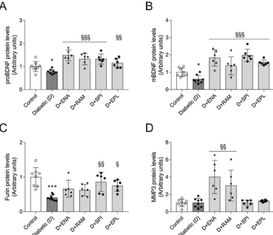
3.5. RAASi restore hippocampal BDNF production and transformation in diabetes
BDNF and downstream signaling pathways play a pivotal role in the pathophysiology of depression. Hippocampal synthesis of both proBDNF and mBDNF decreased in diabetic rats. These changes were fully reversed by RAASi treatment (Fig. 2A–B). While measuring clea- vage enzymes responsible for BDNF transformation, we found that different RAASi acted on different pathways. Interestingly enough, MR antagonists increased the intracellular enzyme furin, while ACEi ele- vated the extracellular MMP3 (Fig. 2C–D).
3.6. RAASi interact with hippocampal mature BDNF signaling
Precursor and mature BDNF forms bind to different receptors and activate divergent pathways. ProBDNF acts on p75NTRand activates the pro-apoptotic Bax transcription factor through phosphorylated JNK mediator. The activation of this pathway leads to neuronal apoptosis and decreased synaptic plasticity. However, in our model neither dia- betes, nor RAASi induced any changes in this signaling cascade in the hippocampus (Fig. 3A–C).
mBDNF binds to TrkB receptor after which the activated pathway Table 2
Depressive-like behavior of control and RAASi treated rats are unaltered. Openfield test (OFT) and forced swim test (FST) were performed were performed on control (C), enalapril (C + ENA) or ramipril (C + RAM) or spironolactone (C + SPI) or eplerenone (C + EPL) treated control rats. During OFT, locomotor activity was evaluated by measuring the number of grid crossings. In FST each moving pattern is represented as percentage of total test time. Floating time indicates immobility, while active mobility consists of struggling, swimming and diving parameters. Data indicate means ± SDs and were analyzed by one-way ANOVA with Holm-Sidak multiple comparisons test or by Kruskal-Wallis test with Dunn correction. F values are results of Brown-Forsythe tests, followed by DFn and DFd numbers (n = 7 in control and n = 6 in treatment groups).
Behavioral test result Control (C) C + ENA C + RAM C + SPI C + EPL p values F value
Number of grid crossings 336.1 ± 104.8 395.3 ± 123.9 368.3 ± 65.4 410.3 ± 104.3 315.2 ± 144.9 Not significant 0.489 (4, 26)
Floating time (%) 38.8 ± 10.4 30.9 ± 14.3 33.1 ± 14.6 38.9 ± 10.1 41.9 ± 10.8 Not significant 0.339 (4, 26)
Mobility time (%) 61.2 ± 10.4 69.1 ± 14.3 66.9 ± 14.6 61.0 ± 10.1 58.1 ± 10.8 Not significant 0.339 (4, 26)
Struggling time (%) 29.3 ± 7.9 27.9 ± 7.8 23.2 ± 7.4 22.7 ± 6.5 24.2 ± 5.4 Not significant 0.362 (4, 26)
Swimming time (%) 31.4 ± 5.0 41.1 ± 14.2 43.2 ± 13.2 37.5 ± 8.7 32.6 ± 10.9 Not significant 1.413 (4, 26)
Diving time (%) 0.4 ± 0.5 0.2 ± 0.4 0.6 ± 0.3 1.5 ± 0.7 1.5 ± 0.6 Not significant 1.448 (4, 34)
Fig. 1. Regulation of cerebral perfusion is unaltered by RAASi, but they ameliorate proinflammatory responses in hippocampi of diabetic rats.(A) Mean arterial pressure (MAP) was calculated in control, vehicle-treated diabetic (D), enalapril (D + ENA) or ramipril (D + RAM) or spironolactone (D + SPI) or eplerenone (D + EPL) treated diabetic rats. (B–D) Fold changes of protein levels of bloodflow regulating phosphorylated endothelial nitric oxide synthase (p-eNOS), phosphorylated neuronal nitric oxide synthase (p-nNOS) and endothelin-1 were evaluated in hippocampi by Western blot. (E) Fold changes of protein levels of proinflammatory nuclear factor kappa-light-chain-enhancer of activated B cells (NF-κB). (F–H) Hippocampal mRNA expression of proinflammatory cytokine in- terleukin-1α(Il1a), interleukin-6(Il6)and tumor necrosis factor-α(Tnf)were measured by qRT-PCR. All proteins were normalized to total protein Ponceau S staining as loading control. All mRNAs were normalized toRn18SmRNA expression. Bars indicate means ± SDs and data were analyzed by one-way ANOVA with Holm-Sidak multiple comparisons test or by Kruskal-Wallis test with Dunn correction (n = 8 in control and diabetic and n = 6 in treatment groups). *p < 0.05vs.Control;
**p < 0.01vs.Control; ***p < 0.001vs.Control; §p < 0.05vs.Diabetic; §§p < 0.01vs.Diabetic; §§§p < 0.001vs.Diabetic.
phosphorylates extracellular ERK and CREB factors and leads to anti- apoptotic Bcl-2 increment. TrkB, p-ERK, p-CREB protein levels andBcl2 mRNA expression were decreased in the hippocampi of diabetic rats.
RAASi treatment reversed the decrement in all cases (Fig. 3D–G).
4. Discussion
The possible association between diabetes and depression was re- cognized as early as the 17th century, however it has only been in- tensively studied in the last decade. RAASi are the cornerstones of the Fig. 2. Hippocampal BDNF production is increased by RAASi in diabetic rats. (A–D) Fold changes of protein levels of precursor brain-derived neurotrophic factor (BDNF), mature BDNF (mBDNF) and its cleavage en- zymes furin and matrix metalloproteinase-3 (MMP3) were evaluated in hippocampi by Western blot in control, vehicle-treated dia- betic (D), enalapril (D + ENA) or ramipril (D + RAM) or spironolactone (D + SPI) or eplerenone (D + EPL) treated diabetic rats. All proteins were normalized to total protein Ponceau S staining as loading control. Bars indicate means ± SDs and data were an alyzed by one-way ANOVA with Holm-Sidak multiple comparisons test or by Kruskal-Wallis test with Dunn correction (n = 8 in control and diabetic and n = 6 in treatment groups). *p < 0.05vs.
Control; ***p < 0.001vs. Control; §p < 0.05 vs. Diabetic; §§p < 0.01 vs. Diabetic;
§§§p < 0.001vs. Diabetic.
Fig. 3. Neuronal response in diabetes can be altered by RAASiviamBDNF-TrkB signaling in the hippocampus. (A–B) Fold changes of protein levels in proBDNF pathway: p75 neurotrophin receptor (p75NTR) and phosphorylated c-Jun N-terminal kinase (p-JNK) were evaluated in hippocampi by Western blot; (C) Hippocampal mRNA expression of pro-apoptotic Bcl-2-associated X protein (Bax) was measured by qRT-PCR in control, vehicle-treated diabetic (D), enalapril (D + ENA) or ramipril (D + RAM) or spironolactone (D + SPI) or eplerenone (D + EPL) treated diabetic rats. (D–F) Fold changes of protein levels in downstream elements of mBDNF pathway: tropomyosin receptor kinase B (TrkB), phosphorylated extracellular-signal-regulated kinase (p-ERK) and phosphorylated cAMP response ele- ment binding protein (p-CREB) were measured in hippocampi by Western blot. (G) Hippocampal mRNA expression of pro-survival B-cell lymphoma 2 (Bcl2) was measured by qRT-PCR. All proteins were normalized to total protein Ponceau S staining as loading control. All mRNAs were normalized to Rn18S mRNA expression.
Bars indicate means ± SDs and data were analyzed by one-way ANOVA with Holm-Sidak multiple comparisons test or by Kruskal-Wallis test with Dunn correction (n
= 8 in control and diabetic and n = 6 in treatment groups). *p < 0.05vs. Control; **p < 0.01vs. Control; §p < 0.05vs. Diabetic; §§p < 0.01vs. Diabetic;
§§§p < 0.001vs. Diabetic.
management of diabetes-related complications. All components of RAAS have been identified in the brain and are suggested to be involved in the pathomechanism of depression. Thefirst evidence that RAAS in the brain is involved in depression was observed in hypertensive pa- tients who showed reduced depressive symptoms after receiving ACEi (Germain and Chouinard, 1988,1989). Recently, our group reported that losartan minimizes depressive-like behavior via modulating the BDNF pathway in diabetic rats, independently of serum glucose and blood pressure. To investigate whether all types of RAASi exhibit these beneficial effects, here we tested the efficacy of enalapril, ramipril, spironolactone or eplerenone treatment in diabetes-associated depres- sion.
The main finding of the present study is that ACEi and MR an- tagonists have antidepressant-like effect in diabetic rats, which is characterized by decreased total immobility (floating) time in FST.
Immobility reflects behavioral despair and it is the most relevant parameter in the characterization of depression-like behavior. Others showed that ACEi reduce depression-like behavior in healthy animals subjected to FST (Giardina and Ebert, 1989;Nayak and Patil, 2008).
Further, spironolactone has been reported to decrease immobility time in FST in a corticosterone-induced depression model (Wu et al., 2013).
Beside immobility, active parameters (swimming or struggling) reflect escape-seeking behavior and have been shown to be relevant as well. A paper reported that increased time of swimming is connected to the activation of serotonergic system, while longer struggling is associated with increased catacholaminergic neurotransmission (Cryan et al., 2005). The time of swimming was increased significantly only by ra- mipril, while struggling remained unaltered in all RAASi-treated groups. Further, locomotion was not affected by RAASi confirming that the results are independent of the rats' physical condition.
The ‘vascular depression hypothesis’ highlights the link between cerebral vascular disease and depression. Diabetes-related changes in cerebral hemodynamics contribute to the development of depression.
Reduced eNOS levels (Demir et al., 2015;Utkan et al., 2015), elevated endothelin-1 and activation of RAAS resulted in detrimental vascular alterations and local vasoconstriction in the diabetic brain (Saavedra, 2005). Here, the eNOS/nNOS pathway and endothelin-1 were not al- tered significantly in the hippocampus in all groups. This was not un- expected in light of our recentin vivoSPECT-CT measurements showing that losartan did not improve decreased CBF (Lenart et al., 2019). These results together with unchanged MAP indicate that the beneficial an- tidepressant-like effect of ACEi and MR antagonist are not due to a vasoactive action.
Inflammation has been implicated in the pathophysiology of both depression and diabetes, which may be one link between the two dis- eases (Leonard and Maes, 2012;Shoelson et al., 2006). Overactivation of RAAS has been identified in diabetes and neuropsychiatric disorders (e.g.Alzheimer’s disease, depression), which are frequently associated with neurodegeneration and neuroinflammation (Gebre et al., 2018;
Vargas et al., 2012; Yagi et al., 2013). The activation of local brain RAAS is characterized by elevated ACE expression, AngII generation and AT1R expression (Gong et al., 2019). Cerebral Ang II induces mi- croglial activation and release of proinflammatory cytokines. Data concerning the role of mineralocorticoids in depression-associated in- flammation is limited, but aldosterone has been demonstrated to in- crease proinflammatory cytokine levels in the brain (Dinh et al., 2016).
In our model hippocampal elevation ofIl1a, Il6andTnfindicate the activation of the NF-κB-mediated inflammatory pathway in diabetes- associated depression, similarly to models of other psychiatric disorders (Asraf et al., 2018;Dinh et al., 2016;Torika et al., 2016). Furthermore, we showed that both ACEi and MR antagonists mitigate inflammatory response. Together with our previous results, thesefindings support the anti-inflammatory role of RAASi as a novel mechanism of action in diabetes comorbid depression.
Neuroinflammation interacts with neurotransmitter metabolism, neuroendocrine function, synaptic plasticity and alters behavior. IL-1,
IL-6 and TNF have profound stimulatory effects on hypothalamic-pi- tuitary-adrenal axis and contribute to changes in neuronal growth and survival (Silverman et al., 2005). Studies demonstrated that adminis- tration of IL-1 decreases the neurotrophin BDNF in rat hippocampus (Lapchak et al., 1993; Zhang et al., 2014) leading to cognitive dys- function. Earlier we reported that hippocampal BDNF is reduced in diabetes-associated depression (Lenart et al., 2016) and the alteration is reversed by losartan (Lenart et al., 2019). Here we showed that not only ARBs, but also ACEi and MR antagonists suspend BDNF decrement.
These data provide novel evidence of the link between diabetes-induced neuroinflammation and BDNF abatement.
BDNF is initially synthesized as pre-pro-BDNF, then the pre-region is removed yielding the pro-BDNF. The N-terminal region is proteolyti- cally cleaved by intracellular furin and/or extracellular matrix me- talloproteases (MMPs) to produce the mature form (Lu et al., 2005).
Here we found that ACEi and MR antagonists act on different proteo- lytic cleavage pathways. ACEi elevated extracellular MMP3, while the intracellular enzyme furin was increased by MR antagonists. Interest- ingly enough, according to our previous paper ARBs exert their effect on both. These cleavage enzymes may be targets of RAASi and might suggest a possible synergistic antidepressant performance of combina- tion therapy.
Identifying molecular pathways by which altered BDNF signaling could lead to depression in diabetes is of paramount importance. Lu et al. propose a‘yin and yang’ model of neurotrophin action, since BDNF forms have opposing effects on cell survival. Hippocampal p75NTRactivated JNK-Bax signaling leads to neuronal apoptosis, while the TrkB-ERK-CREB pathway mediates cell survival and has a crucial role in antidepressant effects (Chen et al., 2001). Here, the pro-apop- totic pathway remained unchanged, while the pro-survival pathway was impaired. We demonstrated that TrkB and its downstream ele- ments, p-ERK and p-CREB were repressed in the diabetic hippocampus.
This is in line with our previous results and other studies showing im- peded hippocampal BDNF signaling in diabetes (Agrawal et al., 2014;
Lenart et al., 2019;Qin et al., 2016). Both ACEi and MR antagonists increased TrkB, p-ERK and p-CREB levels. CREB-dependent transcrip- tional mechanism activates Bcl-2, which possesses anti-apoptotic effects and enhances neuronal survival (Riccio et al., 1999). In accordance with this, here we showed thatBcl2expression was higher in diabetic rats treated with ACEi or MR antagonists. These results suggest that activated BDNF-mediated pro-survival pathway contributes to hippo- campal plasticity and thus moderates depressive-like behavior.
In conclusion, the present study provides experimental data for the antidepressant-like effect of RAASi in diabetes comorbid depression.
We hypothesize that diabetes-induced neuroinflammation decreases BDNF level, which contributes to neuronal damage and subsequently to the development of depression-like behavior. Our results indicate that both ACEi and MR antagonists reduce neuroinflammation and promote BDNF maturation resulting in the activation of BDNF-mediated pro- survival pathway. According to this, we propose two additional possible mechanisms by which RAASi may exert neuroprotection and anti- depressant-like action. Ultimately, various RAASi may provide a novel therapeutic opportunity to treat and improve outcomes of both dis- orders simultaneously.
Author contributions
Author Dora B. Balogh made substantial contributions to collection, analysis and interpretation of data and wrote the first draft of the manuscript. Authors Andrea Fekete and Lilla Lenart designed the study, drafted and revised the article critically for important intellectual content. Authors Agnes Molnar, Tamas Lakat and Judit Hodrea man- aged the literature searches and undertook the statistical and experi- mental data analysis. Authors Adam Hosszu and Attila J. Szabo made substantial contributions revising the article critically for important intellectual content. All authors contributed to and have approved the
final manuscript. Andrea Fekete is the guarantor of this work and, as such, had full access to all the data in the study and is responsible for the integrity of the work as a whole.
Funding
Fundings for this study was provided by National Research, Development and Innovation Office (FK-124491, NN-114607, 2017- 1.3.1-VKE-2017-00006), Semmelweis University (FIKP) and Ministry of Innovation and Technology (UNKP-19-3-III-SE-6).
The funding sources had no further involvement in the study design;
in the collection, analysis and interpretation of data; in the writing of the report; and in the decision to submit the paper for publication.
Declaration of Competing Interest
All other authors declare that they have no conflicts of interest.
Acknowledgements
The authors are grateful to Maria Bernath, Renata Gellai and Sandor Koszegi who provided excellent technical assistance. We specifically thank Eva Mikics and Adrienn Barczi for constant help on behavior studies.
Appendix A. Supplementary data
Supplementary material related to this article can be found, in the online version, at doi:https://doi.org/10.1016/j.psyneuen.2020.
104705.
References
Agrawal, R., Zhuang, Y., Cummings, B.P., Stanhope, K.L., Graham, J.L., Havel, P.J., Gomez-Pinilla, F., 2014. Deterioration of plasticity and metabolic homeostasis in the brain of the UCD-T2DM rat model of naturally occurring type-2 diabetes. Biochim.
Biophys. Acta 1842, 1313–1323.
Ahola, A.J., Harjutsalo, V., Forsblom, C., Groop, P.H., 2014. Renin-angiotensin-aldos- terone-blockade is associated with decreased use of antidepressant therapy in pa- tients with type 1 diabetes and diabetic nephropathy. Acta Diabetol. 51 (Aug (4)), 529–533.https://doi.org/10.1007/s00592-013-0547-x.Epub 2014 Jan 17.
Asraf, K., Torika, N., Apte, R.N., Fleisher-Berkovich, S., 2018. Microglial activation is modulated by captopril: in vitro and in vivo studies. Front. Cell. Neurosci. 12.
Atwal, J.K., Massie, B., Miller, F.D., Kaplan, D.R., 2000. The TrkB-Shc site signals neu- ronal survival and local axon growth via MEK and P13-kinase. Neuron 27, 265–277.
Chen, A.C., Shirayama, Y., Shin, K.H., Neve, R.L., Duman, R.S., 2001. Expression of the cAMP response element binding protein (CREB) in hippocampus produces an anti- depressant effect. Biol. Psychiatry 49, 753–762.
Chen, S., Zhang, Q., Dai, G., Hu, J., Zhu, C., Su, L., Wu, X., 2016. Association of depression with pre-diabetes, undiagnosed diabetes, and previously diagnosed diabetes: a meta- analysis. Endocrine 53, 35–46.
Coppey, L.J., Davidson, E.P., Rinehart, T.W., Gellett, J.S., Oltman, C.L., Lund, D.D., Yorek, M.A., 2006. ACE inhibitor or angiotensin II receptor antagonist attenuates diabetic neuropathy in streptozotocin-induced diabetic rats. Diabetes 55, 341–348.
Cryan, J.F., Valentino, R.J., Lucki, I., 2005. Assessing substrates underlying the beha- vioral effects of antidepressants using the modified rat forced swimming test.
Neurosci. Biobehav. Rev. 29, 547–569.
Demir, R., Cadirci, E., Akpinar, E., Cayir, Y., Atmaca, H.T., Un, H., Kunak, C.S., Yayla, M., Bayraktutan, Z., Demir, I., 2015. Does bosentan protect diabetic brain alterations in rats? The role of endothelin-1 in the diabetic brain. Basic Clin. Pharmacol. Toxicol.
116, 236–243.
Denenberg, V.H., 1969. Open-field bheavior in the rat: what does it mean? Ann. N. Y.
Acad. Sci. 159, 852–859.
Dinh, Q.N., Young, M.J., Evans, M.A., Drummond, G.R., Sobey, C.G., Chrissobolis, S., 2016. Aldosterone-induced oxidative stress and inflammation in the brain are mediated by the endothelial cell mineralocorticoid receptor. Brain Res. 1637, 146–153.
Egede, L.E., Zheng, D., Simpson, K., 2002. Comorbid depression is associated with in- creased health care use and expenditures in individuals with diabetes. Diabetes Care 25, 464–470.
Gebre, A.K., Altaye, B.M., Atey, T.M., Tuem, K.B., Berhe, D.F., 2018. Targeting renin- angiotensin system against Alzheimer’s disease. Front. Pharmacol. 9, 440.
Germain, L., Chouinard, G., 1988. Treatment of recurrent unipolar major depression with captopril. Biol. Psychiatry 23, 637–641.
Germain, L., Chouinard, G., 1989. Captopril treatment of major depression with serial
measurements of blood cortisol concentrations. Biol. Psychiatry 25, 489–493.
Giardina, W.J., Ebert, D.M., 1989. Positive effects of captopril in the behavioral despair swim test. Biol. Psychiatry 25, 697–702.
Gong, X., Hu, H., Qiao, Y., Xu, P., Yang, M., Dang, R., Han, W., Guo, Y., Chen, D., Jiang, P., 2019. The involvement of renin-angiotensin system in lipopolysaccharide-induced behavioral changes, neuroinflammation, and disturbed insulin signaling. Front.
Pharmacol. 10, 318.
Grima, M., Anjuere, J., Ingert, C., Coquard, C., Steger, J., Barthelmebs, M., Imbs, J.L., 2001. [Effect of a non-antihypertensive dose of ramipril on the plasma and tissue renin-angiotensin system in 27 TGR (mRen2) rats]. Arch. Mal. Coeur Vaiss. 94, 805–812.
Karege, F., Perret, G., Bondolfi, G., Schwald, M., Bertschy, G., Aubry, J.M., 2002.
Decreased serum brain-derived neurotrophic factor levels in major depressed pa- tients. Psychiatry Res. 109, 143–148.
Lapchak, P.A., Araujo, D.M., Hefti, F., 1993. Systemic interleukin-1-beta decreases brain- derived neurotrophic factor messenger-Rna expression in the rat hippocampal-for- mation. Neuroscience 53, 297–301.
Lenart, L., Hodrea, J., Hosszu, A., Koszegi, S., Zelena, D., Balogh, D., Szkibinszkij, E., Veres-Szekely, A., Wagner, L., Vannay, A., Szabo, A.J., Fekete, A., 2016. The role of sigma-1 receptor and brain-derived neurotrophic factor in the development of dia- betes and comorbid depression in streptozotocin-induced diabetic rats.
Psychopharmacology (Berl.) 233, 1269–1278.
Lenart, L., Balogh, D.B., Lenart, N., Barczi, A., Hosszu, A., Farkas, T., Hodrea, J., Szabo, A.J., Szigeti, K., Denes, A., Fekete, A., 2019. Novel therapeutic potential of angio- tensin receptor 1 blockade in a rat model of diabetes-associated depression parallels altered BDNF signalling. Diabetologia 62, 1501–1513.
Leonard, B., Maes, M., 2012. Mechanistic explanations how cell-mediated immune acti- vation, inflammation and oxidative and nitrosative stress pathways and their sequels and concomitants play a role in the pathophysiology of unipolar depression. Neurosci Biobehav R 36, 764–785.
Lu, B., Pang, P.T., Woo, N.H., 2005. The yin and yang of neurotrophin action. Nat. Rev.
Neurosci. 6, 603–614.
Majewski, C., Bakris, G.L., 2016. Has RAAS blockade reached its limits in the treatment of diabetic nephropathy? Curr. Diab. Rep. 16, 24.
Matsubara, B.B., Matsubara, L.S., Franco, M., Padovani, J.C., Janicki, J.S., 1999. The effect of non-antihypertensive doses of angiotensin converting enzyme inhibitor on myocardial necrosis and hypertrophy in young rats with renovascular hypertension.
Int. J. Exp. Pathol. 80, 97–104.
Nayak, V., Patil, P.A., 2008. Antidepressant activity of fosinopril, ramipril and losartan, but not of lisinopril in depressive paradigms of albino rats and mice. Indian J. Exp.
Biol. 46, 180–184.
Pandey, G.N., Ren, X.G., Rizavi, H.S., Conley, R.R., Roberts, R.C., Dwivedi, Y., 2008.
Brain-derived neurotrophic factor and tyrosine kinase B receptor signalling in post- mortem brain of teenage suicide victims. Int. J. Neuropsychophol. 11, 1047–1061.
Paul, M., Poyan Mehr, A., Kreutz, R., 2006. Physiology of local renin-angiotensin systems.
Physiol. Rev. 86, 747–803.
Pavlatou, M.G., Mastorakos, G., Lekakis, I., Liatis, S., Vamvakou, G., Zoumakis, E., Papassotiriou, I., Rabavilas, A.D., Katsilambros, N., Chrousos, G.P., 2008. Chronic administration of an angiotensin II receptor antagonist resets the hypothalamic-pi- tuitary-adrenal (HPA) axis and improves the affect of patients with diabetes mellitus type 2: preliminary results. Stress 11, 62–72.
Porsolt, R.D., Le Pichon, M., Jalfre, M., 1977. Depression: a new animal model sensitive to antidepressant treatments. Nature 266, 730–732.
Porsolt, R.D., Anton, G., Blavet, N., Jalfre, M., 1978. Behavioural despair in rats: a new model sensitive to antidepressant treatments. Eur. J. Pharmacol. 47, 379–391.
Qin, L., Chong, T., Rodriguez, R., Pugazhenthi, S., 2016. Glucagon-like peptide-1-medi- ated modulation of inflammatory pathways in the diabetic brain: relevance to Alzheimer’s disease. Curr. Alzheimer Res. 13, 1346–1355.
Renn, B.N., Feliciano, L., Segal, D.L., 2011. The bidirectional relationship of depression and diabetes: a systematic review. Clin. Psychol. Rev. 31, 1239–1246.
Ribeiro-Oliveira Jr., A., Nogueira, A.I., Pereira, R.M., Boas, W.W., Dos Santos, R.A., Simoes e Silva, A.C., 2008. The renin-angiotensin system and diabetes: an update.
Vasc. Health Risk Manag. 4, 787–803.
Riccio, A., Ahn, S., Davenport, C.M., Blendy, J.A., Ginty, D.D., 1999. Mediation by a CREB family transcription factor of NGF-dependent survival of sympathetic neurons.
Science 286, 2358–2361.
Rossi, C., Angelucci, A., Costantin, L., Braschi, C., Mazzantini, M., Babbini, F., Fabbri, M.E., Tessarollo, L., Maffei, L., Berardi, N., Caleo, M., 2006. Brain-derived neuro- trophic factor (BDNF) is required for the enhancement of hippocampal neurogenesis following environmental enrichment. Eur. J. Neurosci. 24, 1850–1856.
Rubin, R.R., Ma, Y., Marrero, D.G., Peyrot, M., Barrett-Connor, E.L., Kahn, S.E., Haffner, S.M., Price, D.W., Knowler, W.C., Grp, D.P.P.R., 2008. Elevated depression symp- toms, antidepressant medicine use, and risk of developing diabetes during the dia- betes prevention program. Diabetes Care 31, 420–426.
Saavedra, J.M., 2005. Brain angiotensin II: new developments, unanswered questions and therapeutic opportunities. Cell. Mol. Neurobiol. 25, 485–512.
Saavedra, J.M., 2012. Angiotensin II AT(1) receptor blockers ameliorate inflammatory stress: a beneficial effect for the treatment of brain disorders. Cell. Mol. Neurobiol.
32, 667–681.
Shoelson, S.E., Lee, J., Goldfine, A.B., 2006. Inflammation and insulin resistance. J. Clin.
Invest. 116, 1793–1801.
Silverman, M.N., Pearce, B.D., Biron, C.A., Miller, A.H., 2005. Immune modulation of the hypothalamic-pituitary-adrenal (HPA) axis during viral infection. Viral Immunol. 18, 41–78.
Stuart, M.J., Baune, B.T., 2012. Depression and type 2 diabetes: inflammatory mechan- isms of a psychoneuroendocrine co-morbidity. Neurosci. Biobehav. Rev. 36, 658–676.
Taira, M., Toba, H., Murakami, M., Iga, I., Serizawa, R., Murata, S., Kobara, M., Nakata, T., 2008. Spironolactone exhibits direct renoprotective effects and inhibits renal renin-angiotensin-aldosterone system in diabetic rats. Eur. J. Pharmacol. 589, 264–271.
Tong, L., Balazs, R., Soiampornkul, R., Thangnipon, W., Cotman, C.W., 2008. Interleukin- 1 beta impairs brain derived neurotrophic factor-induced signal transduction.
Neurobiol. Aging 29, 1380–1393.
Tong, L.Q., Prieto, G.A., Kramar, E.A., Smith, E.D., Cribbs, D.H., Lynch, G., Cotman, C.W., 2012. Brain-derived neurotrophic factor-dependent synaptic plasticity is suppressed by Interleukin-1 beta via p38 mitogen-activated protein kinase. J. Neurosci. 32, 17714–17724.
Torika, N., Asraf, K., Roasso, E., Danon, A., Fleisher-Berkovich, S., 2016. Angiotensin converting enzyme inhibitors ameliorate brain inflammation associated with micro- glial activation: possible implications for Alzheimer’s disease. J. Neuroimmune Pharmacol. 11, 774–785.
Utkan, T., Yazir, Y., Karson, A., Bayramgurler, D., 2015. Etanercept improves cognitive performance and increases eNOS and BDNF expression during experimental vascular dementia in streptozotocin-induced diabetes. Curr. Neurovasc. Res. 12, 135–146.
van Dieren, S., Beulens, J.W., van der Schouw, Y.T., Grobbee, D.E., Neal, B., 2010. The
global burden of diabetes and its complications: an emerging pandemic. Eur. J.
Cardiovasc. Prev. Rehabil. 17 (Suppl. 1), S3–S8.
Vargas, R., Rincon, J., Pedreanez, A., Viera, N., Hernandez-Fonseca, J.P., Pena, C., Mosquera, J., 2012. Role of angiotensin II in the brain inflammatory events during experimental diabetes in rats. Brain Res. 1453, 64–76.
Williams, L.J., Pasco, J.A., Kessing, L.V., Quirk, S.E., Fernandes, B.S., Berk, M., 2016.
Angiotensin converting enzyme inhibitors and risk of mood disorders. Psychother.
Psychosom. 85, 250–252.
Wu, T.C., Chen, H.T., Chang, H.Y., Yang, C.Y., Hsiao, M.C., Cheng, M.L., Chen, J.C., 2013.
Mineralocorticoid receptor antagonist spironolactone prevents chronic corticosterone induced depression-like behavior. Psychoneuroendocrinology 38, 871–883.
Yagi, S., Akaike, M., Ise, T., Ueda, Y., Iwase, T., Sata, M., 2013. Renin-angiotensin-al- dosterone system has a pivotal role in cognitive impairment. Hypertens. Res. 36, 753–758.
Zhang, J.C., Wu, J., Fujita, Y., Yao, W., Ren, Q., Yang, C., Li, S.X., Shirayama, Y., Hashimoto, K., 2014. Antidepressant effects of TrkB ligands on depression-like be- havior and dendritic changes in mice after inflammation. Int. J.
Neuropsychopharmacol. 18.