Review
Endoplasmic Reticulum-Plasma Membrane Contact Sites as an Organizing Principle for Compartmentalized Calcium and
cAMP Signaling
Tim Crul1,2,3,* and József Maléth1,2,3,*
Citation: Crul, T.; Maléth, J.
Endoplasmic Reticulum-Plasma Membrane Contact Sites as an Organizing Principle for Compartmentalized Calcium and cAMP Signaling.Int. J. Mol. Sci.2021, 22, 4703. https://doi.org/10.3390/
ijms22094703
Academic Editor: Henrique Girao
Received: 25 February 2021 Accepted: 27 April 2021 Published: 29 April 2021
Publisher’s Note:MDPI stays neutral with regard to jurisdictional claims in published maps and institutional affil- iations.
Copyright: © 2021 by the authors.
Licensee MDPI, Basel, Switzerland.
This article is an open access article distributed under the terms and conditions of the Creative Commons Attribution (CC BY) license (https://
creativecommons.org/licenses/by/
4.0/).
1 First Department of Medicine, University of Szeged, H6720 Szeged, Hungary
2 HAS-USZ Momentum Epithelial Cell Signaling and Secretion Research Group, University of Szeged, H6720 Szeged, Hungary
3 HCEMM-SZTE Molecular Gastroenterology Research Group, University of Szeged, H6720 Szeged, Hungary
* Correspondence: crul.tim@gmail.com (T.C.); jozsefmaleth1@gmail.com (J.M.)
Abstract:In eukaryotic cells, ultimate specificity in activation and action—for example, by means of second messengers—of the myriad of signaling cascades is primordial. In fact, versatile and ubiquitous second messengers, such as calcium (Ca2+) and cyclic adenosine monophosphate (cAMP), regulate multiple—sometimes opposite—cellular functions in a specific spatiotemporal manner.
Cells achieve this through segregation of the initiators and modulators to specific plasma membrane (PM) subdomains, such as lipid rafts and caveolae, as well as by dynamic close contacts between the endoplasmic reticulum (ER) membrane and other intracellular organelles, including the PM.
Especially, these membrane contact sites (MCSs) are currently receiving a lot of attention as their large influence on cell signaling regulation and cell physiology is increasingly appreciated. Depletion of ER Ca2+stores activates ER membrane STIM proteins, which activate PM-residing Orai and TRPC Ca2+channels at ER–PM contact sites. Within the MCS, Ca2+fluxes relay to cAMP signaling through highly interconnected networks. However, the precise mechanisms of MCS formation and the influence of their dynamic lipid environment on their functional maintenance are not completely understood. The current review aims to provide an overview of our current understanding and to identify open questions of the field.
Keywords:membrane contact sites; SOCE; STIM1; Orai1; calcium; cAMP
1. Introduction
Eukaryotic cells are characterized by a myriad of metabolic and signaling pathways in which pleiotropic second messengers—such as calcium (Ca2+) and cAMP—function as important connecting hubs between different cascades [1–4]. Although pleiotropic second messengers might generally offer flexibility to cells as to how to respond to certain stimuli, the logistic dilemma is, however, enormous and requires a strict spatiotemporal control in order to preserve the specificity of the intended cascade and avoid the development of pathological conditions [5–8].
Plasma membrane (PM) nanodomains—lipid rafts, caveolae, and membrane contact sites (MCSs)—serve as excellent tools allowing compartmentalization of initiators and modulators. For example, lipid rafts and caveolae are dynamic cholesterol-enriched PM microdomains, which play an important role in the initiation of many signaling path- ways [9–11]. MCSs are dynamic close contacts between intracellular organelles with a major regulatory role on signaling cascades and cell physiology [12–14]. Affinity between the different membrane types as well as the maintenance of the MCS is mediated by a large variety of tether proteins, including, for example, extended synaptotagmins (E-Syts).
Next to being a general mediator of proper protein synthesis and transport, the endo- plasmic reticulum (ER) acts as an important Ca2+store [15]. Intracellular Ca2+fluxes are,
Int. J. Mol. Sci.2021,22, 4703. https://doi.org/10.3390/ijms22094703 https://www.mdpi.com/journal/ijms
Int. J. Mol. Sci.2021,22, 4703 2 of 26
among others, generated through agonist-mediated release of cellular compartmentalized Ca2+and subsequent activation of extracellular Ca2+influx through plasma membrane (PM) channels through so-called store-operated Ca2+entry (SOCE) within ER–PM contact sites.
Based on their biophysical properties, store-operated currents can be roughly divided into two different types: (a) the highly Ca2+selective Ca2+release-activated Ca2+current (ICRAC), which is a non-voltage activated, inwardly rectifying current; and (b) a variety of store-operated currents classified as ISOC, which differ from ICRACin that, for instance, these currents are non-selective for Ca2+and exhibit greater conductance compared to ICRAC(reviewed in [16]).
Subsequent Ca2+-mediated activation of adenylyl cyclases within these MCSs allows specific spatiotemporal regulation of Ca2+-induced cAMP generation. Although a complete understanding of MCS functioning is still lacking, it is now clear that an intricate interplay between the MCS lipid environment and its resident proteins is crucial for it to function properly. Hence, considering that the activity of multiple ion channels and transporters is modulated by lipid species that are specifically enriched at ER–PM contact sites, this points towards a general role of the MCSs microenvironment in ion transport and cell physiology.
In the current review, the dynamics of ER–PM contact sites on SOCE-mediated cAMP signaling will be elaborated. Based on the current state-of-the-art, open questions will be identified that might enable the field to be pushed forward.
2. STIM/Orai/TRPC-Mediated Store-Operated Ca2+Entry
Activation of G-protein-coupled receptors—seven-transmembrane receptors, includ- ingβ2 adrenergic-like receptors activated by, for example, epinephrine; glucagon receptors activated by, for example, glucagon; and metabotropic neuroreceptors activated by, for example, small molecule neurotransmitters (such as dopamine or acetylcholine) or neu- ropeptides (such as encephalin [17])—results in synthesis of inositol triphosphate (IP3).
Subsequent binding of IP3to the inositol triphosphate receptor (IP3R) in the ER membrane induces Ca2+efflux from the ER into the cytoplasmic space, enabling a myriad of cellular responses, including transcriptional regulation, exocytosis, and contraction.
Changes in ER luminal Ca2+levels are monitored by single transmembrane-spanning stromal interaction molecule (STIM1 and -2) proteins and subsequently translated to the PM-residing Ca2+ uptake channels Orai and TRPC. Whereas Orai channels are highly selective for Ca2+, TRP channels are mostly non-selective cation channels permeable to both monovalent and divalent cations, including Ca2+and Na+[18,19].
At the protein level, STIM proteins contains an ER signal peptide, a canonical EF- hand Ca2+-binding motif as well as a hidden EF-hand, and a sterile alpha motif (SAM) at the N-terminal region, a transmembrane domain, and an ezrin/radixin/moesin domain, including three coiled-coil domains, a CRAC-modulatory domain, a proline/serine-rich region, and a polybasic lysine-rich region at the cytosolic C-terminus [20–27].
STIM1-mediated sensing of the ER Ca2+levels and subsequent activation of CRAC channels relies on extensive conformational changes of STIM1 (Figure1).
At the luminal site, interaction of Ca2+with the negatively charged aspartates and glutamates induces a helix-loop-helix conformation of the EF hand. Agonist-induced ER discharge and subsequent dissociation of Ca2+from the STIM EF hand destabilizes this conformation, resulting in activation of STIM1 [28]. Interestingly, while the STIM1 TM domains are not in close proximity in resting conditions, the presence of three TM domain- localized glycine residues (G223, G225, and G226) aid in the movement of the STIM1 TM domains towards each other upon Ca2+release from the luminal EF hand [29–31]. Subse- quent homomerization of STIM1 is mediated by CC2, CC3, and SHD, which is essential for coupling to and activation of Orai1 [32,33]. X-ray crystallographic structural data of the STIM1 cytosolic region revealed a dimeric assembly with an antiparallel arrangement of CC2 and CC3 and two inter-adjacent shortα-helices [34]. From these crystal structures, it could be derived that the physiological V-shaped conformation of the critical STIM-Orai1- activating region (SOAR) [26] (also referred to as channel-activating domain (CAD) [35],
Orai-activating small fragment (OASF) [33,36], or coiled-coil domain-containing region b9 (CCb9) [37]) dimer is established through coiled-coil interactions of one monomer (residues R429, W430, I433, and L436) and the other (residues T354, L351, W350, and L347). Of note, mutation-induced disruption of this SOAR binding interface abrogated Orai1activation [34]. Even though only partial structural data is currently available of the STIM1 CC1 domain, this region comprises three alpha helical segments CC1α1 (aa238- 271), CC1α2 (aa278-304), and the so-called inhibitory helix CC1α3 (aa308-337) [34,38,39].
While bridging the distance within ER–PM junctions mediating Orai1 interaction, the CC1 domain equally keeps STIM1 in a tight and compact conformation at high ER Ca2+
levels [29,36,39,40]. The polybasic cluster (K382, K384, K385, K386, and R387) located at the STIM1 C-terminal is—together with the SOAR domain—crucial for puncta formation through a diffusion-trap mechanism via interaction with PM-resident phospholipids and Orai channels; however, it does not mediate STIM1 oligomerization [32,35,41–43]. Overall, high ER Ca2+levels preserve the STIM1 C-terminal region—including the CC1, SOAR, and polybasic region—in a folded (inactive) configuration. Depletion of the ER Ca2+stores induces conformational changes of STIM1, resulting in extension of its C-terminal region, which exposes the SOAR and polybasic domain through the release of an intramolecular CC1-CC3 clamp, allowing interactions with PIP2 moieties in the inner leaflet of the PM (mediated by the polybasic region) as well as interaction and activation of CRAC channels (mediated by the SOAR region) [29,36,39,40].
Int. J. Mol. Sci. 2021, 22, x FOR PEER REVIEW 3 of 26
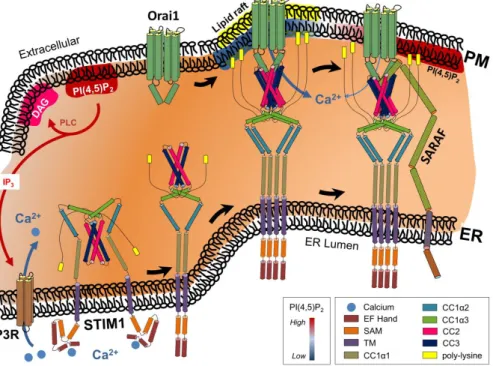
Figure 1. Receptor-induced activation of phospholipase C (PLC) generates inositol-triphosphate (IP3) and diacylglycerol (DAG) from phosphatidylinositol 4,5-bisphosphate (PI(4,5)P2). Binding of IP3 to its ER membrane-localized cognate receptor releases calcium (Ca2+) from ER into the cytosol.
Subsequent depletion of the ER Ca2+ store induces conformational changes in the ER Ca2+ sensor stromal interaction molecule-1 (STIM1). Binding of STIM1 to the Ca2+ release-activated Ca2+ channel Orai1 within phosphatidylinositol 4,5-bisphosphate (PI(4,5)P2)-poor PM microdomains activates the channel, resulting in Ca2+ from the extracellular milieu into the cytosol. Interaction of store-operated Ca2+ entry-associated regulatory factor (SARAF) with STIM1 induces transition of the STIM1-Orai1 complex towards PI(4,5)P2-enriched PM microdomains, resulting in attenuation of Ca2+ influx. See the text for more details. Although the presence of cholesterol-enriched lipid rafts is needed for proper STIM1-Orai1 functioning, the precise interaction of lipid rafts and PI(4,5)P2 microdomains as depicted still needs to be clarified. Additional abbreviations: ER, endoplasmic reticulum; PM, plasma membrane.
At the luminal site, interaction of Ca2+ with the negatively charged aspartates and glutamates induces a helix-loop-helix conformation of the EF hand. Agonist-induced ER discharge and subsequent dissociation of Ca2+ from the STIM EF hand destabilizes this conformation, resulting in activation of STIM1 [28]. Interestingly, while the STIM1 TM domains are not in close proximity in resting conditions, the presence of three TM domain- localized glycine residues (G223, G225, and G226) aid in the movement of the STIM1 TM domains towards each other upon Ca2+ release from the luminal EF hand [29–31].
Subsequent homomerization of STIM1 is mediated by CC2, CC3, and SHD, which is essential for coupling to and activation of Orai1 [32,33]. X-ray crystallographic structural data of the STIM1 cytosolic region revealed a dimeric assembly with an antiparallel arrangement of CC2 and CC3 and two inter-adjacent short α-helices [34]. From these crystal structures, it could be derived that the physiological V-shaped conformation of the critical STIM-Orai1-activating region (SOAR) [26] (also referred to as channel-activating domain (CAD) [35], Orai-activating small fragment (OASF) [33,36], or coiled-coil domain- containing region b9 (CCb9) [37]) dimer is established through coiled-coil interactions of one monomer (residues R429, W430, I433, and L436) and the other (residues T354, L351, W350, and L347). Of note, mutation-induced disruption of this SOAR binding interface
Figure 1. Receptor-induced activation of phospholipase C (PLC) generates inositol-triphosphate (IP3) and diacylglycerol (DAG) from phosphatidylinositol 4,5-bisphosphate (PI(4,5)P2). Binding of IP3to its ER membrane-localized cognate receptor releases calcium (Ca2+) from ER into the cytosol.
Subsequent depletion of the ER Ca2+store induces conformational changes in the ER Ca2+sensor stromal interaction molecule-1 (STIM1). Binding of STIM1 to the Ca2+release-activated Ca2+channel Orai1 within phosphatidylinositol 4,5-bisphosphate (PI(4,5)P2)-poor PM microdomains activates the channel, resulting in Ca2+from the extracellular milieu into the cytosol. Interaction of store-operated Ca2+entry-associated regulatory factor (SARAF) with STIM1 induces transition of the STIM1-Orai1 complex towards PI(4,5)P2-enriched PM microdomains, resulting in attenuation of Ca2+influx. See the text for more details. Although the presence of cholesterol-enriched lipid rafts is needed for proper STIM1-Orai1 functioning, the precise interaction of lipid rafts and PI(4,5)P2microdomains as depicted still needs to be clarified. Additional abbreviations: ER, endoplasmic reticulum; PM, plasma membrane.
Int. J. Mol. Sci.2021,22, 4703 4 of 26
In addition, STIM1 activity is regulated by a variety of post-translational modifications, including phosphorylation on serine [44] and tyrosine residues [45,46], and N-linked glycosylation at Asn131and Asn171located within the SAM domain [47].
Alternative splicing of STIM1 generates two splice variants: STIM1L, which is mainly expressed in skeletal muscle in human, and STIM1S, which has a more general expres- sion profile [48]. Although both STIM1S and STIM1L interact with and activate Orai1 and TRPC, STIM1L does so with a much higher affinity and activating potential com- pared to STIM1S [49]. Alternative splicing of STIM2 gives rise to three splice variants:
STIM2α/Stim2.2, which is able to interact with Orai1 and TRPC [44]; STIM2β/Stim2.1, a larger splicing variant with additional amino acids in the SOAR-region, which impairs its association with Orai and TRP channels [50,51]; and Stim2.3, a shorter variant lacking a lysine-rich region and the calmodulin (CaM)-binding domain at the cytosolic C-terminus (see further) [51]. ER store depletion-induced activation of STIM is modelled as a biphasic process [52]. Due to its lower affinity for Ca2+, STIM2 is more sensitive to changes in ER Ca2+levels. Thus, the initial minimal Ca2+ depletion from the ER—approximately below 400µM—is first sensed by STIM2 which—through triggering small and prolonged store-operated currents—corrects small fluctuations in ER Ca2+levels [53]. Extended ER Ca2+depletion activates STIM1, which initiates larger and transient store-operated cur- rents to replenish the ER [41,52,53]. Physical interaction between both isoforms allows fine-tuning of the Ca2+sensing process [44]. Moreover, heteromerization of STIM2.1 with STIM2.2 and STIM1 inhibits the function of the latter two, suggesting that variations in the expression ratio of these STIM variants might have a facilitative, redundant, or inhibitory effect on SOCE [50,51,54].
The three mammalian Orai subtypes—Orai1, -2, and -3—consist of a cytosolic N termi- nus followed by four transmembrane regions (TM1-4) and a cytosolic C-terminal extension (M4x). Crystallographic as well as functional studies revealed a hexameric arrangement of the Orai1 channel in which juxtaposition of the TM1 helices from all six Orai subunits form a highly selective Ca2+pore [55–58]. Although the M4x region was originally identified as a STIM binding site [24,35,59], other Orai cytosolic domains—in particular, the N-terminus and intracellular TM2-TM3 loop—have been implicated [35,60–63], suggesting that multi- ple cytosolic domains may contribute to the binding with STIM. As such, by acting as a bridge between hexameric Orai channels, STIM facilitates the formation of a lattice structure consisting of STIM dimers crosslinking a multitude of Orai channel hexamers, implying the ability of STIM to ultimately coordinate oligomerization and hence control the density of Orai channels. Interestingly, liquid-phase electron microscopy indicated that a fraction of the present Orai1 channels form STIM-independent distinct supra-molecular clusters, suggesting that supra-molecular ORAI1 clusters might fulfill an amplifying function for creating dense ORAI1 accumulations upon SOCE activation [64]. Although initial studies indicated that association of STIM with both the N-terminal TM1-extension (M1x) and M4x region of Orai is essential for initiating the gating process [35,62,65–67], the current model proposes the M4x region as the prime interaction site for STIM. Next, interaction of STIM1 SOAR with Orai M4x induces a conformational change, which acts as a coupling trigger between the TM4 and TM3 helices to propagate structural alterations through the TM2 helix and ultimately pore-opening through conformational alterations of the neighboring TM1 pore helix [68–71].
TRPC channels are ubiquitously expressed and display diverse roles in most cell types [16,72,73]. When activated by receptor stimulation, TRPC channels function in STIM1- dependent or -independent modes. STIM1-mediated gating of the channels depends on specific TRPC multimer formation and consequently on the complement of TRPC channels present in the cells. In fact, when expressed alone, certain TRPCs (TRPC1, -2, -4, and -5) interact with STIM1, whereas others (TRPC3, -6, and -7) do not [32]. However, although characterized by a lack of interaction with STIM1 when expressed alone, TRPC3 and -6 do so when present in complex with the STIM1-interacting channels TRPC1 and -4 and consequently function as STIM1-dependent channels [74]. As such, the cellular
composition as well as their ratio determines whether TRPC channels function in an STIM1- dependent or -independent manner. Interestingly, next to interaction with STIM1, parallel interaction of the STIM1/TRPC complex with Orai channels enhances the store dependence of TRPC [75–77]. Based on this, two mechanisms accounting for the observed channel interdependence are proposed: targeting of the channel to the PM from an intracellular pool and/or formation of STIM1/TRPC/Orai1 complexes at the ER–PM contact sites [78–80].
Direct gating of TRPC is regulated by STIM1 through interaction of positively charged lysines of STIM1 with negatively charged residues at the C-terminus of TRPC [35,81]. The precise mechanism of how such an interaction might result in channel opening is currently not entirely understood, although STIM1-mediated recruitment to PI(4,5)P2-rich domains has been proposed (see further).
Of note, spatiotemporal regulation of Orai1-STIM1 or STIM1-Orai1-TRPC1 ternary complex formation underlies the activation of different transcriptional programs driven by, for instance, NFAT (triggered by Orai1-STIM1+) or nuclear factor-kappaB (NF-κB) (triggered by STIM1-Orai1-TRPC1) [71].
Major regulation of STIM1 and STIM1-regulated Ca2+entry is mediated through an interplay between the EF-hand domain family member B (EFHB) [82] and the single-pass type I membrane protein SARAF, which is expressed in the ER membrane [83] as well as in the PM [84]. Under basal conditions, E-Syt-dependent interaction of SARAF with SOAR maintains STIM1 in an inactive state and prevents STIM1–Orai communication [83].
Upon store depletion, STIM1 dissociates from SARAF and associates with the cytosolic Ca2+sensor EFHB [82], forms STIM1-Orai1 complexes at ER–PM contact sites, and initiates SOCE [85]. Additionally, STIM1-independent interaction of SARAF with TRPC1 adjusts the Ca2+influx and protects against Ca2+overload [86]. By attenuating STIM1/Orai1-mediated Ca2+entry, SARAF contributes to so-called slow Ca2+-dependent inhibition (SCDI) of Ca2+
influx through a destabilization of STIM1/Orai1 complexes [83,85]. Interestingly, SARAF- induced SCDI depends on the STIM1 CTID domain whereas Ca2+/calmodulin-induced SCDI does not, suggesting that both proteins operate through different mechanisms to attenuate Ca2+influx [85,87]. On the contrary, STIM1-independent interaction of SARAF with Orai1 enhances Orai1-mediated Ca2+entry [88]. These observations suggest that SARAF-mediated regulation of Orai1 and TRPC1 channels depends on the presence of STIM1. In cells co-expressing STIM1, SARAF regulates inactivation of SOCE [83,85];
in STIM1-deficient cells, SARAF rather enhances the Orai1 activity while attenuating TRPC1 function [88].
Next to attenuating STIM1/Orai complex-activity, increased [Ca2+]iactivates cytosolic Ca2+clearance mechanisms [89] through which part of the Ca2+re-enters the ER through ER-localized SERCA channels, while most of the Ca2+is extruded out of the cytosol by PM-localized PMCA channels [90,91].
3. Ca2+and cAMP Signaling Cascades form a Highly Interconnected Network
Cyclic AMP (cAMP)—another highly abundant and versatile cellular second messenger—
is generated from ATP through the activity of adenylyl cyclases (ACs) and metabolized by phosphodiesterases (PDEs). SOCE-induced Ca2+influx directly modulates cAMP signaling cascades through Ca2+/calmodulin-mediated activation of Ca2+-dependent AC-5, -6, and -8 [92–97] (Figure2).
Interestingly, although highly responsive to SOCE-mediated Ca2+influx, these ACs are insensitive to alternative sources of cytosolic Ca2+increase—for example, Ca2+release from intracellular stores or those induced by diacylglycerol or arachidonate [93,96–98]—
suggesting a close functional apposition of ACs with SOCE channels. In fact, even in the absence of SOCE, a close interaction of AC8 with Orai1 has been shown, suggesting stable complex formation between the AC and its regulator [99]. Of note, although direct interaction between AC8 and TRPC1 has not been evidenced yet, colocalization of AC8, TRPC1, and STIM1 was shown by TIRF analysis [100].
Int. J. Mol. Sci.2021,22, 4703 6 of 26
Int. J. Mol. Sci. 2021, 22, x FOR PEER REVIEW 6 of 26
ER-localized SERCA channels, while most of the Ca2+ is extruded out of the cytosol by PM-localized PMCA channels [90,91].
3. Ca2+ and cAMP Signaling Cascades form a Highly Interconnected Network
Cyclic AMP (cAMP)—another highly abundant and versatile cellular second messenger—is generated from ATP through the activity of adenylyl cyclases (ACs) and metabolized by phosphodiesterases (PDEs). SOCE-induced Ca2+ influx directly modulates cAMP signaling cascades through Ca2+/calmodulin-mediated activation of Ca2+- dependent AC-5, -6, and -8 [92–97] (Figure 2).
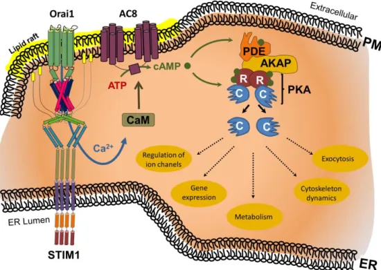
Figure 2. ER store depletion-induced Ca2+ influx triggers Ca2+/calmodulin (CaM)-mediated activation of adenylcyclase 8 (AC8) within lipid rafts. The resulting cAMP binds to the regulatory unit (R) of protein kinase A (PKA), resulting in the release of the PKA catalytic unit (C), and enabling regulation of ion channels, gene expression, metabolism, cytoskeleton dynamics, or exocytosis.
Excess cAMP is broken down by phosphodiesterases (PDE). The range and selectivity of the cAMP- mediated biological effect is regulated by A-kinase anchoring protein (AKAP) through linkage of PKA and PDEs. Additional abbreviations: ER, endoplasmic reticulum; PM, plasma membrane.
Interestingly, although highly responsive to SOCE-mediated Ca2+ influx, these ACs are insensitive to alternative sources of cytosolic Ca2+ increase—for example, Ca2+ release from intracellular stores or those induced by diacylglycerol or arachidonate [93,96–98]—
suggesting a close functional apposition of ACs with SOCE channels. In fact, even in the absence of SOCE, a close interaction of AC8 with Orai1 has been shown, suggesting stable complex formation between the AC and its regulator [99]. Of note, although direct interaction between AC8 and TRPC1 has not been evidenced yet, colocalization of AC8, TRPC1, and STIM1 was shown by TIRF analysis [100].
The efficiency and specificity of cAMP signaling is provided through A-kinase (PKA) anchoring protein (AKAP)-mediated multiprotein complex formation of cAMP signaling components. As such, AKAPs anchor the cAMP-regulated PKA in the vicinity of its substrates and ensure preferential phosphorylation of selected targets [101,102]. Next to
Figure 2.ER store depletion-induced Ca2+influx triggers Ca2+/calmodulin (CaM)-mediated activa- tion of adenylcyclase 8 (AC8) within lipid rafts. The resulting cAMP binds to the regulatory unit (R) of protein kinase A (PKA), resulting in the release of the PKA catalytic unit (C), and enabling regulation of ion channels, gene expression, metabolism, cytoskeleton dynamics, or exocytosis. Excess cAMP is broken down by phosphodiesterases (PDE). The range and selectivity of the cAMP-mediated biological effect is regulated by A-kinase anchoring protein (AKAP) through linkage of PKA and PDEs. Additional abbreviations: ER, endoplasmic reticulum; PM, plasma membrane.
The efficiency and specificity of cAMP signaling is provided through A-kinase (PKA) anchoring protein (AKAP)-mediated multiprotein complex formation of cAMP signal- ing components. As such, AKAPs anchor the cAMP-regulated PKA in the vicinity of its substrates and ensure preferential phosphorylation of selected targets [101,102]. Next to PKA, AKAPs interact with regulatory proteins, including PDEs, protein kinase C (PKC), calcineurin, as well as all Ca2+-sensitive ACs, ultimately resulting in the establishment of cAMP microdomains [103]. For example, cytoplasmic retention of nuclear factor of acti- vated T cells (NFATs)—due to extensive phosphorylation masking its nuclear localization sequence—is reversed by calcineurin-mediated dephosphorylation, enabling NFAT nuclear translocation and transcriptional induction of its responsive genes [104,105]. Upon store depletion, Ca2+nanodomain formation near open store-operated Orai1 initiates AKAP79- mediated complex formation of calcineurin with Orai-tethered calmodulin, thus linking Ca2+signaling with NFAT-mediated transcriptional regulation [106]. Of note, whereas knockdown of STIM2 expression had relatively little effect on Orai1/STIM1 clustering and subsequent Ca2+influx, it significantly impaired NFAT1 activation and assembly of Orai1 with AKAP79, suggesting an important role for STIM2 in coupling Orai1-mediated influx to NFAT1 activation [107].
Obviously, the close interconnected character of both Ca2+- and cAMP-mediated signaling cascades provides multiple opportunities to mutually regulate the activity and outcome of each other. For example, Ca2+-mediated regulation of cAMP signaling at the level of ACs depends on AKAP-mediated compartmentalization of specific ACs in close proximity of Ca2+transport proteins. In fact, the effect of Ca2+on cAMP signaling is AC dependent as increased [Ca2+]iresults in activation of AC1, -3, and -8 but inhibition of AC5 and -6 [94,99]. Moreover, AC3 activity is indirectly attenuated through Ca2+/calmodulin protein kinase (CaMKII)-dependent phosphorylation of Ca2+/calmodulin [108]. In human
melanocytes, binding of a-melanocyte-stimulating hormone (aMSH)—one of the major physiological determinants of melanogenesis—to G-coupled receptor melanocortin-1 re- ceptor (MC1R) depletes Ca2+ER stores and recruits STIM1 to ER–PM junctions. Next to Orai-mediated SOCE activation, STIM1 equally interacts with and activates AC6 at the PM, resulting in sustained elevated cAMP levels required to signal the induction of pigmentation genes [109]. Similarly, a crucial role of SOCE in fatty acid metabolism is evidenced by pathological amounts of lipid droplets (LDs) in skeletal and heart muscles of ORAI1- or STIM1/ STIM2-deficient mice or in isolated fibroblasts from human patients with loss-of-function mutations in STIM1 or ORAI1. In fact, SOCE regulates the expression of the neutral lipases hormone-sensitive lipase (HSL) and adipose triglyceride lipase as well as the cAMP-dependent activation of HSL and thereby controls lipolysis [110].
Of note, store-operated cAMP signaling (SOcAMPS) refers to the regulation of AC3 through ER Ca2+depletion and subsequent clustering of STIM1 at PM Ca2+microdomains [111].
Interestingly, this type of regulation is independent of Orai1, or elevation of cytosolic Ca2+
and could involve other ACs in a cell type-dependent manner [112,113]. For example, exposure of glucagon-releasing alpha cells to epinephrine stimulates sub-plasmalemmal translocation of STIM1 in a cAMP-dependent manner without formation of Orai1 protein clusters or SOCE, suggesting the involvement of additional factors in the formation of functional Orai1 channels and activation of Ca2+entry [114]. Similarly, synaptic plastic- ity in excitatory neurons is mediated by cAMP-mediated migration of STIM2—but not STIM1—and the AMPA receptor (AMPAR) subunit GluA1 to ER–PM contact sites in den- dritic spines. At these junctions, STIM2 promotes GluA1 phosphorylation though coupling of PKA to AMPAR in a SOCE-independent manner [115].
Next to modulating the activity of ACs, Ca2+controls the extent of cAMP signaling through regulation of PDE activity. For example, while the affinity of Ca2+/calmodulin for PDEs is controlled by calmodulin kinase (CaMKII)-dependent phosphorylation [116], functional interaction of Ca2+/calmodulin with PDEs attenuates their auto-inhibition and increases their Vmax [117–119]. Moreover, analysis of SOCE-induced modulations on PDE1 isoform activity suggests that fluctuations in [Ca2+]imight facilitate the formation of intracellular cAMP domains [120].
In parallel, alterations in cAMP generation and subsequent changes in PKA activity equally regulate the magnitude and extent of [Ca2+]iat multiple levels. For example, PKA- mediated phosphorylation of IP3Rs at multiple sites affects its activity in an isoform- and cell type-specific manner. In fact, whereas the output of IP3R1 is enhanced through PKA- mediated phosphorylation on Ser1589and Ser1755[121,122], neuronal IP3Rs are equally activated by phosphorylation of Ser1755alone [123]. Moreover, PKA-mediated phosphoryla- tion of IP3R2 Ser937increases its activity in exocrine cells [124]. Interestingly, at submaximal IP3concentrations, PKA-mediated phosphorylation enhances the open probability of IP3R1, suggesting a cAMP/PKA-mediated increased affinity of the IP3Rs for IP3[121]. Combined with the observation that the affinity of IP3R1 for its inhibitor IRBIT is reduced through PKA-mediated phosphorylation [125], this indicates that PKA might regulate IP3R activity through sequential alterations in the affinity of IP3Rs for IP3 and IRBIT. Furthermore, AKAP79-mediated positioning of PKA in the proximity of Orai1 results in Orai1 Ser34 phosphorylation and subsequent Ca2+-dependent inactivation (CDI) [126]. While Orai1 channels exist as store-operated CRAC channels and store-independent arachidonic acid- activated ARC channels, CRAC channels are activated by ER membrane-localized STIM1 proteins whereas ARC channels are activated by a small PM-associated pool of STIM1 [127].
Interestingly, selective activation of ARC channels requires PKA-mediated phosphoryla- tion of STIM1 Thr389, resulting in a conformational change of its SOAR region [127]. As such, PKA-mediated structural changes underlie the selective activation of STIM1-induced CRAC or ARC channels and determine the specific stimulation of these two functionally distinct Ca2+entry pathways [128]. The cAMP/PKA pathway equally modulates [Ca2+]i
clearance. In fact, PKA-mediated phosphorylation of the SERCA inhibitor phospholam- ban (PLN) at Ser16dissociates the PLN/SERCA complex and results in SERCA-mediated
Int. J. Mol. Sci.2021,22, 4703 8 of 26
clearance of [Ca2+]i into the ER [129]. Additionally, while calmodulin acts as the main activator of PMCA, PKA-mediated phosphorylation enhances the activity of PMCA in a Ca2+-dependent manner, suggesting that it might work through a Ca2+-dependent AC- mediated mechanism [130].
4. Compartmentalization of Ca2+/cAMP Signaling in Membrane Nanodomains at ER–PM Contact Sites
ER–PM contact sites are characterized by highly dynamic modulations of its lipid environment with a huge regulatory function on its resident proteins (Figure3).
Int. J. Mol. Sci. 2021, 22, x FOR PEER REVIEW 9 of 26
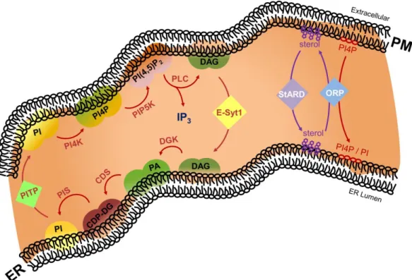
Figure 3. In the phosphoinositide cycle, phosphatidylinositol (PI) is sequentially converted to phosphatidylinositol 4-phosphate (PI4P) and phosphatidylinositol 4,5-bisphosphate (PI(4,5)P2) by PI kinase activities at the PM. Upon physiological triggers, phospholipase C (PLC) hydrolyzes PI(4,5)P2, generating the second messenger molecules diacylglycerol (DAG) and inositol trisphosphate (IP3). Extended synaptotagmin 1 (E-Syt1)-mediated non-vesicular transport of DAG from PM allows conversion of DAG into PI, PI4P, and PI(4,5)P2 at the ER [131]. PI4P species are transferred from the PM to the ER by oxysterol-binding protein-related protein (ORP). Cholesterol is cycled between the PM and the ER through the sequential actions of Star-related lipid transfer protein/Gram domain-containing protein (StARD/GRAMd) and ORP proteins. Additional abbreviations: ER, endoplasmic reticulum; PM, plasma membrane.
In fact, through spatiotemporal fluctuations of specific lipid species, ER–PM contact sites have a high degree of plasticity, which is a critical denominator of how they respond to physiological stimuli.
Through the so-called phosphoinositide cycle, phosphatidylinositol (PI) species in the PM are phosphorylated by phosphatidylinositol 4-kinase (PI4K) to generate phosphatidylinositol 4-phosphate (PI4P), which is subsequently phosphorylated by phosphatidylinositol 4-phophate 5-kinase (PIP5K) to generate phosphatidylinositol 4,5- bisphosphate (PI(4,5)P
2). The latter is subsequently hydrolyzed to diacylglycerol (DAG) and inositol 1,4,5-trisphosphate (IP
3) by phospholipase C (PLC). Next, DAG activates PKC whereas IP
3binds its cognate receptor in the ER membrane and activates Ca
2+release.
Finally, upon E-Syt-mediated non-vesicular transport from PM to ER, DAG is converted to PI through the sequential action of diacylglycerol kinase (DGK), CDP-diacylglycerol synthase (CDS), and PI synthase (PIS). Finally, non-vesicular phosphatidylinositol transfer protein (PITP)-mediated transfer of PI from the ER to PM closes the circle, allowing additional rounds through the PI cycle. As such, the ER–PM contact site is an important regulator of Ca
2+signaling through spatiotemporal modulation of the levels of specific phosphoinositide lipid species.
In addition, the membranes that constitute the MCS are linked through protein–
protein and protein–lipid interactions. MCS tethering proteins can be roughly subdivided into (i) structural proteins, tethers that hold the two organelles together; (ii) functional
Figure 3. In the phosphoinositide cycle, phosphatidylinositol (PI) is sequentially converted to phosphatidylinositol 4- phosphate (PI4P) and phosphatidylinositol 4,5-bisphosphate (PI(4,5)P2) by PI kinase activities at the PM. Upon physiological triggers, phospholipase C (PLC) hydrolyzes PI(4,5)P2, generating the second messenger molecules diacylglycerol (DAG) and inositol trisphosphate (IP3). Extended synaptotagmin 1 (E-Syt1)-mediated non-vesicular transport of DAG from PM allows conversion of DAG into PI, PI4P, and PI(4,5)P2 at the ER [131]. PI4P species are transferred from the PM to the ER by oxysterol-binding protein-related protein (ORP). Cholesterol is cycled between the PM and the ER through the sequential actions of Star-related lipid transfer protein/Gram domain-containing protein (StARD/GRAMd) and ORP proteins. Additional abbreviations: ER, endoplasmic reticulum; PM, plasma membrane.In fact, through spatiotemporal fluctuations of specific lipid species, ER–PM contact sites have a high degree of plasticity, which is a critical denominator of how they respond to physiological stimuli.
Through the so-called phosphoinositide cycle, phosphatidylinositol (PI) species in the PM are phosphorylated by phosphatidylinositol 4-kinase (PI4K) to generate phosphatidyli- nositol 4-phosphate (PI4P), which is subsequently phosphorylated by phosphatidyli- nositol 4-phophate 5-kinase (PIP5K) to generate phosphatidylinositol 4,5-bisphosphate (PI(4,5)P2). The latter is subsequently hydrolyzed to diacylglycerol (DAG) and inositol 1,4,5-trisphosphate (IP3) by phospholipase C (PLC). Next, DAG activates PKC whereas IP3
binds its cognate receptor in the ER membrane and activates Ca2+release. Finally, upon E-Syt-mediated non-vesicular transport from PM to ER, DAG is converted to PI through the sequential action of diacylglycerol kinase (DGK), CDP-diacylglycerol synthase (CDS), and PI synthase (PIS). Finally, non-vesicular phosphatidylinositol transfer protein (PITP)- mediated transfer of PI from the ER to PM closes the circle, allowing additional rounds
through the PI cycle. As such, the ER–PM contact site is an important regulator of Ca2+
signaling through spatiotemporal modulation of the levels of specific phosphoinositide lipid species.
In addition, the membranes that constitute the MCS are linked through protein–protein and protein–lipid interactions. MCS tethering proteins can be roughly subdivided into (i) structural proteins, tethers that hold the two organelles together; (ii) functional proteins facilitating the initially intended function of the MCS; (iii) regulatory proteins modulating the extent and function of the MCS; and (iv) recruitment/sorting proteins, which define the MCS-specific proteome and lipidome. Of note, this subdivision is most probably artificial as multiple proteins might in fact belong to multiple classes [132,133].
Extended-synaptotagmins (E-Syts) are ER-resident proteins characterized by a Synap- totagmin-like, mitochondrial and lipid-binding protein (SMP) domain. [134]. In mammalian cells, three E-Syts (E-Syt1, -2, and -3) are described [135]. In fact, through dimerization of their SMP domains, E-Syts function as homo- and heterodimers, which tether the ER to the PM through interaction with PM phospholipids, in particular PI(4,5)P2 [136,137].
Interestingly, PM interaction of E-Syt1 is enhanced by Ca2+, suggesting that [Ca2+]iregu- lates the fraction of E-Syt1 present throughout the ER or concentrated at ER–PM contact sites [136,138,139]. Thus, considering heteromeric complex formation, both changes in [Ca2+]iand PM PI(4,5)P2 levels regulate the formation/maintenance of E-Syt-dependent ER–PM contact sites [136,139,140]. Of note, although the rather high [Ca2+]iconcentrations—
low micromolar Ca2+range—required for E-Syt1 recruitment to the PM are established through activation of extracellular Ca2+influx, including SOCE, E-Syts-dependent contacts are not required for SOCE itself [136].
At the MCS, E-Syts are essential for non-vesicular transport of DAG from PM to ER and for PI(4,5)P2re-synthesis during repetitive rounds of PLC signaling [138]. However, interaction of E-Syts with other lipids was evidenced, suggesting their involvement in the transfer of as yet unidentified phospholipids [131,141]. Non-vesicular exchange of PI for phosphatidic acid (PA) between ER and PM is maintained by Nir2. PLC activation induces transfer of Nir2 into MCSs, where it interacts with vesicle-associated membrane protein (VAMP)-associated protein (VAP)—a tail-anchored ER membrane protein—and PA mi- crodomains in the PM. Functional interaction between E-Syt1 and Nir2 has been suggested as E-Syt1 facilitates Nir2 recruitment and function at ER–PM contact sites [138]. Addition- ally, interaction of F-actin with Nir2 stabilizes the resulting ER–PM contact sites [142].
Next to agonist-induced modulations of the phosphoinositide cycle, the ER–PM contact site lipidome is equally characterized by alterations in PS and sterol levels. For example, VAP recruits ORP3 and ORP6 to ER–PM contact sites [143] which—together with the ER-embedded ORP5 and ORP8—mediate exchange of ER-derived PS and sterols for PM-localized PI4P [143–147]. Subsequently, PI4P is hydrolyzed by the MCS-residing Sac1 phosphatase [148]. Additionally, StARD/GRAM proteins extract accessible cholesterol from the cytoplasmic leaflet of the PM and transfer it to the ER [149]. Together, these lipidome alterations have a huge regulatory effect on the flux through the PI cycle. For example, ORP-induced lipid environment modulations through PS and sterol enrichments in the PM synergistically activate PIP5K and subsequent PI(4,5)P2synthesis [143,150].
Through assembly into multimeric complexes and formation of linear filaments or other higher-order structures, septins operate as diffusion barriers and intracellular scaf- folds during various cellular processes, including SOCE [151]. Loss of septin filaments dSEPT1 and dSEPT4 in Drosophila results in loss of the diffusion barrier and impaired dOrai activation by dSTIM, suggesting their role as positive regulators of SOCE [152,153].
Similarly, in human cells, SEPT4 promotes interaction between STIM1 and Orai1 through limiting the lateral mobility of Orai1 in the PM [154]. On the contrary, loss of dSEPT7 in Drosophila enhanced the intensity of dSTIM and resulting dSTIM-dOrai clusters near the ER–PM region [153]. Similar results were observed in SEPT7-knockdown human neurons differentiated from neural progenitor cells [155]. These observations suggest that—through
Int. J. Mol. Sci.2021,22, 4703 10 of 26
uncoupling septin heteromers from ER-PM junctions—loss of SEPT7 influences the consti- tutive activation of Orai channels, thus allowing the STIM interaction with Orai [153,155].
The family of anoctamins—ER–PM contact site tethers homologous to yeast Ist2 [156,157]—
consists of 10 members. Although some have been reported to function as Ca2+-activated Cl−channels (ANO1 and ANO2) [158–161] or lipid scramblase and Cl-channel (ANO6) [162–164], little is known about their cellular functions. Recently, ANO8 was reported to regulate multiple steps of ER depletion-induced Ca2+signaling, including the formation of STIM1 dimers and puncta, STIM1–Orai1 interaction, SOCE, and SCDI [165]. Importantly, a crucial role for ANO8 in assembling the core Ca2+signaling complex into PI(4,5)P2-rich domains at the ER–PM contact sites was evidenced [165].
As part of the cytoskeleton, microtubules—tubulin polymers—provide structure and shape to eukaryotic cells. Plus-ends of microtubules associate with end-binding proteins (EB1, -2, and -3), which in turn interact with STIM1 [166]. Complex formation between EB1/EB3 and STIM1 mediates ER movement and prevents excessive SOCE activation through sequestering STIM1 in the microtubules [167–170].
5. Regulatory Interplay between ER–PM Contact Site Lipid Species and Residing Proteins
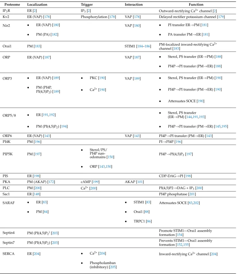
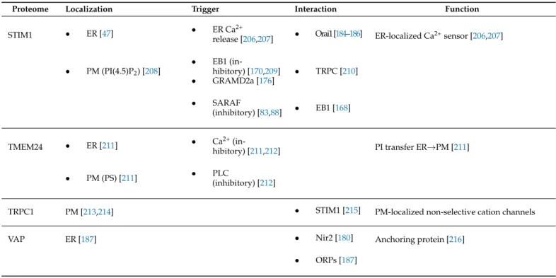
5.1. ER–PM Contact Site-Residing Proteins Modulate Their Surrounding Lipid Environment ER–PM contact sites are characterized by a dynamic spatiotemporal interplay of its residing proteins and its lipid microenvironment [171]. Not only are proteins attracted to and/or remain inside the ER–PM contact site based on their specific interactions with selected lipid microdomains, the lipid environment of ER–PM contact sites is equally modulated by ER–PM contact site-residing proteins (Table1).
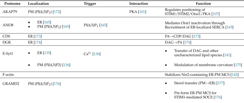
Table 1.Overview of ER–PM contact site-residing proteins. Abbreviations: AKAP79, A-kinase Anchor Protein 79; ANO8, Anoctamine-8; CDS, CDP-Diacylglycerol Synthase; DGK, Diacylglycerol Kinase; E-Syt1, Extended Synaptotagmin-1;
GRAMd2, GRAM domain-containing protein 2; IP3R, Inositol Triphosphate Receptor; Kv2, Delayed rectifier potassium channel; Nir2, Pyk2 N-terminal Domain-Interacting Receptor 2; Orai1, Calcium Release-Activated Calcium Channel Protein 1; ORP, Oxysterol-Binding Protein-Related Protein; PI4K, Phosphatidylinositol 4-kinase; PIP5K, Phosphatidyli- nositol 4-Phosphate 5-Kinase; Phosphatidylinositol Synthase; PKA, Protein Kinase A; PLC, Phospholipase C; Sac1, Phosphatidylinositol-3-Phosphatase; SARAF, Store-Operated Calcium Entry-Associated Regulatory Factor; SERCA, Sar- coplasmic/Endoplasmic Reticulum Calcium ATPase 3; STIM1, Stromal Interaction Molecule 1; TMEM24, Transmembrane Protein 24; TRPC1, Transient Receptor Potential Channel 1; VAP, Vesicle-Associated Membrane Protein-Associated Protein.
Proteome Localization Trigger Interaction Function
AKAP79 PM (PI(4,5)P2) [172] PKA [101] Regulates positioning of
STIM1/STIM2/Orai1/PKA [107]
ANO8 • ER [165]
• PM (PI(4,5)P2) [165] PI(4,5)P2[165] Mediates Orai1 inactivation through Recruitment of ER-localized SERCA [165]
CDS ER [173] PA→CDP-DAG [173]
DGK ER [174] DAG→PA [174]
E-Syt1 • ER [135] Ca2+[136] • Transfer of DAG and other
uncharacterized lipid species [141]
• PM (PI(4,5)P2) [136] • Modulation of membrane curvature [175]
F-actin Stabilizes Nir2-containing ER-PM MCS [142]
GRAMD2 PM (PI(4,5)P2) [176] • Sterol transfer (PM→ER) [177]
• Pre-form ER-PM MCS for STIM1-mediated SOCE [176]
Table 1.Cont.
Proteome Localization Trigger Interaction Function
IP3R ER [2] IP3[2] Outward-rectifying Ca2+channel [2]
Kv2 ER (VAP) [178] Phosphorylation [178] VAP [178] Delayed rectifier potassium channel [179]
Nir2 • ER (VAP) [180] VAP [180] • PI transfer ER→PM [181]
• PM (PA) [182] • PA transfer PM→ER [181]
Orai1 PM [183] STIM1 [184–186] PM-localized inward-rectifying Ca2+
channel [183]
ORP ER (VAP) [187] VAP [187] • Sterol, PS transfer (ER→PM) [188]
• PI4P→PI transfer (PM→ER) [188]
ORP3 • ER (VAP) [189] • PKC [190] VAP [189] • Sterol, PS transfer (ER→PM) [190]
• PM (PI4P,
PI(4,5)P2) [189] • Ca2+[190] • PI4P→PI transfer (PM→ER) [190]
• Attenuates SOCE [190]
ORP5/8 • ER [191,192] • Sterol, PS transfer
(ER→PM) [144,191,193]
• PM (PI(4,5)P2) [194] • PI4P→PI transfer (PM→ER) [145,195]
ORP6 ER (VAP) [143] VAP [143] PI4P→PI transfer (PM→ER) [143]
PI4K PM [196] PI→PI4P [196]
PIP5K PM [197]
• Sterol/PS/
PI4P nan-
odomains [150] PI4P→PI(4,5)P2[197]
• ORP [143,150]
PIS ER [198] CDP-DAG→PI [198]
PKA PM (AKAP) [172] cAMP [199] AKAP [101]
PLC PM [200] Ca2+[200] PI(4,5)P2→DAG + IP3[200]
Sac1 ER [148] PI4P phosphatase [201]
SARAF • ER [83] • STIM1 [83] Attenuates SOCE [83,202]
• PM [84] • Orai1 [88]
• TRPC1 [86]
Septin4 PM (PI(4,5)P2)[203] Promote STIM1—Orai1 assembly
formation [154]
Septin7 PM (PI(4,5)P2) [203] Prevents STIM1—Orai1 assembly
formation [152,155]
SERCA ER [204] • Ca2+[204] Inward-rectifying Ca2+channel [204]
• Phospholamban (inhibitory) [205]
Int. J. Mol. Sci.2021,22, 4703 12 of 26
Table 1.Cont.
Proteome Localization Trigger Interaction Function
STIM1 • ER [47] • ER Ca2+
release [206,207] • Orai1[184–186] ER-localized Ca2+sensor [206,207]
• PM (PI(4.5)P2) [208]
• EB1 (in-
hibitory) [170,209]
• GRAMD2a [176]
• TRPC [210]
• SARAF
(inhibitory) [83,88] • EB1 [168]
TMEM24 • ER [211] • Ca2+(in-
hibitory) [211,212] PI transfer ER→PM [211]
• PM (PS) [211] • PLC
(inhibitory) [212]
TRPC1 PM [213,214] • STIM1 [215] PM-localized non-selective cation channels
VAP ER [187] • Nir2 [180] Anchoring protein [216]
• ORPs [187]
For example, upon association of E-Syts with the PM, micromolar [Ca2+]iconcentra- tion stimulates E-Syts-mediated bidirectional transport of glycerolipids—including DAG—
between ER and PM driven by the lipid concentration gradient in the membranes [141,217].
As such, E-Syts might be part of a homeostatic response needed to reset the PM lipid composition to normal levels after acute perturbations by transferring excess DAG from the PM to the ER for its metabolic recycling [218]. Additionally, ORP5 and -8 are recruited to ER–PM contact sites through interaction of their pleckstrin homology domain with PI(4,5)P2 pools in the PM, where it subsequently modulates PI(4,5)P2 levels [194]. Addi- tionally, association of ORP3 with the PM is determined by both PI(4,5)P2 and PI4P. Upon activation, ORP3 extracts PI4P and PA from the PM while increasing its cholesterol and PS levels [150,190]. Similarly, the recruitment of ORP6 to ER–PM contact sites is involved in the turnover of PI4P [143,150]. Subsequently, clusters of PS and PI4P activate PIP5K, suggesting that ORP/Osh proteins create a PM lipid environment favoring PIP5K activity and PI(4,5)P2 synthesis [147,150]. Similarly, StARD/GRAM-induced cholesterol extraction from the PM might influence the organization and biophysical properties of the PM and thus the dynamic events taking place at the PM [131].
5.2. ER–PM Contact Site-Residing Lipid Species Modulate Protein Activity
Membrane composition is crucial in defining ion channel structure and function, either through specific interactions between lipids and proteins or nonspecific through changes in membrane physicochemical properties—thickness, fluidity, and curvature—that affect ion channel dynamics. Lipids directly interacting with membrane-embedded proteins are defined as annular lipids, those lipids in the first ring surrounding the protein, and non-annular lipids, lipids present in clefts or at the protein subunit interface. For example, molecular dynamics (MD) simulations emphasize the crucial influence of membrane struc- ture and thickness on the function of membrane channels and provide important insights into the influence of the lipid microenvironment on protein function (see [219–221] for recent reviews). In fact, surrounding lipids directly affect stimulus-induced conformational changes necessary for efficient gating of channels while providing a specific/selective medium for small molecules to approach and interact with them.
MD simulations capturing cholesterol-induced modulation of protein structure and dynamics indicated a cholesterol-mediated effect through both specific binding interactions or through alterations in membrane bulk properties as well as through the formation of lipid domains with saturated phospholipids and sphingolipids [221–224].
Initial studies indicated that cholesterol depletion impairs interaction between STIM1 and both Orai1 and TRPC1 upon emptying of intracellular Ca2+stores, suggesting their association with lipid raft domains [225]. Moreover, lipid raft domains were shown to be involved in the activation but not the maintenance of SOCE, probably due to the support of the formation of Ca2+signaling complexes involving STIM1, Orai1, and TRPCs [226].
In fact, TRPC channels are characterized by a conserved Cav-1 binding domain allowing Cav-1-mediated targeting of TRPC proteins into lipid raft/caveolae domains [227]. Silenc- ing of Cav-1 results in attenuation of SOCE and misallocation of TRPC1 and TRPC4 in airway smooth muscle and endothelial cells, respectively [228,229]. Interestingly, although TRPC1 interacts with Cav-1 [227–231], once inside the raft region, TRPC1 is regulated by STIM1 [232,233]. In fact, interaction of TRPC1 with STIM1 results in its integration into lipid raft domains, where it functions as a store-operated channel; in the absence of STIM1 interaction, TRPC1 functions as an agonist-activated channel outside rafts [232].
Interestingly, SOCE attenuates TRPC1-Cav1 complex formation in an STIM1-dependent manner [234], allowing interaction between TRPC1 and STIM1 in the lipid rafts [233]. Next to TRPC proteins, Orai1 also possesses a Cav-1-binding domain allowing Orai1-Cav-1 com- plex formation [235,236], suggesting a role for Cav-1 in the proper targeting of Orai1 into the lipid raft/caveolae regions [237]. Interestingly, the absence of Cav-1 results in impaired ER depletion-induced interaction of STIM1 with TRPC1 and microdomain localization in salivary gland cells of Cav-1-negative mice, although the interaction between STIM1 and Orai1 was unaffected [230]. Of note, interaction of Cav-1 with the STIM1-Orai1 complex selectively regulates the SOCE-induced activation of downstream NFAT- or c-fos-mediated transcriptional programs. In fact, phosphorylation of Cav-1 Tyr14 impairs c-fos activa- tion without impacting the NFAT pathway or Orai1 activity indicating that—potentially through selective targeting towards specific PM microdomains—structurally distinct re- gions of Cav-1 selectively regulate the ability of local Ca2+to activate distinct downstream transcriptional cascades [238].
In addition, cyclodextrin-induced cholesterol depletion attenuates SOCE in a wide variety of cells [226,233,239] accompanied with the dissociation of STIM1, Orai1, and TRPC1 [240]. Interestingly, whereas STIM1 puncta formation is absent in cyclodextrin- treated cells [233], overexpression of Orai1 and STIM1 is able to overrule the cyclodextrin- induced cholesterol depletion effect on SOCE [239]. However, lipid rafts are only essential during STIM1-Orai1-TRPC1 complex formation during initial activation of SOCE; when the complex is assembled, cyclodextrin-induced cholesterol depletion has no effect on SOCE nor on the complex itself [226]. However, recent studies suggest that the amount of cholesterol in the PM is monitored by the Orai1 amino terminus, which could modulate its activity during SOCE [241]. Whereas, under basal cholesterol conditions, most Orai1 channels are restricted in a confined space, reducing PM cholesterol induces Orai1 internal- ization and affects the lateral movement of Orai1, resulting in unobstructed diffusion in the plane of the PM. Since overexpression of Cav-1 during cholesterol depletion maintained Orai1 into a confined area and movement, this suggests a direct cholesterol-mediated regu- latory effect on Orai1 localization and compartmentalization and SOCE [242]. Moreover, STIM1 possesses a cholesterol-binding motif inside the SOAR domain. Reduction of PM cholesterol levels detaches SOAR from PM while enhancing its association to Orai1 [243].
Anionic lipids, such as PI species, are asymmetrically distributed over the inner and outer leaflets of the PM by active transporters maintaining a higher concentration of these lipid species in the inner leaflet, a process of high physiological importance [244]. Due to its highly negatively charged nature, MD simulations evidenced, for example, PIP2 clustering around integral membrane proteins and its capacity to regulate their activity through interaction with both their transmembrane domain and their cytosolic linker/domain
Int. J. Mol. Sci.2021,22, 4703 14 of 26
or juxtamembrane domain [219–221]. The lysine-rich polybasic motif located at the C- terminus of STIM1 allows interaction of STIM1 with anionic phospholipids, such as PIP2 and PIP3, in the PM [40,245,246]. Interestingly, whereas efficient binding of STIM1 to PIP2 requires tetramerization of its lysine-rich domain, association of STIM2 with PIP2 in contrast relies on dimerization of its lysine-rich domain, resulting in enhanced affinity for PIP2 and a lower activation threshold of STIM2 [247]. Upon deletion of this polybasic motif, ER depletion-induced translocation of STIM1 to the PM is impaired although STIM1 is still able to form oligomers in puncta [41]. Moreover, phosphatidyl kinase inhibitor-induced depletion of PI4P results in decreased Orai1-mediated Ca2+entry although STIM1 puncta formation is preserved [248]. In fact, STIM1 first associates with PM-localized PIP2 and PIP3 pools in raft domains before its actual interaction with Orai1 [43], suggesting that cholesterol indirectly orchestrates proper STIM1-mediated targeting of SOCE components into lipid rafts through preservation of the required phosphoinositide pools. In addition, an N-terminal polybasic arginine-rich motif of Orai1 is involved in directing the channel to distinct PIP2 pools [249]. Interestingly, transient shuttling between PI(4,5)P2-rich and -poor domains is an important regulator of Ca2+transport proteins and Ca2+signaling. In fact, ER store depletion induces STIM1/Orai1 complex formation in PI(4,5)P2-poor regions. Due to the lack of PI(4,5)P2, SARAF is unable to interact with STIM1, thus allowing maximal Ca2+
influx. Next, the STIM1-Orai1 complex migrates into PI(4,5)P2-rich domains (or PI(4,5)P2 translocates to the STIM1-Orai1 complex), allowing SARAF to interact with STIM1 followed by SCDI-mediated Ca2+influx attenuation [202]. Thus, rather than going through cycles of PI(4,5)P2 hydrolysis and re-synthesis, STIM1/SARAF-mediated regulation of Ca2+influx through Orai1 operates by translocation between PI(4,5)P2 domains.
Additionally, TRPC channels are regulated by PI(4,5)P2 [250–253]. As such, although the STIM1-mediated gating of TRPC is not entirely understood, STIM1-mediated targeting of TRPC channels into PI(4,5)P2-rich domains could be essential for keeping the TRPC channels in an active state [254].
Ca2+-regulated adenylyl cyclases—AC1, -5, -6, and -8—are targeted to plasma mem- brane rafts, whereas Ca2+-independent adenylyl cyclases—AC2 and -7—are not [255].
Cyclodextrin-induced cholesterol depletion destroys these interactions and disrupts the regulation of the raft-localized ACs by SOCE, indicating that the presence of these com- plexes in raft domains is essential for their assembly and functioning [28]. The underlying mechanism targeting ACs to rafts is not completely understood. Although protein–protein interactions with the cytosolic region of AC5 and AC6 seemingly underlie their residence in rafts [47,48], the involvement of additional—weaker—protein–lipid interactions has been suggested [256]. In fact, a dynamic interaction between Cav-1 and the raft-targeting sequence located on the cytoplasmic domain of AC8 affects its processing, targeting, and responsiveness in plasma membrane lipid rafts in an N-glycosylation-specific man- ner [257,258]. Moreover, at the PM, AC8 enhances cortical actin and directly associates with cholesterol, suggesting that AC8 tracks along the cytoskeleton into cholesterol-enriched domains, where the produced cAMP modulates its association with the actin cytoskeleton.
As such, AC8 actively orchestrates its microenvironment into a highly organized signaling hub [259]. The responsiveness of AC8 to SOCE is regulated through its direct interaction with AKAP79. Proper targeting of AKAP79 to lipid rafts is mediated by the association of three N-terminal polybasic regions with PM phospholipids as well as specific palmi- toylation of specific cysteine residues, which regulate SOCE-dependent AC8 activity and subsequent PKA-mediated phosphorylation of raft-residing proteins, including AC8 [260].
Moreover, AKAP-mediated targeting of AC8 (and potentially other ACs) to PM raft regions enables direct interaction with Orai1, where they regulate the activity of each other [99].
6. Physiological Effects of ER–PM Contact Sites
Similar to Orai, additional ion transporters and channels are structurally and func- tionally modulated through alterations in their lipid environment [261–263]. Although currently understudied, the regulation of these transporters and channels by MCS-enriched
lipid species strongly suggests their presence at MCSs. For example, arachidonic acid (AA) modulates the activity of the AA-regulated channels (ARCs), which are composed of STIM1-assembled Orai1/Orai3 heteromers [264]. As STIM1-mediated Orai activation occurs at ER–PM contact sites, ARC activation within this MCS is anticipated [131]. Ad- ditionally, whereas most TRPC channels are regulated by DAG, a clear connection with ER–PM contact sites is of yet not proven [122]. Furthermore, several channels, including K+, Ca2+, TRP, ANO1, and ENaC Na+channels [265]; Na+/H+exchangers [266]; Na+.HCO3− cotransporters [267,268]; and PMCA pumps [269], are regulated by PI(4,5)P2through, for example, direct interaction of PI(4,5)P2with the transporter [270,271] or receptor-stimulated reduction in PM PI(4,5)P2levels [272]. Together, these observations clearly suggest that the dynamic modulation of the ER–PM contact site lipid environment has a huge effect on transporter function, signaling, and cell physiology.
7. Conclusions and Outlook
It is clear that, through dynamic modulations of its lipid environment and associated proteins, the ER–PM contact site plays a huge regulatory role on the spatiotemporal activity of the versatile second messengers Ca2+and cAMP. Therefore, a better understanding of this complex regulation may also reveal unknown aspects of crucial cell functions, such as ion and fluid secretion [273–278]. However, as we only start to appreciate its complex- ity, many open questions still need to be addressed. For example, identifying additional MCS-resident proteins and their function(s) might give clues on how the formation and dis- solution of membrane contact sites is regulated. Additionally, a clear understanding of the interplay between lipid environment modulations and protein function within the MCS is still missing, mainly due to the highly dynamic character of these modulations and the lack of high-resolution fractionation methods as well as the complexity of lipidomic analyses.
Finally, knowledge of how (disrupted) MCSs function in pathological states could provide insight in the underlying mechanism and offer new functional therapeutic interventions.
Author Contributions:T.C. and J.M. conceptualized the review. T.C. performed literature search, data analysis, and manuscript writing, table and figure designs and construction. J.M. critically revised the work. Both authors have read and agreed to the published version of the manuscript.
Funding:The research was supported by funding from the Hungarian National Research, Develop- ment and Innovation Office (GINOP-2.3.2–15–2016–00048 to JM), the Ministry of Human Capacities (EFOP 3.6.2-16-2017-00006 to JM), the Hungarian Academy of Sciences (LP2017–18/2017 to JM), by the National Excellence Program (20391-3/2018/FEKUSTRAT, TUDFO/47138-1/2019/ITM and TKP2020) to JM. This work was supported by Albert Szent-Györgyi Research Grant (to JM) by the Faculty of Medicine. The project has received funding from the EU’s Horizon 2020 research and innovation program under grant agreement No. 739593.
Acknowledgments: We apologize to all the investigators whose original work could not be cited due to space limitations.
Conflicts of Interest:The authors declare no conflict of interest.
References
1. Raffaello, A.; Mammucari, C.; Gherardi, G.; Rizzuto, R. Calcium at the Center of Cell Signaling: Interplay between Endoplasmic Reticulum, Mitochondria, and Lysosomes.Trends Biochem. Sci.2016,41, 1035–1049. [CrossRef]
2. Berridge, M.J. The Inositol Trisphosphate/Calcium Signaling Pathway in Health and Disease.Physiol. Rev.2016,96, 1261–1296.
[CrossRef] [PubMed]
3. Gold, M.G.; Gonen, T.; Scott, J.D. Local CAMP Signaling in Disease at a Glance. J. Cell Sci. 2013,126, 4537–4543. [CrossRef]
[PubMed]
4. Ahuja, M.; Jha, A.; Maléth, J.; Park, S.; Muallem, S. CAMP and Ca2+Signaling in Secretory Epithelia: Crosstalk and Synergism.
Cell Calcium2014,55, 385–393. [CrossRef] [PubMed]
5. Maléth, J.; Hegyi, P. Ca2+Toxicity and Mitochondrial Damage in Acute Pancreatitis: Translational Overview.Philos. Trans. R. Soc.
Lond. B Biol. Sci.2016,371. [CrossRef] [PubMed]
6. Madácsy, T.; Pallagi, P.; Maleth, J. Cystic Fibrosis of the Pancreas: The Role of CFTR Channel in the Regulation of Intracellular Ca2+Signaling and Mitochondrial Function in the Exocrine Pancreas.Front. Physiol.2018,9, 1585. [CrossRef]