R E V I E W A R T I C L E O p e n A c c e s s
Current translational potential and underlying molecular mechanisms of necroptosis
Tamás Molnár
1,2, Anett Mázló
1,2,3, Vera Tslaf
1, Attila Gábor Szöll ő si
1, Gabriella Emri
4and Gábor Koncz
1Abstract
Cell death has a fundamental impact on the evolution of degenerative disorders, autoimmune processes,
in fl ammatory diseases, tumor formation and immune surveillance. Over the past couple of decades extensive studies have uncovered novel cell death pathways, which are independent of apoptosis. Among these is necroptosis, a tightly regulated, in fl ammatory form of cell death. Necroptosis contribute to the pathogenesis of many diseases and in this review, we will focus exclusively on necroptosis in humans. Necroptosis is considered a backup mechanism of apoptosis, but the in vivo appearance of necroptosis indicates that both caspase-mediated and caspase-independent mechanisms control necroptosis. Necroptosis is regulated on multiple levels, from the transcription, to the stability and posttranslational modifications of the necrosome components, to the availability of molecular interaction partners and the localization of receptor-interacting serine/threonine-protein kinase 1 (RIPK1), receptor-interacting serine/threonine- protein kinase 3 (RIPK3) and mixed lineage kinase domain-like protein (MLKL). Accordingly, we classified the role of more than seventy molecules in necroptotic signaling based on consistent in vitro or in vivo evidence to understand the molecular background of necroptosis and to find opportunities where regulating the intensity and the modality of cell death could be exploited in clinical interventions. Necroptosis speci fi c inhibitors are under development, but >20 drugs, already used in the treatment of various diseases, have the potential to regulate necroptosis. By listing necroptosis-modulated human diseases and cataloging the currently available drug-repertoire to modify necroptosis intensity, we hope to kick-start approaches with immediate translational potential. We also indicate where necroptosis regulating capacity should be considered in the current applications of these drugs.
Facts
●
Necroptosis is closely associated with the pathogenesis of many human diseases.
●
The in vivo appearance of necroptosis indicates that both caspase-independent and caspase-dependent mechanisms control this cell death pathway.
●
More than 70 human molecules play a role in the regulation of necroptosis.
●
More than 20 approved drugs have the potential to regulate necroptosis.
Open Questions
●
How can we monitor and regulate necroptosis in human diseases?
●
What are the main molecular targets in caspase independent regulatory mechanisms of necroptosis?
●
How effective can the off-label use of already approved drugs in necroptosis-driven diseases be?
Introduction
The development and homeostasis of multicellular organisms depends on the balance between cell pro- liferation and cell death. In the past few years new regu- lated cell death pathways have been discovered and
© The Author(s) 2019
Open AccessThis article is licensed under a Creative Commons Attribution 4.0 International License, which permits use, sharing, adaptation, distribution and reproduction in any medium or format, as long as you give appropriate credit to the original author(s) and the source, provide a link to the Creative Commons license, and indicate if changes were made. The images or other third party material in this article are included in the article’s Creative Commons license, unless indicated otherwise in a credit line to the material. If material is not included in the article’s Creative Commons license and your intended use is not permitted by statutory regulation or exceeds the permitted use, you will need to obtain permission directly from the copyright holder. To view a copy of this license, visithttp://creativecommons.org/licenses/by/4.0/.
Correspondence: Gábor Koncz (konczgb@gmail.com)
1Department of Immunology, Faculty of Medicine, University of Debrecen, Debrecen, Hungary
2Doctoral School of Molecular Cellular and Immune Biology, University of Debrecen, Debrecen, Hungary
Full list of author information is available at the end of the article These authors contributed equally: Tamás Molnár, Anett Mázló Edited by A. Oberst
1234567890():,; 1234567890():,; 1234567890():,; 1234567890():,;
classified
1. One of these tightly controlled inflammatory cell death pathways – necroptosis – has come to the center of attention because of its known contribution to the pathogenesis of many diseases
1,2.
Many death-, pattern recognition-, DNA binding-, adhesion, and dependence-receptors, immune reactions, pathogens and various drugs have been identi fi ed as necroptosis triggers
1,3. Necroptosis utilizes a signaling pathway requiring the involvement of receptor interacting protein kinase 3 (RIPK3)
4, mixed lineage kinase domain- like protein (MLKL)
5and upon stimulation of death receptors (DR)
2RIPK1. RIPK3 oligomerization and its subsequent phosphorylation allows the RIPK3-MLKL interaction and the double phosphorylation of MLKL by RIPK3
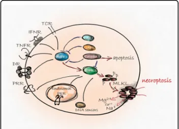
6. After this step, MLKL forms oligomers and translocates to the plasma membrane to execute necroptosis (Fig. 1). Generally, necroptosis requires inhi- bition of caspases
3,7or the absence of the pro-caspase-8- activating adaptor Fas-associated protein with death domain (FADD)
8, demonstrating the crucial role of the apoptotic platform in the negative regulation of necrop- tosis. Active caspases block necroptosis
2preferentially through the cleavage of RIPK1
9, RIPK3
3,10, and cylin- dromatosis (CYLD) protein
11which acts as the de- ubiqutinase enzyme of RIPK1. During DR-mediated sig- naling, inhibitors of apoptosis proteins (IAPs) initiate the ubiquitination of RIPK1 and this process favors cell sur- vival
12. Blockage of IAPs or the subsequent events of IAP- induced signaling strongly support necroptosis
13. Various molecular pathways have been documented as regulators of downstream necroptotic events beside MLKL- mediated membrane rupture, but the complexity of the signaling and regulation network of necroptosis are still not fully understood.
The immunological outcome of cell death can be clas- si fi ed as anti-in fl ammatory or pro-in fl ammatory and tol- erogenic or immunogenic
1. Dominance of apoptosis ensures the tolerogenic outcome of cell death under physiological conditions. When apoptosis signaling is blocked, necroptotic pathways are activated and the dying cells have the potential to initiate innate immune responses via production of damage associated molecules (DAMPs) resulting in an in fl ammatory response
14. Sig- naling in necroptotic cells also supports the cross priming capacity of dendritic cells (DCs)
15.
In this review our goal was to understand the molecular background of necroptosis in humans and to find potential points of clinical intervention. We summarized how the expression, posttranslational modification, and localization of necroptotic molecules are regulated and what the interaction partners of the necrosome complex are. Finally, we provide an overview of drugs, which are already used in the clinic and have been shown to affect necroptosis.
Necroptosis involved in human diseases
Currently, necroptosis is mainly documented in various in vivo mice models
16,17, but regulated necrosis con- tributes to the pathogenesis of many human diseases (Table 1). Both up and down-regulation of necroptosis and misregulation of the apoptosis-necroptosis transition which modifies the immunological outcome of cell death contribute to the evolution of degenerative disorders, autoimmune processes, inflammatory diseases or the immune surveillance of tumors.
Some physiological processes such as alteration of glu- cose level, oxygen deprivation or immune reactions resulted in elevated RIPK3 expression allowing in vivo emergence of necroptosis. Hyperglycemia (35 – 40 mM glucose) markedly enhanced the expression of RIPK3 in various cell lines and primed cells for necroptosis
18,19. Similarly, upregulated expression of RIPK1, RIPK3 and MLKL, and increased RIPK1/3 complex formation have been observed in hypoxic cells
20–22. At the same time caspase-8 mRNA, functioning as a negative regulator of necroptosis, was reported to be transiently decreased following the deprivation of oxygen and glucose (OGD)
23. These processes are also involved in brain injury caused by hypoxia-ischemia and OGD-induced necroptosis
24,25. Type I
26–28and type II
27,29interferons have been pub- lished to induce increased expression of RIPK3, while constitutive IFN β signaling was demonstrated to increase the intracellular level of MLKL
28. CD8 + T lymphocytes can trigger both apoptosis and necroptosis, which make these cells capable of killing tumor cells, even those that
Fig. 1 Backbone of necroptosis signaling.Various extra - or intracellular signals activates the RIPK3 protein directly or through RIPK1. RIPK3-mediated phosphorylation induces MLKL membrane translocation and consequently, ion influx results in necroptosis147. Survival signals through upregulation of IAPs or activation of TAK1 kinase pathway blocks RIPK1-induced signaling and protects cells from unwanted necroptosis. Caspase-8-mediated cleavage of pro- necroptotic RIPK1 and RIPK3 ensures the dominance of
immunologically silent apoptosis to immune stimulant necroptosis
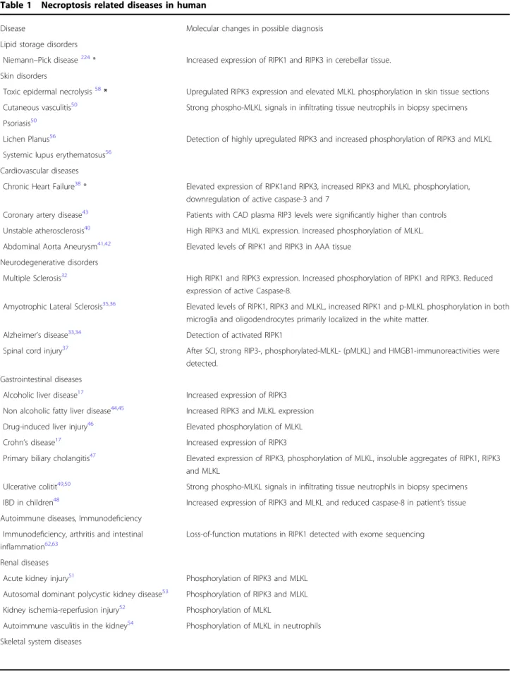
Table 1 Necroptosis related diseases in human
Disease Molecular changes in possible diagnosis
Lipid storage disorders
Niemann–Pick disease224* Increased expression of RIPK1 and RIPK3 in cerebellar tissue.
Skin disorders
Toxic epidermal necrolysis58* Upregulated RIPK3 expression and elevated MLKL phosphorylation in skin tissue sections Cutaneous vasculitis50 Strong phospho-MLKL signals in infiltrating tissue neutrophils in biopsy specimens Psoriasis50
Lichen Planus56 Detection of highly upregulated RIPK3 and increased phosphorylation of RIPK3 and MLKL Systemic lupus erythematosus56
Cardiovascular diseases
Chronic Heart Failure38* Elevated expression of RIPK1and RIPK3, increased RIPK3 and MLKL phosphorylation, downregulation of active caspase-3 and 7
Coronary artery disease43 Patients with CAD plasma RIP3 levels were significantly higher than controls Unstable atherosclerosis40 High RIPK3 and MLKL expression. Increased phosphorylation of MLKL.
Abdominal Aorta Aneurysm41,42 Elevated levels of RIPK1 and RIPK3 in AAA tissue Neurodegenerative disorders
Multiple Sclerosis32 High RIPK1 and RIPK3 expression. Increased phosphorylation of RIPK1 and RIPK3. Reduced expression of active Caspase-8.
Amyotrophic Lateral Sclerosis35,36 Elevated levels of RIPK1, RIPK3 and MLKL, increased RIPK1 and p-MLKL phosphorylation in both microglia and oligodendrocytes primarily localized in the white matter.
Alzheimer’s disease33,34 Detection of activated RIPK1
Spinal cord injury37 After SCI, strong RIP3-, phosphorylated-MLKL- (pMLKL) and HMGB1-immunoreactivities were detected.
Gastrointestinal diseases
Alcoholic liver disease17 Increased expression of RIPK3
Non alcoholic fatty liver disease44,45 Increased RIPK3 and MLKL expression
Drug-induced liver injury46 Elevated phosphorylation of MLKL
Crohn’s disease17 Increased expression of RIPK3
Primary biliary cholangitis47 Elevated expression of RIPK3, phosphorylation of MLKL, insoluble aggregates of RIPK1, RIPK3 and MLKL
Ulcerative colitit49,50 Strong phospho-MLKL signals in infiltrating tissue neutrophils in biopsy specimens IBD in children48 Increased expression of RIPK3 and MLKL and reduced caspase-8 in patient’s tissue Autoimmune diseases, Immunodeficiency
Immunodeficiency, arthritis and intestinal inflammation62,63
Loss-of-function mutations in RIPK1 detected with exome sequencing
Renal diseases
Acute kidney injury51 Phosphorylation of RIPK3 and MLKL
Autosomal dominant polycystic kidney disease53 Phosphorylation of RIPK3 and MLKL Kidney ischemia-reperfusion injury52 Phosphorylation of MLKL
Autoimmune vasculitis in the kidney54 Phosphorylation of MLKL in neutrophils Skeletal system diseases
escaped apoptosis
30. T cell-mediated necroptotic cytolysis also plays a role in activation induced cell death, and can be critical in the development of autoimmune reactions
31.
Upregulation of necroptosis in human diseases
Necroptosis takes part in the pathogenesis of human neurodegenerative disorders, such as Multiple Sclerosis (MS)
32, Alzheimer ’ s disease (AD)
33,34, and Amyotrophic Lateral Sclerosis (ALS)
35,36. Defects in the activation of caspase-8 were demonstrated in the pathologic process of MS. Additionally, activated forms of RIPK1, RIPK3 and MLKL were detected in the cortical lesions of human MS samples
32. Activated RIPK1 as a marker of necroptosis was also observed in human AD brains correlating posi- tively with Braak stage and negatively with brain mass and cognition
33,34. In ALS samples, multiple biochemical hallmarks of necroptosis including increased levels of RIPK1, RIPK3 and MLKL and elevated pRIPK1 and pMLKL were detected in both microglia and oligoden- drocytes. Importantly, pMLKL was primarily localized in the white matter, where demyelination was found
35. In spinal cord injury strong RIPK3 expression and MLKL phosphorylation were detected
37.
In certain cardiovascular diseases, such as chronic heart failure (HF) cell loss and subsequent deterioration of contractile function is associated with elevated expression of RIPK1, RIPK3, and pRIPK3. On the other hand, the expression of caspase-8 was downregulated suggesting activation of necroptosis signaling. MLKL expression did not differ among the control and HF groups; however, pMLKL were present in all HF samples, which is in contrast to the controls where this was almost undetect- able
38. A genetic variant in the RIP3 promoter region was associated with increased RIPK3 transcription, which contributed to the poor prognosis of HF patients
39.
In humans with unstable carotid atherosclerosis, expression of RIPK3 and MLKL was increased, while the phosphorylation of MLKL was detected in advanced atheromas
40. In patients with abdominal aorta aneurysm, the tissue showed elevated levels of RIPK1 and RIPK3 proteins
41,42. In coronary artery disease higher plasma RIPK3 levels were detected than in controls
43.
Regarding gastrointestinal diseases, increased RIPK3 expression was detected in liver biopsies from patients with alcoholic liver disease
17, while both RIPK3 and MLKL expression was increased in non-alcoholic fatty liver diseases
44,45, as well as elevated MLKL phosphor- ylation in drug-induced liver injury
46. High levels of RIPK3 and MLKL phosphorylation were also detected in the liver biopsies of patients with primary biliary cho- langitis, in contrast with its low hepatic expression in healthy controls
47. Similarly, increased levels of RIPK3 were documented in the terminal ileum of patients with Crohn ’ s disease
17and elevated RIPK3 and MLKL levels were observed in in fl amed tissues of in fl ammatory bowel disease (IBD) and allergic colitis patients, whereas the expression of caspase-8 in these tissues was reduced
48. The migration of human neutrophils to sites of inflam- mation was found to activate the RIPK3-MLKL pathway: a strong pMLKL signal was observed in infiltrating tissue neutrophils in samples collected from patients with cutaneous vasculitis, ulcerative colitis, and psoriasis
49,50.
Phosphorylation of MLKL molecules was also detected in human acute kidney injury biopsies
51, in biopsies taken immediately after excision for transplantation
52and in autosomal dominant polycystic kidney disease
53repre- senting involvement of necroptosis in renal disorders.
Antineutrophil cytoplasmic antibody (ANCA) induces neutrophil extracellular traps via necroptosis and causes subsequent endothelial cell damage. ANCA-associated vasculitis exhibited a speci fi c p-MLKL staining in glo- merular neutrophils in human kidney biopsies
54.
Concerning skin diseases, human biopsy samples obtained from patients with Lichen Planus (LP) and Sys- temic lupus erythematosus (SLE) confirm the role of necroptosis in their development. RIPK3 and MLKL acti- vation was demonstrated in podocytes in renal biopsies from patients with lupus nephritis
55. LP and SLE tissue sections showed enhanced epidermal expression of phos- phorylated RIPK3
56. B cells from SLE patients also sig- ni fi cantly displayed high expression levels of necroptosis- related genes
57. As we already mentioned, phosphorylation of MLKL in the in fi ltrated human neutrophils was also found in cutaneous vasculitis and psoriasis
49,50.
Table 1continuedKashin‐Beck disease60 High RIPK3 expression and necrotic cell death morphology in the middle zones of KBD samples.
Negative staining for caspase‐3 Dental diseases
Chronic periodontitis61 Elevated levels of RIPK1, phosphorylated RIPK3, MLKL, phosphorylated MLKL and cFLIPLin gingival tissues
Pulmonary diseases
Chronic obstructive pulmonary disease59 Increase in expression of RIPK3 and PINK1 using confocal imaging
Upregulation of RIPK3, and elevated MLKL phosphor- ylation were observed in the skin samples from patients with toxic epidermal necrolysis in correlation with unwanted necroptosis and subsequent inflammation
58.
Expression of RIPK3 and dynamin-related protein 1 (Drp1) was increased in lung tissue homogenates col- lected from patients suffering from chronic obstructive pulmonary disease, proving the role of necroptotic cell death in pulmonary diseases
59. In Kashin – Beck disease (KBD) necroptosis dominates as a cell death mechanism in the middle zone of cartilage from KBD children
60. Necroptotic cell death is involved in the progression of chronic periodontitis, as gingival tissue in patients showed increased levels of RIPK1, RIPK3, and MLKL, as well as increased phosphorylation of MLKL
61.
Although RIPK1 is one of the key molecules required for execution of necroptosis, patients with its complete deficiency due to homozygous mutations suffered from recurrent infections, early-onset of IBD and progressive polyarthritis. In vitro, cells with RIPK1 de fi ciency showed impaired mitogen-activated protein kinase activation and cytokine secretion and were prone to necroptosis
62,63.
Role of necroptosis in cancers
An increasing number of studies have been published about the importance of necroptotic cell death in anti- cancer therapies, which have been extensively reviewed in recent papers
64,65.
Briefly, both pro- and anti-tumoral effects have been demonstrated following necroptosis in cancer develop- ment and progression. The anti-tumoral effect of necroptosis has been shown in many types of cancer in which the expression of RIPK3
66,67or MLKL
68was silenced or polymorphisms in their coding genes lead to modified expression of necrosomal components
66,69. In general, necroptosis resistance of cancer cells is a com- mon process, and escape from necroptosis was suggested to be a potential hallmark of cancer, similar to the escape from apoptosis
64. Additionally, effective anti-cancer agents trigger immunogenic cell death, inducing the kill- ing of the transformed cells and provoking the members of innate and adaptive immune system to attack. Beside the massive release of DAMPs, necroptotic cells create a great possibility to trigger the activation of CD8 + T cells via cross presentation
15,70. The dual ability of necroptosis to activate innate and adaptive immunity simultaneously makes this cell death pathway a promising therapeutic target.
However, the tumor-promoting outcome of necroptosis has also been shown. RIPK3 and MLKL expression seems to vary among tissue samples from different subtypes and stages of cancer, and downregulation of necroptosis mediators has also been published in various cancers
71–73. Upregulated RIPK3 expression is a general phenomenon
in tumor necrotic areas playing a critical role in tumor growth and metastasis
74. Necroptosis-induced inflamma- tion contributes to tumorigenesis and necroptosis can also lead to an immunosuppressive tumor micro- environment
75. The immune-suppressing environment was associated with necroptosis-induced expression of the chemokine attractant CXCL1
71. It has also been shown that tumor cells induce necroptosis of endothelial cells, which promotes tumor cell extravasation and metas- tasis
76. Thus, we can conclude that necroptosis occurs in different phases during tumorigenesis and plays an ambivalent role in tumor formation.
Molecular mechanisms in the regulation of necroptosis
To understand the molecular background of necropto- sis and to find potential points of clinical intervention we summarize below how the expression, the posttransla- tional modification, and the localization of key necrop- totic molecules (RIPK1, RIPK3 and MLKL) are regulated, while also highlighting the interaction partners of the necrosome complex.
Regulation the expression level of necroptotic proteins
RIPK3-RIPK3 homodimerization is suf fi cient to induce necroptosis; after which, its kinase domain stimulates the activation of RIPK3 through cis-autophosphorylation; a prerequisite step for the recruitment of MLKL
77–79. Thus, RIPK3 dimerization is probably the most critical point of necroptosis induction. Several lines of evidence support the idea that increased expression of RIPK3 can induce its oligomerization and can initiate necroptosis
42,80. RIPK1 dimerization, and accordingly upregulation of RIPK1, facilitates RIPK3 oligomerization, mainly upon death receptor stimuli.
All aspects of necroptotic protein expression are inten- sely regulated, including their transcriptional activity, the stability of the expressed molecules and their degradation.
Speci fi city protein 1 (Sp1), a zinc- fi nger transcription factor, directly regulates RIPK3 expression in cancer cells.
Knockdown of endogenous Sp1 signi fi cantly decreases the
transcription of RIPK3, while re-expression of Sp1 restores
necroptotic response in vitro
81. Induction of necroptosis
by interferon gamma (IFN-γ) resulted in elevated levels of
RIPK3
27and MLKL
28,29,82. This effect was found to
depend on janus kinase 1 (JAK1) and its substrates: the
signal transducer and activator of transcription 1 (STAT1)
and interferon regulatory factor (IRF) transcription factors,
pinpointing interferon-stimulated gene factor 3 (ISGF3) as
a critical promoter
83. Bromodomain-containing protein 4
(BRD4), a member of the bromodomain and extraterminal
domain (BET) family, has been shown to interact IRF1 and
to upregulate MLKL transcription
84. Oncogenes such as
BRAF and AXL have also been implicated in the regulation
of RIPK3 expression
67. The activity of RIPK3 promoter is tightly controlled by methylation
67,85–87(Fig. 2a).
Ubiquitin-like PHD and RING finger domain-containing protein 1 (UHRF1) is essential for the maintenance of the hypermethylation of the RIPK3 promoter and thus con- tributes to the silencing of RIPK3 expression in quiescent cells.
Following transcriptional regulation multiple processes control the protein level of necrosome components. The heat shock protein 90 (HSP90) and CDC37 co-chaperone complex increases the stability of all RIPK1
88, RIPK3
89, and MLKL
90proteins. Consequently, inhibitors of HSP90 facilitated the degradation of these necroptotic compo- nents and potently blocked necroptosis
91. Protein levels of RIPK1 and RIPK3 also decreased in FK506-binding pro- tein 12 (FKBP12) knockdown cells
92.
On the contrary, cells treated with Hsp70 inhibitors underwent cell death, because Hsp70 enhances the sta- bility of necroptosis antagonists, the RIPK1 regulators:
cIAP1/2, x-linked inhibitor of apoptosis protein (XIAP), and the cellular FLICE-like inhibitor protein (cFLIP)
93.
The expression of necroptotic molecules are down- regulated by cleavage and proteosomal degradation. The most well-known inhibitor of necroptosis, caspase-8 cleaves both RIPK1
9, RIPK3
94, and the necroptosis pro- moting deubiquitinase CYLD proteins
11. In macrophages, cathepsins were also reported to be capable of processing RIPK1, which resulted in significant decrease in necrop- totic cell death
95.
Several ubiquitin-ligases mediate K48-linked poly- ubiquitylation and the subsequent proteasome dependent degradation of necroptotic molecules: RIPK1 is regulated by A20
96, carboxyl terminus of Hsp70-interacting protein (CHIP; also known as STUB1)
97, optineurin (Optn)
35, Triad3a
98, RIPK3 by CHIP
97, Optn
35, E3 ubiquitin ligase Pellino 1 (PELI1)
99, and MLKL by Optn (Table 2)
35. Knock down of any of these K48 ubiquitin-ligases increased the sensitivity of necroptosis in both in vitro and in vivo studies. (Fig. 2b).
Posttranslational modifications in the regulation of necroptosis
Accumulating evidence suggests that cell death path- ways are finely tuned by posttranslational modifications, such as ubiquitination and phosphorylation. Multiple excellent recent reviews go into extensive detail about the role of these processes in necroptosis
100, therefore we only provide a brief overview of these processes below.
These pathways are mentioned in the tables and figures of this manuscript in the interest of providing a compre- hensive visual guide to these processes as well (Fig. 2c).
The necrosome is formed due to the phosphorylation driven assembly of RIPK1, RIPK3, and MLKL
4,80,101. However several phosphorylation steps have been
published to inhibit necroptosis, chief among them the transforming growth factor beta-activated kinase 1 (TAK1) complex, which is the most important hub for these necroptosis-dampening signals
102,103. Various pro- tein complexes are assembled along TNFR signaling;
namely the survival (complex I), the apoptotic (complex IIa and IIb) and the necroptosis inducer (complex IIc) complexes. Upon activation TNFR recruits TRADD, RIPK1, TRAF2, TRAF5 proteins. The gathered E3 ubi- quitin ligases, cIAP-1 and cIAP-2 molecules, and the linear ubiquitin chain assembly complex LUBAC (con- sisting of HOIP, HOIL-1L and Sharpin)
104poly- ubiquitinates RIPK1, and modified RIPK1 can now act as a scaffold for TAK1 and the IKK complex
105which molecules in many ways block RIPK1-mediated cell death pathways, and thus the formation of complex II:
106–108These mechanism are: (1) By inducing the activation of NFκB and MAPK signaling pathways and thereby increasing the transcription of several survival molecules such as cIAP1/2
109and FLIP
110(2) by blocking the binding of cell death related molecules to RIPK1
111and (3) by phosphorylating RIPK1
106,108.
Interaction partners of necrosome components
The activity of necrosome components are also mediated by molecular interactions (Fig. 2d). Three molecules, aur- ora kinase A (AURKA), PPM1b, and HSP90 have been recently identified as binding partners of RIPK3
90,91,112,113and/ or RIPK1
91,112in resting cells. AURKA
112and PPM1b
113act as local inhibitors against spontaneous necroptosis, since their silencing induces necroptosis.
PPM1b as a phosphatase prevents RIPK3 autopho- sphorylation in resting cells
113. AURKA together with its downstream target, Glycogen synthase kinase 3β (GSK3β) regulates the formation of RIPK1-RIPK3 and RIPK3-MLKL complexes
112. Silencing or blocking of AURKA, or inhibi- tors of GSK3 β result in necroptosis without any other stimuli. Phosphorylation of GSK3 β at Ser9 suppresses necroptosis through interfering with the formation of RIPK3-MLKL complex, however the direct targets of GSK3 β still have not been identi fi ed. The third molecule which associates with RIPK3 in resting cells, HSP90, is required for proper activation of necroptosis. Formation of the HSP90–CDC37 complex is necessary for RIPK1–RIPK3 interaction, thus it mediates RIPK3 activa- tion during necroptosis. Unsurprisingly HSP90 inhibitors can block TNF-induced systemic inflammatory response syndrome (SIRS) in rats
91. Additionally, membrane teth- ered mucins have been shown to interact with RIPK1 to block necroptosis in human bronchial epithelial cells in vitro
114.
The nuclear retinoic acid receptor gamma (RAR γ ) is
released from the nucleus to initiate the formation of cell
death signaling complexes by mediating RIPK1
dissociation from TNFR when cIAP activity is blocked. In vitro silencing of RAR γ inhibited necroptosis and in vivo results also confirmed that RARγ was essential for TNF- induced RIPK1-initiated apoptosis and necroptosis (Table 2)
115.
Although RIPK1 initiates RIPK3 activation during death receptor driven necroptosis, it plays an ambivalent role in the regulation of RIPK3 aggregation. Under special cir- cumstances instead of activation, RIPK1 acts to suppress the spontaneous activation of RIPK3 by TIR-domain- containing adapter-inducing interferon- β (TRIF)
116or DNA-dependent activator of IFN-regulatory factors (DAI;
also known as ZBP1)
78,117. RIPK3 oligomerization is able to
seed a RHIM dependent oligomer and this process is both suf fi cient and a necessary step in necroptosis. RHIM domains of RIPK1 intrinsically inhibit RHIM-mediated RIPK3 aggregation by competing with the RHIM domain of TRIF or DAI; conversely death domain-driven RIPK1 oligomerization results in RIPK3 aggregation and necrop- tosis. In vivo results also reveal a kinase-independent function for RIPK1 in inhibiting necroptosis. Caspase-8/
RIPK1 double-knockout animals die shortly after birth, however, additional ablation of RIPK3 to make caspase-8/
RIPK1/RIPK3 triple knockouts rescues the viability of these animals
117–120. These data undoubtedly prove the anti- necroptotic activity of RIPK1 under special conditions
78.
Fig. 2 Direct interacting partners of main necroptotic signaling molecules.Sp1 transcription factor increases RIPK3 expression. INFγ-mediated up-regulation of RIPK3 and MKLK level depend on JAK1 kinase, and STAT1 and IRF transcription factors. BRD4 cooperating with IRF1 also increase MLKL transcription. Hypermethylation of the RIPK3 promoter by UHRF1 results in silenced RIPK3 expression. The stability of all RIPK1, RIPK3 and MLKL proteins are increased by HSP90 and CDC37 co-chaperone complex and by FKBP12. The level of both RIPK1 and RIPK3 are down-regulated by caspase-8-mediated cleavage. Cathepsins are also capable of processing RIPK1. A20, CHIP, Optn, PELI1 and Triad3a ubiquitin-ligases mediate K48- linked polyubiquitylation and the subsequent proteasome dependent degradation of: RIPK1, RIPK3 and/or MLKL Upon necroptosis human RIPK1 is autophosphorylated at ser14, ser15, ser161, ser166 and RIPK3 at ser199 and ser227 and ser277. The transient phosphorylation of RIPK1 at ser321 is phosphorylated transiently by TAK1 leads to RIPK1-independent apoptosis and the sustained phosphorylation of RIPK1 by TAK1 at ser321, ser332, ser334 and ser336 induces RIPK1 kinase activation106. IKKα/IKKβalso phosphorylate RIPK1 at ser25 and thereby block RIPK1 activity108,214,215. Mitogen- activated protein kinase-activated protein kinase 2 (MK2) mediates phosphorylation of RIPK1 at ser321 and ser336 and restrains integration of RIPK1 into the cytosolic death complex107,216,217. The phosphorylation at ser89 by a currently unknown kinase inhibits the RIPK1 kinase activity218. Ubiquitylation of RIPK1 at Lys115 by PELI219or Lys377 by cIAP1, cIAP2 and Parkin220promotes necroptosis. LUBAC complex and the deubiquitinase CYLD regulates M1 ubiquitination of RIPK1221. Lys363 ubiquitylation of RIPK3 leads to its proteasomal degradation. RIPK3 is responsible for the phosphorylation of MLKL at thr357 and ser358. TAM (Tyro3, Axl, and Mer) family of receptor tyrosine kinases phosphorylate MLKL on Tyr376 to facilitate MLKL oligomerization145. MLKL is also phosphorylated on Ser441 by a still unidentified kinase222. Caspase-8 mediates the cleavage and inactivation of RIPK1 at asp324 and RIPK3 at asp328. O-GlcNAcylation of the RIPK3 at thr467 by OGT prevents necroptosis223. Red names indicate interaction partners of RIPK1, RIPK3, MLKL which activate necroptosis, blue marks necroptosis inhibitors
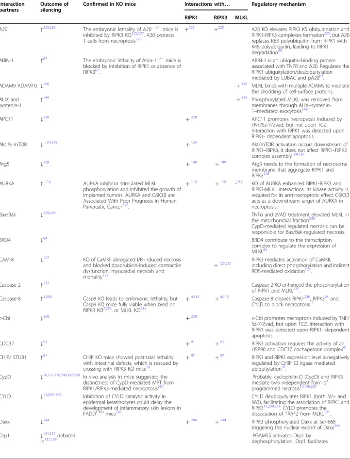
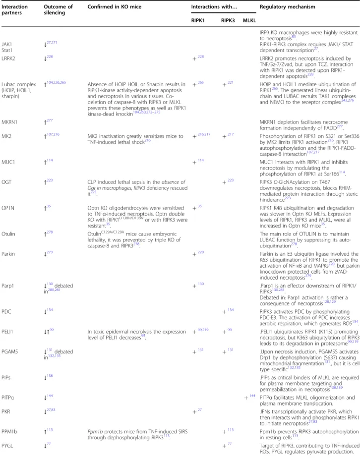
Table 2 Molecules in necroptotic signaling Interaction
partners
Outcome of silencing
Confirmed in KO mice Interactions with… Regulatory mechanism RIPK1 RIPK3 MLKL
A20 ↑225,226 The embryonic lethality of A20−/−mice is
inhibited by RIPK3 KO225,227. A20 protects T cells from necroptosis225
+225 +225 A20 KO elevates RIPK3 K5 ubiquitination and RIPK1-RIPK3 complexes formation225, but A20 replaces K63 polyubiquitin from RIPK1 with K48 polyubiquitin, leading to RIPK1 degradation49.
ABIN-1 ↑67 The embryonic lethality of Abin-1−/−mice is blocked by inhibition of RIPK1 or absence of RIPK367.
ABIN-1 is an ubiquitin-binding protein associated with TNFR and A20. Regulates the RIPK1 ubiquitylation/deubiquitylation mediated by LUBAC and pA2067.
ADAM9 ADAM10 ↓150 +150 MLKL binds with multiple ADAMs to mediate
the shedding of cell-surface proteins.
ALIX and
syntenin-1 ↑149 +149 Phosphorylated MLKL was removed from
membranes through ALIX–syntenin- 1–mediated exocytosis149.
APC11 ↓228 +228 APC11 promotes necroptosis induced by
TNF/5z-7/Zvad, but not upon TCZ.
Interaction with RIPK1 was detected upon RIPK1- dependent apoptosis
Akt ½ mTOR ↓124,125 +124 Akt/mTOR activation occurs downstream of
RIPK1–RIPK3, it does not affect RIPK1–RIPK3 complex assembly124,126
Atg5 ↓128 +199 +199 Atg5 needs to the formation of necrosome
membrane that aggregate RIPK1 and RIPK3128.
AURKA ↑112 AURKA inhibitor stimulated MLKL
phosphorylation and inhibited the growth of implanted tumors. AURKA and GSK3βare Associated With Poor Prognosis in Human Pancreatic Cancer112.
+112 +112 -112 KO of AURKA enhanced RIPK1-RIPK3 and RIPK3-MLKL interactions. Its kinase activity is required for its anti-necroptotic effect. GSK3β acts as a downstream target of AURKA in necroptosis.
Bax/Bak ↓229,230 TNFαand zVAD treatment elevated MLKL in
the mitochondrial fraction229.
CypD-mediated regulated necrosis can be responsible for Bax/Bak-regulated necrosis.
BRD4 ↓84 BRD4 contribute to the transcription
complex to regulate the expression of MLKL84.
CAMKII ↓127 KO of CaMKII abrogated I/R-induced necrosis and blocked doxorubicin-induced contractile dysfunction, myocardial necrosis and mortality127
+127,231 RIPK3-mediates activation of CaMKII, including direct phosphorylation and indirect ROS-mediated oxidation127.
Caspase-2 ↑232 Caspase-2 KO enhanced the phosphorylation
of RIPK1 and MLKL232. Caspase-8 ↑2,233 Casp8 KO leads to embryonic lethality, but
Casp8 KO mice fully viable when bred on RIPK3 KO7,234. or MLKL KO235.
+4,115 +4,115 Caspase-8 cleaves RIPK1236, RIPK394and CYLD to block necroptosis11.
c-Cbl ↓228 +228 c-Cbl promotes necroptosis induced by TNF/
5z-7/Zvad, but upon TCZ. Interaction with RIPK1 was detected upon RIPK1- dependent apoptosis
CDC37 ↓91 +91 +91 RIPK3 activation requires the activity of an
HSP90 and CDC37 cochaperone complex91 CHIP/ STUB1 ↑97 CHIP KO mice showed postnatal lethality
with intestinal defects, which is rescued by crossing with RIPK3 KO mice97.
+97 +97 RIPK3 and RIPK1 expression level is negatively regulated by CHIP E3 ligase mediated ubiquitylation97.
CypD ↓10,127,176,196,237,238 In vivo analysis in mice suggested the distinctness of CypD-mediated MPT from RIPK1/RIPK3-mediated necroptosis237.
.Probably, cyclophilin-D (CypD) and RIPK3 mediate two independent form of programmed necrosis10,176,227 CYLD ↓11,239–242 Inhibition of CYLD catalytic activity in
epidermal keratinocytes could delay the development of inflammatory skin lesions in FADDE-KOmice241.
CYLD deubiquitylates RIPK1 (both M1- and K63), facilitating the association of RIPK1 and RIPK311,239,243. CYLD promotes the dissociation of TRAF2 from MLKL121.
Daxx ↓244 +244 +244 RIPK3 phosphorylated Daxx at Ser-668
triggering the nuclear export of Daxx244.
Drp1 ↓131,132debated
in132,135
.PGAM5S activates Drp1 by dephosphorylation, Drp1 facilitates
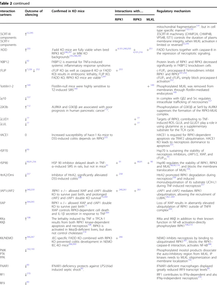
Table 2continued Interaction partners
Outcome of silencing
Confirmed in KO mice Interactions with… Regulatory mechanism RIPK1 RIPK3 MLKL
mitochondrial fragmentation131. but in cell type specific manner132,135
ESCRT-III components ESCRT-I components
↑52,245 .ESCRT-III machinery (CHMP2A, CHMP4B,
VPS4B, IST1) controls the duration of plasma membrane integrity, when MLKL activation is limited or reversed52,245
FADD ↑233 .Fadd KO mice are fully viable when bred
RIPK3 KO246,247or Mlkl KO backgrounds235,248,249
+4,101,240,250 +4,101,216 +251 FADD functions together with caspase-8 in the repression of necroptotic signaling.
FKBP12 ↓92 FKBP12 is essential for TNFα-induced systemic inflammatory response syndrome.
Protein levels of RIPK1 and RIPK3 decreased significantly in FKBP12 knockdown cells cFLIP ↑7,226↓252 cFLIP KO (as well as caspase-8 KO or FADD
KO) results in embryonic lethality, FLIP KO, FADD KO, RIPK3 KO mice are viable7,247
+253 c-FLIPL: procaspase-8 heterodimers inhibit RIPK1 and RIPK3247,254.
cFLIPSand cFLIPRsimply block procaspase-8 activation252.
Flottilin1-2 ↑149 Flotillin-null mice were highly senstitive to
TZ-induced SIRS149 +149 Phosphorylated MLKL was removed from
membranes throughflotillin-mediated endocytosis149
Gγ10 ↓157 In complex with Gβ2 and Src regulates
intracellular trafficking of necrosomes157
GSK3b ↑112 AURKA and GSK3βare associated with poor
prognosis in human pancreatic cancer112.
Phosphorylation of GSK3βat Ser9 by AURKA suppresses the formation of the RIPK3-MLKL complex.
GLUD1 ↓77 +77 Targets of RIPK3, contributing to TNF-
induced ROS. GLUL and GLUD1 play a role in using glutamine as a supplementary substrate for the TCA cycle.
GLUL ↓77 +77
HACE1 0 Increased susceptibility of hace-1 Ko mice to
DSS-induced colitis depends on RIPK3255
HACE1 is required for RIPK1-dependent apoptosis via TRAF2 ubiquitination. HACE1 KO leads to necroptosis dominance to apoptosis255.
HSP70 ↑93 Hsp70 is sustaining the stability of
necroptosis inhibitors, cIAP1/2, XIAP, and cFLIPS/L93.
HSP90 ↓90,91,256 HSP 90 inhibitor delayed death in TNF-
α–induced SIRS in rats, but not in mice91 +91 +90,91 +90 Hsp90 regulates the stability of RIPK1, RIPK3 and MLKL88,90,174. and blocks the membrane translocation of MLKL256.
HtrA2/Omi ↓257 Inhibitor of HtrA2, significantly alleviated
DSS-induced colitis258 +258 HtrA2 promoted RIPK1 degradation during
necroptosis258and induced
monoubiquitination of its substrate UCH-L1 during TNF-induced necroptosis257 cIAP1cIAP2 ↑240,259 .RIPK1+/−allowed XIAP and cIAP1 double
KO to survive past birth, and prolonged cIAP2 and cIAP1 double KO survival13,260
+240,261 +261 .cIAP1 and cIAP2 mediates RIPK1 ubiquitination, allowing the recruitment of LUBAC262–264
XIAP ↑264,265 RIPK1+/−allowed XIAP and cIAP1 double
KO to survive past birth13
XIAP controls RIPK3-dependent cell death and IL-1βsecretion in response to TNF264
Loss of XIAP results in aberrantly elevated ubiquitylation of RIPK1 outside of TNFR complex264.
IKKα
IKKβ ↑108 The lethality induced by TNF+TPCA-1
results from both RIPK1 kinase-dependent apoptosis and necroptosis108. RIPK3 is activated in Ikkα/β‐deficient livers, but does not control cholestasis214
IKKαand IKKβin addition to their known function in NF-κB activation-directly phosphorylate RIPK1108,214
IKK/NEMO ↑266,267 .IEC-specific FADD KO combined with RIPK3 KO prevented colitis development in NEMO IEC-KO mice268,269
+266 NEMO inhibits necroptosis by binding to
ubiquitinated RIPK1267, blocks the RIPK1- caspase-8 interaction, activates NF-kB266. IPMK
IPTK IPPK
↓142,143 Phosphorylated inositol products dissociate
the auto-inhibitory region from MLKL. IP kinases needs to MLKL oligomerization and membrane localization142.
IFNAR1 ↓83 IFNAR1-deficiency protects against LPS/zVad induced septic shock83.
IFNAR1-deficient macrophages displayed greatly reduced IRF9 transcript levels83.
IRF1 ↓270 IRF1 contributes to IFNγ-dependent and also
IFNγ-independent necroptosis270.
IRF9 ↓83
Table 2continued Interaction partners
Outcome of silencing
Confirmed in KO mice Interactions with… Regulatory mechanism RIPK1 RIPK3 MLKL
IRF9 KO macrophages were highly resistant to necroptosis83.
JAK1
Stat1 ↓27,271 RIPK1-RIPK3 complex requires JAK1/ STAT
dependent transcription27.
LRRK2 ↓228 +228 LRRK2 promotes necroptosis induced by
TNF/5z-7/Zvad, but upon TCZ. Interaction with RIPK1 was detected upon RIPK1- dependent apoptosis228.
Lubac complex (HOIP, HOIL1, sharpin)
↑104,226,265
Absence of HOIP HOIL or Sharpin results in RIPK1-kinase activity-dependent apoptosis and necroptosis in various tissues. Co- deletion of caspase-8 with RIPK3 or MLKL prevents these phenotypes as well as RIPK1 kinase-dead knockin104,260,272–275
+265 +221 HOIP and HOIL1 mediate ubiquitination of RIPK1265. The generated linear ubiquitin- chain and LUBAC recruits TAK1 complexes and NEMO to the receptor complex243,276
MKRN1 ↑277 MKRN1 depletion facilitates necrosome
formation independently of FADD277.
MK2 ↑107,216 MK2 inactivation greatly sensitizes mice to
TNF-induced lethal shock216. +216,217 +217 Phosphorylation of RIPK1 on S321 or Ser336 by MK2 limits RIPK1 activation216, RIPK1 autophosphorylation and the RIPK1-FADD- caspase-8 interaction107,217
MUC1 ↑114 +114 MUC1 interacts with RIPK1 and inhibits
necroptosis by modulating the phosphorylation of RIPK1 at Ser166114.
OGT ↑223 CLP induced lethal sepsis in theabsence of
Ogt in macrophages, RIPK3deficiency rescued it223.
+223 RIPK3O-GlcNAcylation on T467 downregulates necroptosis, blocks RHIM- mediated protein interaction through steric hinderance223
OPTN ↑35 Optn KO oligodendrocytes were sensitized
to TNFα-induced necroptosis. Optn double KO with RIPK1D138N/D138Nor with RIPK3 were resistant35.
+35 RIPK1 K48 ubiquitination and degradation
was slower in Optn KO MEFs. Expression levels of RIPK1, RIPK3 and MLKL, were all increased in Optn KO mice35.
Otulin ↑278 OtulinC129A/C129A
mice cause embryonic lethality, it was prevented by triple KO of caspase-8 and RIPK3278.
The main role of OTULIN is to maintain LUBAC function by suppressing its auto- ubiquitination278.
Parkin ↓279 +220 Parkin is an E3 ubiquitin ligase involved the
K63 ubiquitination of RIPK1 to promote the activation of NF-κB and MAPKs220, but parkin knockdown protected cells from zVAD- induced necroptosis279.
Parp1 ↓130debated
in280,281 +130 .Parp1 is an effector downstream of RIPK1/
RIPK3130,281.
Debated in: Parp1 activation is rather a consequence of necroptosis128,129
PDC ↓134 +134 RIPK3 activates PDC by phosphorylating
PDC-E3. The activation of PDC increases aerobic respiration, which generates ROS134. PELI1 ↓↑99 In toxic epidermal necrolysis the expression
level of PELI1 decreases99. +99,219 +99 .PELI1 ubiquitinates RIPK1 (K115) promoting necroptosis, but K363 ubiquitylation of RIPK3 leads to its degradation in proteasome99,219
PGAM5 ↓131debated
in132,135 +131 +131 .Upon necrosis induction, PGAM5S activates
Drp1 by dephosphorylation (S637) causing mitochondrial fragmentation131., but it is cell type specific132,135
PIPs ↓138 .PIPs as critical binders of MLKL are required
for plasma membrane targeting and permeabilization in necroptosis138,139
PITPα ↓144 +144 PITPαfacilitates MLKL oligomerization and
plasma membrane translocation.
PKR ↓27,83 +27 .IFNs transcriptionally activate PKR, which
then interacts with and phosphorylates RIPK1 to initiate necroptosis27,83
PPM1b ↑113 Ppm1bprotects mice from TNF-induced SIRS
through dephosphorylating RIPK3113. +113 Ppm1b prevents RIPK3 autophosphorylation in resting cells113.
PYGL ↓77 +77 Target of RIPK3, contributing to TNF-induced
ROS. PYGL regulates pyruvate production.
MLKL association with RIPK3 is also suppressed by a constitutive interaction of MLKL with a competitive inhibitor, TRAF2, in resting cells. TRAF2 deubiquitina- tion by CYLD promotes the dissociation of TRAF2 from
MLKL and allows necroptosis
121. Two other molecules inhibit cell death by blocking MLKL association with pro- necroptotic components: Repulsive guidance molecule b (RGMb) inhibits MLKL membrane translocation or
Table 2continuedInteraction partners
Outcome of silencing
Confirmed in KO mice Interactions with… Regulatory mechanism RIPK1 RIPK3 MLKL
RARγ ↓115 RARγKO mice are protected from TNF+Z-
vad induced death115. +115 RARγfacilitates RIPK1 dissociation from TNF
receptor and the formation of death signaling complexes115
RelA ↑282 Embryonic lethality of RelA KO mice is
partially prevented by the KO of RIPK3 or MLKL, and it is fully rescued by the combined ablation of Fadd and RIPK3 or MLKL or RIPK1K459A282.
RelA KO leads to TNF-induced activation of FADD-dependent apoptosis and RIPK3- dependent necroptosis.
RGMb ↑122 Renal tubule-specific RGMB knockout mice
exhibited severe tubular injury, after renal ischemia/reperfusion122
RGMb inhibits MLKL membrane translocation or membrane binding122.
RIPK1 ↓↑78,118 .Caspase-8/RIPK1 double-knockout animals die shortly after birth, ablation of RIPK3 to triple knockouts, rescues the viability of these animals. Deficiency in either RIPK3 or MLKL prevented the development of skin lesions in RIPK1E-KO mice117–120
+4 +283 In a kinase-independent function of RIPK1 the RHIM domains of RIPK1 competes with RHIM domain of TRIF or DAI to RHIM- mediated RIPK3 aggregation, but RIPK1 oligomerization is initiative of death domain driven necroptosis78.
Sp1 ↓81 Sp1 specifically binds to RIPK3 promoter and
regulates transcription81. SPATA2 ↓284,285 In contrary to the in vitro data Spata2
deficiency sensitizes mice to SIRS induced by TNFα221.
.SPATA2 binds CYLD into the TNF-RSC and to HOIP. SPATA2 KO reduces phosphorylation of RIPK1 and MLKL in TNF‐α‐induced necroptosis284,285
Src ↓157 Interacting with Gγ10-Gβ2 complex regulates
intracellular trafficking of necrosomes157 STAT1 ↓27,83,271 IFN-γfailed to induce Mlkl transcription in
Stat1–/–mice29
.RIPK1, RIPK3 and MLKL requires JAK1/STAT1- dependent transcription27,235
TAB1/2 ↑286 +259 .TAB1/2 function to maintain TAK1 activity,
which is required for the survival of naive macrophages286,287
TAK1 ↑102,103 Various tissue injuries have been published in the absence of Tak1, These symptoms are associated primarily with apoptosis and were not rescued byRIPK3deletion288.
+102,103,259 TAK1 inhibition triggered the degradation of cIAP2, FLIP, and NFκB-p65. TAK1 blocks RIPK1-RIPK3-FADD complex formation102,111. Intermediate domain of RIPK1 is
phosphorylated transiently by TAK1106,289. Downstream targets of TAK1 phosphorylates RIPK1 (see, MK2, IKK, RelA)
TAM kinases ↓145 Tyro3,Axl,Mertk tripla KO mice were completely resistant to the TZ-induced SIRS145.
+145 TAM (Tyro3, Axl, and Mer) receptor tyrosine kinases phosphorylate MLKL to protmote MLKL oligomerizatin and necroptosis145 TRAF2 ↑121,290 TRAF2 deletion causes morbidity, RIPK3 KO
delays TRAF2 KO mortality121,291and suppressing TRAF2 augments ischemic brain damage through necroptosis mechanism292
+121 TRAF2-MLKL association suppresses the interaction of MLKL with RIPK3121.
Triad3a ↑98 Triad3a induces K48 ubiquitination and the
degradation of RIPK1, FADD and Caspase-898.
TRIF ↓83,116 Mice without functional TRIF did not show
macrophage loss and elevation of inflammatory cytokines upon LPS/zVad293.
+294
+116,294 Activates necroptosis through RHIM dependent association of TRIF with RIPK3 kinase116.
TRPM7 ↓146 +146 +146 TRPM7 is a target of MLKL for the induction
of Ca (2+) influx146.
TRX1 ↑123 +123 TRX1 blocksnecroptosisby maintaining MLKL
in a reduced inactive state123.
UCH-L1 ↓128,257 HtrA2/Omi induces monoubiquitination of
UCH-L1257
UHRF1 ↑81 UHRF1 silences RIPK3 expression via
promoter hypermethylation. Sp1 initiates RIPK3 transcription in the absence of UHRF181.
membrane binding
122and Redox regulator thioredoxin-1 (TRX1) blocks MLKL disulfide bond formation, and through it the critical polymerization of MLKL
123.
Various molecules have been published to act as downstream targets of RIPK3 and others to regulate MLKL localization and/or activation. RIPK3 constitutes an important upstream kinase of death associated protein (Daxx), triggering its nuclear export. The Akt/mTOR pathway
124–126, and Ca
2+/calmodulin-dependent protein kinase II (CaMKII)
127are also active effectors of down- stream necroptotic signaling. Accordingly, several models suggest that effects on these signaling routes modify necroptotic intensity. Poly [ADP-ribose] polymerase 1 (PARP-1)
128(debated in ref.
129,130) and phosphoglycerate mutase family member 5 (PGAM5)
131(debated in ref.
132) have been documented as cell type specific regulators of downstream necroptotic events (Table 2).
Glucose metabolism and ROS production in necroptosis
Reactive oxygen species (ROS) have long been con- sidered to contribute to necroptosis
49,133–135. Oxidation of speci fi c cysteine residues in RIPK1 by ROS activates RIPK1 autophosphorylation. A positive feedback loop is generated because silencing of RIPK1 or RIPK3 reduces ROS production. RIPK1 autophosphorylation is also promoted by mitochondrial ROS and is essential for RIPK3 recruitment into the necrosome. However, necroptosis could occur without ROS induction in some cell lines
135,136.
Metabolic enzymes − human liver glycogen phosphor- ylase (PYGL), glutamate-ammonia ligase (GLUL), gluta- mate dehydrogenase 1 (GLUD1) − increase pyruvate production from glycogen or play a role in glutamine catabolism. These enzymes are activated by RIPK3, resulting in enhancement of aerobic respiration and thus likely contribute to TNF-induced ROS production
80. Pyruvate dehydrogenase complex (PDC) converts pyr- uvate to acetyl-CoA, and triggers the entrance of meta- bolic fl ux into the tricarboxylic acid cycle. Activated RIPK3 in the necrosome enhances PDC activity by phosphorylating the PDC E3 at T135 and plays a major role in increasing aerobic respiration. Based on in vitro studies, activation of these enzymes has additive effects to aerobic respiration and ROS production (Table 2)
80,134.
Intracellular localization of necrosome components