Original Article
Posttranslational modifications of
calcium/calmodulin-dependent protein kinase IIδ and its downstream signaling in human failing hearts
Tomas Rajtik1, Eva Goncalvesova2, Zoltan V Varga3, Przemyslaw Leszek4, Mariusz Kusmierczyk4, Michal Hulman5, Jan Kyselovic1, Peter Ferdinandy3, Adriana Adameova1
1Department of Pharmacology & Toxicology, Faculty of Pharmacy, Comenius University, Bratislava, Slovak Republic; 2Department of Heart Failure & Transplantation, 5Clinic of Heart Surgery, The National Institute of Cardiovascular Diseases, Bratislava, Slovak Republic; 3Department of Pharmacology & Pharmacotherapy, Semmelweis University, Budapest, Hungary; 4Institute of Cardiology, Warszawa, Poland
Received April 7, 2017; Accepted July 16, 2017; Epub August 15, 2017; Published August 30, 2017
Abstract: Background: In human failing hearts (HF) of different origin (coronary artery disease-CAD, dilated-DCM, re- strictive and hypertrophic cardiomyopathy-OTHER), we investigated the active forms of Ca2+/calmodulin-dependent protein kinase IIδ (p-Thr287-CaMKIIδ, oxMet281/282-CaMKIIδ) and their role in phenotypes of the disease. Methods and results: Although basic diagnostic and clinical markers indicating the attenuated cardiac contractility and remodel- ing were comparable in HF groups, CaMKIIδ-mediated axis was different. P-Thr287-CaMKIIδ was unaltered in CAD group, whereas it was upregulated in non-ischemic cardiomyopathic groups. No correlation between the upregu- lated p-Thr287-CaMKIIδ and QT interval prolongation was detected. Unlike in DCM, oxMet281/282-CaMKIIδ did not differ among HF groups. Independently of CaMKIIδ phosphorylation/oxidation, activation of its downstreams-phosphol- amban and cardiac myosin binding protein-C was significantly downregulated supporting both diminished cardiac lusitropy and inotropy in all hearts. Content of sarcoplasmic reticulum Ca2+-ATPase 2a in all HF was unchanged.
Protein phosphatase1β was upregulated in CAD and DCM only, while 2A did not differ among groups. Conclusion:
This is the first demonstration that the posttranslational activation of CaMKIIδ differs in HF depending on etiology.
Lower levels of downstream molecular targets of CaMKIIδ do not correlate with either activation of CaMKIIδ or the expression of major protein phosphatases in the HF. Thus, it is unlikely that these mechanisms exclusively underlie failing of the heart.
Keywords: Human heart failure, Ca2+/calmodulin-dependent protein kinase II, sarcoplasmic reticulum calcium handling, cardiac myosin binding protein-C
Introduction
Heart failure (HF) is a progressive cardiac dis- ease ranging from stages which impair the patient’s quality of life and finally progress to a stage that is characterized by symptoms resis- tant to treatment. HF can occur as a result of coronary artery disease (CAD), dilated cardio- myopathy (DCM) and other hereditary and idio- pathic causes [1]. In contrast to the most preva- lent HF due to CAD, the age-adjusted preva- lence of other forms of HF is lower (1:500 to 1:2500) [1-3]. Regardless of etiology, all types of systolic HF are characterized by seriously impaired cardiac contractility leading into inad- equate perfusion of organs. Great effort has
been undertaken to understand the pathome- chanisms of systolic myocardial dysfunction.
The abnormal Ca2+ cycling and defects in the sarcomeric proteins are proposed to be one of the crucial players in the adverse remodeling and dysfunction of the heart [4].
Ca2+/calmodulin-dependent protein kinase IIδ (CaMKIIδ) has emerged as an important Ca2+
handling protein regulating excitation-contrac- tion coupling. Alterations in its activity have been proposed to exaggerate mishandling in Ca2+ homeostasis, and thereby underlie the de- pressed cardiac contractile function and arrhy- thmogenesis [5, 6]. Overactivation of CaMKIIδ can arise from its capability to undergo the vari-
ous posttranslational modifications, including autophosphorylation (pThr287-CaMKIIδ) and oxi- dation (oxMet281/282-CaMKIIδ) due to oxidative stress [7]. As CaMKIIδ phosphorylates a wide variety of proteins involved in the regulation of both Ca2+ handling (phospholamban-PLN), and contractile proteins (cardiac myosin binding protein-C-cMyBP-C), this protein kinase might be an interesting target in the management of HF treatment [8]. In fact, it has been previously reported that in failing hearts, sarcoplasmic reticulum (SR) Ca2+ uptake is significantly re- duced due to the inhibitory effects of PLN on SERCA2a [9, 10]. This may occur as a result of a decreased activity/expression of SERCA2a [11] or a higher expression of PLN [12, 13]. In addition to Ca2+ dysregulation, attenuation of cMyBP-C function, which serves as a regulatory and structural protein of the sarcomere, due to its dephosphorylation and subsequent degra- dation of cMyBP-C is associated with the dimin- ished cardiac contractile function and poor prognosis of patients with HF [14]. However, it is not known if the above-mentioned link involv- ing SR Ca2+ proteins, cMyBP-C and CaMKIIδ, as a central signal-transducing element, is altered depending on the certain type of systolic ven- tricular dysfunction. Therefore, the objective of this study was to provide a comprehensive analysis of the posttranslational activation of CaMKIIδ and if it is associated with the altera- tions of downstream target proteins in the SR and sarcomere in human failing heart samples.
We also investigated a potential role of protein phosphatases PP1 and PP2A counterbalanc- ing the effects of CaMKIIδ and its downstream proteins [15]. As CaMKIIδ has been suggested as a pro-arrhythmogenic marker [16, 17], and arrhythmias are a common complication of HF [18], therefore we also analyzed a correlation between the levels of the active forms of CaMKIIδ and QT interval duration.
Methods Study design
All procedures were in accordance with ethical standards for human experiments based on Heslinki declaration. All experiments were ap- proved by national and institutional ethical commissions (statement no. IK-NP-0021-24/
1426/14 and NUSCH EK 126/180509). Sam- ples of left ventricles were obtained from ex- planted hearts of patients with diagnosed ter-
minal stadium of HF (NYHA III-IV) and reduced left ventricle ejection fraction (LVEF<25%). We employed the failing hearts due to coronary artery disease (CAD, n=6), dilated cardiomyop- athy (DCM, n=10), and other cardiomyopathies being either restrictive or hypertrophied cardio- myopathy (OTHER, n=6). All patients underwent echocardiographical, hemodynamical and bio- chemical examination before transplantation.
Additionally, the HF pharmacotherapy was ana- lyzed from the last medical records. Samples of left ventricles from healthy donor patients (C, n=4), whose hearts cannot have been used for transplantation from various medical reasons (CMV infection, size/donor recipient mismatch, major damage during procedure), served as a control group. Tissues from the walls of left ventricles have been harvested in the time of explanation avoiding the scared, fibrotic and adipose tissues. They were afterwards rinsed, dried and snap-frozen in liquid nitrogen until further processing.
Immunoblotting
The samples of left ventricles were processed for the immunoblotting analysis based on our standard laboratory protocol for SDS-PAGE and Western Blotting [19]. Proteins were trans- ferred on PVDF membranes (Immobilon-P, EMD Millipore, USA) and incubated with primary anti- bodies against p-Thr287-CaMKII (Cell Signaling, USA), oxMet281/282-CaMKII (EMD Millipore, USA), total CaMKIIδ (Santa Cruz, USA), p-Thr17-PLN (Badrilla, UK), p-Ser16-PLN (Badrilla, UK), total PLN (Badrilla, UK), p-Ser282(284)-cMyBP-C (Enzo, USA), total cMyBP-C (Santa Cruz, USA), total SERCA2a (Badrilla, UK), PP1β (Abcam, USA) and PP2A (Sigma-Aldrich, USA). Afterwards, the incubation with HRP-conjugated secondary antibodies, donkey anti-rabbit IgG (GE Health- care Life Sciences, UK) and rabbit anti-goat IgG (Sigma-Aldrich, USA) was performed. Signals were detected by enhanced chemiluminiscen- ce (Crescendo Luminata, EMD Millipore, USA) and chemiluminiscence imaging system (my- ECL imager, Thermo Scientific, USA). Total pro- tein staining with Coomasive Brilliant Blue R- 250 or Reactive Brown 10B evaluated by scan- ning densitometry was used as the loading con- trol as a substitution to the housekeeping pro- tein immunodetection [20-22]. Intensity of a particular protein band was compared to a whole lane.
Statistical analysis
The results are expressed as means ± stan- dard error of means (S.E.M.), unless stated oth- erwise. One-way ANOVA analysis with Newman- Keuls and Tukey’s post-hoc tests and two-tailed unpaired Student’s t-test were used for evalua- tion of group differences in variables with nor- mal distribution. In case of the non-normal dis- tribution, a Man-Whitney’s analysis was used.
Correlation between biochemical parameters, QT duration or N-terminal pro-B type natriuretic peptide (NT-proBNP) and the levels of certain proteins was analyzed by Pearson’s test. All analyses were performed with GraphPad Prism 6.00 for Windows (GraphPad Software, USA).
Differences between groups were considered significant when P<0.05.
Results
Characteristics of study subjects
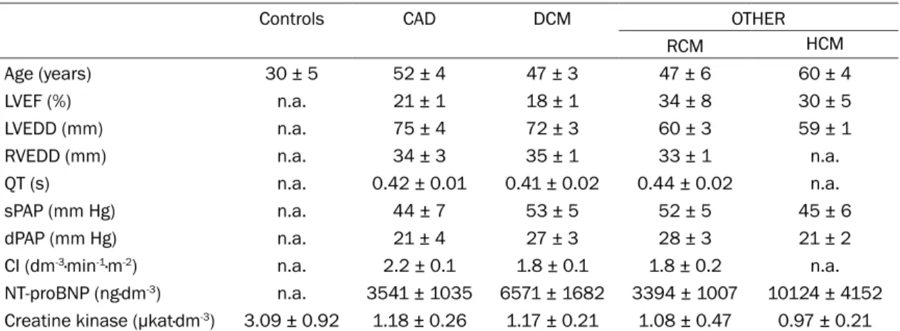
A summary of the main pre-transplant data of the study subjects are presented in Table 1. All patients irrespective of the etiology of HF were in either NYHA III or IV class. Among the HF groups, there were no significant differences in neither of the values of left ventricular ejection fraction (LVEF), parameters of cardiac remodel- ing (the size of left and right end-diastolic ven- tricular diameters-LVEDD, RVEDD), markers of pulmonary hypertension (systolic and diastolic pulmonary pressures-sPAP, dPAP) nor NT-pro- BNP, a common biochemical marker of HF. HF
patients were treated at least with ACE inhibi- tors or AT1R-blockers, beta-blockers and diuret- ics. The control subjects were treated with the infusion of noradrenaline and dopamine and fluid balance was maintained with desmopres- sin and hydroxyethyl starch, an intravenous col- loid volume expander. They presented pre- served systolic/diastolic function. No metabo- lic disorders were diagnosed in the study sub- jects.
Posttranslationally modified forms of CaMKIIδ in end-stage human HF show no association with the QT interval duration
Western blot analysis of the posttranslationally modified forms of CaMKIIδ by phosphorylation at Thr287 and oxidation at Met281/282 is shown in Figures 1, 2.
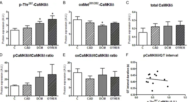
The content of p-Thr287-CaMKIIδ in the group of CAD did not differ from the values of the control subjects. On the other hand, in all cardiomyop- athic groups of HF, the expression of p-Thr287- CaMKIIδ was significantly increased as com- pared with the control hearts and CAD (Figures 1, 2A). Of note, the levels of oxMet281/282-Ca- MKIIδ did not differ among HF group. However, by using Student’s t-test, significantly lower ex- pression of oxMet281/282-CaMKIIδ was observed in the DCM as compared to control group (Figures 1, 2B). This is the first analysis of the oxidized form of this protein kinase in human end-stage HF.
Table 1. Echocardiographic, hemodynamic and biochemical parameters of probands
Controls CAD DCM OTHER
RCM HCM
Age (years) 30 ± 5 52 ± 4 47 ± 3 47 ± 6 60 ± 4
LVEF (%) n.a. 21 ± 1 18 ± 1 34 ± 8 30 ± 5
LVEDD (mm) n.a. 75 ± 4 72 ± 3 60 ± 3 59 ± 1
RVEDD (mm) n.a. 34 ± 3 35 ± 1 33 ± 1 n.a.
QT (s) n.a. 0.42 ± 0.01 0.41 ± 0.02 0.44 ± 0.02 n.a.
sPAP (mm Hg) n.a. 44 ± 7 53 ± 5 52 ± 5 45 ± 6
dPAP (mm Hg) n.a. 21 ± 4 27 ± 3 28 ± 3 21 ± 2
CI (dm-3·min-1·m-2) n.a. 2.2 ± 0.1 1.8 ± 0.1 1.8 ± 0.2 n.a.
NT-proBNP (ng·dm-3) n.a. 3541 ± 1035 6571 ± 1682 3394 ± 1007 10124 ± 4152 Creatine kinase (µkat·dm-3) 3.09 ± 0.92 1.18 ± 0.26 1.17 ± 0.21 1.08 ± 0.47 0.97 ± 0.21 End-stage HF groups of ischemic and non-ischemic origin did not significantly differ in neither echocardiographic nor hemody- namic parameters. CAD-coronary artery disease; DCM-dilated cardiomyopathy; RCM-restrictive cardiomyopathy; HCM-hypertro- phied cardiomyopathy. LVEF-left ventricular ejection fraction; LVEDD-left ventricular end-diastolic diameter; RVEDD-right ventric- ular end-diastolic diameter; QT-QT interval; sPAP-systolic pulmonary artery pressure; dPAP-diastolic pulmonary artery pressure;
CI-cardiac index; NT-proBNP-N-terminal pro-B type natriuretic peptide. Data are expressed as the means ± SEM.
The total CaMKIIδ was comparable in all HF groups and did not differ from the values of controls (Figures 1, 2C). Thus, the ratio of both p-Thr287/total CaMKIIδ and oxMet281/282-Ca- MKIIδ/total CaMKIIδ mimicked the pattern of
diseased groups indicating that PKA does not substitute a role of CaMKIIδ to maintain the function of PLN (Figures 1, 3B). The expression of the total PLN (Figures 1, 3C) was unchanged in the HF groups, thereby the ratio of both Figure 1. Representative blots of evaluated proteins in control and human fail-
ing hearts. Total CaMKIIδ-total Ca2+/calmodulin-dependent protein kinase 2δ;
p-Thr287-CaMKIIδ-phospho-Thr287-Ca2+/calmodulin-dependent protein kinase 2δ; ox-Met281/282-CaMKIIδ-oxidized-Met281/282-Ca2+/calmodulin-dependent pro- tein kinase 2δ; total PLN-total phospholamban; p-Thr17-PLN-phospho-Thr17- phospholamban; p-Ser16-PLN-phospho-Ser16-phospholamban; SERCA2a-sar- coplasmic/endoplasmic reticulum Ca2+-ATPase; total cMyBP-C-total cardiac myosin-binding protein C; p-Ser284-cMyBP-C-phospho-Ser284-cardiac myosin- binding protein C; PP1β-protein phosphatase 1β; PP2A-protein phosphatase 2A.
changes of the particular posttranslational modifica- tions (Figures 1, 2D, 2E).
QT interval prolongation is known as a predisposing factor for increased ventricu- lar arrhythmia risk and a higher activity and/or expres- sion of CaMKIIδ has been associated with arrhythmia triggering [23, 24]. Therefore in the groups of HF with the altered expression of post- translationally modified form of CaMKIIδ (DCM, OTHER), a potential correlation between the content of phosphorylat- ed form and prolongation of QT interval was analyzed.
However, we did not identify any link between p-Thr287- CaMKIIδ and the prolonga- tion of QT interval (Figures 1, 2F).
Regulation of SR Ca2+ uptake in end-stage human HF As SR Ca2+ cycling is known to be diminished in HF [25]
and CaMKII activates certain SR proteins [26] we further analyzed p-Thr17-PLN, a down- stream of this protein kinase, and SERCA2a. In spite of the different state of the phos- phorylation of CaMKIIδ in the particular HF types (ischemic vs. non-ischemic) (Figures 1, 2A), the levels of p-Thr17-PLN were greatly downregulated in all HF groups in compari- son to non-failing hearts (Figures 1, 3A). Likewise, PKA-dependent phosphoryla- tion of PLN at Ser16 residue was decreased and there was no difference among the
p-Thr17-PLN/total PLN and p-Ser16-PLN/total PLN showed the same pattern as the respec- tive active forms of this protein (Figures 1, 3D, 3E). These data, as general characteristics of diastolic dysfunction, indicates no important role of the expression of either of active form of the upstream kinase in terminal HF.
Non-phosphorylated form of PLN produces inhibitory effects on SERCA2a activity and restricts Ca2+ filling back into the SR [27, 28].
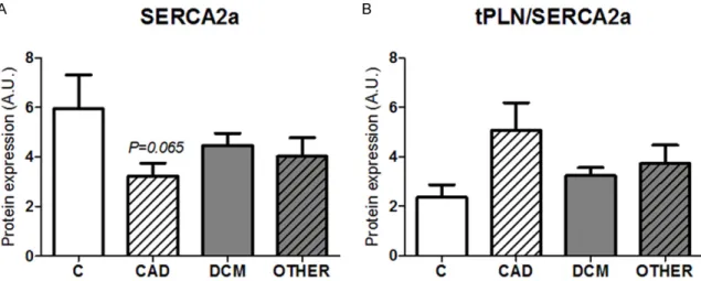
Therefore, we estimated a ratio between the total PLN and SERCA2a. Similarly to the total PLN, the levels of total SERCA2a were unchan- ged in all HF groups when compared to control Figure 2. Phosphorylation (p-Thr287-CaMKIIδ) and oxidation (oxMet281/282-CaMKIIδ) of CaMKIIδ in different types of terminal human HF. (A) Levels of p-Thr287-CaMKIIδ; (B) Content of oxMet281/282-CaMKIIδ; (C) Expression of total CaMKIIδ; (D) Ratio of phosphorylated to total CaMKIIδ; (E) Ratio of oxidized to total CaMKIIδ; (F) Comparison be- tween QT interval duration and phosphorylation of CaMKIIδ (n=12). Data for (A-E) are expressed as the means ± S.E.M. n=4-10 hearts per group. *P<0.05 vs. control hearts.
Figure 3. CaMKIIδ-dependent (p- Thr17-PLN) and PKA-dependent phos- phorylation (p-Ser16-PLN) of PLN in human failing hearts. (A) Expression of p-Thr17-PLN; (B) Expression of p- Ser16-PLN; (C) Expression of total PLN;
(D) Ratio of p-Thr17-PLN to total PLN;
(E) Ratio of p-Ser16-PLN to total PLN.
Data for (A-E) are expressed as the means ± S.E.M. n=4-10 hearts per group. *P<0.05 vs. control hearts.
group (Figures 1, 4A). Accordingly, no signifi- cant changes were found in the PLN/SERCA2a ratio in HF groups as compared with non-failing hearts (Figures 1, 4B).
Cardiac MyBP-C in human HF
In addition to the altered Ca2+ cycling, the res- ponse of the contractile proteins is diminished in HF [29]. Therefore, we evaluated expression of total cMyBP-C and its phosphorylated form at Ser284. Unlike the expression of the total protein (Figure 5B), the levels of p-Ser284-cMy- BP-C were significantly downregulated in all HF groups regardless the expression of particular posttranslationally modified forms of CaMKIIδ (Figures 1, 5A, 5C). These results indicate serious systolic dysfunction in HF and similarly to the status of p-Thr17-PLN propose no role of the levels of the active forms of CaMKIIδ in this context.
To further expand the knowledge about this active form of cMyBP-C in end-stage human HF, a correlation between the serum NT-proBNP and p-Ser284-cMyBP-C was evaluated. Interest- ingly, the levels of NT-proBNP positively corre- Figure 4. Content of SERCA2a in human failing hearts and ratio of total PLN to SERCA2a. (A) Expression of SERCA2a.
(B) Ratio of total PLN to SERCA2a. Values for (A) and (B) are expressed as the means ± S.E.M. n=4-10 hearts per group.
Figure 5. Expression and phosphorylation of cMyBP-C in human failing hearts. (A) Expression of p-Ser284-cMyBP-C;
(B) Content of total cMyBP-C; (C) Ratio of p-Ser284-cMyBP-C to total protein. Values for (A-C) are expressed as the means ± S.E.M. n=4-10 hearts per group. *P<0.05 vs. control hearts.
Figure 6. Correlation between the expression of p- Ser284-cMyBP-C and marker of volume overload and myocyte stretch NT-proBNP in failing hearts. n=13 hearts. Correlation was considered as positively sig- nificant (P=0.0181).
lated with the levels of p-Ser284-cMyBP-C in the HF groups (Figures 1, 6). This may indicate a compensatory increase in cMyBP-C phosphory-
lation in response to increased myocyte stretch documented by the higher levels of NT-proBNP.
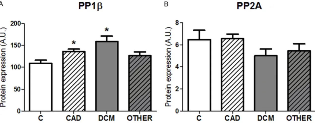
Modulation of protein phosphorylation status by protein phosphatases in failing hearts We also analyzed certain protein phosphatases which counterbalance the phosphorylation of PLN, cMyBP-C and CaMKIIδ itself. Indeed, two major isoforms of protein phosphatases in human myocardium, PP1 subunit β (PP1β) and PP2A were investigated. PP1β, proposed to dephosphorylate the most of PLN as well as cMyBP-C [30], was increased in the CAD and DCM group but not in other cardiomypathic groups (Figures 1, 7A). PP2A sharing similar catalytic sites with PP1 [31, 32] proposed also to dephosphorylate cMyBP-C [33, 34] and most of CaMKII in cytosolic and membrane fraction [35] did not differ among the diseased groups nor in comparison with control healthy hearts (Figures 1, 7B).
Discussion
In the present comprehensive study we have shown that the levels of both active, posttrans- lationally modified forms of CaMKIIδ, p-Thr287- CaMKIIδ and oxMet281/282-CaMKIIδ in failing hearts due to CAD were comparable to those of healthy hearts. On the other hand, in HF of non- ischemic origin (DCM, RCM and HCM), p-Thr287- CaMKIIδ was upregulated whereas expression of oxMet281/282-CaMKIIδ was unchanged. Re- gardless the levels of these active forms of the kinase, the downstream targets of CaMKIIδ, such as p-Thr17-PLN or p-Ser284-cMyBP-C were Figure 7. Expression of protein phosphatases PP1β and PP2A in left ventricles of various types of human heart failure. (A) Expression of PP1β; (B) Expression of PP2A. Values for (A) and (B) are expressed as the means ± S.E.M.
n=4-10 hearts per group. *P<0.05 vs. control hearts.
Table 2. Expression of evaluated proteins in left ventricles of human failing hearts of dif- ferent origin
Proteins CAD DCM OTHER
Total CaMKIIδ ≈ ≈ ≈
p-Thr287-CaMKIIδ ≈ ↑ ↑
ox-Met281/282-CaMKIIδ ≈ ↓ ≈
Total PLN ≈ ≈ ≈
p-Thr17-PLN ↓ ↓ ↓
p-Ser16-PLN ↓ ↓ ↓
SERCA2a ≈ ≈ ≈
Total cMyBP-C ≈ ≈ ≈
p-Ser284-cMyBP-C ↓ ↓ ↓
PP1β ↑ ↑ ≈
PP2A ≈ ≈ ≈
Table summarizes protein expression detected by immunoblot analysis between particular HF groups:
CAD-coronary artery disease; DCM-dilated cardiomyopa- thy; RCM-restrictive cardiomyopathy; HCM-hypertrophied cardiomyopathy and control group. Total CaMKIIδ, p-Thr286-CaMKIIδ, ox-Met281/282-CaMKIIδ-total, phospho- Thr286 and oxidized-Met281/282-Ca2+/calmodulin-dependent protein kinase IIδ; total PLN, p-Thr17-PLN, p-Ser16-PLN- total, phospho-Thr17 and phospho-Ser16-phospholam- ban; SERCA2a-sarcoplasmic/endoplasmic reticulum Ca2+-ATPase; total cMyBP-C, p-Ser284-cMyBP-C-total and phospho-Ser284-cardiac myosin-binding protein C; PP1β- protein phosphatase 1β; PP2A-protein phosphatase 2A.
(≈)-no difference in expression; (↑)-significantly increased expression in comparison to control group; (↓)-significant- ly decreased expression in comparison to controls. Differ- ences between the groups were evaluated by ANOVA test and considered significant when P<0.05.
significantly downregulated in all HF types.
Evaluation of protein phosphatases revealed the upregulated content of PP1β in CAD and DCM but not in other types of HF. Expression of PP2A in failing hearts did not differ of the levels of controls. Thus, this study employing the samples of various types of HF (both of a high and low incidence) provides the first demonstration that i) the oxidative activation of CaMKIIδ does not go hand in hand with its activation through phosphorylation, ii) CaMKIIδ activation through phosphorylation and oxida- tion differs depending on the etiology of HF, iii) the lower phosphorylation of the downstream proteins indicating diminished inotropy and lusitropy in these diseased hearts is not paralleled with either active forms of CaMKIIδ or with the expression of the main cardiac protein phosphatases (Table 2).
Under physiological conditions, the activation of CaMKII in heart is mainly regulated by auto- phosphorylation in the presence of Ca2+/CaM [36]. This mechanism allows the kinase to acti- vate various Ca2+-sensitive proteins to ensure proper Ca2+ handling, including proteins regu- lating the function of the sarcoplasmic reticu- lum and sarcomere [37-39]. Therefore, the observed higher activation state of CaMKIIδ and Ca2+ mishandling may contribute to the dis- turbances in contraction and relaxation in HF [40]. Considering these deleterious effects of the overactivated protein kinase, increased p- Thr287-CaMKIIδ could be expected in the failing hearts. However, in spite of this logical hypoth- esis, data shown in this study as well as in others [41, 42] are unequivocal. In fact, all HF hearts characterized by depressed cardiac contractility and remodeling have shown the different pattern of oxidation and phosphoryla- tion of CaMKIIδ. Moreover, the pattern of changes in the oxidative activation of the kinase did not mimic the changes in phosphory- lation and vice versa. Interestingly, in spite of this observation the markers of HF such as NT-proBNP, pulmonary arterial pressures, EF or cardiac indices did not differ among the types of HF.
So far published papers have mainly been deal- ing with CaMKII in human HF of a higher inci- dence. In study of Miyamoto et al. [43], in DCM, the increased phosphorylation of CaMKII has been reported what is in line with our data. On the other hand, similarly to Fisher et al [42] the
ratio of the phosphorylated form/total protein kinase has not indicated the changes among the groups. In another study, the activity of Ca- MKII measured by enzymatic assay was found to be increased in DCM while unchanged in CAD [41]. By predicting a linear link between phosphorylation and catalytic activity of the kinase this observation is in line with results reported in this study. In the present study, we have extended the current knowledge on the activation of CaMKIIδ and reported for the first time the content of p-Thr287-CaMKIIδ being also upregulated in HF types of a lower incidence. It has been suggested that the overactivated/
hyperphosphorylated CaMKII is associated with electrical instability along with contractile dysfunction [19, 42, 44]. However, we have been unable to found any correlation between p-Thr287-CaMKIIδ expression and the duration of QT interval in the HF groups of non-ischemic origin which exerted the higher phosphoryla- tion of this protein kinase.
In addition to phosphorylation, CaMKIIδ can undergo other posttraslational modifications [7]. In the presence of oxidative stress, which is commonly observed in HF [45], the activation of CaMKIIδ through oxidation might be of a great relevance thereby suggesting the levels of oxMet281/282-CaMKIIδ being increased in our samples of human HF. Of note, the increased levels of oxMet281/282-CaMKIIδ have been shown to underlie electrical remodeling and hypertrophy in cardiac myocytes and isoprena- line-treated rats [46, 47]. Surprisingly, no changes in the expression of oxMet281/282- CaMKIIδ were found among HF groups.
However, while comparing DCM and control group, oxMet281/282-CaMKIIδ was found to be decreased in the diseased hearts. It has been proposed that methionine residues at 281/282 can undergo oxidation by superoxide produced by NADPH oxidases what leads into the kinase activation [48, 49]. Another enzyme, methio- nine sulfoxide reductase A is able to reverse this effect and thereby decrease pro-oxidant potential of the kinase [48]. It would be inter- esting to measure the activity of this enzyme;
however, at the time of performing the study it was impossible due to the unavailability of assay and antibody. Thus, the aforementioned data indicate that a role of CaMKIIδ including its posttranslational forms should be carefully investigated and the particular etiology of HF should be taken into account while assessing
its involvement in the pathologic mechanisms of the disease. Of note, in our recent study, we have shown that both p-Thr287-CaMKIIδ and oxMet281/282-CaMKIIδ are not elevated, rather markedly decreased in the late phase of reper- fusion of previously ischemic hearts which experienced contractile dysfunction and higher oxidative stress [19].
CaMKIIδ is one of critical proteins regulating proper Ca2+ cycling. For instance, by phosphory- lation of PLN at Thr17 it relieves the inhibitory effects on SERCA2a and thereby mediates lusi- tropic effects [50]. Since HF is characterized by significantly abolished cardiac contractility and relaxation we have hypothesized CaMKIIδ- dependent phosphorylation of PLN being de- creased. In agreement with this hypothesis, the expression of CaMKIIδ-phosphorylated form of p-Thr17-PLN was decreased to almost non- detectable levels in all HF groups. Similarly to our study, Dash et al. [12] and Miyamoto et al.
[43] have also reported CaMKII-mediated phos- phorylation of PLN being decreased in DCM.
The content of p-Thr17-PLN was parallel with the levels of p-Ser16-PLN regulated by PKA. Addi- tionally, we have shown that SERCA2a levels did not differ among the groups what is in agreement with some previous studies [11, 51]. On the other hand, downregulation of SERCA2a in heart failure resulting from DCM has also been reported [12, 52]. The expres- sion of the ratio of total PLN to SERCA2a was unaltered in all diseased groups. The levels of non-phosphorylated PLN have been suggested to act as a marker of decreased SERCA2a pumping activity [53]. However, our data and of others [39, 43, 54] do support the role of phos- phorylated PLN rather than the non-phosphory- lated protein in diminished Ca2+ SR handling in HF.
CaMKIIδ is also known to directly phosphory- late some proteins of contractile apparatus and thereby regulates cardiac contractility in- dependently of Ca2+ cycling modulation. In fact, it phosphorylates cMyBP-C, regulates actin- myosin cross-bridging, and controls force gen- eration within the sarcomere [29]. Recently, cMyBP-C has been proposed to serve as a no- vel biomarker of cardiac injury [14, 55]. De- phosphorylation and subsequent degradation of this protein may indicate cardiac dysfunction and HF [56, 57]. Likewise, several gene muta- tions of this protein have been reported in CAD,
DCM and HCM [58, 59]. 17 phosphorylation sites of cMyBP-C being phosphorylated by four various kinases have been identified so far;
however, Ser282residue (Ser284 in humans), which can be phosphorylated by CaMKII [60], is the most frequent target of phosphorylation in vivo [61]. Here, we have shown that phos- phorylation of Ser284 residue was significantly downregulated while the total protein was unal- tered in all HF groups. In support, other studies dealing with genetic mutations promoting car- diac dysfunction have reported total cMyBP-C being downregulated [59]. In context with p-Thr287-CaMKIIδ, these observations open several questions. Similarly to p-Thr17-PLN, p-Ser284-cMyBP-C was decreased independent- ly of the phosphorylation/oxidation of the upstream protein kinase CaMKIIδ. However, as indicated above, it should be mentioned that the Ser284 residue of cMyBP-C can also be phosphorylated by some other protein kinases such as PKA, PKC or ribosomal S6 kinase [29].
Nevertheless, the phosphorylation of Ser284 mediated by CaMKII seems to play an impor- tant role because its inhibition resulted in the decreased phosphorylation of cMyBP-C [62, 63].
As the ability of the kinase to phosphorylate the proteins is counterbalanced by phosphatases we investigated the expression levels of main cardiac protein phosphatases PP1 and PP2A.
The PP1 has been proposed to serve as a nega- tive regulator of cardiac function. It is able to dephosphorylate substrates of CaMKIIδ and thus may play an important role in gradual blunting of heart function during the develop- ment of HF [15, 64]. In this and in previously published studies [30, 43, 65], the expression of β subunit of PP1 was increased only in CAD and DCM failing hearts. Interestingly, the most abundant PP, PP2A did not differ among the HF groups and was comparable to the value of controls. The data about PP2A are controver- sial. The increased mRNA levels have been reported in DCM [43], while the protein content of this phosphatase was unchanged [66] sup- porting the findings of our current study.
Although this study has reported several novel findings and raised some new questions, there are few limitations. Firstly, the conclusions are based on proteomic data only. Nevertheless, we aimed to investigate an association between certain posttranslational modifications of Ca-
MKIIδ and its downstream proteins, with cardi- ac function of all known types of HF. Secondly, phosphorylation status of proteins can be affected by certain drugs, however, by assum- ing that all HF patients were given the standard, almost the same therapy, this is unlikely to influence our present data.
Conclusion
In failing hearts of ischemic and non-ischemic origin, not differing in any main diagnostic char- acteristics, we have detected a different phos- phorylation and oxidation status of CaMKIIδ.
The changes in posttranslational status of the kinase have not reflected the phosphorylation of its downstream targets neither in p-Ser284- cMyBP-C nor p-Thr17-PLN which, however were significantly downregulated and thereby could underlie the diminished cardiac contractile and relaxation function in HF. By investigating a link between the duration of the QT interval and the posttranslationally modified levels of CaMKIIδ we have been unable to confirm that the altered activation of CaMKIIδ promotes pro-arrhythmo- genic environment. Thus, the assessment of a role of CaMKIIδ, its active forms and potential consequences on cardiac function in particular forms of HF needs more detailed investiga- tions.
Acknowledgements
This study was supported by Slovak Scientific Grant Agency (VEGA 1-0271-16), Slovak Rese- arch and Development Agency (APVV 15-0607), Slovak Society of Cardiology and Comenius University in Bratislava (UK/402/2016) and by the National Research, Development, and Innovation Office of Hungary (OTKA K 109737, National Heart Program NVKP 16-1-2016- 0017). Authors would like to thank Dr. Gabriel Doka for organizing clinical patient data and Mrs. Veronika Hassova for her technical assistance.
Disclosure of conflict of interest None.
Address correspondence to: Dr. Adriana Adameova, Department of Pharmacology and Toxicology, Fa- culty of Pharmacy, Comenius University, Bratislava, Odbojarov 10, 832 32, Slovak Republic. Tel: +421 2 50117 366; Fax: +421 2 50 117 100; E-mail:
adameova@fpharm.uniba.sk
References
[1] Yancy CW, Jessup M, Bozkurt B, Butler J, Casey DE Jr, Drazner MH, Fonarow GC, Geraci SA, Horwich T, Januzzi JL, Johnson MR, Kasper EK, Levy WC, Masoudi FA, McBride PE, McMurray JJ, Mitchell JE, Peterson PN, Riegel B, Sam F, Stevenson LW, Tang WH, Tsai EJ, Wilkoff BL;
American College of Cardiology Foundation;
American Heart Association Task Force on Practice Guidelines. 2013 ACCF/AHA guideline for the management of heart failure: a report of the American college of cardiology founda- tion/American heart association task force on practice guidelines. Circulation 2013; 128:
e240-327.
[2] Gersh BJ, Maron BJ, Bonow RO, Dearani JA, Fi- fer MA, Link MS, Naidu SS, Nishimura RA, Om- men SR, Rakowski H, Seidman CE, Towbin JA, Udelson JE, Yancy CW; American College of Cardiology Foundation/American Heart Asso- ciation Task Force on Practice Guidelines;
American Association for Thoracic Surgery;
American Society of Echocardiography; Ameri- can Society of Nuclear Cardiology; Heart Fail- ure Society of America; Heart Rhythm Society;
Society for Cardiovascular Angiography and In- terventions; Society of Thoracic Surgeons.
2011 ACCF/AHA guideline for the diagnosis and treatment of hypertrophic cardiomyopa- thy: a report of the American college of cardiol- ogy foundation/American heart association task force on practice guidelines. Circulation 2011; 124: e783-831.
[3] Kushwaha SS, Fallon JT, Fuster V. Restrictive cardiomyopathy. N Engl J Med 1997; 336:
267-276.
[4] Stienen GJ. Pathomechanisms in heart failure:
the contractile connection. J Muscle Res Cell Motil 2015; 36: 47-60.
[5] Hund TJ, Mohler PJ. Role of CaMKII in cardiac arrhythmias. Trends Cardiovasc Med 2015;
25: 392-397.
[6] Swaminathan PD, Purohit A, Soni S, Voigt N, Singh MV, Glukhov AV, Gao Z, He BJ, Luczak ED, Joiner ML, Kutschke W, Yang J, Donahue JK, Weiss RM, Grumbach IM, Ogawa M, Chen PS, Efimov I, Dobrev D, Mohler PJ, Hund TJ, An- derson ME. Oxidized CaMKII causes cardiac sinus node dysfunction in mice. J Clin Invest 2011; 121: 3277-3288.
[7] Erickson JR. Mechanisms of CaMKII activation in the heart. Front Pharmacol 2014; 5: 59.
[8] Marks AR. Calcium cycling proteins and heart failure: mechanisms and therapeutics. J Clin Invest 2013; 123: 46-52.
[9] Haghighi K, Bidwell P, Kranias EG. Phosphol- amban interactome in cardiac contractility and survival: a new vision of an old friend. J Mol Cell Cardiol 2014; 77: 160-167.
[10] Schmitt JP, Kamisago M, Asahi M, Li GH, Ah- mad F, Mende U, Kranias EG, MacLennan DH, Seidman JG, Seidman CE. Dilated cardiomy- opathy and heart failure caused by a mutation in phospholamban. Science 2003; 299: 1410- 1413.
[11] Frank K, Bölck B, Bavendiek U, Schwinger R.
Frequency dependent force generation corre- lates with sarcoplasmic calcium ATPase activi- ty in human myocardium. Basic Res Cardiol 1998; 93: 405-411.
[12] Dash R, Frank KF, Carr AN, Moravec CS, Kra- nias EG. Gender influences on sarcoplasmic reticulum Ca2+-handling in failing human myo- cardium. J Mol Cell Cardiol 2001; 33: 1345- 1353.
[13] Kadambi VJ, Ponniah S, Harrer JM, Hoit BD, Dorn GW 2nd, Walsh RA. Cardiac-specific over- expression of phospholamban alters calcium kinetics and resultant cardiomyocyte mechan- ics in transgenic mice. J Clin Invest 1996; 97:
533-539.
[14] Baker JO, Tyther R, Liebetrau C, Clark J, How- arth R, Patterson T, Möllmann H, Nef H, Sicard P, Kailey B, Devaraj R, Redwood SR, Kunst G, Weber E, Marber MS. Cardiac myosin-binding protein C: a potential early biomarker of myo- cardial injury. Basic Res Cardiol 2015; 110:
23.
[15] Weber S, Meyer-Roxlau S, Wagner M, Dobrev D, El-Armouche A. Counteracting protein ki- nase activity in the heart: the multiple roles of protein phosphatases. Front Pharmacol 2015;
6: 270.
[16] Koval OM, Guan X, Wu Y, Joiner ML, Gao Z, Chen B, Grumbach IM, Luczak ED, Colbran RJ, Song LS, Hund TJ, Mohler PJ, Anderson ME.
CaV1.2-subunit coordinates CaMKII-triggered cardiomyocyte death and afterdepolarizations.
Proc Natl Acad Sci U S A 2010; 107: 4996- 5000.
[17] Sag CM, Wadsack DP, Khabbazzadeh S, Abesser M, Grefe C, Neumann K, Opiela MK, Backs J, Olson EN, Brown JH, Neef S, Maier SK, Maier LS. Calcium/calmodulin-dependent pro- tein kinase II contributes to cardiac arrhythmo- genesis in heart failure. Circ Heart Fail 2009;
2: 664-675.
[18] Lip GY, Heinzel FR, Gaita F, Juanatey JR, Le Heuzey JY, Potpara T, Svendsen JH, Vos MA, Anker SD, Coats AJ, Haverkamp W, Manolis AS, Chung MK, Sanders P, Pieske B, Gorenek B, Lane D, Boriani G, Linde C, Hindricks G, Tsut- sui H, Homma S, Brownstein S, Nielsen JC, Lainscak M, Crespo-Leiro M, Piepoli M, Se- ferovic P, Savelieva I; EP-Europace. European Heart Rhythm Association/Heart Failure Asso- ciation joint consensus document on arrhyth- mias in heart failure, endorsed by the Heart
Rhythm Society and the Asia Pacific Heart Rhythm Society. Europace 2016; 18: 12-36.
[19] Rajtik T, Carnicka S, Szobi A, Giricz Z, O-Uchi J, Hassova V, Svec P, Ferdinandy P, Ravingerova T, Adameova A. Oxidative activation of CaMKIIδ in acute myocardial ischemia/reperfusion in- jury: a role of angiotensin AT1 receptor-NOX2 signaling axis. Eur J Pharmacol 2016; 771:
114-122.
[20] Romero-Calvo I, Ocón B, Martínez-Moya P, Suárez MD, Zarzuelo A, Martínez-Augustin O, de Medina FS. Reversible ponceau staining as a loading control alternative to actin in West- ern blots. Anal Biochem 2010; 401: 318-320.
[21] Gilda JE, Gomes AV. Stain-Free total protein staining is a superior loading control to β-actin for Western blots. Anal Biochem 2013; 440:
186-188.
[22] Welinder C, Ekblad L. Coomassie staining as loading control in Western blot analysis. J Pro- teome Res 2011; 10: 1416-1419.
[23] Anderson ME, Braun AP, Wu Y, Lu T, Wu Y, Schulman H, Sung RJ. KN-93, an inhibitor of multifunctional Ca++/calmodulin-dependent protein kinase, decreases early afterdepolar- izations in rabbit heart. J Pharmacol Exp Ther 1998; 287: 996-1006.
[24] Kirchhof P, Fabritz L, Kilić A, Begrow F, Breithar- dt G, Kuhn M. Ventricular arrhythmias, in- creased cardiac calmodulin kinase II expres- sion, and altered repolarization kinetics in ANP receptor deficient mice. J Mol Cell Cardiol 2004; 36: 691-700.
[25] Luo M, Anderson ME. Mechanisms of altered Ca2+ handling in heart failure. Circ Res 2013;
113: 690-708.
[26] Mattiazzi A, Mundiña-Weilenmann C, Guoxiang C, Vittone L, Kranias E. Role of phospholam- ban phosphorylation on Thr17 in cardiac physi- ological and pathological conditions. Cardio- vasc Res 2005; 68: 366-375.
[27] Kimura Y, Kurzydlowski K, Tada M, MacLennan DH. Phospholamban inhibitory function is acti- vated by depolymerization. J Biol Chem 1997;
272: 15061-15064.
[28] Smeazzetto S, Saponaro A, Young HS, Moncelli MR, Thiel G. Structure-function relation of phospholamban: modulation of channel activ- ity as a potential regulator of SERCA activity.
PLoS One 2013; 8: e52744.
[29] Moss RL, Fitzsimons DP, Ralphe JC. Cardiac MyBP-C regulates the rate and force of con- traction in mammalian myocardium. Circ Res 2015; 116: 183-192.
[30] Aoyama H, Ikeda Y, Miyazaki Y, Yoshimura K, Nishino S, Yamamoto T, Yano M, Inui M, Aoki H, Matsuzaki M. Isoform-specific roles of protein phosphatase 1 catalytic subunits in sarcoplas- mic reticulum-mediated Ca2+ cycling. Cardio- vasc Res 2011; 89: 79-88.
[31] Terrak M, Kerff F, Langsetmo K, Tao T, Domin- guez R. Structural basis of protein phospha- tase 1 regulation. Nature 2004; 429: 780- 784.
[32] Cho US, Xu W. Crystal structure of a protein phosphatase 2A heterotrimeric holoenzyme.
Nature 2007; 445: 53-57.
[33] Schlender KK, Hegazy MG, Thysseril TJ. De- phosphorylation of cardiac myofibril C-protein by protein phosphatase 1 and protein phos- phatase 2A. Biochim Biophys Acta 1987; 928:
312-319.
[34] Noguchi T, Hünlich M, Camp PC, Begin KJ, El- Zaru M, Patten R, Leavitt BJ, Ittleman FP, Alpert NR, LeWinter MM, VanBuren P. Thin-filament- based modulation of contractile performance in human heart failure. Circulation 2004; 110:
982-987.
[35] Strack S, Barban MA, Wadzinski BE, Colbran RJ. Differential inactivation of postsynaptic density-associated and soluble Ca2+/calmod- ulin-dependent protein kinase II by protein phosphatases 1 and 2A. J Neurochem 1997;
68: 2119-2128.
[36] Meyer T, Hanson PI, Stryer L, Schulman H.
Calmodulin trapping by calcium-calmodulin- dependent protein kinase. Science 1992; 256:
1199-1202.
[37] Guo T, Zhang T, Mestril R, Bers DM. Ca2+/
calmodulin-dependent protein kinase II phos- phorylation of ryanodine receptor does affect calcium sparks in mouse ventricular myocytes.
Circ Res 2006; 99: 398-406.
[38] Simmerman HK, Collins JH, Theibert JL, We- gener AD, Jones LR. Sequence analysis of phospholamban. Identification of phosphoryla- tion sites and two major structural domains. J Biol Chem 1986; 261: 13333-13341.
[39] Schlender KK, Bean LJ. Phosphorylation of chicken cardiac C-protein by calcium/calmodu- lin-dependent protein kinase II. J Biol Chem 1991; 266: 2811-2817.
[40] Anderson ME, Brown JH, Bers DM. CaMKII in myocardial hypertrophy and heart failure. J Mol Cell Cardiol 2011; 51: 468-473.
[41] Kirchhefer U, Schmitz W, Scholz H, Neumann J.
Activity of cAMP-dependent protein kinase and Ca2+/calmodulin-dependent protein kinase in failing and nonfailing human hearts. Cardio- vasc Res 1999; 42: 254-261.
[42] Fischer TH, Eiringhaus J, Dybkova N, Förster A, Herting J, Kleinwächter A, Ljubojevic S, Schmit- to JD, Streckfuß-Bömeke K, Renner A, Gum- mert J, Hasenfuss G, Maier LS, Sossalla S.
Ca2+/calmodulin-dependent protein kinase II equally induces sarcoplasmic reticulum Ca2+ leak in human ischaemic and dilated cardio- myopathy. Eur J Heart Fail 2014; 16: 1292- 1300.
[43] Miyamoto SD, Stauffer BL, Nakano S, Sobus R, Nunley K, Nelson P, Sucharov CC. Beta-adren- ergic adaptation in paediatric idiopathic dilat- ed cardiomyopathy. Eur Heart J 2014; 35: 33- [44] Zhang T, Maier LS, Dalton ND, Miyamoto S, 41.
Ross J Jr, Bers DM, Brown JH. The deltaC iso- form of CaMKII is activated in cardiac hypertro- phy and induces dilated cardiomyopathy and heart failure. Circ Res 2003; 92: 912-919.
[45] Dhalla NS, Temsah RM, Netticadan T. Role of oxidative stress in cardiovascular diseases. J Hypertens 2000; 18: 655-673.
[46] Wagner S, Ruff HM, Weber SL, Bellmann S, Sowa T, Schulte T, Anderson ME, Grandi E, Bers DM, Backs J, Belardinelli L, Maier LS. Re- active oxygen species-activated Ca/calmodu- lin kinase IIδ is required for late I(Na) augmen- tation leading to cellular Na and Ca overload.
Circ Res 2011; 108: 555-565.
[47] Velez Rueda JO, Palomeque J, Mattiazzi A. Ear- ly apoptosis in different models of cardiac hy- pertrophy induced by high renin-angiotensin system activity involves CaMKII. J Appl Physiol (1985) 2012; 112: 2110-2120.
[48] Erickson JR, Joiner ML, Guan X, Kutschke W, Yang J, Oddis CV, Bartlett RK, Lowe JS, O’Donnell SE, Aykin-Burns N, Zimmerman MC, Zimmerman K, Ham AJ, Weiss RM, Spitz DR, Shea MA, Colbran RJ, Mohler PJ, Anderson ME.
A dynamic pathway for calcium-independent activation of CaMKII by methionine oxidation.
Cell 2008; 133: 462-474.
[49] Palomeque J, Rueda OV, Sapia L, Valverde CA, Salas M, Petroff MV, Mattiazzi A. Angiotensin II-induced oxidative stress resets the Ca2+ de- pendence of Ca2+-calmodulin protein Kinase II and promotes a death pathway conserved across different species. Circ Res 2009; 105:
1204-1212.
[50] Mundiña-Weilenmann C, Vittone L, Ortale M, de Cingolani GC, Mattiazzi A. Immunodetection of phosphorylation sites gives new insights into the mechanisms underlying phospholam- ban phosphorylation in the intact heart. J Biol Chem 1996; 271: 33561-3367.
[51] Schwinger RH, Böhm M, Schmidt U, Karczews- ki P, Bavendiek U, Flesch M, Krause EG, Erd- mann E. Unchanged protein levels of SERCA II and phospholamban but reduced Ca2+ up- take and Ca(2+)-ATPase activity of cardiac sar- coplasmic reticulum from dilated cardiomyop- athy patients compared with patients with nonfailing hearts. Circulation 1995; 92: 3220- 3228.
[52] Meyer M, Schillinger W, Pieske B, Holubarsch C, Heilmann C, Posival H, Kuwajima G, Miko- shiba K, Just H, Hasenfuss G. Alterations of sarcoplasmic reticulum proteins in failing hu-
man dilated cardiomyopathy. Circulation 1995;
92: 778-784.
[53] Luo W, Wolska BM, Grupp IL, Harrer JM, Haghighi K, Ferguson DG, Slack JP, Grupp G, Doetschman T, Solaro RJ, Kranias EG. Phos- pholamban gene dosage effects in the mam- malian heart. Circ Res 1996; 78: 839-847.
[54] Clarke JD, Caldwell JL, Horn MA, Bode EF, Rich- ards MA, Hall MC, Graham HK, Briston SJ, Greensmith DJ, Eisner DA, Dibb KM, Trafford AW. Perturbed atrial calcium handling in an ovine model of heart failure: potential roles for reductions in the l-type calcium current. J Mol Cell Cardiol 2015; 79: 169-179.
[55] Lynch TL, Sadayappan S. Surviving the infarct:
a profile of cardiac myosin binding protein-C pathogenicity, diagnostic utility, and pro- teomics in the ischemic myocardium. Pro- teomics Clin Appl 2014; 8: 569-577.
[56] Stathopoulou K, Wittig I, Heidler J, Piasecki A, Richter F, Diering S, van der Velden J, Buck F, Donzelli S, Schröder E, Wijnker PJ, Voigt N, Do- brev D, Sadayappan S, Eschenhagen T, Carrier L, Eaton P, Cuello F. S-glutathiolation impairs phosphoregulation and function of cardiac myosin-binding protein C in human heart fail- ure. FASEB J 2016; 30: 1849-1864.
[57] van Dijk SJ, Holewijn RA, Tebeest A, Dos Reme- dios C, Stienen GJ, van der Velden J. A piece of the human heart: variance of protein phos- phorylation in left ventricular samples from end-stage primary cardiomyopathy patients. J Muscle Res Cell Motil 2009; 30: 299-302.
[58] Gajendrarao P, Krishnamoorthy N, Selvaraj S, Girolami F, Cecchi F, Olivotto I, Yacoub M. An investigation of the molecular mechanism of double cMyBP-C mutation in a patient with end-stage hypertrophic cardiomyopathy. J Car- diovasc Transl Res 2015; 8: 232-243.
[59] Kuster DW, Govindan S, Springer TI, Martin JL, Finley NL, Sadayappan S. A Hypertrophic Car- diomyopathy-associated MYBPC3 mutation common in populations of south Asian descent causes contractile dysfunction. J Biol Chem 2015; 290: 5855-5867.
[60] Gautel M, Zuffardi O, Freiburg A, Labeit S.
Phosphorylation switches specific for the car- diac isoform of myosin binding protein-C: a modulator of cardiac contraction? EMBO J 1995; 14: 1952-1960.
[61] Kooij V, Holewinski RJ, Murphy AM, Van Eyk JE.
Characterization of the cardiac myosin binding protein-C phosphoproteome in healthy and fail- ing human hearts. J Mol Cell Cardiol 2013; 60:
116-120.
[62] Sadayappan S, Gulick J, Osinska H, Barefield D, Cuello F, Avkiran M, Lasko VM, Lorenz JN, Maillet M, Martin JL, Brown JH, Bers DM, Molkentin JD, James J, Robbins J. A critical function for Ser-282 in cardiac myosin binding protein-C phosphorylation and cardiac func- tion. Circ Res 2011; 109: 141-150.
[63] Tong CW, Gaffin RD, Zawieja DC, Muthuchamy M. Roles of phosphorylation of myosin binding protein-C and troponin I in mouse cardiac mus- cle twitch dynamics. J Physiol 2004; 558: 927- 941.
[64] Carr AN, Schmidt AG, Suzuki Y, del Monte F, Sato Y, Lanner C, Breeden K, Jing SL, Allen PB, Greengard P, Yatani A, Hoit BD, Grupp IL, Hajjar RJ, DePaoli-Roach AA, Kranias EG. Type 1 phosphatase, a negative regulator of cardiac function. Mol Cell Biol 2002; 22: 4124-4135.
[65] Hamdani N, Borbély A, Veenstra SP, Kooij V, Vrydag W, Zaremba R, Dos Remedios C, Nies- sen HW, Michel MC, Paulus WJ, Stienen GJ, van der Velden J. More severe cellular pheno- type in human idiopathic dilated cardiomyopa- thy compared to ischemic heart disease. J Muscle Res Cell Motil 2010; 31: 289-301.
[66] Wijnker PJ, Boknik P, Gergs U, Müller FU, Neu- mann J, dos Remedios C, Schmitz W, Sinder- mann JR, Stienen GJ, van der Velden J, Kirch- hefer U. Protein phosphatase 2A affects myofilament contractility in non-failing but not in failing human myocardium. J Muscle Res Cell Motil 2011; 32: 221-233.